Anatomy & Examination of the Athlete s Hip
|
|
|
- Griffin Mervin Greer
- 5 years ago
- Views:
Transcription


1 Anatomy & Examination of the Athlete s Hip Shane J. Nho, MD, MS Hip Preservation Center, Division of Sports Medicine, Department of Orthopedic Surgery, Rush University Medical Center


2 Layered Anatomical Approach to the Hip Layer 2: Inert Layer Layer 3: Dynamic Layer Layer 1: Osteochondral Layer Mechanics of joint Layer 4: Neural Layer Draovitch et al. Curr MSK Med 2012.


3 Hip Differential Diagnosis Is the hip the SOURCE of the problem? Is the hip the SITE of the problem? Is the hip the SOLUTION of the problem?
4 Layer 1: Osteochondral Layer Structures: Femur, Pelvis, Acetabulum Purpose: Joint congruence and normal osteo / arthro kinematics Static Overload Acetabular Dysplasia Acetabular Protrusio Femoral Anteversion Femoral Valgus Dynamic Impingement Cam Impingement Rim Impingement Femoral Retroversion Femoral Varus
5 What is Dysplasia? Klaue JBJS-Br 1991


6 Pre- and Post-Op

7 Layer 2: Inert Layer Structures: Labrum, joint capsule, ligamentous complex, ligamentum teres Purpose: Static stability of the joint Labral Injury Cartilage Injury Capsular Injury Instability Adhesive capsulitis
8 Acetabular Labrum Deepens the socket allowing for greater coverage of the femoral head Maintain stability Decrease contact pressure Provides a fluid seal for the hip joint **Most common area of injury is at the capsulolabral junction Parvizi et al. JAAOS 2012.


9 Hip Labrum Microscopic Structure 3 Layers by SEM First: 10 μm without distinct orientation Second: 40 μm with lamellar orientation Third: μm with circumferential orientation Petersen et al. Arch Orthop Trauma Surg 2003.


10 Hip Labrum Microscopic Structure Physiologic cleft at the chondro-labral junction Chondral: hyaline cartilage Histology Articular side: fibrocartilage with chondrocytes Capsular side: dense connective tissue with fibroblasts Petersen et al. Arch Orthop Trauma Surg 2003.


11 Hip Labrum Vasculature Increased vascularity at the capsular side Anastamosis between Medial/lateral circumflex, deep branch of superior gluteal, inferior gluteal arteries to provide branches to the capsule and synovium Kalhour M et al. JBJS 2009; Kelly BT et al. Arthroscopy 2005.
12 Hip Labrum Innervation Hip pain related to labral injury Torn Impingement Originates from the nerve to the quadratus femoris and obturator nerve Multiple nerve endings Pain: unmyelinated nerve endings in the anterosuperior labrum Pressure: corpuscles (Vater-Pacini, Golgi- Mazzoni, Ruffini, Krause) Propioception: mechanoreceptors Kim YT et al. CORR 1995.

13 Anterior Hip Capsule and Ligamentous Support Anterior Static Stabilizers: restrains extension & external rotation Iliofemoral ligament (Y Ligament of Bigelow): strongest hip ligaments Originates from AIIS and inserts on the intertrochanteric line of femur. Terminal fibers form zona orbicularis Screw home mechanism with hip extension / ER Pubofemoral ligament Originates from the pubic rami and inserts on the intertrochanteric crest

14 Posterior Hip Capsule and Ligamentous Support Posterior Static Stabilizers: restrains internal rotation in flexion and extension Ischiofemoral ligament: originates from the ischial rim and inserts on the posterosuperior base of the GT Blends with zona orbicularis posteriorly Zona Orbicularis Encircles entire femoral neck Functions as locking ring around the femoral neck and provides stability with distraction
.")
15 Ligamentum Teres Ligamentum Teres Travels from the inferior aspect of the acetabulum at the transverse acetabular ligament to fovea of the femoral head (fovea capitis). Tension with adduction and ER May serve as a secondary stabilizing structure Torn LT has been described as a source of hip pain (Byrd & Jones. Arthroscopy 2004). Some have recommended debridement (Haviv & O Donnell. KSSTA 2011) Some have recommended LT reconstruction (Amenabar et al. Arth Tech 2012; Lindner Arth Tech 2012; Philippon et al. JBJS Br 2013.)
16 Layer 3: Contractile Layer Structures: All musculature including lumbosacral musculature Purpose: Dynamic stability Enthesopathies / Tendinopathy: Anterior: Hip flexor tendonitis or Psoas dysfunction or subspine impingement Posterior: Deep Gluteal Syndrome (Proximal Hamstring Syndrome or Paraspinal dysfunction) * SI Joint & Lumbar Spine Medial: Athletic Pubalgia or Osteitis Pubis Lateral: GTPS / ITB

17 27 Muscles Cross the Hip Joint Muscles supporting and stabilizing the lumbopelvic complex Core canister : Transversus abdominis, multifidi, pelvic floor, diaphragm All other muscles with direct or fascial attachments to the pelvis

18 Anterior Enthesopathy: Hip Flexors Primary: Iliopsoas Sartorius Tensor fascia lata Rectus femoris Adductor longus Pectineus Secondary: Adductor brevis, gracilis, anterior fibers of gluteus minimus

19 Medial Enthesopathy: Hip Adductors Primary: Pectineus Adductor longus Gracilis Adductor brevis Adductor magnus Secondary: Biceps femoris, gluteus maximus, quadratus femoris

20 Medial Enthesopathy: Hip Adductors Athletic pubalgia Weakness or tear of the posterior inguinal wall (injury to rectus abdominus, avulsion of internal oblique muscle FAI associated with AP (Larson et al. Arthroscopy 2011) 25% RTP with AP alone 50% RTP with HA alone 89% RTP with HA and AP surgery

21 Lateral Enthesopathy: Hip Abductors Primary: Gluteus medius Gluteus minimus Tensor fascia lata Secondary: Piriformis, sartorius
 External coxa saltans 10-25% General population 2,4,8 Middle aged population Females > males 4,9-12 Association with low back pain 4,9-12 20-22% of Gluteus Medius tears are found incidentally at")
22 Lateral Enthesopathy: Greater Trochanteric Pain Syndrome GTPS : disorders of the peritrochanteric space of the hip Trochanteric bursitis Gluteus medius and minimus tears ( rotator cuff tear of the hip) External coxa saltans 10-25% General population 2,4,8 Middle aged population Females > males 4,9-12 Association with low back pain 4, % of Gluteus Medius tears are found incidentally at time of THA

23 Anatomy
24 Anatomy


25 Anatomy
26 Anatomy


27 Anatomy

 Robertson WR &")
28 Anatomy 2 Distinct Footprints Lateral: 438 mm 2 (34.8mm x 11.2mm) Superoposterior: mm 2 (17mm diameter) Robertson WR & Kelly BT. Arthroscopy 2008.

29 Posterior Enthesopathy: Hip Extensors Primary: Gluteus maximus Hamstrings Posterior head of the adductor magnus Secondary: Posterior fibers of the gluteus medius, anterior fibers of the adductor magnus


30 Proximal Hamstring Anatomy Guanche. Op Tech Sports 2012.

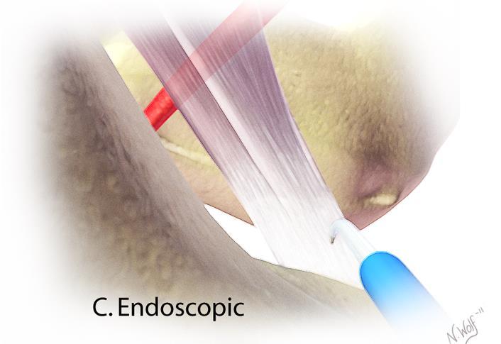
31 Endoscopic HS Repair

 can be caused by fibrous bands,")
32 Posterior Enthesopathy: Deep Gluteal Space Syndrome Sciatic Nerve (L4 to S3) exits through sciatic notch inferior to the piriformis muscle Covered by the G. Max and passes between the ischial tuberosity and greater trochanter 28mm of excursion with hip flexion Nerve is able to stretch and glide with joint movement Deep gluteal syndrome (DGS) can be caused by fibrous bands, gluteal, piriformis, or HS muscles Martin et al. Arthroscopy 2011.

33 Physical Examination of the Hip Critical for accurate diagnosis of hip pain Can help to differentiate intra-articular and extraarticular etiologies of symptoms

34 Laxity Asymptomatic passive translation of the femoral head relative to the acetabulum Beighton criteria: Placing flat hands on the floor with straight legs Left knee bending backward Right knee bending backward Left elbow bending backward Right elbow bending backward Left thumb touching the forearm Right thumb touching the forearm Left little finger bending backward past 90 degrees Right little finger bending backward past 90 degrees
35 Gait: Trendelenburg Abductor lurch Antalgic Foot progression angle Excessive ER Excessive IR Short leg limp Physical Exam


36 20 patients with FAI underwent gait analysis Alpha Angle neg correlated with peak hip abd and IR moment CEA correlated with hip flexion moment FAI produces characteristic gait deviations including gluteus weakness and HF hypertonicity

37 Range of Motion
38 Extra-Articular Hip Exam

39 Impingement Tests

40 Lateral Hip Exam
41 Conclusions Hip and pelvis dysfunction can be a challenging diagnostic dilemma. Understand the pathomorphology of the hip joint as well as the surrounding neuromuscular anatomy. Failure to recognize and treat compensatory injury patterns may result in continued pain and dysfunction. FAI generally produces characteristic gait deviations due to gluteal weakness and hip flexor hypertonicity
42 Conclusions Recognition of the detrimental mechanical overload from alterations in layers 1 & 2 is important in both the treatment and prevention of these injuries. Surgical correction of layers 1 & 2 will require a necessary Layer 3 & 4 accommodation period

43

44 Shane J. Nho, MD, MS

45 Provocative Maneuvers Passive ROM of both hips is critical for diagnosis (Normal : Flexion 120, Extension 10, ER 50, IR 30, ABD 45, ADD
46 Provocative Maneuvers Impingement Test FADDIR Painful Arc

47 Provocative Maneuvers Posterior Impingement Test Hip extension & ER

48 Provocative Maneuvers Lateral Rim Impingement Hyperabduction

49 Provocative Maneuvers Psoas Impingement FABER

50 Provocative Maneuvers Instability Test Extension and ER Loss of normal recoil

51 Circumduction maneuver for snapping psoas tendon Provocative Maneuvers

52 Provocative Maneuvers Trochanteric Pain Sign Abduction and ER
53 Provocative Maneuvers Peritrochanteric Space Exam Palpation of the anterior, lateral, and posterior trochanter Exam of areas of bony prominence
54 Provocative Maneuvers Hip Abduction strength with knee in flexion


55 Hip Abduction strength with knee in extension Provocative Maneuvers

56 Bicycle maneuver for snapping ITB Provocative Maneuvers
57 Stability Arc: Capsule and Muscular Attachments Walters et al. Arthroscopy 2014.
 Ischial")
58 Topographic Bony Anatomy Anterior Anterior Superior Iliac Spine (ASIS) Pubic Tubercle Lateral Iliac Crest Greater Trochanter Posterior Posterior Superior Iliac Spine (PSIS) Ischial Tuberosity
59 Surface Anatomy ASIS Anterior Portal Anterolateral Portal DALA Portal Greater trochanter


60 Hip Arthroscopy Portals ASIS Anterior Portal Anterolateral Portal DALA Portal Greater trochanter
61 Neurovascular Structures Byrd. JAAOS 2006.
62 Neurovascular Structures Byrd. JAAOS 2006.
63 Neurovascular Structures Byrd. JAAOS 2006.
64 Considerations for Difficult Access : Portal Distance ASIS Anterior Portal Anterolateral Portal DALA Portal Greater trochanter


65 Capsulotomy Interportal Capsulotomy T-Capsulotomy

66 Vascular Safe Zones


67


68 Layer 4: Neuromechanical Layer Structures: TLS Plexus, Lumbopelvic structures, LE structures Purpose: Neuromuscular linking and functional control of the entire segment as it functions within its environment Nerve compression syndromes Pain syndromes Neuromuscular dysfunction Spine referral patterns

69 Blood Supply External Iliac a. Deep femoral a. 4 Vessels to the hip capsule Superior gluteal a Inferior gluteal a. MFCA LFCA

70 Blood Supply

71 Blood Supply MFCA provides primary blood supply to femoral head in most cases. Inferior gluteal artery provides primary blood supply in some cases. Kalhor et al. JBJS 2009.
The Hip (Iliofemoral) Joint. Presented by: Rob, Rachel, Alina and Lisa
 Joint. Presented by: Rob, Rachel, Alina and Lisa") The Hip (Iliofemoral) Joint Presented by: Rob, Rachel, Alina and Lisa Surface Anatomy: Posterior Surface Anatomy: Anterior Bones: Os Coxae Consists of 3 Portions: Ilium Ischium Pubis Bones: Pubis Portion
The Hip (Iliofemoral) Joint Presented by: Rob, Rachel, Alina and Lisa Surface Anatomy: Posterior Surface Anatomy: Anterior Bones: Os Coxae Consists of 3 Portions: Ilium Ischium Pubis Bones: Pubis Portion
Joints of the lower limb
 Joints of the lower limb 1-Type: Hip joint Synovial ball-and-socket joint 2-Articular surfaces: a- head of femur b- lunate surface of acetabulum Which is deepened by the fibrocartilaginous labrum acetabulare
Lectures of Human Anatomy
 Lectures of Human Anatomy Lower Limb Gluteal Region and Hip Joint By DR. ABDEL-MONEM AWAD HEGAZY M.B. with honor 1983, Dipl."Gynecology and Obstetrics "1989, Master "Anatomy and Embryology" 1994, M.D.
Bony Anatomy. Femur. Femoral Head Femoral Neck Greater Trochanter Lesser Trochanter Intertrochanteric Crest Intertrochanteric Line Gluteal Tuberosity
 Hip Anatomy Bony Anatomy Femur Femoral Head Femoral Neck Greater Trochanter Lesser Trochanter Intertrochanteric Crest Intertrochanteric Line Gluteal Tuberosity Bony Anatomy Pelvic Girdle Acetabulum 3 bones
Evaluation of the Hip
 Evaluation of the Hip Adam Lewno, DO PCSM Fellow, University of Michigan Primary Care Sports Update 2017 Disclosures Financial: None Images: I would like to acknowledge the work of the original owners
SURGICAL AND APPLIED ANATOMY
 Página 1 de 6 Copyright 2001 Lippincott Williams & Wilkins Bucholz, Robert W., Heckman, James D. Rockwood & Green's Fractures in Adults, 5th Edition SURGICAL AND APPLIED ANATOMY Part of "37 - HIP DISLOCATIONS
Lesson 24. A & P Hip
 Lesson 24 A & P Hip 1 Aims of the Session This session will allow candidates to have an understanding of the bony prominences and soft tissues of the hip 2 Learning Outcomes By the end of the lesson the
Overview. Overview. Introduction. Introduction Anatomy History Examination Common Disorders. Introduction Anatomy History Examination Common Disorders
 Common Hip Disorders in Figure Skaters 14 th Annual Meeting of Sports Medicine and Science in Figure Skating January 25, 2009 8:15-8:45am Robert J. Dimeff, MD Medical Director of Sports Medicine Overview
Main Menu. Joint and Pelvic Girdle click here. The Power is in Your Hands
 1 Hip Joint and Pelvic Girdle click here Main Menu K.6 http://www.handsonlineeducation.com/classes//k6entry.htm[3/23/18, 2:01:12 PM] Hip Joint (acetabular femoral) Relatively stable due to : Bony architecture
The Lower Limb. Anatomy RHS 241 Lecture 2 Dr. Einas Al-Eisa
 The Lower Limb Anatomy RHS 241 Lecture 2 Dr. Einas Al-Eisa The bony pelvis Protective osseofibrous ring for the pelvic viscera Transfer of forces to: acetabulum & head of femur (when standing) ischial
Hip Injuries & Arthroscopy in Athletes
 Hip Injuries & Arthroscopy in Athletes John P Salvo, MD Sports Medicine Rothman Institute Philadelphia, PA EATA Annual Meeting January, 2011 Hip Injuries & Arthroscopy in Anatomy History Physical Exam
The Hip Joint. Shenequia Howard David Rivera
 The Hip Joint Shenequia Howard David Rivera Topics Of Discussion Movement Bony Anatomy Ligamentous Anatomy Muscular Anatomy Origin/Insertion/Action/Innervation Common Injuries MOVEMENT Flexion Extension
MR Imaging in Athlete s Hip/Pelvis
 MR Imaging in Athlete s Hip/Pelvis Tara Lawrimore, MD FRCPC Department of Radiology Musculoskeletal Division Massachusetts General Hospital Harvard Medical School No disclosures MR and Hip Pain in the
Figure 1 - Hip and Pelvis
 Hip Figure 1 - Hip and Pelvis The terms hip and pelvis are frequently used interchangeably, but strictly speaking, the pelvis is a girdle of bones and the hip is a joint. The pelvis consists of The sacrum
Gluteal region DR. GITANJALI KHORWAL
 Gluteal region DR. GITANJALI KHORWAL Gluteal region The transitional area between the trunk and the lower extremity. The gluteal region includes the rounded, posterior buttocks and the laterally placed
rotation of the hip Flexion of the knee Iliac fossa of iliac Lesser trochanter Femoral nerve Flexion of the thigh at the hip shaft of tibia
 Anatomy of the lower limb Anterior & medial compartments of the thigh Dr. Hayder The fascia lata encloses the entire thigh like a sleeve/stocking. Three intramuscular fascial septa (lateral, medial, and
The thigh. Prof. Oluwadiya KS
 The thigh Prof. Oluwadiya KS www.oluwadiya.com The Thigh: Boundaries The thigh is the region of the lower limb that is approximately between the hip and knee joints Anteriorly, it is separated from the
Hip joint and pelvic girdle. Lower Extremity. Pelvic Girdle 6/5/2017
 Hip joint and pelvic girdle Lower Extremity The relationship between the pelvic girdle and hip is similar to that between the shoulder girdle and shoulder joint. The lower limbs are attached to the axial
The Evaluation of Hip pain in the Athlete
 The Evaluation of Hip pain in the Athlete DREW ROGERS,MD The Evaluation of Hip pain in the Athlete Andrew Rogers, MD (Drew) Orthopedic Care Physician Network Chief of Orthopedics Morton Hospital Team Physician
FUNCTIONAL ANATOMY AND EXAM OF THE HIP, GROIN AND THIGH
 FUNCTIONAL ANATOMY AND EXAM OF THE HIP, GROIN AND THIGH Peter G Gerbino, MD, FACSM Orthopedic Surgeon Monterey Joint Replacement and Sports Medicine Monterey, CA TPC, San Diego, 2017 The lecturer has no
Hip Anatomy. Bony. The Athletic Hip: Anatomy and Common Injuries. Kyle Wilkens MSPAS, PA-C, ATC/L October 8, 2013
 The Athletic Hip: Anatomy and Common Injuries Kyle Wilkens MSPAS, PA-C, ATC/L October 8, 2013 Hip Anatomy Bony Femur Ischium Ilium Pubis Sacrum Coccyx Soft Tissue Joints Muscles -Pubic Symphisis Labrum
To classify the joints relative to structure & shape
 To classify the joints relative to structure & shape To describe the anatomy of the hip joint To describe the ankle joint To memorize their blood & nerve supply JOINTS: Joints are sites where skeletal
Non-arthritic anterior hip pain in the younger patient: examination and intervention strategies
 Non-arthritic anterior hip pain in the younger patient: examination and intervention strategies Melodie Kondratek, PT, DScPT, OMPT Bryan Kuhlman, PT, DPT, OMPT Oakland University Orthopedic Spine and Sports
Applied anatomy of the hip and buttock
 CHAPTER CONTENTS The hip joint e9 Capsule and ligaments e9 s e0 Flexor muscles................... e0 Extensor muscles.................. e Abductor muscles.................. e Adductor muscles..................
Pelvis and hip joints: from anatomy to sports trauma
 Pelvis and hip joints: from anatomy to sports trauma Sports Medicine Program, Sackler Faculty of Medicine, Tel-Aviv University, Israel Iftach Hetsroni, MD Sports Medicine Injuries Service Meir General
THE HIP. Cooler than cool, the pinnacle of what is "it". Beyond all trends and conventional coolness.
 THE HIP Cooler than cool, the pinnacle of what is "it". Beyond all trends and conventional coolness. Objectives Hip anatomy Causes of hip pain Hip exam Anatomy Bones Ilium Anterior Superior Iliac Spine
RN(EC) ENC(C) GNC(C) MN ACNP *** MECHANISM OF INJURY.. MOST IMPORTANT ***
 ENC(C) GNC(C) MN ACNP *** MECHANISM OF INJURY.. MOST IMPORTANT ***") HISTORY *** MECHANISM OF INJURY.. MOST IMPORTANT *** Age of patient - Certain conditions are more prevalent in particular age groups (Hip pain in children may refer to the knee from Legg-Calve-Perthes
HISTORY *** MECHANISM OF INJURY.. MOST IMPORTANT *** Age of patient - Certain conditions are more prevalent in particular age groups (Hip pain in children may refer to the knee from Legg-Calve-Perthes
CLINICS IN SPORTS MEDICINE
 Clin Sports Med 25 (2006) 365 369 CLINICS IN SPORTS MEDICINE A Acetabular labrum, tears of, hip arthroscopy in, 264 Acetabular rim, trimming of, and labral repair, new method for, 293 297 Acetabulum, femoral
The Young Adult Hip: FAI. Jason Snibbe, M.D. Snibbe Orthopedics Team Physician, University of Southern California
 The Young Adult Hip: FAI Jason Snibbe, M.D. Snibbe Orthopedics Team Physician, University of Southern California Introduction Femoroacetabular Impingment(FAI) Presentation and Exam Imaging Surgical Management
Human Anatomy Biology 351
 Human Anatomy Biology 351 Lower Limb Please place your name on the back of the last page of this exam. You must answer all questions on this exam. Because statistics demonstrate that, on average, between
Muscles of the lower extremities. Dr. Nabil khouri MD, MSc, Ph.D
 Muscles of the lower extremities Dr. Nabil khouri MD, MSc, Ph.D Posterior leg Popliteal fossa Boundaries Biceps femoris (superior-lateral) Semitendinosis and semimembranosis (superior-medial) Gastrocnemius
Identify the muscles associated with the medial compartment of the thigh. Identify the attachment points of the medial thigh muscles.
 L 8 A B O R A T O R Y Thigh MEDIAL THIGH Identify the muscles associated with the medial compartment of the thigh. Identify the attachment points of the medial thigh muscles. Identify the actions of these
Thigh, Hip, & Low Back Evaluation.
 Thigh, Hip, & Low Back Evaluation www.fisiokinesiterapia.biz Thigh Injuries Quad contusions - Myositis Ossificans Trochanteric bursitis - snapping hip Ischial bursitis - bench-warmer s bursitis Strains
Sports Medicine and Radiology
 Sports Medicine and Radiology The judicious utilization of a thorough history and physical examination and appropriately applied imaging studies will allow for accurate diagnosis and treatment of athletic
Human Anatomy Biology 255
 Human Anatomy Biology 255 Exam #4 Please place your name and I.D. number on the back of the last page of this exam. You must answer all questions on this exam. Because statistics demonstrate that, on average,
Diagnostic Approach to Hip Pain. Zoë J. Foster, MD October 3, 2018
 Diagnostic Approach to Hip Pain Zoë J. Foster, MD October 3, 2018 Disclosures I have nothing to disclose. Objectives Review examination of the hip, including special tests Discuss differential diagnosis
Review causes of hip and groin pain in athlete Discuss indications for hip arthroscopy Review, if any, history & physical findings of a patient who
 Review causes of hip and groin pain in athlete Discuss indications for hip arthroscopy Review, if any, history & physical findings of a patient who may benefit from hip arthroscopy Review portal placement
بسم هللا الرحمن الرحيم
 بسم هللا الرحمن الرحيم Laboratory RHS 221 Manual Muscle Testing Theory 1 hour practical 2 hours Ali Aldali, MS, PT Tel# 4693601 Department of Physical Therapy King Saud University Content Outline Brief
lesser trochanter of femur lesser trochanter of femur iliotibial tract (connective tissue) medial surface of proximal tibia
 medial surface of proximal tibia") LOWER LIMB MUSCLES OF THE APPENDICULAR SKELETON The muscles that act on the lower limb fall into three groups: those that move the thigh, those that move the lower leg, and those that move the ankle, foot,
LOWER LIMB MUSCLES OF THE APPENDICULAR SKELETON The muscles that act on the lower limb fall into three groups: those that move the thigh, those that move the lower leg, and those that move the ankle, foot,
Lower limb summary. Anterior compartment of the thigh. Done By: Laith Qashou. Doctor_2016
 Lower limb summary Done By: Laith Qashou Doctor_2016 Anterior compartment of the thigh Sartorius Anterior superior iliac spine Upper medial surface of shaft of tibia 1. Flexes, abducts, laterally rotates
Young Adult Hip problems. Aresh Hashemi-Nejad FRCS(Orth)
") Young Adult Hip problems Aresh Hashemi-Nejad FRCS(Orth) RNOH founded 1837 by William Little 14 year old presenting with limp Knee pain on and off 4 months Limps Aresh Hashemi-Nejad FRCS(Orth) The Royal
Young Adult Hip problems Aresh Hashemi-Nejad FRCS(Orth) RNOH founded 1837 by William Little 14 year old presenting with limp Knee pain on and off 4 months Limps Aresh Hashemi-Nejad FRCS(Orth) The Royal
What s Hip: Common Hip Problems and Kids and Adults
 What s Hip: Common Hip Problems and Kids and Adults Alan Zhang MD Assistant Professor Sports Medicine and Hip Arthroscopy UCSF Department of Orthopaedic Surgery I have no relevant disclosures. 2 1 Most
The University Of Jordan Faculty Of Medicine THE LOWER LIMB. Dr.Ahmed Salman Assistant Prof. of Anatomy. The University Of Jordan
 The University Of Jordan Faculty Of Medicine THE LOWER LIMB Dr.Ahmed Salman Assistant Prof. of Anatomy. The University Of Jordan Gluteal Region Cutaneous nerve supply of (Gluteal region) 1. Lateral cutaneous
DISSECTION SCHEDULE. Session I - Hip (Front) & Thigh (Superficial)
 & Thigh (Superficial)") DISSECTION SCHEDULE Session I - Hip (Front) & Thigh (Superficial) Surface anatomy Inguinal region Gluteal region Thigh Leg Foot bones Hip bone Femur Superficial fascia Great saphenous vein Superficial
DISSECTION SCHEDULE Session I - Hip (Front) & Thigh (Superficial) Surface anatomy Inguinal region Gluteal region Thigh Leg Foot bones Hip bone Femur Superficial fascia Great saphenous vein Superficial
The Muscular System. Chapter 10 Part D. PowerPoint Lecture Slides prepared by Karen Dunbar Kareiva Ivy Tech Community College
 Chapter 10 Part D The Muscular System Annie Leibovitz/Contact Press Images PowerPoint Lecture Slides prepared by Karen Dunbar Kareiva Ivy Tech Community College Table 10.14: Muscles Crossing the Hip and
Hip & Groin pain. M Hassabi (MD) Assistant professor Department of Sports & Exercise Medicine Shahid Beheshti University of Medical Sciences
 Assistant professor Department of Sports & Exercise Medicine Shahid Beheshti University of Medical Sciences") Hip & Groin pain M Hassabi (MD) Assistant professor Department of Sports & Exercise Medicine Shahid Beheshti University of Medical Sciences EPIDEMIOLOGY Groin pain and injury is common with sports that
Hip & Groin pain M Hassabi (MD) Assistant professor Department of Sports & Exercise Medicine Shahid Beheshti University of Medical Sciences EPIDEMIOLOGY Groin pain and injury is common with sports that
Muscles of Lesson Five. Muscular Nomenclature and Kinesiology - Two. Muscles of Lesson Five, cont. Chapter 16
 Chapter 16 Muscular Nomenclature and Kinesiology - Two Lessons 5-6 Muscles of Lesson Five Iliopsoas (psoas major, iliacus) Hip outward rotators (piriformis, gemellus superior, gemellus inferior, obturator
Femoroacetabular Impingement in the Throwing Athlete. Michael Banffy, MD Sports Medicine, Hip Preservation Kerlan Jobe Institute
 Femoroacetabular Impingement in the Throwing Athlete Michael Banffy, MD Sports Medicine, Hip Preservation Kerlan Jobe Institute Disclosures None Baseball Hip Injuries - Background Abdominal/groin injuries
Muscles of the Thigh. 6.1 Identify, describe the attachments of and deduce the actions of the muscles of the thigh: Anterior group
 Muscles of the Thigh 6.1 Identify, describe the attachments of and deduce the actions of the muscles of the thigh: Anterior group Sartorius: This is a long strap like muscle with flattened tendons at each
ANATOMY TEAM GLUTEAL REGION & BACK OF THIGH
 ANATOMY TEAM GLUTEAL REGION & BACK OF THIGH OBJECTIVES By the end of this lecture, the student should be able to identify and discuss: Contents of gluteal region: Groups of Glutei muscles and small muscles
Muscles of the Hip 1. Tensor Fasciae Latae O: iliac crest I: lateral femoral condyle Action: abducts the thigh Nerve: gluteal nerve
 Muscles of the Hip 1. Tensor Fasciae Latae O: iliac crest I: lateral femoral condyle Action: abducts the thigh Nerve: gluteal nerve 2. Gluteus Maximus O: ilium I: femur Action: abduct the thigh Nerve:
Hip Impingement and Arthritis: Preservation vs. Total Hip Arthroplasty. Faculty Disclosures. Objectives 11/17/2017
 Hip Impingement and Arthritis: Preservation vs. Total Hip Arthroplasty Jonathan R. Schiller, MD Assistant Professor of Orthopedics Warren Alpert Medical School of Brown University Director, Adolescent
Acland's DVD Atlas of Human Anatomy. Transcript for Volume Robert D Acland
 Acland's DVD Atlas of Human Anatomy Transcript for Volume 2 2007 Robert D Acland This free downloadable pdf file is to be used for individual study only. It is not to be reproduced in any form without
HIP ANATOMY. The hip or coxafemoral joint includes the concave acetabulum of the innominate of the
 HIP ANATOMY The Joint The hip or coxafemoral joint includes the concave acetabulum of the innominate of the pelvis and the convex head of the femur (1) (Table 12.1). Structurally, the hip is a very stable
Human Anatomy Biology 351
 Human Anatomy Biology 351 Lower Limb Please place your name on the back of the last page of this exam. You must answer all questions on this exam. Because statistics demonstrate that, on average, between
First practical session. Bones of the gluteal region
 First practical session 2017 Bones of the gluteal region The Hip bone The hip bone is made of: 1 The ilium: superior in position 2 The ischium:postero-inferior in position 3 The pubis: antero-inferior
Baraa Ayed حسام أبو عوض. Ahmad Salman. 1 P a g e
 4 Baraa Ayed حسام أبو عوض Ahmad Salman 1 P a g e Today we are going to cover these concepts: Iliotibial tract Anterior compartment of the thigh and the hip Medial compartment of the thigh Femoral triangle
Anterior and Medial compartments of the thigh. Dr. Heba Kalbouneh Associate Professor of Anatomy and Histology
 Anterior and Medial compartments of the thigh Dr. Heba Kalbouneh Associate Professor of Anatomy and Histology Terms Related to Movements Movement Flexion Extension Abduction Adduction Medial (internal)
ANATYOMY OF The thigh
 ANATYOMY OF The thigh 1- Lateral cutaneous nerve of the thigh Ι) Skin of the thigh Anterior view 2- Femoral branch of the genitofemoral nerve 5- Intermediate cutaneous nerve of the thigh 1, 2 and 3 are
CONSERVATIVE MANAGEMENT OF FEMOROACETABULAR IMPINGEMENT
 SPORTS REHABILITATION CONSERVATIVE MANAGEMENT OF FEMOROACETABULAR IMPINGEMENT A case study and rationale for treatment Written by Joanne Kemp and Kay Crossley, Australia BACKGROUND The hip joint and FAI
musculoskeletal system anatomy nerves of the lower limb 2 done by: Dina sawadha & mohammad abukabeer
 musculoskeletal system anatomy nerves of the lower limb 2 done by: Dina sawadha & mohammad abukabeer #Sacral plexus : emerges from the ventral rami of the spinal segments L4 - S4 and provides motor and
Hip Injuries in the Workers Compensation Arena: Diagnosis and Treatment. Joshua S Hornstein, MD TOG Institute
 Hip Injuries in the Workers Compensation Arena: Diagnosis and Treatment Joshua S Hornstein, MD TOG Orthopaedics@Rothman Institute Disclosures No Relevant Disclosures Objectives Basic Anatomy Pathology
The hip: Built for endurance and mobility
 The hip: Built for endurance and mobility The hip joint Some anatomical landmarks Innominate Ilium, pubis, ischium Sacrum Iliac crests Asis Psis Pubic tubercle Acetabulum Femur Head of femur Neck of femur
Organization of the Lower Limb
 Organization of the Lower Limb Most illustrations from: Thieme Atlas of Anatomy: Musculoskeletal System. M Schuenke, et al, 2006. Anatomy: A Regional Atlas of the Human Body. Carmine Clemente, 4th edition.
SURGICAL EXPOSURES SURGERY OF THE HIP
 1 of 24 11/19/03 1:11 PM SURGICAL EXPOSURES SURGERY OF THE HIP by R. CALANDRUCCIO In: Atlas of Orthopaedic Surgery Volume 3 Lower Extremity; Editors: Laurin, CA, Riley Jr. LH, Roy-Camille R Reprinted with
Snapping Hip and Impingement
 Snapping Hip and Impingement Jon A. Jacobson, M.D. Professor of Radiology Director, Division of Musculoskeletal Radiology University of Michigan Disclosures: Consultant: Bioclinica Advisory Board: GE,
APPLICATION OF THE MOVEMENT SYSTEMS MODEL TO THE MANAGEMENT COMMON HIP PATHOLOGIES
 APPLICATION OF THE MOVEMENT SYSTEMS MODEL TO THE MANAGEMENT COMMON HIP PATHOLOGIES Tracy Porter, PT, DPT Des Moines University Department of Physical Therapy Objectives Review current literature related
Evaluation of Posterior Hip Pain
 Evaluation of Posterior Hip Pain Anthony J. Ferretti, D.O., MHSA Hip Pain in the Adult Various etiologies: Traumatic Infectious Neurovascular Degenerative Congenital Pathologic 1 Hip Pain Complex interaction
Female Athlete Hip Injuries: Exploring the CORE of Patterns and Prevention
 Female Athlete Hip Injuries: Exploring the CORE of Patterns and Prevention Kelly C. McInnis DO Irene Davis, PhD, PT, FAPTA, FACSM, FASB David Nolan, PT, DPT, MS, OCS, SCS, CSCS Your name and credentials
musculoskeletal system anatomy nerves of the lower limb 1 done by: dina sawadha & mohammad abukabeer
 musculoskeletal system anatomy nerves of the lower limb 1 done by: dina sawadha & mohammad abukabeer What is the importance of plexuses? plexuses provides us the advantage of a phenomenon called convergence
Topic 7: Hip and pelvis. Parts of the hip. Parts of the femur
 Topic 7: Hip and pelvis Parts of the hip Parts of the femur Classifying the hip joint Ball and socket Synovial Multiaxial Movements of the hip: Abduction/adduction Flexion/extension Medial/lateral rotation
Stephanie W. Mayer, MD. Director of Child and Young Adult Hip Preservation Sports Medicine Center Children s Hospital Colorado
 Stephanie W. Mayer, MD Director of Child and Young Adult Hip Preservation Sports Medicine Center Children s Hospital Colorado University of Colorado Sports Medicine Assistant Team Physician, Colorado Avalanche
CHAPTER 8: THE BIOMECHANICS OF THE HUMAN LOWER EXTREMITY
 CHAPTER 8: THE BIOMECHANICS OF THE HUMAN LOWER EXTREMITY _ 1. The hip joint is the articulation between the and the. A. femur, acetabulum B. femur, spine C. femur, tibia _ 2. Which of the following is
Lecture 10 Arteries and veins of the upper limb
 Lecture 10 Arteries and veins of the upper limb 1. Identify the Subclavian, axillary, brachial (deep and superficial), radial and ulnar arteries and superficial/deep palmar arches 2. Describe the major
Anatomage Table Instructors Guide- Lower Limb
 The Lower Limb Anatomage Table Instructors Guide- Lower Limb Table of Contents Lower Limb 1- The Skeletal System...3 1: Hip Bone...3 2: Hip Joint and Femur...4 3: Patella and Knee Joint...7 4: Tibia, Fibula,
The Lower Limb II. Anatomy RHS 241 Lecture 3 Dr. Einas Al-Eisa
 The Lower Limb II Anatomy RHS 241 Lecture 3 Dr. Einas Al-Eisa Tibia The larger & medial bone of the leg Functions: Attachment of muscles Transfer of weight from femur to skeleton of the foot Articulations
Lower Limb Dr. Robin Paudel
 Lower Limb n What is a limb? n Skeleton n Joints n Pelvis or limb girdle n Hip/Hip Muscles n Lumber and sacral plexus getting spinal nerves out onto limb n Muscles anterior and posterior compartments n
Lumbar Plexus. Ventral rami L1 L4 Supplies: Major nerves.. Abdominal wall External genitalia Anteromedial thigh
 Lower Limb Nerves Lectures Objectives Describe the structure and relationships of the plexuses of the lower limb. Describe the course, relationships and structures supplied for the major nerves of the
Hip Arthroscopy. Christopher J. Utz, MD. Assistant Professor of Orthopaedic Surgery University of Cincinnati
 Hip Arthroscopy Christopher J. Utz, MD Assistant Professor of Orthopaedic Surgery University of Cincinnati Disclosures I have no disclosures relevant to this topic. Outline 1. Brief History 2. Review of
Rehabilitation Guidelines for Open Hip Abductor (Gluteus Medius) Repair
 Repair") UW HEALTH SPORTS REHABILITATION Rehabilitation Guidelines for Open Hip Abductor (Gluteus Medius) Repair The hip joint is composed of the femur (the thigh bone) and the acetabulum (the socket which is from
UW HEALTH SPORTS REHABILITATION Rehabilitation Guidelines for Open Hip Abductor (Gluteus Medius) Repair The hip joint is composed of the femur (the thigh bone) and the acetabulum (the socket which is from
Mr Simon Jennings BSc, MB BS, FRCS, Dip Sports Med FRCS (Trauma & Orthopaedics)
") Mr Simon Jennings BSc, MB BS, FRCS, Dip Sports Med FRCS (Trauma & Orthopaedics) Consultant Orthopaedic Surgeon Northwick Park Hospital 107 Harley Street RSM 16 th September 2010 Orthopaedic Surgeon Knee
Mr Simon Jennings BSc, MB BS, FRCS, Dip Sports Med FRCS (Trauma & Orthopaedics) Consultant Orthopaedic Surgeon Northwick Park Hospital 107 Harley Street RSM 16 th September 2010 Orthopaedic Surgeon Knee
Mohammad Ashraf. Abdulrahman Al-Hanbali. Ahmad Salman. 1 P a g e
 - 7 Mohammad Ashraf Abdulrahman Al-Hanbali Ahmad Salman 1 P a g e Structures under the cover of Gluteus Maximus: 1-Bones: Ileum, Femur (Head, greater trochanter and gluteal tuberosity), Ischium (ischial
Conservative Interventions for the Hip Region
 Anatomical Considerations Conservative Interventions for the Hip Region Shoulder vs. Hip RobRoy Martin, PhD PT Associate Professor Duquesne University Staff Physical Therapist UPMC/CRS Center for Sports
Muscles of the Gluteal Region
 Muscles of the Gluteal Region 1 Some of the most powerful in the body Extend the thigh during forceful extension Stabilize the iliotibial band and thoracolumbar fascia Related to shoulders and arms because
Copyright 2003 Pearson Education, Inc. publishing as Benjamin Cummings. Dr. Nabil Khouri MD, MSc, Ph.D
 Dr. Nabil Khouri MD, MSc, Ph.D Pelvic Girdle (Hip) Organization of the Lower Limb It is divided into: The Gluteal region The thigh The knee The leg The ankle The foot The thigh and the leg have compartments
Muscles of Gluteal Region
 1 The Gluteal Region In the gluteal region the skin is tough with many layers underneath. Directly under it is the superficial fascia followed by the deep fascia then the muscles and the bones of the thigh.
Biomechanics of the Upper Extremity Shoulder and Hip
 Biomechanics of the Upper Extremity Shoulder and Hip www.fisiokinesiterapia.biz The Shoulder Common Injuries Shoulder Joint - Bones Anatomical Structures Bursa - Fibrous, fluid-filled sac that reduces
Myology of the Knee. PTA 105 Kinesiology
 Myology of the Knee PTA 105 Kinesiology Objectives Describe the planes of motion and axes of rotation of the knee joint Visualize the origins and insertions of the muscles about the knee List the innervations
The psoas minor is medial to the psoas major. The iliacus is a fan-shaped muscle that when contracted helps bring the swinging leg forward in walking
 1 p.177 2 3 The psoas minor is medial to the psoas major. The iliacus is a fan-shaped muscle that when contracted helps bring the swinging leg forward in walking and running. The iliopsoas and adductor
Lower Limb Nerves. Clinical Anatomy
 Lower Limb Nerves Clinical Anatomy Lumbar Plexus Ventral rami L1 L4 Supplies: Abdominal wall External genitalia Anteromedial thigh Major nerves.. Lumbar Plexus Nerves relation to psoas m. : Obturator n.
Lecture 08 THIGH MUSCLES ANTERIOR COMPARTMENT. Dr Farooq Khan Aurakzai. Dated:
 Lecture 08 THIGH MUSCLES ANTERIOR COMPARTMENT BY Dr Farooq Khan Aurakzai Dated: 11.02.2017 INTRODUCTION to the thigh Muscles. The musculature of the thigh can be split into three sections by intermuscular
lower limb Anterior Compartment: lecture 3 The deep fascia ( fascia lata) divides the thigh into 3 compartments:
 divides the thigh into 3 compartments:") lower limb lecture 3 The deep fascia ( fascia lata) divides the thigh into 3 compartments: 1. Anterior Extensor compartment 2. Medial Adductor compartment 3. Posterior Flexor compartment Anterior Compartment:
lower limb lecture 3 The deep fascia ( fascia lata) divides the thigh into 3 compartments: 1. Anterior Extensor compartment 2. Medial Adductor compartment 3. Posterior Flexor compartment Anterior Compartment:
Rehabilitation Considerations Following Surgical Arthroscopy of the Hip. Joy Anderson PT, ATC, CSCS
 Rehabilitation Considerations Following Surgical Arthroscopy of the Hip Joy Anderson PT, ATC, CSCS 1 Best Rehab Program? Review of the Evidence paucity of evidence surrounding post-operative rehabilitation
Organization of the Lower Limb
 Organization of the Lower Limb Limb Development Lower limb develops in an aterolateral position at the level of the L2 to S3 trunk segments Great toe positioned cephalic direction with the soles of the
Scapula Spine Lateral edge of clavicle. Medial border Scapula. Medial border of Scapula, between superior angle and root of spine. Scapula.
 Muscle attachments and actions answer sheet Muscle Origins insertions Movements Joints crossed Trapezius Base of skull Spinous process of C7 Thoracic Spine Lateral edge of clavicle Elevation Retraction
this makes sense, however this is lower order thinking and does not solve the lower leg
 Functional Knee Valgus in a Barbell Squat 1 One of the most common lower leg dysfunction we see in athletes, particularly general population is functional knee valgus, or better referred to as the knees
In-Depth Foundations: Anatomy Terms to Know
 Be familiar with / able to identify and define all the following parts. The Spine Cranium Vertebrae Cervical, Thoracic, Lumbar Sacrum Coccyx Bones of Upper Body Cranium Mastoid process; Occipital condyle,
Coxarthrosis: a proposal to avoid prosthesis
 Coxarthrosis: a proposal to avoid prosthesis Authors: prof. Daniele RAGGI Degree in Sport Science, Physiotherapist, Posturologist, Mézières practitioner. Lecturer in Posturology c/o School of Medicine
LAB Notes#1. Ahmad Ar'ar. Eslam
 LAB Notes#1 Ahmad Ar'ar Eslam 1 P a g e Anatomy lab Notes Lower limb bones :- Pelvic girdle: It's the connection between the axial skeleton and the lower limb; it's made up of one bone called the HIP BONE
5 Testing the Muscles of the Lower Extremity
 C H A P T E R 5 Testing the Muscles of the Lower Extremity Hip Flexion Hip Flexion, Abduction, and External Rotation with Knee Flexion Hip Extension Hip Abduction Hip Abduction from Flexed Position Hip