Motor system. Guo-Fang Tseng
|
|
|
- Helena Juliana Watts
- 5 years ago
- Views:
Transcription
1 Motor system Guo-Fang Tseng 1

2 Lower motoneuron: ~ final common path α-motoneuron γ-motoneuron Motor Unit Size principle Muscle spindle: nuclear bag ff. nuclear chain ff. Sensory: 1) from spindle type Ia: (annulospiral endings) type II: (flower-spray endings) 2) from tendon type Ib 2

3 Spinal Reflex: sensory + motor Muscle tone γ-loop α-γ coactivation 3
4 Lower motoneurons in ventral horn (spinal cord) 4
5 Lower Motoneurons in brainstem: * * 1) SE column: * CN III, IV, VI, XII 2) SVE column: CN V, VII, IX, X, (XI) * * 5


6 Interneurons Renshaw cells: recurrent inhibition Circuits of interneurons: pattern generators Central pattern generator: spinal locomotor circuits 6
7 Spinal Reflex 1) monosynaptic knee jerk reflex (patellar reflex; stretch reflex) 2) polysynaptic: antagonistic reflex withdrawal reflex 7
8 Withdrawal reflex flexor reflex with crossed extensor reflex Muscle Tone abnormality: hypotonia hypertonia 8
9 Upper motoneurons Supraspinal descending pathways: 1) ventromedial group: balance, posture antigravity 2) lateral group: limb control 9
10 Origin of corticospinal tract Layer Vb of primary motor cortex (4, M1), premotor cortex (6, PMC) (note: PMC projects more to brainstem reticular formation) supplementary motor cortex (SMC), cingulate motor area, 1st somatosensory area (3a in particular) 10
 pyramid pyramidal decussation Corticospinal tract")
11 pyramidal axons corona radiata internal capsule crus cerebri (middle 3/5) pyramid pyramidal decussation Corticospinal tract 11


12 Corticospinal tract Laminae V, VI, VII, VIII, (IX) 12

13 Topography at the internal capsule 13
 slightly exaggerated stretch reflexes?? C) impaired discrete movements of fingers or toes?")
14 CS tract: control the precision & speed of skilled movement SELECTIVE LESION: Pyramidal tract lesion: A) Babinski sign B) slightly exaggerated stretch reflexes?? C) impaired discrete movements of fingers or toes? 14
 from: frontal (areas 8 & 46) & parietal eye fields brainstem gaze centers: horizontal: rostral and caudal PPRF vertical: interstitial nucl.")
15 Corticobulbar control: 1) To CNIII, IV, VI nucl. (eye moment control) from: frontal (areas 8 & 46) & parietal eye fields brainstem gaze centers: horizontal: rostral and caudal PPRF vertical: interstitial nucl. of Cajal at the rostral end of MLF CN III, IV, VI nucl. *conjugate eye movements; frontal eye field (saccade); parietal eye field (pursuit) bilateral control; polysynaptic eye movement toward the side contralateral to the initiating cortex **Convergence (close vision): disconjugate (control center at midbrain, rostral to oculomotor nucl.) 15
16 2) To CNV, VII, IX, X, XI, CN V: ends in nearby reticular formation (bilateral The rest end in nucleus directly 16
17 Central control of CN VII (cortical input to cranial nucl.: bilateral; to caudal nucl.: contralateral) Central Seven: unilateral upper MN lesion : paralyzes only lower face of opposite side Bell s palsy: unilateral lower MN lesion (e.g., facial nerve cut) : paralyzes half the face of the same side. 17


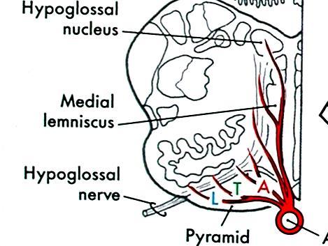
18 @ To nucl. ambiguus: bilateral; but uvula & soft palate are contralateral X: deviation of uvula to cortical lesion side on to hypoglossal nucleus: contralateral X: deviation of tongue to the opposite (weak) side on protrusion (genioglossus) 18
19 The location of corticobulbar and corticopontine fibers in the crus cerebri 19
20 Rubrospinal: to cervical enlargement only Ventral tegmental decussation Rubrospinal tract Termination: spinal laminae V,VI,VII proximal upper limb flexor Inputs: ipsilateral corticorubral contralateral cerebellorubral 20
21 Medial (pontine) reticulospinal (RS) tract: oral & caudal pontine reticular nucl. chiefly ipsi.; vent. funiculus; excite extensor -MN; Excitatory center; inhibition from higher centers Lateral (medullary) RS tract: gigantocellular reticular nucl. bilateral; lat. funiculus inhibit extensor -MN(?); Inhibitory center; excitation from higher centers Characteristics: rather diffuse little somatotopy posture modulate muscle tone 21


22 Vestibulospinal tract Lateral VS tract: lat. vest. nucl. ipsi., ventral funiculus VII,VIII, excite paravertebral & proximal limb extensor -MN antigravity: posture, balance Medial VS tract: med. + inf. vest. nucl.; bilaterally MLF to upper T (on -MN) head & neck movements in response to vestibular input (MLF: ascending part, vestibulo-ocular reflex) 22
23 Vestibular nuclei inputs: 1) cerebellar fastigial nucl. 2) inhibitory: cerebellar Purkinje axons (No direct cerebral cortical lat. vest. nucl. ipsi. extensor contraction & flexor Decerebellate rigidity (extensor rigidity): lesion lat. vest. nucl. rigidity abolished 23
24 Mesencephalic projections to the spinal cord a) Periventricular (periaqueductal) central pain integrate autonomic and somatomotor activities in emotional behavior b) Tectospinal tract: superior colliculus dorsal tegmental decussation cervical cord neck muscles; br. to interneurons in gaze centers conjugate eye movement Orienting reflex: visual, auditory, vestibular, somatosensory inputs eye, head & neck movements 24
25 Propriospinal (Spinospinal) a) short: limb mm. lateral b) long: axial mm. medial 25


26 A: decerebration: X between SC and IC: decerebrate rigidity gamma rigidity C: X cerebellar ant. lobe: decerebellate rigidity alpha rigidity D: decorticate rigidity: decorticate posturing Decerebrate posturing Excitatory inhibitory Decorticate posturing 26
27 Lower motoneuron lesion e.g., poliomyelitis A) Flaccid paralysis B) Loss of all reflexes hyporeflexia, areflexia Syndromes C) Pronounced Partial paralysis = paresis (part of the motor innervations of a muscle is lost) D) Hypotonia: decreased muscle tone E) Fibrillation potential: contraction of isolated denervated muscle fiber fasciculation: spontaneous contraction of motor unit(s) F) Sprouting of nearby intact nerve fibers 27
28 Upper motor neuron syndromes: spastic paralysis initially weak and flaccid, eventually spastic and hypertonia A) No profound atrophy of affected muscle: slow wasting B) Increased muscle tone: spasticity exaggerated stretch reflex (hyperreflexia): pronounced in antigravity mm. Clasp-knife rigidity Clonus : repetitive, alternating contraction of flexors and extensors C) Abnormal plantar reflex: Babinski sign 28
29 Clinical Terms: Pyramidal syndrome: e.g., what happens following capsular loss of cortical outputs as well as ascending characterized by losses of voluntary movements and spasticity Extrapyramidal syndrome: basal ganglia, cerebellum, reticular formation, or vestibular disorders with abnormal spontaneous characterized by involuntary difficulties in movement initiation and control, and changes in muscle tone 29
 Striatum: the receiving part")
: external (lateral, GPe) (GPi & SNpr:")
30 BASAL GANGLION Caudate nucleus Lentiform nucleus: putamen globus pallidus (pallidum) Striatum: the receiving part caudate + putamen: Globus pallidus (GP): internal (medial, GPi): external (lateral, GPe) (GPi & SNpr: output part) 30


31 31
 1) Ventral striatopallidal")
32 Ventral striatum (VS) = nucleus accumbens Ventral pallidum (VP) = substantia innominata Basal Forebrain (substantia innominata) 1) Ventral striatopallidal system 32
 nigrostriatal fibers: SNpc (dopaminergic) dopaminergic cells in ventral tegmental area ventral striatum 5) hippo form; temporal & prefrontal ctx, BL amygdala ventral striatum Inputs SNpc 2 parts")
33 1) motor, somatosensory cortex putamen 2) associational cortex caudate 3) thalamostriatal fiber: intralaminar & midline thalamic nucl. 4) nigrostriatal fibers: SNpc (dopaminergic) dopaminergic cells in ventral tegmental area ventral striatum 5) hippo form; temporal & prefrontal ctx, BL amygdala ventral striatum Inputs SNpc 2 parts in Substantia Nigra: SNpc (pars compacta) SNpr (pars reticulata) (also pars reticularis) 33
 GABA/enkephalin neurons GPe 2) GABA/substance P neurons GPi + SNpr GPe GPi")
34 Striatopallidal projections: Striatal GABAergic cells: the receiving and output neurons 1) GABA/enkephalin neurons GPe 2) GABA/substance P neurons GPi + SNpr GPe GPi SNpr 34
![Pallidofugal projections Direct pathways: from GPi and SNpr 1) GPi VA-VLa [ premotor ctx, SMC, frontal eye field, prefrontal ctx] 2) SNpr sup.](/docs-images/82/86934390/images/35-0.jpg "colliculus (orienting/eye movement) 3) GPi pedunculopontine nucl. (cholinergic neurons) 4)?? habenular nucl. [ limbic sys., autonomic activity] 5) vent.")
35 Pallidofugal projections Direct pathways: from GPi and SNpr 1) GPi VA-VLa [ premotor ctx, SMC, frontal eye field, prefrontal ctx] 2) SNpr sup. colliculus (orienting/eye movement) 3) GPi pedunculopontine nucl. (cholinergic neurons) 4)?? habenular nucl. [ limbic sys., autonomic activity] 5) vent. pallidum MD [ prefrontal ctx, ant. cingulate gyrus 35
 GPe subthalamus GPi,")
36 Pallidofugal- Indirect pathway: (Motor cortex) GPe subthalamus GPi, SNpr 36
37 upper GPi field H2 of Forel (lenticular fasciculus) field H of Forel + ansa lenticularis (from lower GPi) thalamic fasciculus (field H1 of Forel) VA-VLa *VLp receives cbr ff 37

38 Differences in circuitry Between Basal ganglia Cerebellum 38
 thalamic fibers to cortex d) subthalamic output fibers GABAergic: strial efferents pallidal efferents")
39 Transmitter phenotypes of basal ganglion connections Glutamatergic: a) cortical fibers to striatum b) cortical fibers to subthalamus c) thalamic fibers to cortex d) subthalamic output fibers GABAergic: strial efferents pallidal efferents 39
40 Differences in the action of GABA/subs P neurons: (direct pathway) GPi/SNpr D1 receptor: GABA/enkephalin neurons: (indirect pathway) GPe D2 receptor: inhibitory 40


41 Parkinson s Dis. loss of dopaminergic neurons in SNpc 41


42 Disinhibition: thalamic disinhibition subthalamic disinhibition Parkinson s disease Huntington s disease Hemiballismus 42

43 Direct pathway: Facilitates movement 43
44 Pyramidal syndromes: sensory and motor losses, clasp-knife rigidity Extrapyramidal syndromes: 1:Dyskinesia: unwanted superfluous movements 1a) Hypokinetic: Parkinson's dis. loss of dopaminergic neurons in SNpc: akinesia; bradykinesia, tremor at rest, cogwheel rigidity 1b) Hyperkinetic: Huntington's dis. loss of striatal GABAergic cells; hereditary chorea, athetosis, dystonia, dementia, die in years 1c) Hemiballismus: lesion of contralateral subthalamus 2. Tardive (late) dyskinesia: abnormal face & tongue movements + cogwheel rigidity long-term antipsychotic drug treatment: DR3 hypersensitivity Cerebellar lesion syndromes: ataxia, hypotonia, intention tremor (ipsilateral) *dysmetria: such as past pointing 44
 prefrontal cortex, ant.")
45 BASAL FOREBRAIN 1) Ventral striatopallidal system hippocampal formation, basolateral amygdala, temporal & prefrontal asso. cortices & neocortex ventral striatum ventral pallidum MD (thalamus) prefrontal cortex, ant. cingulate gyrus 2) Basal nucleus of Meynert (B) cholinergic neurons; GABAergic neurons diffuse projection: to all cortical areas + thalamus 3) Extended amygdala (BST: Bed nucleus of stria terminalis) : striatal cholinergic interneurons : corticopetal cholinergic neurons 45


 Level: slightly caudal to")
46 Extended amygdala cortical areas similar to ventral striatopallidal system centromedial amygdala stria terminalis Bed nucleus of Stria Terminalis (BST) neuroendocrine, autonomic, somatomotor centers in hypothalamus & brainstem (emotional behavior) Level: slightly caudal to ventral striato-pallidal system 46
47 Central autonomic control Cortical regions: Insula, frontoparietal operculum, Medial prefrontal cortex Septal area (back end of the inner part of the frontal lobe)( pleasure zone in animals; role in reward and reinforcement along with the nucleus accumbens) Hypothalamus: esp. PV & lat. hypothalamic area Extended amygdala Periaqueductal gray 47
 dorsal vagal nucl. (2) autonomic nucl. of spinal cord (Basal forebrain) Locus coeruleus 48")
48 Medial forebrain bundle (1) dorsal vagal nucl. (2) autonomic nucl. of spinal cord (3) LC, Raphe, NTS (4) other reticular nucl. Dorsal longitudinal fasciculus (1) dorsal vagal nucl. (2) autonomic nucl. of spinal cord (Basal forebrain) Locus coeruleus 48
Motor System I Lower motor neurons: typed according to axon diameters
 Motor System I Lower motor neurons: typed according to axon diameters MOTOR UNIT: a motoneuron + supplied m.f. -motoneuron: extrafusal muscle fibers -motoneuron: intrafusal muscle fibers, control the sensitivity
Motor System Hierarchy
 Motor Pathways Lectures Objectives Define the terms upper and lower motor neurons with examples. Describe the corticospinal (pyramidal) tract and the direct motor pathways from the cortex to the trunk
I: To describe the pyramidal and extrapyramidal tracts. II: To discuss the functions of the descending tracts.
 Descending Tracts I: To describe the pyramidal and extrapyramidal tracts. II: To discuss the functions of the descending tracts. III: To define the upper and the lower motor neurons. 1. The corticonuclear
CN V! touch! pain! Touch! P/T!
 CN V! touch! pain! Touch! P/T! Visual Pathways! L! R! B! A! C! D! LT! E! F! RT! G! hypothalamospinal! and! ALS! Vestibular Pathways! 1. Posture/Balance!!falling! 2. Head Position! 3. Eye-Head Movements
 Spinal Cord Tracts DESCENDING SPINAL TRACTS: Are concerned with somatic motor function, modification of ms. tone, visceral innervation, segmental reflexes. Main tracts arise form cerebral cortex and others
Functional Distinctions
 Functional Distinctions FUNCTION COMPONENT DEFICITS Start Basal Ganglia Spontaneous Movements Move UMN/LMN Cerebral Cortex Brainstem, Spinal cord Roots/peripheral nerves Plan Cerebellum Ataxia Adjust Cerebellum
Biological Bases of Behavior. 8: Control of Movement
 Biological Bases of Behavior 8: Control of Movement m d Skeletal Muscle Movements of our body are accomplished by contraction of the skeletal muscles Flexion: contraction of a flexor muscle draws in a
Spinal Interneurons. Control of Movement
 Control of Movement Spinal Interneurons Proprioceptive afferents have a variety of termination patterns in the spinal cord. This can be seen by filling physiologically-identified fibers with HRP, so their
VL VA BASAL GANGLIA. FUNCTIONAl COMPONENTS. Function Component Deficits Start/initiation Basal Ganglia Spontan movements
 BASAL GANGLIA Chris Cohan, Ph.D. Dept. of Pathology/Anat Sci University at Buffalo I) Overview How do Basal Ganglia affect movement Basal ganglia enhance cortical motor activity and facilitate movement.
Motor tracts Both pyramidal tracts and extrapyramidal both starts from cortex: Area 4 Area 6 Area 312 Pyramidal: mainly from area 4 Extrapyramidal:
 Motor tracts Both pyramidal tracts and extrapyramidal both starts from cortex: Area 4 Area 6 Area 312 Pyramidal: mainly from area 4 Extrapyramidal: mainly from area 6 area 6 Premotorarea: uses external
COGNITIVE SCIENCE 107A. Motor Systems: Basal Ganglia. Jaime A. Pineda, Ph.D.
 COGNITIVE SCIENCE 107A Motor Systems: Basal Ganglia Jaime A. Pineda, Ph.D. Two major descending s Pyramidal vs. extrapyramidal Motor cortex Pyramidal system Pathway for voluntary movement Most fibers originate
A. General features of the basal ganglia, one of our 3 major motor control centers:
 Reading: Waxman pp. 141-146 are not very helpful! Computer Resources: HyperBrain, Chapter 12 Dental Neuroanatomy Suzanne S. Stensaas, Ph.D. April 22, 2010 THE BASAL GANGLIA Objectives: 1. What are the
Role of brainstem in somatomotor (postural) functions
 functions") Role of brainstem in somatomotor (postural) functions (vestibular apparatus) The muscle tone and its regulation VESTIBULAR SYSTEM (Equilibrium) Receptors: Otolith organs Semicircular canals Sensation (information):
Role of brainstem in somatomotor (postural) functions (vestibular apparatus) The muscle tone and its regulation VESTIBULAR SYSTEM (Equilibrium) Receptors: Otolith organs Semicircular canals Sensation (information):
Brain Stem and cortical control of motor function. Dr Z Akbari
 Brain Stem and cortical control of motor function Dr Z Akbari Brain stem control of movement BS nuclear groups give rise to descending motor tracts that influence motor neurons and their associated interneurons
Voluntary Movement. Ch. 14: Supplemental Images
 Voluntary Movement Ch. 14: Supplemental Images Skeletal Motor Unit: The basics Upper motor neuron: Neurons that supply input to lower motor neurons. Lower motor neuron: neuron that innervates muscles,
Basal ganglia Sujata Sofat, class of 2009
 Basal ganglia Sujata Sofat, class of 2009 Basal ganglia Objectives Describe the function of the Basal Ganglia in movement Define the BG components and their locations Describe the motor loop of the BG
A. General features of the basal ganglia, one of our 3 major motor control centers:
 Reading: Waxman pp. 141-146 are not very helpful! Computer Resources: HyperBrain, Chapter 12 Dental Neuroanatomy Suzanne S. Stensaas, Ph.D. March 1, 2012 THE BASAL GANGLIA Objectives: 1. What are the main
Basal Ganglia. Today s lecture is about Basal Ganglia and it covers:
 Basal Ganglia Motor system is complex interaction between Lower motor neurons (spinal cord and brainstem circuits) and Upper motor neurons (pyramidal and extrapyramidal tracts) plus two main regulators
Chapter 8. Control of movement
 Chapter 8 Control of movement 1st Type: Skeletal Muscle Skeletal Muscle: Ones that moves us Muscles contract, limb flex Flexion: a movement of a limb that tends to bend its joints, contraction of a flexor
Connections of basal ganglia
 Connections of basal ganglia Introduction The basal ganglia, or basal nuclei, are areas of subcortical grey matter that play a prominent role in modulating movement, as well as cognitive and emotional
Basal nuclei, cerebellum and movement
 Basal nuclei, cerebellum and movement MSTN121 - Neurophysiology Session 9 Department of Myotherapy Basal Nuclei (Ganglia) Basal Nuclei (Ganglia) Role: Predict the effects of various actions, then make
Basal Ganglia George R. Leichnetz, Ph.D.
 Basal Ganglia George R. Leichnetz, Ph.D. OBJECTIVES 1. To understand the brain structures which constitute the basal ganglia, and their interconnections 2. To understand the consequences (clinical manifestations)
Strick Lecture 4 March 29, 2006 Page 1
 Strick Lecture 4 March 29, 2006 Page 1 Basal Ganglia OUTLINE- I. Structures included in the basal ganglia II. III. IV. Skeleton diagram of Basal Ganglia Loops with cortex Similarity with Cerebellar Loops
1. The cerebellum coordinates fine movement through interactions with the following motor-associated areas:
 DENT/OBHS 131 2009 Take-home test 4 Week 6: Take-home test (2/11/09 close 2/18/09) 1. The cerebellum coordinates fine movement through interactions with the following motor-associated areas: Hypothalamus
PETER PAZMANY CATHOLIC UNIVERSITY Consortium members SEMMELWEIS UNIVERSITY, DIALOG CAMPUS PUBLISHER
 PETER PAZMANY CATHOLIC UNIVERSITY SEMMELWEIS UNIVERSITY Development of Complex Curricula for Molecular Bionics and Infobionics Programs within a consortial* framework** Consortium leader PETER PAZMANY
CNS consists of brain and spinal cord PNS consists of nerves
 CNS consists of brain and spinal cord PNS consists of nerves 1 As with sensory input, motor output is organized in central nervous system Peripheral Nervous system divides efferent signals somatotopically
Damage on one side.. (Notes) Just remember: Unilateral damage to basal ganglia causes contralateral symptoms.
 Just remember: Unilateral damage to basal ganglia causes contralateral symptoms.") Lecture 20 - Basal Ganglia Basal Ganglia (Nolte 5 th Ed pp 464) Damage to the basal ganglia produces involuntary movements. Although the basal ganglia do not influence LMN directly (to cause this involuntary
Lecture 20 - Basal Ganglia Basal Ganglia (Nolte 5 th Ed pp 464) Damage to the basal ganglia produces involuntary movements. Although the basal ganglia do not influence LMN directly (to cause this involuntary
skilled pathways: distal somatic muscles (fingers, hands) (brainstem, cortex) are giving excitatory signals to the descending pathway
 (brainstem, cortex) are giving excitatory signals to the descending pathway") L15 - Motor Cortex General - descending pathways: how we control our body - motor = somatic muscles and movement (it is a descending motor output pathway) - two types of movement: goal-driven/voluntary
L15 - Motor Cortex General - descending pathways: how we control our body - motor = somatic muscles and movement (it is a descending motor output pathway) - two types of movement: goal-driven/voluntary
BASAL GANGLIA. Dr JAMILA EL MEDANY
 BASAL GANGLIA Dr JAMILA EL MEDANY OBJECTIVES At the end of the lecture, the student should be able to: Define basal ganglia and enumerate its components. Enumerate parts of Corpus Striatum and their important
The Nervous System: Sensory and Motor Tracts of the Spinal Cord
 15 The Nervous System: Sensory and Motor Tracts of the Spinal Cord PowerPoint Lecture Presentations prepared by Steven Bassett Southeast Community College Lincoln, Nebraska Introduction Millions of sensory
Teach-SHEET Basal Ganglia
 Teach-SHEET Basal Ganglia Purves D, et al. Neuroscience, 5 th Ed., Sinauer Associates, 2012 Common organizational principles Basic Circuits or Loops: Motor loop concerned with learned movements (scaling
Unit VIII Problem 5 Physiology: Cerebellum
 Unit VIII Problem 5 Physiology: Cerebellum - The word cerebellum means: the small brain. Note that the cerebellum is not completely separated into 2 hemispheres (they are not clearly demarcated) the vermis
The Motor Systems. What s the motor system? Plan
 The Motor Systems What s the motor system? Parts of CNS and PNS specialized for control of limb, trunk, and eye movements Also holds us together From simple reflexes (knee jerk) to voluntary movements
Brainstem: Midbrain. 1. Midbrain gross external anatomy 2. Internal structure of the midbrain:
 Brainstem: Midbrain 1. Midbrain gross external anatomy 2. Internal structure of the midbrain: cerebral peduncles tegmentum tectum (guadrigeminal plate) Midbrain Midbrain general features location between
By Dr. Saeed Vohra & Dr. Sanaa Alshaarawy
 By Dr. Saeed Vohra & Dr. Sanaa Alshaarawy 1 By the end of the lecture, students will be able to : Distinguish the internal structure of the components of the brain stem in different levels and the specific
The Wonders of the Basal Ganglia
 Basal Ganglia The Wonders of the Basal Ganglia by Mackenzie Breton and Laura Strong /// https://kin450- neurophysiology.wikispaces.com/basal+ganglia Introduction The basal ganglia are a group of nuclei
Neuroanatomy. Dr. Maha ELBeltagy. Assistant Professor of Anatomy Faculty of Medicine The University of Jordan
 Neuroanatomy Dr. Maha ELBeltagy Assistant Professor of Anatomy Faculty of Medicine The University of Jordan 2018 Prof Yousry 10/15/17 Types of brain fibers THE WHITE MATTER OF THE BRAIN The white matter
Reflexes. Dr. Baizer
 Reflexes Dr. Baizer 1 Learning objectives: reflexes Students will be able to describe: 1. The clinical importance of testing reflexes. 2. The essential components of spinal reflexes. 3.The stretch reflex.
Spinal Cord Organization. January 12, 2011
 Spinal Cord Organization January 12, 2011 Spinal Cord 31 segments terminates at L1-L2 special components - conus medullaris - cauda equina no input from the face Spinal Cord, Roots & Nerves Dorsal root
Movement Disorders. Psychology 372 Physiological Psychology. Background. Myasthenia Gravis. Many Types
 Background Movement Disorders Psychology 372 Physiological Psychology Steven E. Meier, Ph.D. Listen to the audio lecture while viewing these slides Early Studies Found some patients with progressive weakness
MODULE 6: CEREBELLUM AND BASAL GANGLIA
 MODULE 6: CEREBELLUM AND BASAL GANGLIA This module will summarize the important neuroanatomical and key clinical concepts from Chapters 15 and 16 of the textbook for the course. The first part of this
Page 1 L 58. The University of Connecticut Schools of Medicine and Dental Medicine Humans Systems: Organ Systems /2013 RETICULAR FORMATION
 Page 1 L 58 Douglas L. Oliver, Ph.D. The University of Connecticut Schools of Medicine and Dental Medicine Humans Systems: Organ Systems 1 2012/2013 RETICULAR FORMATION Lecture Lecture: Douglas Oliver
Dr. Farah Nabil Abbas. MBChB, MSc, PhD
 Dr. Farah Nabil Abbas MBChB, MSc, PhD The Basal Ganglia *Functions in association with motor cortex and corticospinal pathways. *Regarded as accessory motor system besides cerebellum. *Receive most of
Orientation, Development, Gross Anatomy, Blood Supply and Meninges References... 3
 Section I Orientation, Development, Gross Anatomy, Blood Supply and Meninges... 1 1 Orientation... 3 References... 3 2 Development... 7 Early Morphogenesis... 7 FormationoftheBrainRegions... 9 Histogenesis...
Physiology of motor control (1)
") Physiology of motor control (1) Physiology of somatomotor system 1. Task: It controls the skeletal muscles 2. Content: Simple reflexes Muscle tone Posture Movement Sexual functions Motor component of emotions
Physiology of motor control (1) Physiology of somatomotor system 1. Task: It controls the skeletal muscles 2. Content: Simple reflexes Muscle tone Posture Movement Sexual functions Motor component of emotions
神經解剖學 NEUROANATOMY BASAL NUCLEI 盧家鋒助理教授臺北醫學大學醫學系解剖學暨細胞生物學科臺北醫學大學醫學院轉譯影像研究中心.
 神經解剖學 NEUROANATOMY BASAL NUCLEI 盧家鋒助理教授臺北醫學大學醫學系解剖學暨細胞生物學科臺北醫學大學醫學院轉譯影像研究中心 http://www.ym.edu.tw/~cflu OUTLINE Components and Pathways of the Basal Nuclei Functions and Related Disorders of the Basal Nuclei
Upper and Lower Motoneurons for the Head Objectives
 Upper and Lower Motoneurons for the Head Objectives Know the locations of cranial nerve motor nuclei Describe the effects of motor cranial nerve lesions Describe how the corticobulbar tract innervates
NS201C Anatomy 1: Sensory and Motor Systems
 NS201C Anatomy 1: Sensory and Motor Systems 25th January 2017 Peter Ohara Department of Anatomy peter.ohara@ucsf.edu The Subdivisions and Components of the Central Nervous System Axes and Anatomical Planes
Biology 218 Human Anatomy
 Chapter 21 Adapted form Tortora 10 th ed. LECTURE OUTLINE A. Overview of Sensations (p. 652) 1. Sensation is the conscious or subconscious awareness of external or internal stimuli. 2. For a sensation
Nsci 2100: Human Neuroanatomy 2017 Examination 3
 Name KEY Lab Section Nsci 2100: Human Neuroanatomy 2017 Examination 3 On this page, write your name and lab section. On your bubble answer sheet, enter your name (last name, space, first name), internet
Motor Functions of Cerebral Cortex
 Motor Functions of Cerebral Cortex I: To list the functions of different cortical laminae II: To describe the four motor areas of the cerebral cortex. III: To discuss the functions and dysfunctions of
Organization of Motor Functions 4.
 Organization of Motor Functions 4. Dr. Attila Nagy 2018 Sensory-motor system Limbic cortex Structure Subcortical Motivational sub areas Frontal cortex Task Motivation Sequence Plan Tim e Ascending system
Anatomy of the basal ganglia. Dana Cohen Gonda Brain Research Center, room 410
 Anatomy of the basal ganglia Dana Cohen Gonda Brain Research Center, room 410 danacoh@gmail.com The basal ganglia The nuclei form a small minority of the brain s neuronal population. Little is known about
CNS MCQ 2 nd term. Select the best answer:
 Select the best answer: CNS MCQ 2 nd term 1) Vestibular apparatus: a) Represent the auditory part of the labyrinth. b) May help in initiating the voluntary movements. c) Contains receptors concerned with
HEAD AND NECK PART 2
 HEAD AND NECK PART 2 INTEGRATED CURRICULUM = Integrate Basic Science and Clinical Training 1- ENT PATIENT EXAM IN ICS COURSE - Today and next week - Review/Preview Anatomy underlying ENT exam 2- NEUROANATOMY/NEUROLOGY
Motor systems.... the only thing mankind can do is to move things... whether whispering or felling a forest. C. Sherrington
 Motor systems... the only thing mankind can do is to move things... whether whispering or felling a forest. C. Sherrington 1 Descending pathways: CS corticospinal; TS tectospinal; RS reticulospinal; VS
The basal forebrain: Questions, chapter 29:
 The basal forebrain: Questions, chapter 29: 7) What is the "basal forebrain", and what is its involvement in Alzheimer' s Disease? The acetylcholine-containing neurons of the nucleus basalis of Meynart
The motor regulator. 1) Basal ganglia/nucleus
 Basal ganglia/nucleus") The motor regulator 1) Basal ganglia/nucleus Neural structures involved in the control of movement Basal Ganglia - Components of the basal ganglia - Function of the basal ganglia - Connection and circuits
The motor regulator 1) Basal ganglia/nucleus Neural structures involved in the control of movement Basal Ganglia - Components of the basal ganglia - Function of the basal ganglia - Connection and circuits
PHYSIOLOHY OF BRAIN STEM
 PHYSIOLOHY OF BRAIN STEM Learning Objectives The brain stem is the lower part of the brain. It is adjoining and structurally continuous with the spinal cord. 1 Mid Brain 2 Pons 3 Medulla Oblongata The
Internal Organisation of the Brainstem
 Internal Organisation of the Brainstem Major tracts and nuclei of the brainstem (Notes) The brainstem is the major pathway for tracts and houses major nuclei, that contain sensory, motor and autonomics
Brainstem. Steven McLoon Department of Neuroscience University of Minnesota
 Brainstem Steven McLoon Department of Neuroscience University of Minnesota 1 Course News Change in Lab Sequence Week of Oct 2 Lab 5 Week of Oct 9 Lab 4 2 Goal Today Know the regions of the brainstem. Know
Reticular Formation George R. Leichnetz, Ph.D.
 Reticular Formation George R. Leichnetz, Ph.D. OBJECTIVES 1. To understand the anatomical and functional organization of the brainstem reticular formation into three general regions: median (raphe), medial
Making Things Happen 2: Motor Disorders
 Making Things Happen 2: Motor Disorders How Your Brain Works Prof. Jan Schnupp wschnupp@cityu.edu.hk HowYourBrainWorks.net On the Menu in This Lecture In the previous lecture we saw how motor cortex and
b. The groove between the two crests is called 2. The neural folds move toward each other & the fuse to create a
 Chapter 13: Brain and Cranial Nerves I. Development of the CNS A. The CNS begins as a flat plate called the B. The process proceeds as: 1. The lateral sides of the become elevated as waves called a. The
Basal Ganglia. Introduction. Basal Ganglia at a Glance. Role of the BG
 Basal Ganglia Shepherd (2004) Chapter 9 Charles J. Wilson Instructor: Yoonsuck Choe; CPSC 644 Cortical Networks Introduction A set of nuclei in the forebrain and midbrain area in mammals, birds, and reptiles.
Department of Neurology/Division of Anatomical Sciences
 Spinal Cord I Lecture Outline and Objectives CNS/Head and Neck Sequence TOPIC: FACULTY: THE SPINAL CORD AND SPINAL NERVES, Part I Department of Neurology/Division of Anatomical Sciences LECTURE: Monday,
Basal Nuclei (Ganglia)
") Doctor said he will not go deep within these slides because we will take them in physiology, so he will explain the anatomical structures, and he will go faster in the functions sheet in yellow Basal Nuclei
Doctor said he will not go deep within these slides because we will take them in physiology, so he will explain the anatomical structures, and he will go faster in the functions sheet in yellow Basal Nuclei
Cortical Control of Movement
 Strick Lecture 2 March 24, 2006 Page 1 Cortical Control of Movement Four parts of this lecture: I) Anatomical Framework, II) Physiological Framework, III) Primary Motor Cortex Function and IV) Premotor
Stretch reflex and Golgi Tendon Reflex. Prof. Faten zakareia Physiology Department, College of Medicine, King Saud University 2016
 Stretch reflex and Golgi Tendon Reflex Prof. Faten zakareia Physiology Department, College of Medicine, King Saud University 2016 Objectives: Upon completion of this lecture, students should be able to
DISORDERS OF THE MOTOR SYSTEM. Jeanette J. Norden, Ph.D. Professor Emerita Vanderbilt University School of Medicine
 DISORDERS OF THE MOTOR SYSTEM Jeanette J. Norden, Ph.D. Professor Emerita Vanderbilt University School of Medicine THE MOTOR SYSTEM To understand disorders of the motor system, we need to review how a
Unit VIII Physiology Review (Problems: 1-6)
") Unit VIII Physiology Review (Problems: 1-6) PROBLEM 1 - What is the difference between receptor potential and generator potential? Receptor potentials: sensory fibers cannot detect the stimuli but must
Unit VIII Physiology Review (Problems: 1-6) PROBLEM 1 - What is the difference between receptor potential and generator potential? Receptor potentials: sensory fibers cannot detect the stimuli but must
2401 : Anatomy/Physiology
 Dr. Chris Doumen Week 7 2401 : Anatomy/Physiology The Cerebrum Central Nervous System TextBook Readings Pages 434-456 and 460-461 Make use of the figures in your textbook ; a picture is worth a thousand
Chapter 3. Structure and Function of the Nervous System. Copyright (c) Allyn and Bacon 2004
 Allyn and Bacon 2004") Chapter 3 Structure and Function of the Nervous System 1 Basic Features of the Nervous System Neuraxis: An imaginary line drawn through the center of the length of the central nervous system, from the
Chapter 3 Structure and Function of the Nervous System 1 Basic Features of the Nervous System Neuraxis: An imaginary line drawn through the center of the length of the central nervous system, from the
Developmental sequence of brain
 Cerebellum Developmental sequence of brain Fourth week Fifth week Location of cerebellum Lies above and behind the medullar and pons and occupies posterior cranial fossa Location of cerebellum External
Gross Morphology of the Brain
 Gross Morphology of the Brain Done by : Marah Marahleh & Razan Krishan *slides in bold Principal Parts of the Brain Cerebrum : largest part of the brain Diencephalon Thalamus & hypothalamus Cerebellum
Lecture XIII. Brain Diseases I - Parkinsonism! Brain Diseases I!
 Lecture XIII. Brain Diseases I - Parkinsonism! Bio 3411! Wednesday!! Lecture XIII. Brain Diseases - I.! 1! Brain Diseases I! NEUROSCIENCE 5 th ed! Page!!Figure!!Feature! 408 18.9 A!!Substantia Nigra in
Oculomotor System George R. Leichnetz, Ph.D.
 Oculomotor System George R. Leichnetz, Ph.D. OBJECTIVES After studying the material of this lecture, the student should be able to: 1. Define different types of eye movement and their underlying neural
Brain anatomy and artificial intelligence. L. Andrew Coward Australian National University, Canberra, ACT 0200, Australia
 Brain anatomy and artificial intelligence L. Andrew Coward Australian National University, Canberra, ACT 0200, Australia The Fourth Conference on Artificial General Intelligence August 2011 Architectures
Auditory and Vestibular Systems
 Auditory and Vestibular Systems Objective To learn the functional organization of the auditory and vestibular systems To understand how one can use changes in auditory function following injury to localize
The Spinal Cord. The Nervous System. The Spinal Cord. The Spinal Cord 1/2/2016. Continuation of CNS inferior to foramen magnum.
 The Nervous System Spinal Cord Continuation of CNS inferior to foramen magnum Simpler than the brain Conducts impulses to and from brain Two way conduction pathway Reflex actions Passes through vertebral
A3.1.7 Motor Control. 10 November 2016 Institute of Psychiatry,Psychology and Neuroscience Marinela Vavla
 A3.1.7 Motor Control 10 November 2016 Institute of Psychiatry,Psychology and Neuroscience Marinela Vavla marinela.vavla@kcl.ac.uk Learning objectives Motor systems: components & organization Spinal cord
Anatomy and Physiology (Bio 220) The Brain Chapter 14 and select portions of Chapter 16
 The Brain Chapter 14 and select portions of Chapter 16") Anatomy and Physiology (Bio 220) The Brain Chapter 14 and select portions of Chapter 16 I. Introduction A. Appearance 1. physical 2. weight 3. relative weight B. Major parts of the brain 1. cerebrum 2.
Anatomy and Physiology (Bio 220) The Brain Chapter 14 and select portions of Chapter 16 I. Introduction A. Appearance 1. physical 2. weight 3. relative weight B. Major parts of the brain 1. cerebrum 2.
Biological Psych Frontal Lobes
 Biological Psych Frontal Lobes Frontal lobe What is it? Home to personality? Lesions: wide variety symptoms More than any part of brain Involved in: motor function problem solving spontaneity memory language
Systems Neuroscience Dan Kiper. Today: Wolfger von der Behrens
 Systems Neuroscience Dan Kiper Today: Wolfger von der Behrens wolfger@ini.ethz.ch 18.9.2018 Neurons Pyramidal neuron by Santiago Ramón y Cajal (1852-1934, Nobel prize with Camillo Golgi in 1906) Neurons
Arterial Blood Supply
 Arterial Blood Supply Brain is supplied by pairs of internal carotid artery and vertebral artery. The four arteries lie within the subarachnoid space Their branches anastomose on the inferior surface of
Located below tentorium cerebelli within posterior cranial fossa. Formed of 2 hemispheres connected by the vermis in midline.
 The Cerebellum Cerebellum Located below tentorium cerebelli within posterior cranial fossa. Formed of 2 hemispheres connected by the vermis in midline. Gray matter is external. White matter is internal,
Sheet lab 3. Page 8B Section1 of medulla at pyramidal {motor} decussation:
 Sheet lab 3 Page 8B Section1 of medulla at pyramidal {motor} decussation: This section is at lower third of medulla and is the most close part to spinal cord and it has some characteristic of spinal cord
Stanley Pruisinger 1980's
 Neuroanatomy Prion disease cerebellum chapter b/c cerebellar ataxia here as a warning for obvious reasons. Creutzfeldt - Jakob Disease (CJD) "Spongiform" (brain turns to sponge) Jews in Lybia who ate
Spinal cord. We have extension of the pia mater below L1-L2 called filum terminale
 Spinal cord Part of the CNS extend from foramen magnum to the level of L1-L2 (it is shorter than the vertebral column) it is covered by spinal meninges. It is cylindrical in shape. It s lower end become
THE BACK. Dr. Ali Mohsin. Spinal Cord
 Spinal Cord THE BACK Dr. Ali Mohsin The spinal cord is the elongated caudal part of the CNS. It starts as the inferior continuation of the medulla oblongata at the level of foramen magnum, & ends as an
Lecturer. Prof. Dr. Ali K. Al-Shalchy MBChB/ FIBMS/ MRCS/ FRCS 2014
 Lecturer Prof. Dr. Ali K. Al-Shalchy MBChB/ FIBMS/ MRCS/ FRCS 2014 Dorsal root: The dorsal root carries both myelinated and unmyelinated afferent fibers to the spinal cord. Posterior gray column: Long
KINE 4500 Neural Control of Movement. Lecture #1:Introduction to the Neural Control of Movement. Neural control of movement
 KINE 4500 Neural Control of Movement Lecture #1:Introduction to the Neural Control of Movement Neural control of movement Kinesiology: study of movement Here we re looking at the control system, and what
MOVEMENT OUTLINE. The Control of Movement: Muscles! Motor Reflexes Brain Mechanisms of Movement Mirror Neurons Disorders of Movement
 MOVEMENT 2 Dr. Steinmetz 3 OUTLINE The Control of Movement: Muscles! Motor Reflexes Brain Mechanisms of Movement Mirror Neurons Disorders of Movement Parkinson s Disease Huntington s Disease 1 4 TYPES
THE CENTRAL NERVOUS SYSTE M
 THE CENTRAL NERVOUS SYSTE M Structure and Functio n THIRD EDITIO N PER BRODAL A Brief Survey, x i Studying the Structures and Function of the Nervous System, xii i Animal Experiments Crucial for Progress,
Lecture 4 The BRAINSTEM Medulla Oblongata
 Lecture 4 The BRAINSTEM Medulla Oblongata Introduction to brainstem 1- Medulla oblongata 2- Pons 3- Midbrain - - - occupies the posterior cranial fossa of the skull. connects the narrow spinal cord
Biological Bases of Behavior. 3: Structure of the Nervous System
 Biological Bases of Behavior 3: Structure of the Nervous System Neuroanatomy Terms The neuraxis is an imaginary line drawn through the spinal cord up to the front of the brain Anatomical directions are
SENSORY (ASCENDING) SPINAL TRACTS
 SPINAL TRACTS") SENSORY (ASCENDING) SPINAL TRACTS Dr. Jamila El-Medany Dr. Essam Eldin Salama OBJECTIVES By the end of the lecture, the student will be able to: Define the meaning of a tract. Distinguish between the different
SENSORY (ASCENDING) SPINAL TRACTS Dr. Jamila El-Medany Dr. Essam Eldin Salama OBJECTIVES By the end of the lecture, the student will be able to: Define the meaning of a tract. Distinguish between the different
Motor control. Proprioception and movement
 Motor control Proprioception and movement 2/24 in general we are not aware of information coming from proprioceptors though they belong to somatosensory receptors these receptors detect stretching of the
DR. JITENDRA PATEL (MBBS, MD) Medical Educator & Researcher
 Medical Educator & Researcher") 1 DR. JITENDRA PATEL (MBBS, MD) Medical Educator & Researcher Associate Professor in Physiology Email: dr.jrpatel84@gmail.com Web: www.esphys.weebly.com 2 OUTLINE Stretch reflex overview Muscle spindle
1 DR. JITENDRA PATEL (MBBS, MD) Medical Educator & Researcher Associate Professor in Physiology Email: dr.jrpatel84@gmail.com Web: www.esphys.weebly.com 2 OUTLINE Stretch reflex overview Muscle spindle
Prof. Saeed Abuel Makarem & Dr.Sanaa Alshaarawy
 Prof. Saeed Abuel Makarem & Dr.Sanaa Alshaarawy 1 Objectives By the end of the lecture, you should be able to: Describe the anatomy and main functions of the thalamus. Name and identify different nuclei