The Lymphatic System
|
|
|
- Delphia Preston
- 5 years ago
- Views:
Transcription

1 94 The Lymphatic System Lymph Lymph is the name for tissue fluid that enters lymph capillaries. Filtration in capillaries creates tissue fluid, most of which returns almost immediately to the blood in the capillaries by osmosis. Some tissue fluid, however, remains in interstitial spaces and must be returned to the blood by way of the lymphatic vessels. Without this return, blood volume and blood pressure would very soon decrease. Lymph vessels The system of lymph vessels begins as dead-end lymph capillaries found in most tissue spaces. Lymph capillaries are very permeable and collect tissue fluid and proteins. Lymphatic capillaries are interlaced with the arterioles and venules of the cardiovascular system. Collagen fibers anchor a lymphatic capillary in the tissue. Interstitial fluid slips through spaces between the overlapping endothelial cells that compose the lymphatic capillary. The overlapping ends are termed 'flaps'. Lymph vessels are located in almost every tissue in the body except the central nervous system, bone marrow, bones, teeth, and the cornea of the eye, which do not contain lymph vessels. Lacteals are specialized lymph capillaries in the villi of the small intestine; they absorb the fat-soluble end products of digestion, such as fatty acids and vitamin A.
, but the lymph is kept moving within lymph vessels by the same mechanisms that promote venous return.")
2 05 Lymph capillaries unite to form larger lymph vessels, whose structure is very much like that of veins. There is no pump for lymph (as the heart is the pump for blood), but the lymph is kept moving within lymph vessels by the same mechanisms that promote venous return. The smooth muscle layer of the larger lymph vessels constricts, and the one-way valves (just like those of veins) prevent backflow of lymph. Lymph vessels in the extremities are compressed by the skeletal muscles that surround them; this is the skeletal muscle pump. The respiratory pump (inflation of lungs) alternately expands and compresses the lymph vessels in the chest cavity and keeps the lymph moving.

3 05 Where is the lymph going? Back to the blood to become plasma again. The lymph vessels from the lower body unite in front of the lumbar vertebrae to form a vessel called cisterna chyli, which continues upward in front of the backbone as the thoracic duct. Lymph vessels from the upper left quadrant of the body join the thoracic duct, which empties lymph into the left subclavian vein. Lymph vessels from the upper right quadrant of the body unite to form the right lymphatic duct, which empties lymph into the right subclavian vein. Flaps in both subclavian veins permit the entry of lymph but prevent blood from flowing into the lymph vessels. Lymphoid organs The lymphoid organs are where lymphocytes mature, proliferate, and are selected, which enables them to attack pathogens without harming the cells of the body. Primary lymphatic organs are where lymphocytes are formed and mature. They provide an environment for stem cells to divide and mature into B- and T- cells. There are two primary lymphatic organs: the red bone marrow
4 05 and the thymus gland. Both T-cell and B-cells are 'born' in the bone marrow. However, whereas B cells also mature in the bone marrow, T-cells have to migrate to the thymus, which is where they mature in the thymus. Secondary lymphoid tissues are arranged as a series of filters monitoring the contents of the extracellular fluids, i.e. lymph, tissue fluid and blood. The lymphoid tissue filtering each of these fluids is arranged in different ways. Secondary lymphoid tissues are also where lymphocytes are activated. Therefore, lymphocytes develop and mature in the primary lymphoid organs, but they mount immune responses from the secondary lymphoid organs. A naïve lymphocyte is one that has left the primary organ and entered a secondary lymphoid organ. Naïve lymphocytes are fully functional immunologically, but have yet to encounter an antigen to respond to. In addition to circulating in the blood and lymph, lymphocytes concentrate in secondary lymphoid organs. Secondary lymphoid organs include: spleen, lymph nodes, lymph nodules, Peyer's patches and mucosa associated lymphoid tissue (MALT). All of these tissues have many features in common, including the following: The presence of lymphoid follicles, the sites of the formation of lymphocytes, with specific B cell-rich and T cell-rich areas An internal structure of reticular fibers with associated fixed macrophages Germinal centers, which are the sites of rapidly dividing B lymphocytes and plasma cells Specialized post-capillary vessels known as high endothelial venules; the cells lining these venules are thicker and more columnar than normal endothelial cells, which allow cells from the blood to directly enter these tissues Thymus The thymus is a bilobed organ found in the space between the sternum and the aorta of the heart, located inferior to the thyroid gland. In the fetus and infant, the thymus is large and extends under the sternum. With increasing age, the thymus shrinks, and relatively little thymus tissue is found in adults (involuted thymus).

5 05 Connective tissue holds the lobes closely together but also separates them and forms a capsule. The connective tissue capsule further divides the thymus into lobules via extensions called trabeculae. The outer region of the organ is known as the cortex and contains large numbers of thymocytes (immature T cells), cortical epithelial cells and some macrophages and dendritic cells. The cortex is densely packed so it stains more intensely than the rest of the thymus. The medulla, where thymocytes migrate before leaving the thymus as naïve T cells, contains a less dense collection of thymocytes, medullary epithelial cells and dendritic cells, in addition to thymic (Hassall's) corpuscles. Hassall's corpuscles are degenerated epithelial cells that do not produce any thymic hormones. They contain some T cells and macrophages.


6 09 Thymocytes proliferate and mature in the thymus but only 1-3% survive the selection process that allows mature T cells to enter the circulation. In the thymus, APCs scan for T cells that may self-react; these cells are killed so as to
.")

7 00 prevent autoimmunity (negative selection). This 'education' of T cells must occur in a very controlled environment. To ensure that no foreign antigens, there is a very tight blood-thymus barrier (note that thymocytes can only enter the thymus via bloodstream; there are no afferent lymph vessels). Thymic hormones (thymosin and thymopoietin) are necessary for what may be called "immunological competence". To be competent means to be able to do something well. Thymic hormones enable the T cells to participate in the recognition of foreign antigens and to provide immunity. This capability of T cells is established early in life and then is perpetuated by the lymphocytes themselves. The newborn's immune system is not yet fully mature, and infants are more susceptible to certain infections than are older children and adults. Usually by the age of 2 years, the immune system matures and becomes fully
8 05 functional. This is why some vaccines, such as measles vaccine, are not recommended for infants younger than 15 to 18 months of age. Their immune systems are not mature enough to respond strongly to the vaccine, and the protection provided by the vaccine may be incomplete. Spleen The spleen is located in the upper left quadrant of the abdominal cavity, just below the diaphragm, behind the stomach. The lower rib cage protects the spleen from physical trauma. It is a major secondary lymphoid organ. It is about 12 cm (5 in) long and is attached to the lateral border of the stomach via the gastrosplenic ligament. The spleen is a fragile organ although it has a connective tissue capsule, and is dark red due to its extensive vascularization. The spleen is sometimes called the filter of the blood because of its extensive vascularization and the presence of macrophages and dendritic cells that remove microbes and other materials from the blood, including dying red blood cells. The functions of the spleen are centered on the systemic circulation. As such, it lacks afferent lymphatic vessels. It is comprised of 2 functionally and morphologically distinct compartments, the red pulp and the white pulp. The red pulp is a blood filter that removes foreign material and damaged erythrocytes. It is also a storage site for iron, erythrocytes, and platelets. The spleen is also the largest secondary lymphoid organ containing about one-fourth of the body s lymphocytes and initiates immune responses to blood-borne antigens. This function is charged to the white pulp which surrounds the central arterioles. In the fetus, the spleen produces red blood cells, a function assumed by the red bone marrow after birth. The functions of the spleen after birth are: 1. Produces lymphoid cells in response to antigens. The newly-formed lymphocytes then enter the blood stream. 2. Contains some fixed plasma cells that produce antibodies to foreign antigens. 3. Contains fixed macrophages (RE cells) that phagocytize pathogens or other foreign materials in the blood. The macrophages of the spleen also

9 05 phagocytize old and damaged red blood cells and form bilirubin. By way of portal circulation, the bilirubin is sent to the liver for excretion in bile. 4. Acts as a reservoir of blood in times of shock or hemorrhage. The spleen is not considered a vital organ, because other organs compensate for its functions if the spleen must be removed. The liver and red bone marrow will remove old red blood cells from circulation, and the many lymph nodes and nodules will produce lymphocytes and monocytes and phagocytize pathogens (as will the liver). Despite this redundancy, a person without a spleen is somewhat more susceptible to certain bacterial infections such as pneumonia and meningitis. The spleen is surrounded by a capsule composed of dense fibrous tissue, elastic fibers, and smooth muscle. The spleen is also divided by extensions of the capsule called trabeculae, and within each splenic nodule is an area of red pulp, consisting of mostly red blood cells, and white pulp, which is composed of three sub-compartments: the periarteriolar lymphoid sheath (PALS), the follicles, and the marginal zone.
, then into several arterioles and eventually into sinusoids.")

10 05 Upon entering the spleen through the hilum, the splenic artery splits into trabecular arteries. Trabecular arteries divide into central arteries (surrounded by white pulp), then into several arterioles and eventually into sinusoids. Blood from the capillaries subsequently collects in the venous sinuses and leaves via the splenic vein. The red pulp has reticular fibers with fixed macrophages attached, free macrophages, and all of the other cells typical of the blood, including some lymphocytes. The red pulp is composed of a three dimensional meshwork of splenic cords and venous sinuses.
.")
11 04 The white pulp surrounds a central arteriole. It is subdivided into the PALS, the follicles, and the marginal zone. The PALS have the structure of diffuse lymphatic tissue (lymphocytes and concentric layers of reticular fibers and flattened reticular cells). The follicles are continuous with the PALS and are typically found at bifurcation sites of the central arterioles. They are composed primarily of B-cells. Follicles may contain germinal centers (dividing B cells surrounded by T cells and accessory cells, including macrophages and dendritic cells), which form upon antigenic stimulation, and stain less intensely. The marginal zone is a unique region of the spleen situated at the interface of the red pulp with the PALS and follicles. Considered by many to be a separate compartment rather than part of the white pulp, it is designed to screen the systemic circulation for antigens and pathogens and plays an important role in antigen processing.

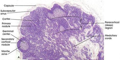
12 55 Lymph nodes and nodules Lymph nodes and nodules are masses of lymphatic tissue. Nodes and nodules differ with respect to size and location. Nodes are usually larger, 10 to 20mm in length; nodules range from a fraction of a millimeter to several millimeters in length. Lymph nodes are found in groups along the pathways of lymph vessels, and lymph flows through these nodes on its way to the subclavian veins. Lymph enters the nodes through several afferent lymph vessels and leaves through one or two efferent vessels. As lymph passes through a lymph node, bacteria and other foreign materials are phagocytized by fixed (stationary) macrophages. Fixed plasma cells (from lymphocytes) produce antibodies to any pathogens in the lymph; these antibodies, as well as lymphocytes and monocytes, will eventually reach the blood. There are many groups of lymph nodes along all the lymph vessels throughout the body, but three paired groups deserve mention because of their strategic locations. These are the cervical, axillary, and inguinal lymph nodes. Notice that these are at the junctions of the head and extremities with the trunk of the body. Breaks in the skin, with entry of pathogens, are much more likely to occur in the arms or legs or head rather than in the trunk. If these pathogens get to the lymph, they will be destroyed by the lymph nodes before they get to the trunk, before the lymph is returned to the blood in the subclavian veins.
, submandibular, sublingual and submental nodes.")
13 55 Cervical Lymph Nodes In the head and neck, lymph nodes are arranged in two horizontal rings and two vertical chains on either side of the neck. The outer, superficial ring consists of the occipital, preauricular (parotid), submandibular, sublingual and submental nodes. The inner, deep ring is formed by clumps of mucosa associated lymphoid tissue (MALT) located primarily in the naso- and oropharynx (Waldeyer s ring). The following describes the main cervical node groups: The occipital nodes are in the superficial group, which includes 3-5 nodes. This group of nodes is localized between the sternocleidomastoid (SCM) and trapezius muscles, at the apex of the posterior triangle. These nodes are superficial to the splenius capitis. The deep posterior cervical group includes 1-3 nodes. This group of nodes is located deep to the splenius capitis and follows the course of the occipital artery. These nodes drain the scalp, the posterior portion of the neck, and the deep muscular layers of the neck. The postauricular nodes vary in number from 2 to 4; they are located in the fibrous portion of the superior attachment of the SCM muscle to the mastoid process. Postauricular nodes drain the posterior parietal scalp and the skin of the mastoid region.
14 55 The preauricular (parotid) nodes can be divided into intraglandular and extraglandular groups. The extraglandular parotid nodes are located outside but adjacent to the parotid gland, where they drain the frontolateral scalp and face, the anterior aspects of the auricle, the external auditory canal, and the buccal mucosa. Embryologically, the lymphatic system develops before the parotid gland, which surrounds the intraglandular nodes as it develops. This explains why the parotid gland contains lymphoid tissue. The intraglandular nodes drain the same regions as the extraglandular nodes, to which they interconnect and then drain into the upper jugular group of lymph nodes. As many as 20 parotid nodes may be found. The submandibular nodes are divided into 5 groups: preglandular, postglandular, prevascular, postvascular, and intracapsular. The preglandular and prevascular groups are located anterior to the submandibular gland and facial artery, respectively. The postglandular and postvascular groups are posterior to these structures. Differing from the parotid gland in embryological development, there is no true intraglandular node; however, occasionally, a node has been identified inside the capsule of the gland. The submandibular nodes drain the ipsilateral upper and lower lip, cheek, nose, nasal mucosa, medial canthus, anterior gingiva, anterior tonsillar pillar, soft palate, anterior two thirds of the tongue, and submandibular salivary gland. The efferent vessels drain into the internal jugular nodes. For the submental nodes, 2-8 nodes are located in the soft tissues of the submental triangle between the platysma and mylohyoid muscles. These nodes drain the chin, the middle portion of the lower lip, the anterior gingiva, and the anterior third of the tongue. The efferent vessels drain into both the ipsilateral and contralateral submandibular nodes or into the internal jugular group. The sublingual nodes are located along the collecting trunk of the tongue and sublingual gland and drain the anterior floor of the mouth and ventral surface of the tongue. These nodes subsequently drain into the submandibular or jugular group of nodes. The retropharyngeal nodes are divided into a medial and lateral group, located between the pharynx and the prevertebral fascia. The lateral group, located at the level of the atlas near the internal carotid artery, consists of 1-3 nodes, which may extend to the skull base. The medial group extends inferiorly to the postcricoid level. This group drains the posterior region of the nasal cavity, sphenoid and ethmoid sinuses, hard and soft palates, nasopharynx, and posterior
15 55 pharynx down to the postcricoid area. Management of these nodes must be considered if any malignancy arises from the mentioned drainage areas. The anterior cervical nodes are divided into the anterior jugular chain and the juxtavisceral chain of nodes. The anterior jugular chain nodes follow the anterior jugular vein, located superficial to the strap muscles. These nodes drain the skin and muscles of the anterior portion of the neck, and the efferent vessels empty into the lower internal jugular nodes. The pretracheal group consists of nodes between the isthmus of the thyroid gland down to the level of the innominate vein. Varying from 2-12 in number, these nodes drain the region of the thyroid gland and the trachea and receive afferent flow from the prelaryngeal group. The pretracheal efferents empty in the internal jugular group and the anterior superior mediastinal nodes. The paratracheal nodes lie near the recurrent laryngeal nerve and drain the thyroid lobes, parathyroid glands, subglottic larynx, trachea, and upper esophagus. The efferent vessels travel to the lower jugular group or directly toward the junction of the internal jugular vein and the subclavian vein. The anterior nodes drain bilaterally because the midline of the neck has no division. Treatment must be planned accordingly when a tumor is located in subjacent draining areas. The lateral cervical nodes are divided into superficial and deep groups. The superficial group follows the external jugular vein and drains into either the internal jugular or transverse cervical nodes of the deep group. The deep group forms a triangle bordered by the internal jugular nodes, the spinal accessory nodes, and the transverse cervical nodes. The transverse cervical nodes, forming the base of the triangle, follow the transverse cervical vessels and may contain as many as 12 nodes. These nodes receive drainage from the spinal accessory group and from collecting trunks of the skin of the neck and upper chest. The spinal accessory chain follows the nerve of the same name and may account for as many as 20 nodes. This chain receives lymph from the occipital, postauricular, and suprascapular nodes and from the posterior aspect of the scalp, nape of the neck, lateral aspect of the neck, and the shoulder. The internal jugular chain consists of a large system covering the anterior and lateral aspects of the internal jugular vein, extending broadly from the digastric muscle superiorly to the subclavian vein inferiorly. As many as 30 of these nodes may exist, and they have been arbitrarily divided into upper, middle, and lower groups. The efferents of these nodes eventually pass into the venous
 and lingual tonsil constitute the major part of Waldeyer's ring, with the tubal tonsils and lateral pharyngeal bands as less prominent components.")
16 59 system via the thoracic duct on the left and multiple lymphatic channels on the right. These nodes drain all the other groups mentioned. The palatine tonsils, nasopharyngeal tonsil (adenoid) and lingual tonsil constitute the major part of Waldeyer's ring, with the tubal tonsils and lateral pharyngeal bands as less prominent components. The lymphoid tissue of Waldeyer's ring is located at the gateway of the respiratory and alimentary tract and belongs to the mucosa-associated lymphoid tissue (MALT). As tonsils (details discussed below) are the first site of encounter with inhaled and ingested micro-organisms, they are considered the first line of defense against exogenous aggressors. The generation of B cells in the germinal centers of the tonsil is one of the most essential tonsillar functions.

17 50 Axillary Lymph Nodes The lymph nodes of the axillary region are responsible for the lymphatic drainage of a large section of human anatomy. Due to this arrangement and duty, they have a particular clinical relevance. This is particularly evident with breast cancer, where axillary lymph node status, with regards to cancer, defines the treatment algorithm and approach. There are lymph nodes divided into five groups; Anterior (pectoral), posterior (subscapular), lateral (humeral), central, and apical. The anterior (pectoral) group is located across the inferior border of the pectoralis minor muscle and the superior border of the pectoralis major muscle. There are usually 4-5 large nodes. The lymph flows from the anterolateral aspect of the abdominal wall superior to the level of the umbilicus and the lateral quadrants of the breast. It conveys the lymph to more central nodes. The posterior (subscapular) group consists of 6-7 nodes that can be found anterior to the subscapularis muscle and receives superficial lymph vessels
18 55 located more commonly within the upper portion of the back and posterior neck. However, these can receive lymph from as far inferior as the superior border of the iliac crests. The lateral (humeral) group is a group of 4-6 nodes that can be found against the axillary vein. The vast majority of the lymph vessels of the upper limb flow into this group. The superficial group of nodes however, drains the lateral aspect of the upper limb and flows into the infraclavicular nodes. The central group consists of 3-4 nodes, and is found at the base and centrally located in the axilla. These nodes are interspread amongst the adipose (fat) of the region. These are the most important group of nodes in terms of drainage because these receive lymph flow from the three groups of nodes mentioned above (anterior, posterior, and lateral). The apical group (4-5 nodes) lies at the apex of the axilla and is located at the lateral border of the first rib. It is also referred to as the subclavicular group. This group receives efferent lymph vessels from the other axillary group of nodes. The apical group of nodes then drains into the subclavian lymph trunk. The drainage is different on the left and right sides. The left side axillary drainage flows into the thoracic duct, whereas on the right side the drainage is into the right lymphatic trunk.

19 55 Inguinal Lymph Nodes The inguinal nodes are found in the upper aspect of the femoral triangle and are around 20 in number. They are subdivided into 2 groupings determined by their position relative to a horizontal line drawn at the level of termination of the great saphenous vein. Those below this line are the sub-inguinal nodes (consisting of a deep and superficial set) and those above are the superficial inguinal nodes. The superficial inguinal nodes form a line directly below the inguinal ligament and receive lymph from the penis, scrotum, perineum, buttock and abdominal wall. The superficial sub-inguinal nodes are located on each side of the proximal section of the great saphenous vein. They receive afferent input primarily from the superficial lymphatic vessels of the lower leg. The deep sub-inguinal nodes are often found in one to three in number and are most commonly found on the medial aspect of the femoral vein. The afferent supply to these nodes is from the deep lymphatic trunks of the thigh which accompany the femoral vessels.

20 55 Lymph nodules are small masses of lymphatic tissue found just beneath the epithelium of all mucous membranes. The body systems lined with mucous membranes are those that have openings to the environment: the respiratory, digestive, urinary, and reproductive tracts. You can probably see that these are also strategic locations for lymph nodules, because any natural body opening is a possible portal of entry for pathogens. For example, if bacteria in inhaled air get through the epithelium of the trachea, lymph nodules with their macrophages are in position to destroy these bacteria before they get to the blood. Some of the lymph nodules have specific names. Those of the small intestine are called Peyer's patches, and those of the pharynx are called tonsils. The palatine tonsils are on the lateral walls of the pharynx, the adenoid (pharyngeal tonsil) is on the posterior wall, and the lingual tonsils are those on the base of the tongue. The tonsils, therefore, form a ring of lymphatic tissue around the

21 54 pharynx, which is a common pathway for food and air and for the pathogens they contain. Histology of lymph nodes The nodes are covered by a capsule of dense connective tissue, and have capsular extensions, of connective tissue, called the trabeculae, which provide support for blood vessels entering into the nodes. Lymph, containing micro-organisms, soluble antigens, antigen presenting cells, and a few B-cells, enters the lymph node via afferent lymphatic vessels which enter the subcapsular sinus. It then runs through cortical sinuses into medullary sinuses and leaves through the efferent lymphatic vessels, at the Hilium as efferent lymph. This contains lots of T-lymphocytes, B-lymphocytes, plasma cells and antibody. All the sinuses are lined by a discontinuous layer of simple squamous endothelium, and they also contain lymphocytes and macrophages. Reticular fibers provide additional support to the matrix/stroma.

22 55 The cortex is divided into an outer and an inner cortex (paracortex). The outer cortex has lymphatic nodules that mostly contain B-cells. Small lymphocytes sit in the spaces between the reticular fiber meshwork in the cortex. The lighter staining areas are germinal centers, where the B-cells proliferate into antibody secreting plasma cells. Macrophages are also present in these regions, together with dendritic cells, and some T-cells. Both the macrophages, and the dendritic cells trap antigens and present them on their surfaces to B-cells. The inner cortex contains mostly T-cells. The deep cortical, and medullary cords contain B-cells and plasma cells. Most of the lymphocytes enter the lymph nodes via blood vessels, and about 10% enter through the lymph. The structure of the post-capillary venule, in the deep cortex (paracortex) is unusual in that it is not lined by simple squamous epithelium, but by a simple cuboidal epithelium. These are called high endothelial venules (HEVs). Lymphocytes recognize and adhere to these endothelial cells, and squeeze

23 55 through them into the deep cortical regions of the nodes. This region of the lymph has lots of T-cells, as well as the antigen presenting dendritic cells. T-cells entering here become activated in the cortex, between lymphoid follicles.


24 55

25 55

26 59 Mucosa-Associated Lymphoid Tissue The mucosal lining of the alimentary canal and airways is in many ways specialized to facilitate the exchange of substances between the external environment and the body. Unfortunately, these specialization do not just apply e.g. to components of the digested food but also pathogens. This is combined with excellent living conditions for bacteria in parts of the alimentary canal - in particular the ileum and the colon. Lymphoid tissue located beneath the mucosal epithelia, mucosa-associated lymphoid tissue (MALT), protects the body against pathogens that may enter the body via the mucosa. The importance of this task is reflected in the mass of the MALT, which corresponds to the combined mass of the other lymphoid organs and tissues. The task that the immune cells of the MALT have to accomplish is different from that of other parts of the immune system. We do need a defense against pathogens, but it would not be a good idea to mount an immune response against components of the food. Immune cell activation therefore differs between the MALT and other lymphoid tissues. This difference is mediated by different receptors expressed by immune cells of the MALT and by different substances which they release upon contact with an antigen. Because of their specific functions, immune cells of the MALT do not mingle with other immune cells. Epithelial cells of the vessels supplying the MALT express specific receptors which are recognized by MALT immune cells and allow their homing to the MALT during recirculation. Lastly, MALT plasma cells produce a secretable form of antibodies, immunoglobulin type A dimers, which can be taken up by epithelial cells and then released onto the epithelial surface. Specialization of MALT immune cells occur at the molecular level. In routine


27 50 histological preparations, immune cells of the MALT look pretty much like immune cells of other lymphoid tissues. Often MALT consists of small accumulations of lymphoid cells or one to a few lymph follicles beneath the epithelium and possibly extending into the submucosa. The tonsils and Peyer's patches are large accumulations of lymphoid tissue with associated specializations of the epithelium.
. The tonsils have many invaginations which form blind crypts.")
28 55 Histology of Tonsils Tonsils are large non-encapsulated (or partially encapsulated) masses of lymphoid tissue, that lie in the walls of the pharynx and nasopharynx and at the base of the tongue. The luminal surface of the tonsils are covered with a stratified squamous epithelium (in common with the oral epithelia). The tonsils have many invaginations which form blind crypts. Below the epithelium, there are many lymphoid follicles beneath which have germinal centers like the lymph nodes. The epithelial cells are able to phagocytose bacteria, and transfer them to macrophages, which then present the foreign antigens to B-cells, which are activated (with the help of T cells). The activated cells mostly secrete IgA type antibodies, which are secreted locally.

29 55 Histology of Peyer's patches Small accumulations of lymphocytes or solitary lymph follicles are found scattered in beneath the epithelium throughout the gastrointestinal tract. However, the most prominent accumulations occur in the ileum and appendix in the form of Peyer's patches. In the ileum, they form dome-shaped protrusions into the lumen. Beneath the epithelial lining of the domes, Peyer's patches extend from the lamina propria to the submucosa. Within Peyer's patches, lymph follicles with germinal centers are typically located deep in the submucosa. The epithelium in contact with the lymphoid tissue is specialised to facilitate the contact of antigens with cells of the immune system. The epithelium appears columnar and contains cells with deeply invaginated basal surfaces - microfold cells or M-cells. Immune system cells can enter these invaginations (intraepithelial pockets) where they are exposed to materials which have been endocytosed by the epithelial cells and then released into the invaginations. Goblet cells are rare or absent in the epithelium which covers the domes.


30 55
Flow Cytometry. Hanan Jafar (2017)
") 1 Flow Cytometry Flow cytometry is a popular laser-based technology to analyze the characteristics of cells or particles. It is predominantly used to measure fluorescence intensity produced by fluorescent-labeled
1 Flow Cytometry Flow cytometry is a popular laser-based technology to analyze the characteristics of cells or particles. It is predominantly used to measure fluorescence intensity produced by fluorescent-labeled
The peripheral (secondary) lymphoid tissues
 lymphoid tissues") The peripheral (secondary) lymphoid tissues The peripheral (secondary) lymphoid tissues : are the lymph nodes, spleen, Mucosal associated lymphoid tissue (MALT). All secondary lymphoid organs have one
The peripheral (secondary) lymphoid tissues The peripheral (secondary) lymphoid tissues : are the lymph nodes, spleen, Mucosal associated lymphoid tissue (MALT). All secondary lymphoid organs have one
Human Anatomy and Physiology - Problem Drill 20: Immunity and the Lymphatic System
 Human Anatomy and Physiology - Problem Drill 20: Immunity and the Lymphatic System Question No. 1 of 10 The lymphatic system is formed early during human development. Which of the following statements
The Lymphoid System Pearson Education, Inc.
 23 The Lymphoid System Introduction The lymphoid system consists of: Lymph Lymphatic vessels Lymphoid organs An Overview of the Lymphoid System Lymph consists of: Interstitial fluid Lymphocytes Macrophages
Lymphoid Organs. Dr. Sami Zaqout. Dr. Sami Zaqout IUG Faculty of Medicine
 Lymphoid Organs Dr. Sami Zaqout Cells of the Immune System Lymphocytes Plasma cells Mast cells Neutrophils Eosinophils Cells of the mononuclear phagocyte system Distribution of cells of the immune system
ANATOMY & PHYSIOLOGY ONLINE COURSE - SESSION 11 THE LYMPHATIC SYSTEM AND IMMUNITY
 ANATOMY & PHYSIOLOGY ONLINE COURSE - SESSION 11 THE LYMPHATIC SYSTEM AND IMMUNITY Functions of the Lymphatic System The lymphatic system has three primary functions. First of all, it returns excess interstitial
Chapter 21 The Lymphatic System Pearson Education, Inc.
 Chapter 21 The Lymphatic System Overview of the Lymphatic System The Lymphatic System Protects us against disease Lymphatic system cells respond to: Environmental pathogens Toxins Abnormal body cells,
The Lymphatic System
 The Lymphatic System The Lymphatic Systems Overview General Functions Organization Components Lymphatic System General Functions Transportation Excess fluid from capillary exchange Fats & fat soluble vitamins
2/19/2018. Lymphatic System and Lymphoid Organs and Tissues. What is Lymph?
 Lymphatic System and Lymphoid Organs and Tissues Lymphatic system a transport system for tissue fluids 1. elaborate network of one-way drainage vessels returning lymph to systemic circulation 2. Lymph:
Lymphatic System and Immunity. Lymphatic System
 Lymphatic System and Immunity Lymphatic System Lymphatic System High hydrostatic pressure in the arterioles and capillaries at the arterial part of the circulation leads to move plasma fluid from the capillaries
Introduction to Lesson 4 - The Lymphatic System
 Introduction to Lesson 4 - The Lymphatic System Your circulatory system is not your body s only vascular transport system. Closely associated with the blood vessels of the circulatory system is the lymphatic
LYMPHATIC ANATOMY LAB. BIO 139 ANATOMY AND PHYSIOLOGY II MARY CATHERINE FLATH, Ph.D.
 LYMPHATIC ANATOMY LAB BIO 139 ANATOMY AND PHYSIOLOGY II MARY CATHERINE FLATH, Ph.D. THE LYMPHATIC SYSTEM ORGANS PRIMARY BONE MARROW THYMUS SECONDARY LYMPH NODES SPLEEN FUNCTIONS CONTROL DISEASE TRANSPORT
A Rough look at the tonsils and adenoids, for Bonny Peppa!
 A Rough look at the tonsils and adenoids, for Bonny Peppa! tonsils (two oval masses in the back of the throat) Lymphoid organs include: adenoids (two glands located at the back of the nasal passage) appendix
CERVICAL LYMPH NODES
 CERVICAL LYMPH NODES (ANATOMY & EXAMINATION) Hemant (DTCD 1 st YEAR) 1. Lymphatic Tissues: A Type of connective tissue that contains large numbers of lymphocytes. 2. Lymphatic Vessels: Are Tubes that assist
Sinusoids and venous sinuses
 LYMPHOID SYSTEM General aspects Consists of organs that are made of lymphoid tissue; Immune defense Breakdown of red blood cells. 1 Sinusoids In place of capillaries Endothelium; often fenestrated More
The Lymphatic System
 الكلية االسالمية الجامعة قسم تقنيات التحليالت المرضية االسم التدريسي: ا.د.عبد الهادي صالل المرحلة الثانية المادة : التشريح / النظري 2018-2017 م. م. عباس حسين عبيد The Lymphatic System The lymphatic system
The Lymphatic and Immune Systems
 PowerPoint Lecture Slides prepared by Leslie Hendon University of Alabama, Birmingham C H A P T E R 21 Part 1 The Lymphatic and Immune Systems The Lymphatic and Immune Systems Lymphatic system Main function
Lymph I: The Peripheral Lymph System
 Lymph I: The Peripheral Lymph System Peripheral = Secondary Primary Immune Organs = bone marrow, thymus Site of maturation of cells of the immune system Secondary Immune Organs = Nodes, MALT, spleen Filter
3/17/2014. The Lymphatic System. Lymphatic System Overview Lymphatic Vessels and Flow of Lymph Lymphoid Cells, Tissues, and Organs
 The Lymphatic System Lymphatic System Overview Lymphatic Vessels and Flow of Lymph Lymphoid Cells, Tissues, and Organs Overview of the Lymphatic System Slide 2 Major Components of the Lymphatic System
Lecture 07. Lymphatic's of Head & Neck. By: Dr Farooq Amanullah Khan PMC
 Lecture 07 Lymphatic's of Head & Neck By: Dr Farooq Amanullah Khan PMC Dated: 28.11.2017 Lymphatic Vessels Of the 800 lymph nodes in the human body, 300 are in the Head & neck region. The lymphatic vessels
Chapt 21: The Lymphatic and Immune Systems
 Chapt 21: The Lymphatic and Immune Systems Goals 1. Discuss the organization of the lymphatic system, including the vessels, principal lymph nodes, thymus, and spleen 2. Explain the relationship between
8: Lymphatic vessels and lymphoid tissue. nur
 8: Lymphatic vessels and lymphoid tissue nur Lymphatic vascular system Functions return to the blood extracellular fluid (Lymph) from connective tissue spaces. ensures the return of water, electrolytes
Chapter10 Immune system
 Chapter10 Immune system Lyu Zhengmei Department of Histology and Embryology, Anhui Medical University Ⅰ.General Introduction Function ------ Defense The human body immune system has the ability to distinguish
Veins of the Face and the Neck
 Veins of the Face and the Neck Facial Vein The facial vein is formed at the medial angle of the eye by the union of the supraorbital and supratrochlear veins. connected through the ophthalmic veins with
Returns fluids that leaked from blood vessels back to blood Consists of three parts
 Lymphatic System Returns fluids that leaked from blood vessels back to blood Consists of three parts 1. Network of lymphatic vessels (lymphatics) 2. Lymph fluid in vessels 3. Lymph cleanse lymph 1 Lymphoid
ANATOMY & PHYSIOLOGY II
 ANATOMY & PHYSIOLOGY II THE BODY SYSTEMS Anatomy & Physiology II The Body Systems Michelle Cochrane 2014 All rights reserved. This material is subject to copyright and may not be reprinted or reproduced
PBS Class #2 Introduction to the Immune System part II Suggested reading: Abbas, pgs , 27-30
 PBS 803 - Class #2 Introduction to the Immune System part II Suggested reading: Abbas, pgs. 15-25, 27-30 Learning Objectives Compare and contrast the maturation of B and T lymphocytes Compare and contrast
Neck-2. Dr. Heba Kalbouneh Associate Professor of Anatomy and Histology
 Neck-2 ` Dr. Heba Kalbouneh Associate Professor of Anatomy and Histology Triangles of the neck Side of the neck Midline Lower border of mandible Line between angle of mandible and mastoid Superior nuchal
Immune - lymphatic system
 Immune system - organisation: Immune - lymphatic system - histology & embryology organised lymphoid structures cell components lymphocytes event. lymphatic follicles accessory cells monocytes-macrophages
The Neck the lower margin of the mandible above the suprasternal notch and the upper border of the clavicle
 The Neck is the region of the body that lies between the lower margin of the mandible above and the suprasternal notch and the upper border of the clavicle below Nerves of the neck Cervical Plexus Is formed
Overview of Anatomy and Physioloy II Second Year Students
 University of Baghdad College of Nursing Department of Basic Medical Sciences Overview of Anatomy and Physioloy II Second Year Students Asaad Ismail Ahmad, Ph.D. Asaad Ismail Ahmad, Ph.D. Electrolyte and
Lymphatic System. Where s your immunity idol?
 Lymphatic System Where s your immunity idol? Functions of the Lymphatic System Fluid Balance Drains excess fluid from tissues Lymph contains solutes from plasma Fat Absorption Lymphatic system absorbs
Cardiovascular & lymphatic system both are supply fluid flow in to the body. but bothe are deferent type of fluid..
 Hap unit 6th Introduction:- All body tissues are bathed in tissue fluid, consisting of the diffusible constituent of blood & waste material from cell. Some tissue fluid returnes to capillaries at their
Chapter 16 Lymphatic System and Immunity. Lymphatic Pathways. Lymphatic Capillaries. network of vessels that assist in circulating fluids
 Chapter 16 Lymphatic System and Immunity network of vessels that assist in circulating fluids closely associated with the cardiovascular system transports excess fluid away from interstitial spaces transports
LYMPHOID ORGANS. Dr. Iram Tassaduq
 LYMPHOID ORGANS Dr. Iram Tassaduq COMPONENTS OF IMMUNE SYSTEM Lymphocytes Diffuse Lymphatic Tissue Lymphatic Nodules Lymph node Spleen Bone marrow Thymus Functions of Immune System Has the ability to distinguish
The Lymphatic System. General Functions of Lymphatic System:
 The Lymphatic System network of tissues, organs and vessels that help to maintain the body s fluid balance & protect it from pathogens lymphatic vessels, lymph nodes, spleen, thymus, tonsils, etc without
Lymphatic and Immune Systems
 Lymphatic and Immune www.vastaccess.com 2 Specialized component of circulatory system Lymphatic system functions: Maintenance of internal fluid balance Immunity Lymph derived from blood and tissue fluid
The Lymphatic System. Dr. Ali Ebneshahidi
 The Lymphatic System Dr. Ali Ebneshahidi Functions of The Lymphatic System Lymphatic capillaries reabsorb excessive tissue fluid and transport the fluid through the lymphatic pathway, and ultimately dispose
OBJECTIVE: To obtain a fundamental knowledge of the root of the neck with respect to structure and function
 The root of the neck Jeff Dupree, Ph.D. e mail: jldupree@vcu.edu OBJECTIVE: To obtain a fundamental knowledge of the root of the neck with respect to structure and function READING ASSIGNMENT: Moore and
2/28/18. Lymphatic System and Immunity. Introduction. Anatomy. Chapter 27. Component of the circulatory system Lymphatic system
 Lymphatic System and Immunity Chapter 27 1 Introduction Component of the circulatory system Lymphatic system - Helps maintain fluid balance - Supports transport of nutrients within the body - Has disease-fighting
- Helps maintain fluid balance - Supports transport of nutrients within the body. - Has disease-fighting functions - Helps maintain homeostasis
 Introduction Lymphatic System and Immunity Chapter 27 Component of the circulatory system Lymphatic system - Helps maintain fluid balance - Supports transport of nutrients within the body - Has disease-fighting
Copy Right- Hongqi ZHANG-Department of Anatomy-Fudan University. Systematic Anatomy
 Systematic Anatomy Department of Anatomy,Histology & Embryology Shanghai Medical College,Fudan University Dr.Hongqi Zhang ( 张红旗 ) Email: Zhanghq58@126.com Office: Building 9,Room308, 54237151-9308 Mobile:13761809799
Lymphatic System. The most important functions of the lymphatic system are: Maintenance of fluid balance in the internal environment
 Lymphatic System Lymphatic System The lymphatic system is a complex network of connective tissue that is composed of: Lymphoid organs Lymph nodes Lymph ducts Lymph vessels Lymph capillaries Lymphatic System
CHAPTER VI PDL 101 HUMAN ANATOMY & PHYSIOLOGY. Ms. K. GOWRI. M.Pharm., Lecturer.
 CHAPTER VI PDL 101 HUMAN ANATOMY & PHYSIOLOGY Ms. K. GOWRI. M.Pharm., Lecturer. Lymphatic System: Lymphatic System which consists of vessels and organs plays two vital roles in our lives: 1) The vessels
BIOH122 Human Biological Science 2
 BIOH122 Human Biological Science 2 Session 7 Lymphatic System & Immune System Bioscience Department Endeavour College of Natural Health endeavour.edu.au Session Plan o Functions and Structure of the lymphatic
Posterior Triangle of the Neck By Prof. Dr. Muhammad Imran Qureshi
 Posterior Triangle of the Neck By Prof. Dr. Muhammad Imran Qureshi For the purpose of anatomical description the neck is sub divided into two major triangles, the Anterior and the Posterior by muscle bellies
Lymphoid tissue. 1. Central Lymphoid tissue. - The central lymphoid tissue (also known as primary) is composed of bone morrow and thymus.
 is composed of bone morrow and thymus.") 1. Central Lymphoid tissue Lymphoid tissue - The central lymphoid tissue (also known as primary) is composed of bone morrow and thymus. Bone Morrow - The major site of hematopoiesis in humans. - Hematopoiesis
1. Central Lymphoid tissue Lymphoid tissue - The central lymphoid tissue (also known as primary) is composed of bone morrow and thymus. Bone Morrow - The major site of hematopoiesis in humans. - Hematopoiesis
Chapter 17. The Lymphatic System and Immunity. Copyright 2010, John Wiley & Sons, Inc.
 Chapter 17 The Lymphatic System and Immunity Immunity Innate Immunity Fast, non-specific and no memory Barriers, ph extremes, Phagocytes & NK cells, fever, inflammation, complement, interferon Adaptive
3 Circulatory Pathways
 40 Chapter 3 Circulatory Pathways Systemic Arteries -Arteries carry blood away from the heart to the various organs of the body. -The aorta is the longest artery in the body; it branches to give rise to
LYMPH GLAND. By : Group 1
 LYMPH GLAND By : Group 1 ANATOMY LYMPH NODE Lymphatic Organs Red bone marrow Thymus gland Lymph nodes Lymph nodules Spleen Primary organs Secondary organs Lymph Nodes Firm, smooth-surfaced, bean-shaped
Human Body Systems Study Guide
 Human Body Systems Study Guide Nervous System 1. Brain stem part of nervous system and controls the heartbeat and breathing by controlling the cardiac muscle and diaphragm. Also receives information from
Lymphatic System. Chapter 14. Introduction. Main Channels of Lymphatics. Lymphatics. Lymph Tissue. Major Lymphatic Vessels of the Trunk
 Lymphatic System Chapter 14 Components Lymph is the fluid Vessels lymphatics Structures & organs Functions Return tissue fluid to the bloodstream Transport fats from the digestive tract to the bloodstream
the liver and spleen. There they will proliferate and differentiate along the various leucocyte lines. Later, bone marrow becomes the predominant
 Chapter 10 Lymphatic System 10.1. General Comments The primary functions of lymphoid organs are protective or immunologic in nature. They are the source of immunocompetent cells which are capable of neutralizing
Organs Histology D. Sahar AL-Sharqi. Respiratory system
 Respiratory system The respiratory system provides for exchange of O2 and CO2 to and from the blood. Respiratory organs include the lungs and a branching system of bronchial tubes that link the sites of
Anatomy: head and Neck (6 questions) 1. Prevertebral Flexor Musculature (lying in front of the vertebrae) include all, EXCEPT: Longus Colli.
 1. Prevertebral Flexor Musculature (lying in front of the vertebrae) include all, EXCEPT: Longus Colli.") Anatomy: head and Neck (6 questions) 1. Prevertebral Flexor Musculature (lying in front of the vertebrae) include all, EXCEPT: Longus Colli. Rectus Capitis Anterior. Rectus Capitis Lateralis. Rectus Capitis
Anatomy: head and Neck (6 questions) 1. Prevertebral Flexor Musculature (lying in front of the vertebrae) include all, EXCEPT: Longus Colli. Rectus Capitis Anterior. Rectus Capitis Lateralis. Rectus Capitis
1. Lymphatic vessels recover about of the fluid filtered by capillaries. A. ~1% C. ~25% E. ~85% B. ~10% D. ~50%
 BIOL2030 Huaman A&P II -- Exam 3 -- XXXX -- Form A Name: 1. Lymphatic vessels recover about of the fluid filtered by capillaries. A. ~1% C. ~25% E. ~85% B. ~10% D. ~50% 2. Special lymphatic vessels called
Biology 323 Human Anatomy for Biology Majors Lecture 11 Dr. Stuart S. Sumida. Peripheral Circulation
 Biology 323 Human Anatomy for Biology Majors Lecture 11 Dr. Stuart S. Sumida Peripheral Circulation Structures of the Splanchnopleure: receive unpaired vessels of the abdominal aorta. Structures of the
Portal System & Lymphatic System. When the vein of any organ of the body does not open in the caval vein or heart.
 1. Introduction of portal system 2. Renal portal system 3. Hepatic portal system 4. Hypophysial portal system 5. Introduction of lymphatic system 6. The lymph 7. Lymph vessels 8. Lymph nodes 9. Lymphoid
When an ordinary man attains knowledge, he becomes a sage. When a sage attains knowledge, he becomes an ordinary man. -Zen saying.
 When an ordinary man attains knowledge, he becomes a sage. When a sage attains knowledge, he becomes an ordinary man. -Zen saying Lymphatic System Lesson Plan: Lymphatic System 5 minutes: Breath of Arrival
The Lymphatic System and Body Defenses
 12 PART A The Lymphatic System and Body Defenses PowerPoint Lecture Slide Presentation by Jerry L. Cook, Sam Houston University ESSENTIALS OF HUMAN ANATOMY & PHYSIOLOGY EIGHTH EDITION ELAINE N. MARIEB
OBJECTIVES. The Amazing Immune System
 The Amazing Immune System Graphic source: (l) Jeanne Kelly, NIAID; (r) Wikimedia Commons OBJECTIVES Describe at least three components of the immune system Describe the role in our immune response of at
KEY - Sample Exam Biology 2050 Circulatory and Lymphatic Systems - KEY
 KEY - Sample Exam Biology 2050 Circulatory and Lymphatic Systems - KEY Note: Not all of the lymphatic system will be on the actual exam next Monday so disregard any questions that deal with something that
Lecture 01. The Thyroid & Parathyroid Glands. By: Dr Farooq Khan PMC Date: 12 th March. 2018
 Lecture 01 The Thyroid & Parathyroid Glands By: Dr Farooq Khan PMC Date: 12 th March. 2018 INTRODUCTION LAYERS OF THE NECK The neck has four major compartments or layer which are enclosed by an outer musculofascial
LECTURE 12: MUCOSAL IMMUNITY GUT STRUCTURE
 LECTURE 12: MUCOSAL IMMUNITY GUT STRUCTURE - Small intestine in humans is around 3-4 metres long - Internal surface of the small intestines are lined by villi o Villi are composed of absorptive cells (epithelial/enterocytes)
Chapter 13 Lecture Outline
 Chapter 13 Lecture Outline See separate PowerPoint slides for all figures and tables preinserted into PowerPoint without notes. Copyright The McGraw-Hill Companies, Inc. Permission required for reproduction
Chapter 14. Lymphatic System and Immunity
 Chapter 14 Lymphatic System and Immunity 1 Introduction A. The lymphatic system is comprised of a network of vessels that transport body fluids, the cells and chemicals in those vessels and the organs
GENERAL ANATOMY OF THE IMMUNE SYSTEM
 GENERAL ANATOMY OF THE IMMUNE SYSTEM 1 THE IMMUNE SYSTEM - A SYSTEM WHICH CONTROLS PRESERVE GENETIC INTEGRITY OF THE ORGANISM. BODILY FUNCTIONS PROTECTION OF ANTIGENS CALLED IMMUNITY (FROM THE LATIN WORD
BIO 116 Anatomy & Physiology II Practice Assignment 3 - The Lymphatic, Immune and Digestive Systems This is not a required assignment
 BIO 116 Anatomy & Physiology II Practice Assignment 3 - The Lymphatic, Immune and Digestive Systems This is not a required assignment 1. Which are components of the lymphatic system? a: Thyroid gland b:
CIRCULATORY SYSTEMS. Has a pump (heart)
") CIRCULATORY SYSTEMS Cardiovascular Derived from mesoderm Transport System Has a pump (heart) Arteries Veins for return Veins have valves Carries RC, WBC, plasma Lymphatic Derived from mesoderm Transport
CIRCULATORY SYSTEMS Cardiovascular Derived from mesoderm Transport System Has a pump (heart) Arteries Veins for return Veins have valves Carries RC, WBC, plasma Lymphatic Derived from mesoderm Transport
Lies in front and sides of the neck. Consists of two lobe connected anterior to the trachea by an isthmus.
 THYROID GLAND 1 Lies in front and sides of the neck. Consists of two lobe connected anterior to the trachea by an isthmus. A small pyramidal lobe projects upwards from the left lobe in 40% of cases. The
Sample Exam Biology 2050 Circulatory and Lymphatic Systems
 Sample Exam Biology 2050 Circulatory and Lymphatic Systems Note: Not all of the lymphatic system will be on the actual exam next Monday so disregard any questions that deal with something that wasn t covered
Chapter 2 (pages 22 33): Cells and Tissues of the Immune System. Prepared by Kristen Dazy, MD, Scripps Clinic Medical Group
: Cells and Tissues of the Immune System. Prepared by Kristen Dazy, MD, Scripps Clinic Medical Group") Allergy and Immunology Review Corner: Cellular and Molecular Immunology, 8th Edition By Abul K. Abbas, MBBS; Andrew H. H. Lichtman, MD, PhD; and Shiv Pillai, MBBS, PhD. Chapter 2 (pages 22 33): Cells and
Allergy and Immunology Review Corner: Cellular and Molecular Immunology, 8th Edition By Abul K. Abbas, MBBS; Andrew H. H. Lichtman, MD, PhD; and Shiv Pillai, MBBS, PhD. Chapter 2 (pages 22 33): Cells and
The Pharynx. Dr. Nabil Khouri MD. MSc, Ph.D
 The Pharynx Dr. Nabil Khouri MD. MSc, Ph.D Introduction The pharynx is the Musculo-fascial halfcylinder that links the oral and nasal cavities in the head to the larynx and esophagus in the neck Common
Overview of the Lymphoid System
 Overview of the Lymphoid System The Lymphoid System Protects us against disease Lymphoid system cells respond to Environmental pathogens Toxins Abnormal body cells, such as cancers Overview of the Lymphoid
Respiratory System. Functional Anatomy of the Respiratory System
 Respiratory System Overview of the Respiratory System s Job Major Duty Respiration Other important aspects ph control Vocalization Processing incoming air Protection Metabolism (ACE) What structures allow
HISTOLOGY OF THE RESPIRATORY SYSTEM I. Introduction A. The respiratory system provides for gas exchange between the environment and the blood. B.
 HISTOLOGY OF THE RESPIRATORY SYSTEM I. Introduction A. The respiratory system provides for gas exchange between the environment and the blood. B. The human respiratory system may be subdivided into two
Chapter 12: The Lymphatic System
 Chapter 12: The Lymphatic System Immune System Composed of many nonspecific and specific defenses Lymphatic System also plays an important role in establishing immunity Lymphatic System Major components
Pearson's Comprehensive Medical Assisting Administrative and Clinical Competencies
 Pearson's Comprehensive Medical Assisting Administrative and Clinical Competencies THIRD EDITION CHAPTER 28 The Immune System Lesson 1: The Immune System Lesson Objectives Upon completion of this lesson,
THYROID & PARATHYROID. By Prof. Saeed Abuel Makarem & Dr. Sanaa Al-Sharawy
 THYROID & PARATHYROID By Prof. Saeed Abuel Makarem & Dr. Sanaa Al-Sharawy 1 OBJECTIVES By the end of the lecture, the student should be able to: Describe the shape, position, relations and structure of
Anatomy & Physiology Lymphatic System and Immunity
 Anatomy & Physiology Lymphatic System and Immunity BELL WORK: DEFINE THE TERMS USING YOUR NEW BOOKS (PAGES 335-336): LYMPHOCYTE PHAGOCYTE PATHOGEN A NTIGEN A NTIBODIES A&P State Standards 33) Describe
Chapter 13 Lymphatic and Immune Systems
 The Chapter 13 Lymphatic and Immune Systems 1 The Lymphatic Vessels Lymphoid Organs Three functions contribute to homeostasis 1. Return excess tissue fluid to the bloodstream 2. Help defend the body against
The PHARYNX. Dr. Nabil Khouri MD Ph.D
 The PHARYNX Dr. Nabil Khouri MD Ph.D PHARYNX Fibromuscular tube lined with mucous membrane extends from base of skull to lower border of cricoid cartilage (C-6). 12-14 cm long At the lower border of cricoid
Tikrit University collage of dentistry Dr.Ban I.S. head & neck anatomy 2 nd y. Lec [5] / Temporal fossa :
![Tikrit University collage of dentistry Dr.Ban I.S. head & neck anatomy 2 nd y. Lec [5] / Temporal fossa :](/thumbs/88/115294566.jpg "Tikrit University collage of dentistry Dr.Ban I.S. head & neck anatomy 2 nd y. Lec [5] / Temporal fossa :") Lec [5] / Temporal fossa : Borders of the Temporal Fossa: Superior: Superior temporal line. Inferior: gap between zygomatic arch and infratemporal crest of sphenoid bone. Anterior: Frontal process of the
Lec [5] / Temporal fossa : Borders of the Temporal Fossa: Superior: Superior temporal line. Inferior: gap between zygomatic arch and infratemporal crest of sphenoid bone. Anterior: Frontal process of the
Cardiovascular System. Chapters 11, 12
 Cardiovascular System Chapters 11, 12 Oxygen enters the cardiovascular system by diffusing from alveoli into blood cells in the capillaries, then binding to hemoglobin in red blood cells. Blood Hematology-
Histology Urinary system
 Histology Urinary system Urinary system Composed of two kidneys, two ureters, the urinary bladder, and the urethra, the urinary system plays a critical role in: 1- Blood filtration,(filtration of cellular
Chapter 12- The Lymphatic System and Body Defenses
 Chapter 12- The Lymphatic System and Body Defenses I. The Lymphatic System a. Consists of two semi-independent parts i. Lymphatic vessels and Lymphoid tissues and organs b. Lymphatic system functions i.
The Respiratory System
 The Respiratory System Cells continually use O2 & release CO2 Respiratory system designed for gas exchange Cardiovascular system transports gases in blood Failure of either system rapid cell death from
Chapter 5: Other mediastinal structures. The Large Arteries. The Aorta. Ascending aorta
 Chapter 5: Other mediastinal structures The Large Arteries The Aorta The aorta is the main arterial trunk of the systemic circulation and in the healthy state its wall contain a large amount of yellow
The Digestive System. Chapter 25
 The Digestive System Chapter 25 Introduction Structure of the digestive system A tube that extends from mouth to anus Accessory organs are attached Functions include Ingestion Movement Digestion Absorption
The Human Body. Lesson Goal. Lesson Objectives 9/10/2012. Provide a brief overview of body systems, anatomy, physiology, and topographic anatomy
 The Human Body Lesson Goal Provide a brief overview of body systems, anatomy, physiology, and topographic anatomy Medial Lateral Proximal Distal Superior Inferior Anterior Lesson Objectives Explain the
Return tissue fluid to the bloodstream (fluid balance) Immunity. Transport fats from the digestive tract to the bloodstream
 Immunity. Transport fats from the digestive tract to the bloodstream") Lymphatic System L Y M P H A T I C C O M P O N E N T S What is a Lymphatic System? The lymphatic system is a complex system of fluid drainage and transport, immune response, and disease resistance. Fluid
Lymphatic System L Y M P H A T I C C O M P O N E N T S What is a Lymphatic System? The lymphatic system is a complex system of fluid drainage and transport, immune response, and disease resistance. Fluid
Lymphatic System and Immune System. Blood capillaries. Lymphatic vessels/ lymph nodes. Then, identify by labeling these specific structures in part B.
 Name: Date: Period: Lymphatic System and Immune System 1. Figure 21.1 provides an overview of the lymphatic vessels. In part A the relationship between lymphatic vessels and the blood vessels of the cardiovascular
The Digestive System. Chapter 16. Introduction. Overview of Digestive System. Histological Organization. Movement and Mixing of Digestive Materials
 The Digestive System Chapter 16 Introduction Structure of the digestive system A tube that extends from mouth to anus Accessory organs are attached Functions include Ingestion Movement Digestion Absorption
REVIEW/PREVIEW OF HEAD AND NECK ANATOMY FOR ENT EXAM
 REVIEW/PREVIEW OF HEAD AND NECK ANATOMY FOR ENT EXAM - 2017 PALPATE CAROTID ARTERY: AT LEVEL OF CAROTID BIFURCATION VERTEBRAL LEVEL C4 Sternocleidomastoid Muscle INTERNAL CAROTID EXTERNAL CAROTID COMMON
Cardiovascular system:
 Cardiovascular system: Mediastinum: The mediastinum: lies between the right and left pleura and lungs. It extends from the sternum in front to the vertebral column behind, and from the root of the neck
Digestive System 7/15/2015. Outline Digestive System. Digestive System
 Digestive System Biology 105 Lecture 18 Chapter 15 Outline Digestive System I. Functions II. Layers of the GI tract III. Major parts: mouth, pharynx, esophagus, stomach, small intestine, large intestine,
ACTIVITY 11: RESPIRATORY AND DIGESTIVE SYSTEMS RESPIRATORY SYSTEM
 ACTIVITY 11: RESPIRATORY AND DIGESTIVE SYSTEMS OBJECTIVES: 1) How to get ready: Read Chapters 25 and 26, McKinley et al., Human Anatomy, 4e. All text references are for this textbook. 2) Identify structures
Ch16: Respiratory System
 Ch16: Respiratory System Function: - O2 in and CO2 out of the blood vessels in the lungs - O2 out and CO2 into the blood vessels around the cells - Gas exchange happens in - Other organs purify, humidify,
The Respiratory System
 PowerPoint Lecture Slide Presentation by Vince Austin Human Anatomy & Physiology FIFTH EDITION Elaine N. Marieb The Respiratory System Dr Nabil Khouri. MD, Ph.D Respiratory System Consists of a conducting