Zoltan Harkanyi M.D., Ph.D. Department of Radiology, Heim Pal Children s Hospital, Budapest, Hungary
|
|
|
- Earl Kelley
- 5 years ago
- Views:
Transcription


1 Zoltan Harkanyi M.D., Ph.D. Department of Radiology, Heim Pal Children s Hospital, Budapest, Hungary


2 CEUS expereince 10 years Department of Radiology, Heim Pal Children s Hospital, Budapest


3 US N o 1 study in pediatric imaging CT/MR are complementary and focused studies after US Courtesy Erika Bartos

4 Leading indications of pediatric CEUS applications based on own experiences and published papers Abdominal trauma Oncology VUR

5 2011 CEUS in paediatric applications remains of critical importance, because of its obvious benefits compared to alternative imaging modalities, which in most cases necessitate exposure to ionizing radiation and the use of potentially harmful contrast agents. Euroson / WFUMB August Vienna.
6 2012 Pediatric Radiology Magyar Radiologia 2008;82: European survey - 45 centers studies - Austria, Finland, France, Germany, Greece, Hungary, Italy, Norway, Poland, Romania, Slovenia, Spain, Sweden, Switzerland IV CEUS applications - 5 pts with minor side effects - 1 severe anaphylactic reaction Magyar Radiologia 2009;83(1):264. Pediatric Radiology Magyar Radiologia 2012;86(1):69 73.


7 EJU references J UltrasoundMed2016; 35:e21 e30 AJR:208, February
8 No ionizing radiation Image gently No nephroxicity, CEUS is independent of renal function Dynamic contrast study: continous observation of vascular changes, no time window, observation of microcirculation CEUS study can be performed in critical care setting Safe examination; low incidence of adverse reactions Examination cost is lower than CT or MRI CEUS can decrease the number of unnecessary MR/CT studies and biopsies
9 Same limitations as with B-mode US: obesity, bowel gas, bones, deep and multiple lesions Studies require patient respiratory cooperation Characterization of small and multiple focal parenchymal lesions is limited IV line / injection is needed No information about the renal function (no excretion) Experience and training in CEUS (and in US) is essential Off-label use and lack of reimbursement
10 Potential Indications of Pediatric CEUS 1 VUR (vesicoureteral reflux) voiding urosonography Blunt abdominal trauma parenchymal injuries Focal hepatic lesions (characterisation and F/U) Abdominal / pelvic / thoracic fluid collections (ICU) Pediatric kidney disease Active bleeding trauma, biopsy, unknown origin Transplant evaluation complications (liver, kidney, BMT)
")
 In selected")
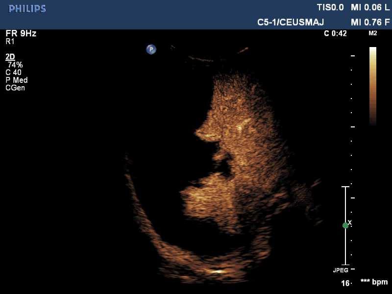
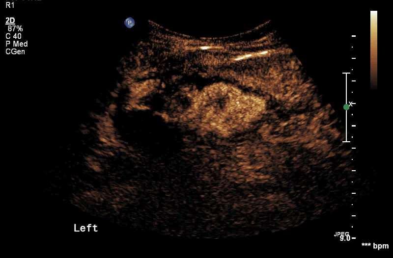
11 Potential Indications of Pediatric CEUS 2 IBD activity and complications Tumor monitoring during treatment Testicular / ovarian torsion (viability) Vascular tumor, vascular malformation Femoral head perfusion, rheumatoid arthritis If CE MR or CT is contraindicated (or not available) In selected cases: ICU, ED

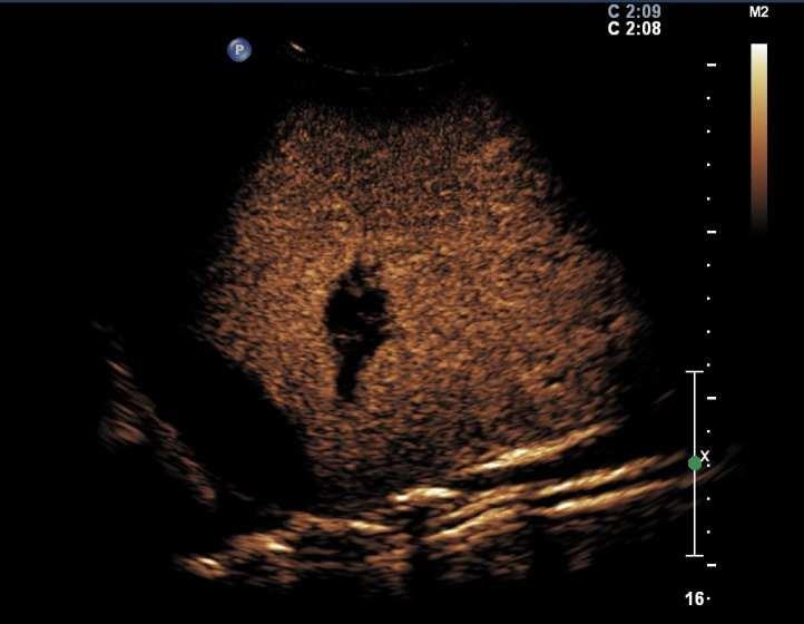
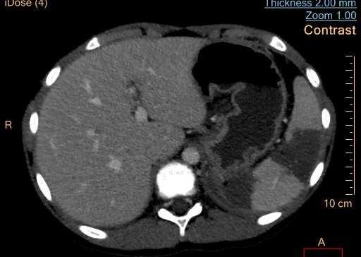
12 T R A U M A and CEUS


13 Liver trauma 9 yr boy motor cycle accident CT at admission
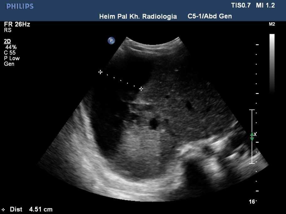
14

15


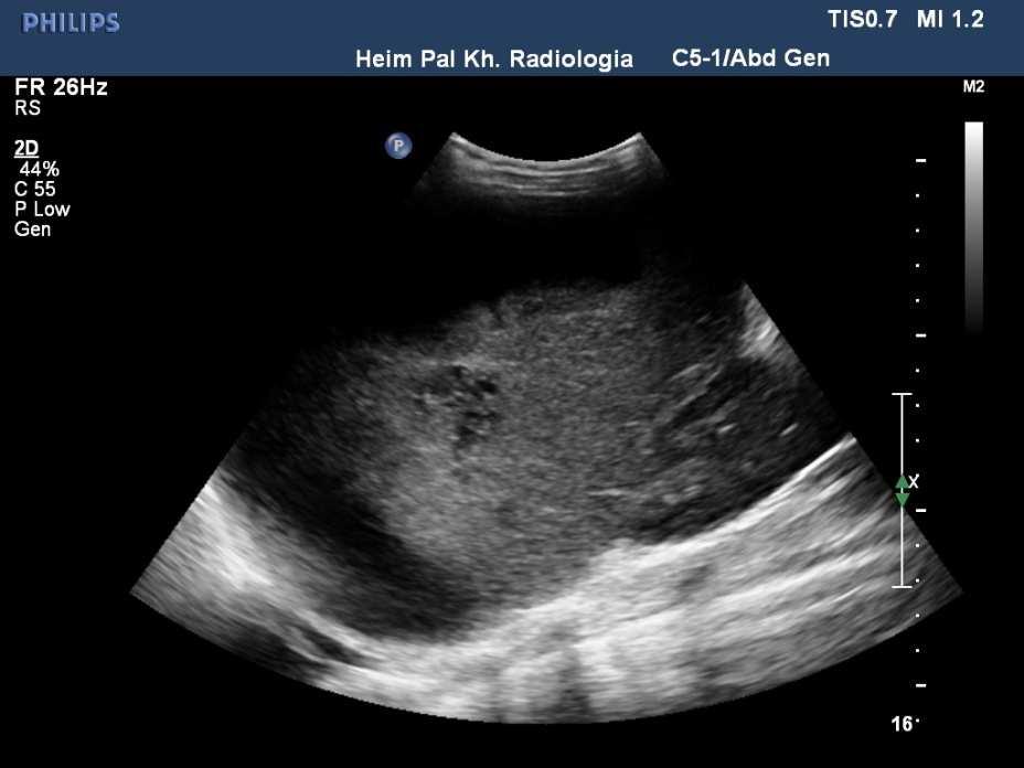
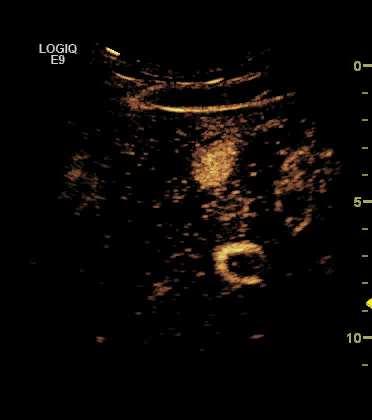
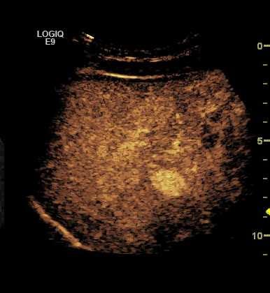
16 Liver injury: follow up with CEUS (12 y f) NC B-mode US + CDI


17 Liver injury: follow up with CEUS (12 y f)
18

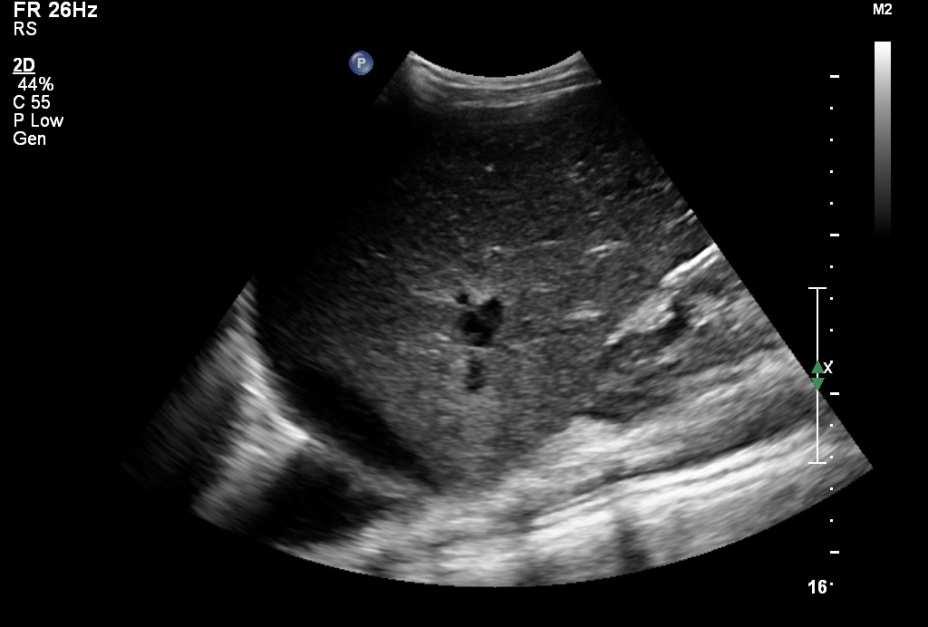
 1 month")
19 Liver injury follow up with CEUS (12 y f) 1 month later


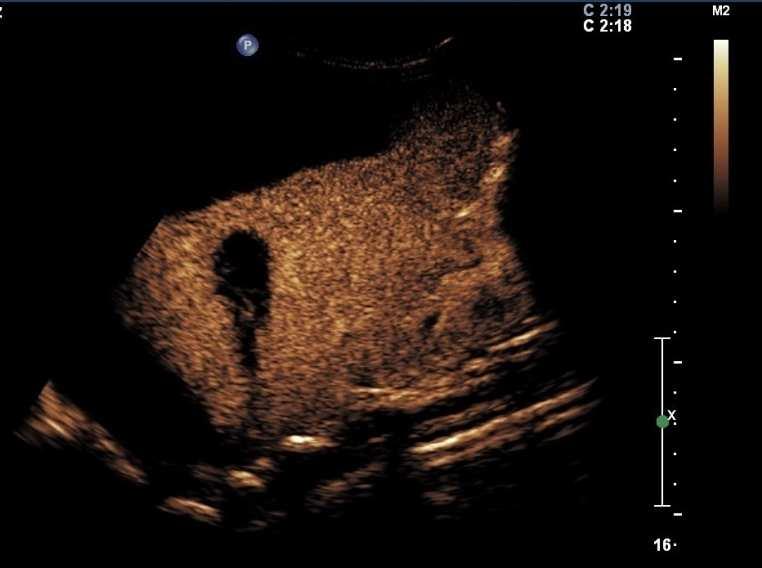
20 Splenic and renal trauma 9 y old girl with blunt abdominal trauma B-mode and CD US


21 9 y old girl, with blunt abdominal trauma - CECT


22 9 y old girl, with blunt abdominal trauma - CEUS

23 9 y old girl, with blunt abdominal trauma CEUS renal cortical necrosis


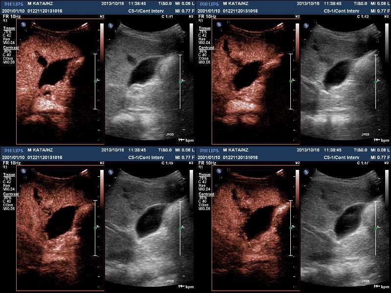
24 11 y old boy, abdominal blunt trauma, suprarenal gland hematoma?


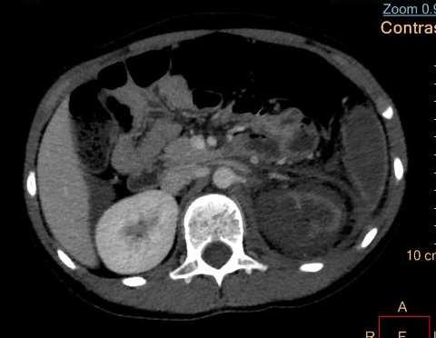
25 11 y old girl, left abdominal blunt trauma, splenic and kidney injury? CT at admission


26 11 y old girl, left abdominal blunt trauma, splenic and kidney injury?


27 11 y old girl, left abdominal blunt trauma, splenic and kidney injury?


28 11 y old girl, left abdominal blunt trauma, splenic and kidney injury?


29 Pediatric abdominal trauma and CEUS Minor abdominal trauma MDCT / NC US / CEUS comparison 30/33 solid injuries were detected by CEUS Solid organ injuires: NC US vs CEUS Miele V. et al.: Role of Contrast Enhanced Ultrasound (CEUS) in the evaluation of localized low-energy abdominal trauma in a pediatric population: our initial experience. ECR C-0873
30 T R A U M A and CEUS INDICATIONS Low energy abdominal trauma with suspected parenchymal injury at admission Follow up CEUS with known injuries detected by CT Detection of complications (re-bleeding, splenic artery pseudoaneurysm, infection)

31 T U M O R and CEUS
 Blunt trauma of the liver Differentiation of focal fatty")
32 Liver CEUS Indications 1. Incidental liver lesion by abdominal US (characterisation, avoid biopsy) Blunt trauma of the liver Differentiation of focal fatty infiltration / sparing and focal neoplasm Follow up of benign liver mass Follow up malignant liver masses during treatment
33 Liver CEUS Indications 2. Equivocal abnormality after MR, CT, or guided biopsy Poor or non-visualization of mass at time of US-guided biopsy US-guided local ablation of focal mass Liver transplant evaluation


34 Incidental liver masses at long term F/U 17 y old girl with with treated neuroblastoma. MR (2015): liver masses Follow up with US/MR + CEUS (2016)


")
35 17 y old girl with with treated neuroblastoma. MR (2015): liver masses At age 18 and 19 yrs follow up with US + CEUS (2016) no change

: liver")

36 17 y old girl with with treated neuroblastoma. MR (2015): liver masses At age 18 and 19 yrs follow up with US + CEUS (2016) no change


37 15 y old boy with multiple liver masses, enlarged lymph nodes. US and MR Surgery + chemotherapy. Histology desmoplastic small-round cell tumor Follow up with MR / US + CEUS


38 6 months F/U, BMT. NC US / MR Liver cyst and viable tumor? 3 months later CEUS


39 6 months F/U, BMT. NC US / MR Liver cyst and viable tumor? 3 months later CEUS, 3 small masses


40 19 y old male with known C F liver mass characterization

41 A F Infantile hepatic hemangioma CEUS: IV. 0,5 ccuca L F


42 Case of MB McCarville / St.Jude Hospital 7 yo girl treated for neuroblastoma at age 13 months. FLL found on CT for abdominal pain


43 Arterial Phase Iso-Enhancing Portal Venous Phase Iso-Enhancing

44 Regenerative Nodule Delayed Phase Iso-Enhancing
45 Our pediatric CEUS liver studies: 22 pediatric patients, between FLL was detected and characterised in 10 patients after chemotherapy Follow up with CEUS and MRI 5 FNH, 1 case residual tumor, 1 case haemangioma Comment: Incidence of FLLs in post-chemo patients can be 100 times higher than in normal population* * Chiorean L et al. Benign liver tumors in pediatric patients - Review with emphasis on imaging features. World J Gastroenterol ; 21(28):


46 Spleen and CEUS Splenomegaly, hypoechoic solid splenic mass, 11 y old boy, NC B-mode and MVI


47 Splenomegaly, hypoechoic solid splenic mass, 11 y old boy CEUS: IV. 0,7 cc SonoVue


48 Spleen and CEUS Splenomegaly, hypoechoic solid splenic mass, 11 y old boy CEUS: IV. 0,7 cc SonoVue


49 Bowel infection or GVH? in a 9 yr old BMT patient
50 V U R and CEUS

51 CE voiding urosonography: Diagnosis and F/U of VUR Voiding sono-cystograhy: detection of V U R VUR detection with VUS (Grade 1-5): Grade 1. Microbubbles in the ureter, only Grade 2. Microbubbles in the urinary tract, no dilatation Grade 3. Microbubbles in the urinary tract, significant pyelectasy and mild calyceal dilatation Grade 4. Microbubbles in the urinary tract, significant pyelectasy and calyceal dilatation Grade 5. Microbubbles in the urinary tract, significant pyelectasy and calyceal dilatation and tortuous ureter Kis É. Magyar Radiológia

52 CE voiding urosonography: Intrarenal reflux (IRR) - 29 patients (18 / 11 F / M), av. age 25 mo - Indications: recurrent UTI, postoperative F/U - IRR: 22 patients Z. Karadi, (SE, 2nd Dept. Of Pediatrics)
 US system, type of")
53 Method of IV pediatric CEUS 1. UCA dose depends on Size / age of the patient Type of UCA Type of the study (depth) US system, type of transducer Software version of the US system Yusuf et al. AJR:208, Febr 2017
54 Method of IV pediatric CEUS 2. Timing of scanning and recording Selection of the ROI / scan plane 2nd person must be present during the study Be prepared for allergic reaction, ICU is available Consider hyperdynamic circulation
55 Potential indications of CEUS in Pediatric Patients: C O N C L U S I O N S Contrast US has a great potential in pediatric imaging in experienced hands No radiation, no sedation, no renal risk Main indications:, trauma, tumor, VUR CEUS methodology needs further studies Potential of US guided local treatments Correlation with other imaging studies


56 Questions and comments? Zoltan Harkanyi MD, PhD
Contrast Enhanced Ultrasound of Parenchymal Masses in Children
 Contrast Enhanced Ultrasound of Parenchymal Masses in Children Sue C Kaste, DO On behalf of Beth McCarville, MD St. Jude Children s Research Hospital Memphis, TN Overview Share St. Jude experience with
Imaging techniques to characterize spleen involvement in patients with Hodgkin lymphoma
 Imaging techniques to characterize spleen involvement in patients with Hodgkin lymphoma Marco Picardi, MD Ematologia, Azienda Ospedaliera Universitaria Federico II, Naples, Italy 5th International Workshop
Vascular Imaging in the Pediatric Abdomen. Jonathan Swanson, MD
 Vascular Imaging in the Pediatric Abdomen Jonathan Swanson, MD Goals and Objectives To understand the imaging approach, appearance, and clinical manifestations of the common pediatric abdominal vascular
Radiation reduction in the follow-up of abdominal trauma imaging using contrast-enhanced ultrasound
 Radiation reduction in the follow-up of abdominal trauma imaging using contrast-enhanced ultrasound Poster No.: B-0892 Congress: ECR 2015 Type: Authors: Keywords: DOI: Scientific Paper A. Deganello, E.
Innovations in HCC Imaging: MDCT/MRI
 Innovations in HCC Imaging: MDCT/MRI Anthony E. Cheng, M.D. Cardinal MRI Center Cardinal Santos Medical Center, Wilson Street, San Juan Innovations in HCC Imaging: Goals/Objectives MDCT/MRI Learn the diagnostic
Off-label-use of SonoVue in voiding urosonography for the diagnosis of vesicoureteric reflux in children: a survey on side effects
 Off-label-use of SonoVue in voiding urosonography for the diagnosis of vesicoureteric reflux in children: a survey on side effects Poster No.: C-0661 Congress: ECR 2014 Type: Scientific Exhibit Authors:
Abdomen Sonography Examination Content Outline
 Abdomen Sonography Examination Content Outline (Outline Summary) # Domain Subdomain Percentage 1 2 3 Anatomy, Perfusion, and Function Pathology, Vascular Abnormalities, Trauma, and Postoperative Anatomy
Appendix 5. EFSUMB Newsletter. Gastroenterological Ultrasound
 EFSUMB Newsletter 87 Examinations should encompass the full range of pathological conditions listed below A log book listing the types of examinations undertaken should be kept Training should usually
Role of imaging in evaluation of genitourinary i trauma Spectrum of GU injuries Relevance of imaging findings in determining management Focus on MDCT
 Genitourinary Tract Injuries 6 th Nordic Course Scott D. Steenburg, MD Assistant Professor University of Maryland Department of Radiology Division of Trauma and Emergency Radiology R Adams Cowley Shock
Imaging of liver and pancreas
 Imaging of liver and pancreas.. Disease of the liver Focal liver disease Diffusion liver disease Focal liver disease Benign Cyst Abscess Hemangioma FNH Hepatic adenoma HCC Malignant Fibrolamellar carcinoma
The use of echo-enhanced voiding urosonography (VUS) to detect VUR is an attractive option.
 to detect VUR is an attractive option.") The role of the echo-enhanced voiding urosonography (VUS) compared to micturating cystourethrography (MCU) in daily practice in the study of vesicoureteral reflux (VUR) Poster No.: C-1201 Congress: ECR
The role of the echo-enhanced voiding urosonography (VUS) compared to micturating cystourethrography (MCU) in daily practice in the study of vesicoureteral reflux (VUR) Poster No.: C-1201 Congress: ECR
Contrast Enhanced Voiding Urosonography (cevus): How we do it
: How we do it") Contrast Enhanced Voiding Urosonography (cevus): How we do it Susan J. Back, MD Department of Radiology, The Children s Hospital of Philadelphia No Disclosures cevus What it is What to do What not to do
Contrast Enhanced Voiding Urosonography (cevus): How we do it Susan J. Back, MD Department of Radiology, The Children s Hospital of Philadelphia No Disclosures cevus What it is What to do What not to do
The role for contrast-enhanced ultrasonography outside of focal liver lesions
 The role for contrast-enhanced ultrasonography outside of focal liver lesions Paul S. Sidhu King s College Hospital, London, UK Introduction Contrast-enhanced ultrasonography (US) of focal liver lesions
Liver Tumors. Prof. Dr. Ahmed El - Samongy
 Liver Tumors Prof. Dr. Ahmed El - Samongy Objective 1. Identify the most important features of common benign liver tumors 2. Know the risk factors, diagnosis, and management of hepatocellular carcinoma
Sulfur hexafluoride-filled microbubbles SonoVue 3-7microns diameter Blood pool agent
 Sulfur hexafluoride-filled microbubbles SonoVue 3-7microns diameter Blood pool agent Extremely good tolerance in clinical practice - No nephrotoxicity, - No thyroid interaction - No need of Blood test
KS Sunny Tse 1, LS Wong 1, TW Fan 1, KY Kwok 1, W Chan 2, MWY Leung 3, NSY Chao 3, TK Tsang 1, HS Fung 1, KW Tang 1, SCH Chan 1
 KS Sunny Tse 1, LS Wong 1, TW Fan 1, KY Kwok 1, W Chan 2, MWY Leung 3, NSY Chao 3, TK Tsang 1, HS Fung 1, KW Tang 1, SCH Chan 1 1 Department of Radiology and Imaging; 2 Department of Paediatrics; 3 Division
Index. Note: Page numbers of article titles are in boldface type.
 Index Note: Page numbers of article titles are in boldface type. A Abdominal injuries clinical presentation of, 23 24 Abdominal trauma evaluation for pediatric surgeon, 59 74 background of, 60 colon and
Rad Lab 4 Unknowns: Genitourinary!
 Rad Lab 4 Unknowns: Genitourinary! Peter Clarke MD! Don Di Salvo, MD! Clerkship Directors for Radiology! Harvard Medical School! Brigham and Women s Hospital! Dana Farber Cancer Institute! Case 1: 69 year
Interesting case. Vikas Kundra, M.D., Ph.D. October Vikas Kundra, M.D., Ph.D.
 Interesting case October 2012 Disclosure Information Vikas Kundra, M.D, Ph.D. I have no financial relationships to disclose. I WILL NOT include discussion of investigational or off-label use of a product
Job Task Analysis for ARDMS Abdomen Data Collected: June 30, 2011
 Job Task Analysis for ARDMS Abdomen Data Collected: June 30, 2011 Reported: Analysis Summary for: Abdomen Examination Survey Dates 06/13/2011-06/26/2011 Invited Respondents 6,000 Surveys with Demographics
Evaluation of Liver Mass Lesions. American College of Gastroenterology 2013 Regional Postgraduate Course
 Evaluation of Liver Mass Lesions American College of Gastroenterology 2013 Regional Postgraduate Course Lewis R. Roberts, MB ChB, PhD Division of Gastroenterology and Hepatology Mayo Clinic College of
Ultrasound of malignant testicular lesions. Arne Hørlyck Department of Radiology Aarhus University Hospital, Skejby
 Ultrasound of malignant testicular lesions Arne Hørlyck Department of Radiology Aarhus University Hospital, Skejby Testis Ultrasound is fantastic!! Scrotum Extratesticular mass: Benign Intratesticular
Index. Note: Page numbers of article titles are in boldface type.
 Magn Reson Imaging Clin N Am 12 (2004) 587 591 Index Note: Page numbers of article titles are in boldface type. A Adenoma(s), adrenal, gadolinium-enhanced MR imaging in, 533 534 hyperfunctioning versus
SELF-ASSESSMENT MODULE REFERENCE SPR 2018 Oncologic Imaging Course Adrenal Tumors November 10, :00 12:10 p.m.
 SELF-ASSESSMENT MODULE REFERENCE SPR 2018 Oncologic Imaging Course Adrenal Tumors November 10, 2018 10:00 12:10 p.m. Staging Susan E. Sharp, MD 1. In the International Neuroblastoma Risk Group Staging
HEPATO-BILIARY IMAGING
 HEPATO-BILIARY IMAGING BY MAMDOUH MAHFOUZ MD PROF.OF RADIOLOGY CAIRO UNIVERSITY mamdouh.m5@gmail.com www.ssregypt.com CT ABDOMEN Indications Patient preparation Patient position Scanogram Fasting 4-6 hours
Case Conference. Discussion. Indications of Trauma Blue. Trauma Protocol In SKH. Trauma Blue VS. Trauma Red. Supervisor:VS 楊毓錚 Presenter:R1 周光緯
 Case Conference Supervisor:VS 楊毓錚 Presenter:R1 周光緯 Discussion 2010.7.14 2/81 Trauma Protocol In SKH Indications of Trauma Blue Trauma Blue VS. Trauma Red 3/81 Severe trauma mechanism : 1. Trauma to multiple
CT abdomen and pelvis
 CT abdomen and pelvis General indications: Assessment of vague abdominal symptoms (pain, colics,distenstion,...) Varifecation of a lesion discovered by other diagnostic modalities as US, barium,ivp, Staging
PREAMBLE GENERAL DIAGNOSTIC RADIOLOGY
 PREAMBLE The General Diagnostic Radiology category is intended to cover the body of knowledge a practicing board certified Diagnostic Radiologist should know. Since the range of content relevant to the
EUROSON SCHOOL 2019 January 18-19, 2019, Athens-Greece Preliminary Programme
 EUROSON SCHOOL 2019 January 18-19, 2019, Athens-Greece Preliminary Programme 08:00-09:00 Registration Friday, January 18 Theoretical Course / PHYSICS AND TECHNOLOGY 09:00-09:15 Basics in US Physics 09:15-09:30
Modern liver imaging techniques - A new era in liver ultrasound
 Modern liver imaging techniques - A new era in liver ultrasound Yuko Kono, M.D., Ph.D. Clinical Professor Departments of Medicine and Radiology University of California, San Diego San Diego, USA How to
Pediatric Hepatobiliary, Pancreatic & Splenic US
 Pediatric Hepatobiliary, Pancreatic & Splenic US Susan J. Back, MD Department of Radiology, The Children s Hospital of Philadelphia No Disclosures Objectives Normal Abnormal: cases and US advances Objectives
CT 101 :Pancreas and Spleen
 CT 101 :Pancreas and Spleen Shikha Khullar,, MD, MPH Division of Radiology University of South Alabama The Pancreas Normal Pancreas 3 Phase Pancreatic CT Non contrast Arterial phase : 30-35 35 second
PDF created with pdffactory Pro trial version
 Neuroblastoma Tumor derived from neural crest cell that form the sympathetic ganglia&adrenal medulla. Causes *unknown. *familial neuroblastoma has been reported but is rare. * The incidence is 1:100,000
12th & 13th May 2016 Weston Education Centre, Kings College Hospital London
 EUROSON SCHOOL CEUS Course How to Incorporate CEUS into your Imaging Practice 12th & 13th Weston Education Centre, Kings College Hospital London CPD points for two days: EFSUMB 16 CME points BMUS 12 CPD
Imaging Ejaculatory Disorders and Hematospermia
 ATHENS 4-6 October 2018 European Society of Urogenital Radiology Imaging Ejaculatory Disorders and Hematospermia Parvati Ramchandani, MD Professor, Radiology and Surgery University of Pennsylvania Medical
Excretory urography (EU) or IVP US CT & radionuclide imaging
 or IVP US CT & radionuclide imaging") Excretory urography (EU) or IVP US CT & radionuclide imaging MRI arteriography studies requiring catherization or direct puncture of collecting system EU & to a lesser extent CT provide both functional
Excretory urography (EU) or IVP US CT & radionuclide imaging MRI arteriography studies requiring catherization or direct puncture of collecting system EU & to a lesser extent CT provide both functional
Urinary tract embolization
 Beograd, 14.10.2012 Urinary tract embolization asist. Peter Popovič, MD, MSc Head of abdominal radiology department, Institute of Radiology, UMC Ljubljana Embolization Who and when procedure: local/general
CONTRAST ENHANCED ULTRASOUND
 Sunrise Session: CONTRAST ENHANCED ULTRASOUND Kassa Darge, MD, PhD Department of Radiology The Children s Hospital of Philadelphia Perelman School of Medicine University of Pennsylvania Philadelphia 1.
Alice Fung, MD Oregon Health and Science University
 Alice Fung, MD Oregon Health and Science University Disclosure Comments The speaker Alice Fung, MD Has relevant financial relationships to disclose. Received honorarium from (Guerbet). This individual
Dr Claire Smith, Consultant Radiologist St James University Hospital Leeds
 Dr Claire Smith, Consultant Radiologist St James University Hospital Leeds Imaging in jaundice and 2ww pathway Image protocol Staging Limitations Pancreatic cancer 1.2.4 Refer people using a suspected
Financial Disclosure
 Benign Liver Masses Adil Abdalla, MBBS Creighton University-CHI Health August 25, 2018 Financial Disclosure Nothing to disclose Financial Disclosure 1 Objectives To assess patients with benign liver tumors
Liver Specific MRI using Gd-EOB-DTPA Disodium (Primovist) Effects Change in Management of Indeterminate Liver Lesions.
 Effects Change in Management of Indeterminate Liver Lesions.") Liver Specific MRI using Gd-EOB-DTPA Disodium (Primovist) Effects Change in Management of Indeterminate Liver Lesions. Poster No.: C-1751 Congress: ECR 2012 Type: Authors: Keywords: DOI: Educational Exhibit
Liver Specific MRI using Gd-EOB-DTPA Disodium (Primovist) Effects Change in Management of Indeterminate Liver Lesions. Poster No.: C-1751 Congress: ECR 2012 Type: Authors: Keywords: DOI: Educational Exhibit
Imaging Guided Biopsy. Edited & Presented by ; Hussien A.B ALI DINAR. Msc Lecturer,Reporting Sonographer
 Imaging Guided Biopsy Edited & Presented by ; Hussien A.B ALI DINAR. Msc Lecturer,Reporting Sonographer Objective By the End of this lessons you should : Define what biopsy Justify Aim to perform biopsy
Hepatic Imaging: What Every Practitioner Should Know
 Hepatic Imaging: What Every Practitioner Should Know Shuchi K. Rodgers, MD Section Chief, Abdominal Imaging Director of Ultrasound Department of Radiology Einstein Medical Center rodgerss@einstein.edu
Sonography of soft-tissue vascular lesions
 Sonography of soft-tissue vascular lesions Oscar M. Navarro Associate Professor, University of Toronto Dept. of Diagnostic Imaging, The Hospital for Sick Children Toronto, Canada Declaration of Disclosure
The UGent Institutional Repository is the electronic archiving and dissemination platform for
 biblio.ugent.be The UGent Institutional Repository is the electronic archiving and dissemination platform for all UGent research publications. Ghent University has implemented a mandate stipulating that
Interesting Cases from Liver Tumor Board. Jeffrey C. Weinreb, M.D.,FACR Yale University School of Medicine
 Interesting Cases from Liver Tumor Board Jeffrey C. Weinreb, M.D.,FACR Yale University School of Medicine jeffrey.weinreb@yale.edu Common Liver Diseases Hemangioma Cyst FNH Focal Fat/Sparing THID Non-Cirrhotic
PROFESSIONAL SKILLS 1 3RD YEAR SEMESTER 6 RADIOGRAPHY. THE URINARY SYSTEM Uz. Fatema shmus aldeen Tel
 PROFESSIONAL SKILLS 1 3RD YEAR SEMESTER 6 RADIOGRAPHY THE URINARY SYSTEM Uz. Fatema shmus aldeen Tel. 0925111552 Professional skills-2 THE URINARY SYSTEM The urinary system (review anatomy and physiology)
The Contribution of Contrast Enhanced Ultrasound for the characterization of benign liver lesions in clinical practice a monocentric experience
 Original papers Med Ultrason 2012, Vol. 14, no. 4, 283-287 The Contribution of Contrast Enhanced Ultrasound for the characterization of benign liver lesions in clinical practice a monocentric experience
Kassa Darge, MD, PhD. Sunrise Workshop UPDATE ON CONTRAST MEDIA: New agents, new concerns & strategies for safe use
 Sunrise Workshop UPDATE ON CONTRAST MEDIA: New agents, new concerns & strategies for safe use Kassa Darge, MD, PhD Professor of Radiology and Surgery Perelman School of Medicine University of Pennsylvania
Imaging of Neuroendocrine Metastases
 Imaging of Neuroendocrine Metastases Aoife Kilcoyne, Shaunagh McDermott, Colin McCarthy,Manuel Patino, Dushyant Sahani, Michael Blake Abdominal Imaging Division Massachusetts General Hospital Disclosure
Ultrasound contrast agents (USCA)
") Ultrasound contrast agents (USCA) Jean-Yves Meuwly, MD, University Hospital Lausanne, Switzerland Ultrasound contrast agents Initially developed in order to enhance the Doppler signal Increase in signal
Ultrasound contrast agents (USCA) Jean-Yves Meuwly, MD, University Hospital Lausanne, Switzerland Ultrasound contrast agents Initially developed in order to enhance the Doppler signal Increase in signal
PET IMAGING (POSITRON EMISSION TOMOGRAPY) FACT SHEET
 FACT SHEET") Positron Emission Tomography (PET) When calling Anthem (1-800-533-1120) or using the Point of Care authorization system for a Health Service Review, the following clinical information may be needed to
Positron Emission Tomography (PET) When calling Anthem (1-800-533-1120) or using the Point of Care authorization system for a Health Service Review, the following clinical information may be needed to
LIVER IMAGING TIPS IN VARIOUS MODALITIES. M.Vlychou, MD, PhD Assoc. Professor of Radiology University of Thessaly
 LIVER IMAGING TIPS IN VARIOUS MODALITIES M.Vlychou, MD, PhD Assoc. Professor of Radiology University of Thessaly Hepatocellular carcinoma is a common malignancy for which prevention, screening, diagnosis,
RADPrimer Curriculum Breast Topics Covered Basic Intermediate 225
 Breast Anatomy & Normal Variants 11 Breast Imaging Modalities 13 BI RADS Lexicon 3 Mammography: Masses 9 Mammography: Calcifications 17 Mammography: Additional Findings 8 Ultrasound Features 10 Ultrasound
Simplifying liver assessment in internal medicine
 Ultrasound Customer story Simplifying liver assessment in internal medicine Philips Affiniti ultrasound for elastography and contrast-enhanced ultrasound (CEUS) Where Sonography Institute, Uster, Switzerland
Liver Cancer (Hepatocellular Carcinoma or HCC) Overview
 Overview") Liver Cancer (Hepatocellular Carcinoma or HCC) Overview Recent advances in liver cancer care seek to address the rising incidence of liver cancer, which has steadily increased over the past three decades.
Liver Cancer (Hepatocellular Carcinoma or HCC) Overview Recent advances in liver cancer care seek to address the rising incidence of liver cancer, which has steadily increased over the past three decades.
Giovanni Montini has documented that he has no relevant financial relationships to disclose or conflict of interest to resolve.
 Giovanni Montini has documented that he has no relevant financial relationships to disclose or conflict of interest to resolve. Imaging in Pediatric UTI Giovanni Montini Milano, Italy giovanni.montini@unimi.it
IT 의료융합 1 차임상세미나 복부질환초음파 이재영
 IT 의료융합 1 차임상세미나 2013-4-3 복부질환초음파 이재영 나는오늘누구를위하여 종을울리나? 전통적의료 의사 공학설계자 의사 최첨단진단장비들 USG, CT, MRI 환자 환자 현대의료 사용자중심의사고 US in the Abdomen Detection DDx Look Behavior Response by external stimuli Guiding Tool
ABDOMINAL DIFFUSION WEIGHTED MR
 ABDOMINAL DIFFUSION WEIGHTED MR Frank Miller, M.D. FACR Professor of Radiology Chief, Body Imaging Section Medical Director, MR Imaging Northwestern University Feinberg School of Medicine fmiller@northwestern.edu
Uroradiology For Medical Students
 Uroradiology For Medical Students Lesson 4: Cystography & Urethrography - Part 2 American Urological Association Review Cystography is useful in evaluating the bladder, the urethra and the competence of
LUS: Laparoscopic Ultrasound
 LUS: Laparoscopic Ultrasound Dr. Bjørn Skjoldbye Herlev Hospital Copenhagen University Training Course for Advanced Oncologic Laparoscopy St. Petersburg - February 14, 2006 Equipment Laparoscopy LUS (B&K
Pediatric Retroperitoneal Masses Radiologic-Pathologic Correlation
 Acta Radiológica Portuguesa, Vol.XVIII, nº 70, pág. 61-70, Abr.-Jun., 2006 Pediatric Retroperitoneal Masses Radiologic-Pathologic Correlation Marilyn J. Siegel Mallinckrodt Institute of Radiology, Washington
Abdominal Solid Organ Injury
 Abdominal Solid Organ Injury 8 th Nordic Course Stockholm, Sweden May 19-22, 2014 K.SHANMUGANATHAN M.D. ABDOMINAL TRAUMA OBJECTIVES Splenic injury Late arterial / early p-v phase imaging Liver injury Blunt
Sonographic Features of Thyroid Nodules & Guidelines for Management
 Sonographic Features of Thyroid Nodules & Guidelines for Management Mark A. Lupo, MD, FACE, ECNU Thyroid & Endocrine Center of Florida Assistant Clinical Professor of Medicine Florida State University,
Looking Outside the Box: Incidental Extracardiac Finding in Echo
 Looking Outside the Box: Incidental Extracardiac Finding in Echo Dr. Aijaz Shah Head of Division, Adult Echocardiography Laboratory Prince Sultan Cardiac Centre Riyadh Case 1 17 year old boy presented
Sex: 女 Age: 51 Occupation: 無 Admission date:92/07/22
 Sex: 女 Age: 51 Occupation: 無 Admission date:92/07/22 Chief complaint Unknown fever for one month Hand tremor and left huge renal tumor was noted Present illness Suffered from fever for one month, hand
Imaging Work-Up of a Neck Mass - Adults & Children
 Disclosures Imaging Work-Up of a Neck Mass - Adults & Children I have nothing to disclose Christine M Glastonbury MBBS Professor of Radiology & Biomedical Imaging Otolaryngology-Head & Neck Surgery and
Liver imaging the revolution
 Liver imaging the revolution Valérie Vilgrain Hôpital Beaujon, Paris, France PHC 2018 - www.aphc.info At the Beginning of the story Radiology in the 1970s US Garrett Radiology 1976 abscess Taylor Radiology
Cortical renal scan in febrile UTI: Established usefulness and future developments
 Cortical renal scan in febrile UTI: Established usefulness and future developments Diego De Palma, MD Nuclear Medicine, Circolo Hospital, Varese, Italy UTI is common in the pediatric population! Girls
RICCARDO LENCIONI,CLOTILDE DELLA PINA, LAURA CROCETTI,DANIA CIONI. Chapter 1
 RICCARDO LENCIONI,CLOTILDE DELLA PINA, LAURA CROCETTI,DANIA CIONI Chapter 1 Impact of European Federation of Societies for Ultrasound in Medicine and Biology (EFSUMB) Guidelines on the Use of Contrast
Gastrointestinal tract
 Gastrointestinal tract Colloidal liver-spleen imaging Presented by: Jehad Felemban Introduction: To obtain better anatomic display of liver and spleen architecture, we use (CT Ultrasound). (Radionuclide
Newcastle HPB MDM updated radiology imaging protocol recommendations. Author Dr John Scott. Consultant Radiologist Freeman Hospital
 Newcastle HPB MDM updated radiology imaging protocol recommendations Author Dr John Scott. Consultant Radiologist Freeman Hospital This document is intended as a guide to aid radiologists and clinicians
Acute renal colic Radiological investigation in patients with renal colic
 Acute renal colic Radiological investigation in patients with renal colic Mikael Hellström Professor Department of Radiology Sahlgrenska University Hospital Göteborg University 0.9-1.8/1.000 inhabitants
Ultrasound of soft-tissue vascular anomalies
 Ultrasound of soft-tissue vascular anomalies Oscar M. Navarro Associate Professor, University of Toronto Dept. of Diagnostic Imaging, The Hospital for Sick Children Toronto, Canada Declaration of Disclosure
Oncology General Principles L A U R I E S I M A R D B R E A S T S U R G I C A L O N C O L O G Y F E L L O W D E C E M B E R
 Oncology General Principles L A U R I E S I M A R D B R E A S T S U R G I C A L O N C O L O G Y F E L L O W D E C E M B E R 2 0 1 2 Objectives Discuss Diagnostic and staging strategies in oncology Know
Guidelines, Policies and Statements D5 Statement on Abdominal Scanning
 Guidelines, Policies and Statements D5 Statement on Abdominal Scanning Disclaimer and Copyright The ASUM Standards of Practice Board have made every effort to ensure that this Guideline/Policy/Statement
ENTEROCOLITIDES CAN YOU TELL THEM APART ON MDCT? Richard M. Gore, MD North Shore University Medical Center University of Chicago Evanston, Illinois
 ENTEROCOLITIDES CAN YOU TELL THEM APART ON MDCT? Richard M. Gore, MD North Shore University Medical Center University of Chicago Evanston, Illinois SCBT/MR 2010 San Diego, California March 8, 2010 13:40-14:00
The clinical sign of pleurisy is characterized by a
 Contrast-Enhanced Sonography for Differential Diagnosis of Pleurisy and Focal Pleural Lesions of Unknown Cause* Christian Görg, MD; Tillmann Bert, MD; and Konrad Görg, MD Background: Ultrasound enables
weighing risks against benefits ALARA principle appropriate activities (radiopharmaceutical doses)
") weighing risks against benefits ALARA principle appropriate activities (radiopharmaceutical doses) based on EANM references adequate appointment method (patient booking system) Appropriate activities (doses)
weighing risks against benefits ALARA principle appropriate activities (radiopharmaceutical doses) based on EANM references adequate appointment method (patient booking system) Appropriate activities (doses)
Extraosseous myeloma: imaging features
 Extraosseous myeloma: imaging features C. Santos Montón, R. Corrales, J. M. Bastida Bermejo, M. Villanueva Delgado, R. E. Correa Soto, J. M. Alonso Sánchez; Salamanca/ES Learning objectives -To review
16.1 Risk of UTI recurrence in children
 16. UTI prognosis 16.1 Risk of UTI recurrence in children Key question: What is the risk of recurrent UTI in children with no known structural or functional abnormalities of the urinary tract with a first
Personal data. Age : 63 Gender : male
 Personal data Age : 63 Gender : male Chief complain No specific symptom or discomfort A hepatic mass, found by abdominal sonography of routine health exam on 88-12-08 Past history 1984-3-3 Old CVA with
ROLE OF MRI IN SCREENING, DIAGNOSIS AND MANAGEMENT OF BREAST CANCER. B.Zandi Professor of Radiology
 ROLE OF MRI IN SCREENING, DIAGNOSIS AND MANAGEMENT OF BREAST CANCER B.Zandi Professor of Radiology Introduction In the USA, Breast Cancer is : The Most Common Non-Skin Cancer The Second Leading cause of
Technological advancements improve the sensitivity of CEUS diagnostics
 Technological advancements improve the sensitivity of CEUS diagnostics. Martegani, MD, L. iani, MD Department of Diagnostic Imaging, Valduce Hospital, Como, Italy Characterization with Ultrasound B C D
Index. Note: Page numbers of article titles are in boldface type.
 Note: Page numbers of article titles are in boldface type. A Adenocarcinoma, pancreatic ductal, laparoscopic distal pancreatectomy for, 61 Adrenal cortical carcinoma, laparoscopic adrenalectomy for, 114
Traumatic and Non Traumatic Adrenal Emergencies
 Traumatic and Non Traumatic Adrenal Emergencies Michael N. Patlas, MD, FRCPC (1), Christine O. Menias, MD (2), Douglas S. Katz, MD, FACR (3), Ania Z. Kielar, MD, FRCPC (4), Alla M. Rozenblit, MD (5), Jorge
Overview. Imaging Indications. Paediatric Radiation Safety 2015/03/12. Paediatric radiation safety General guidelines Protocols
 Overview Paediatric radiation safety General guidelines Protocols Paediatric Radiation Safety Paediatric patients are unique Children are more susceptible to radiation induced cancer than adults Younger
Abdominal Solid Organ Injury
 Abdominal Solid Organ Injury 9th Nordic Trauma Radiology Course Aarhus, Denmark May 23-26, 2016 K.SHANMUGANATHAN M.D. ABDOMINAL TRAUMA OBJECTIVES Splenic injury Late arterial / early p-v phase imaging
Cervical Cancer 3/25/2019. Abnormal vaginal bleeding
 Cervical Cancer Abnormal vaginal bleeding Postcoital, intermenstrual or postmenopausal Vaginal discharge Pelvic pain or pressure Asymptomatic In most patients who are not sexually active due to symptoms
Body MRI from the Liver to the Bladder
 Body MRI from the Liver to the Bladder I Want You! Audience Participation Methodist Hospital Continuing Education Seminar Jordan Swensson, MD November 7, 2015 Objectives Observe the uses of MRI for organs
Abdominal Ultrasound : Aorta, Kidneys, Bladder
 Abdominal Ultrasound : Aorta, Kidneys, Bladder Nilam J. Soni, MD, MSc Associate Professor of Medicine Divisions of Hospital Medicine and Pulmonary/Critical Care Medicine Department of Medicine University
MANAGEMENT RECOMMENDATIONS
 1 MANAGEMENT RECOMMENDATIONS 1. Adrenal masses!!!!!!! page 2 2. Liver Masses!!!!!!! page 3 3. Obstetric US Soft Markers for Aneuploidy!! pages 4-6 4. Ovarian and Adnexal Cysts!!!!! pages 7-10 5. Pancreatic
Audit of Micturating Cystourethrograms performed over 1 year in a Children's Hospital
 Audit of Micturating Cystourethrograms performed over 1 year in a Children's Hospital Poster No.: C-1773 Congress: ECR 2012 Type: Scientific Exhibit Authors: K. Lyons, J. Sorensen, E. L. Twomey, V. Donoghue,
performed to help sway the clinician in what the appropriate diagnosis is, which can substantially alter the treatment of management.
 Hello, I am Maura Polansky at the University of Texas MD Anderson Cancer Center. I am a Physician Assistant in the Department of Gastrointestinal Medical Oncology and the Program Director for Physician
Evaluation of Suspected Pancreatic Cancer
 Evaluation of Suspected Pancreatic Cancer October 15, 2015 If you experience technical difficulty during the presentation: Contact WebEx Technical Support directly at: US Toll Free: 1-866-779-3239 Toll
Radiology of hepatobiliary diseases
 GI cycle - Lecture 14 436 Teams Radiology of hepatobiliary diseases Objectives 1. To Interpret plan x-ray radiograph of abdomen with common pathologies. 2. To know the common pathologies presentation.
Current Treatment of Colorectal Metastases. Dr. Thavanathan Surgical Grand Rounds February 1, 2005
 Current Treatment of Colorectal Metastases Dr. Thavanathan Surgical Grand Rounds February 1, 2005 25% will have metastases at initial presentation 25-50% 50% will develop metastases later 40% of potentially
PEDIATRIC BLUNT TRAUMA WHAT S DIFFERENT? NORDIC TRAUMA COURSE 2016
 PEDIATRIC BLUNT TRAUMA WHAT S DIFFERENT? NORDIC TRAUMA COURSE 2016 Ken F. Linnau, MD, MS Emergency Radiology Harborview Medical Center University of Washington Seattle, WA Thanks to Nupur Verma, MD University