Basal Ganglia. Today s lecture is about Basal Ganglia and it covers:
|
|
|
- Cameron May
- 6 years ago
- Views:
Transcription
1 Basal Ganglia Motor system is complex interaction between Lower motor neurons (spinal cord and brainstem circuits) and Upper motor neurons (pyramidal and extrapyramidal tracts) plus two main regulators (Cerebellum and Basal Ganglia) Today s lecture is about Basal Ganglia and it covers: I. Components of the basal ganglia II. Function of the basal ganglia III. Connection and circuits IV. Functional circuitry of the basal ganglia V. Symptoms and disorders discussed I. Components of the basal ganglia - Basal ganglia comprises multiple subcortical nuclei, most of them were supposed to develop from Telencephalon but only a small part of it does. - Basal ganglia is divided into Striatal Complex Pallidal Complex -Striatal Complex components have the same type of neurons, same type and cyto-architecture of dendrites and the same neurotransmitters and it s furthermore divided into: 1. Dorsal striatal: Caudate and Putamen. 2. Ventral Striatal: Nucleus acumbens and Olfactory tubercle. -Pallidal Complex is also divided into dorsal and ventral pallidal complex and it consists of: Pallidus and Substantia innominata This Substantia innominata is part of Basal Nucleus (also called Nucleus Basalis/ Nucleus of Meynert), and from it Acetylcholine is released. -Basal ganglia is functionally two parts: Ventral Basal nuclei (ventral pallidal substantia innmnata-, ventral striatal Nucleus Accumbens and olfactory tubercle-)
 If the cycle wasn t ending by cortex then the function of basal ganglia is lost.")


2 Dorsal basal nuclei ( dorsal pallidal globus pallidus-, dorsal striatal - caudate and putamen-) Each part has its own circuit (Cortex part-striatal part-pallidal-thalamic- and cortical again) If the cycle wasn t ending by cortex then the function of basal ganglia is lost. -you can see in slides (5-10) MRI of the basal ganglia starting from a very rostral section moving caudally ending these images with an MRI showing the subthalamic nucleus which looks like a lense +: subthalamic nucleus. It lies just above the rostral portion of the substantia nigra II. Function of the basal ganglia Basal ganglia is a sequencer/regulator which acts like air traffic control towers that give airplanes the permission to land, it (basal ganglia) calculates the inputs and the outputs and decides which pathway to be activated and which to be

3 inhibited; accordingly, it has two circuits one for excitation the other is responsible for suppression. (Direct and indirect pathways). The most important component of these circuits is the striatal part which is the main balance keeper, it consists of crucial neurons: the Medium Spiny Neurons. These neurons have a complex of dendrites with a lot of spines meaning that they have a lot of connections; sometimes we might have convergence of these spiny neurons from the cortex Glutamatergic neurons-; many of cortical neurons from layer 5 will converge on one or more of the spiny neurons depending on the distribution of the spiny neurons and the way the EPSP (Excitatory post synaptic potential) comes to the neurons, these medium spiny neurons are GABAergic neurons. These spiny neurons are influenced by modulators two of them are: 1. Dopamine released from Substantia Nigra (regulated through feedback mechanisms from the organ it supplies) 2. Acetylcholine released in local circuits -from nternal neurons within the striatum-(each part of basal nuclei supplies itself with Ach through local circuits plus a small part coming from substantia innominata ). *Substantia Innominata releases Ach mainly in ventral part of Pallidal complex. cortical neuron Glutamate Medium spiny neuron within the striatum (does the calculation) GABA neurons to Globus Pallidus.
 Middle Body of caudate+ Putamen Caudal Tail of caudate (mainly) + ( almost no Putamen at the caudal part) Two areas do not have connections with the Basal ganglia: 1.")
4 III. Connection and Circuits Basal ganglia receive from almost all parts of the cortex (neocortex and paleocortex); Rostral part of cortex rostral part of Basal ganglia Caudate (more involved with head of caudate) Middle Body of caudate+ Putamen Caudal Tail of caudate (mainly) + ( almost no Putamen at the caudal part) Two areas do not have connections with the Basal ganglia: 1. Primary sensory area of vision 2. Primary sensory area of audition
5 Circuits of the basal ganglia: a. Motor Loop: modulates the motor functions Mainly from motor areas; motor cortex (M1 primary motor area-,premotor area and supplementary motor area) sending signals to the body of caudate but mainly Putamen then going to Globus Pallidus to Ventrolateral VL and Ventroanterior VA nuclei of the thalamus then ending in the cortex. b. Visuomotor Loop: Coming from visual associated cortices mainly, Frontal eye field(more rosral to the motor area ) Body of caudate Globus Pallidus Dorsomedial nucleus DM and Ventroanterior VA nucleus of Thalamus ending in the cortex. c. Executive loop: More in the rostral cortices; Prefrontal area Head of Caudate Globus Pallidus Dorsomedial and Ventroanterior nuclei of the thalamus cortex Involved in Cognition and function of prefrontal cortex. This explains why some people with basal ganglia disorders can have psychotic or cognitive dysfunction.
 DM nucleus of the thalamus neocortex Usually the non neocortex is not related to the thalamus and when their tracts return into the neocortex they pass through the DM nucleus")
6 d. Motivational loop: It s from the Non-Neocortical areas, -from the paleocortix or allocortixmainly involved with ventral basal ganglia ventral striatum (Nucleus Acumbens), Ventral pallidal complex (Substantia Innominata) DM nucleus of the thalamus neocortex Usually the non neocortex is not related to the thalamus and when their tracts return into the neocortex they pass through the DM nucleus of the thalamus. * Based on the differences in lamination, the cerebral cortex can be classified into two parts, the large area of neocortex and the much smaller area of allocortex: Wikipedia. *Most of the cerebral cortex is neocortex. However, allocortex represents old functioning cortices such as Hippocampus, some parts of Cingulate Gyrus and cortices involved in emotion especially those with the olfactory function. *Allocortex is related to emotion, limbic system and primitive functions. It (the allocortex) has two components: the paleocortex and archicortex. *Neocortical area has 6 layers while Allocortical areas has 3-5 layers *Less no. of layers less processing older in age *Dorsal basal ganglia works in three loops (motor, visuomotor, executive) which controls Neocortices, while Ventral basal ganglia controls Allo, Pale and those are old, and related to emotion and limbic system *Dorsal and ventral basal ganglia have interconnections they do not function independently. These inter connections are also found in the three aforementioned loops IV. Functional circuitry of the basal ganglia Functional organization of basal ganglia/ internal loops is the direct and indirect pathways. In the direct pathway, the cortical projections to the striatum use the excitatory transmitter glutamate. When they are activated, these cortical projections excite striatal neurons. This excitatory input is enough to turn on the
7 medium spiny neurons. Then medium spiny neurons use the inhibitory transmitter GABA, and they inhibit a cell in GP (internal). The cells in GP (internal) that project to VA/VL of the thalamus also use GABA. So, the cortical signal excites striatal neurons, which results in MORE inhibition from striatum to GP (internal). More inhibition of GP (internal) means LESS inhibition of motor thalamus (VA/VL). leading in the end to excitation of the cortex through thalamocortical feedback neurons. Since the motor thalamus receives LESS inhibition, the VA/VL cells will INCREASE their firing. ( Pallidothalamic neuron, Thalamocortical neuron, Coricospinal, corticobulbar neurons.) So the end result of cortical excitatory input to striatal neurons at the head of the direct pathway is INCREASED FIRING OF VA/VL NEURONS AND IN TURN MOTOR CORTEX. Now let s turn to the Indirect Pathway: Instead of projecting to GP(internal), the striatal neurons of the indirect pathway project to GP(external). In the indirect pathway, cortical fibers excite striatal neurons that project to GP(external). The increased activity of the GABAergic striatal neurons decreases activity in GP(external). The GABAergic cells in GP(external) inhibit
8 cells in the subthalamic nucleus, so the decrease in activity in GP(external) results in less inhibition of cells in the subthalamic nucleus. That is, subthalmic neurons are dis-inhibited and increase their activity. The return projection from the subthalamic nucleus to GP(internal) is excitatory, so the increased activity in the subthalamic nucleus results in more excitation to cells in GP(internal). Thus, the end result of actions of the indirect loop is an increase in activity of the GABAergic cells in GP(internal) that project to VA/VL or an INCREASE in INHIBITION of the thalamic neurons. So, the indirect pathway is striatum to GP(external) to subthalamic nucleus to GP(internal) to VA/VL to motor cortex. The Indirect Pathway turns DOWN the motor thalamus and, in turn, motor cortex. Thus, it TURNS DOWN motor activity. ( Corticosubthalamic, Pallidosubthalamic, Subthalamopallidal, Pallidothalamic, Thalamocortical, Corticospinal, Corticobulbar.) Modulators (associated nuclei): Modulators receive input from other areas or cortices of the brain to modulate the pathway consequently their basic circuit either activate direct pathway or inhibit the indirect pathway - Subthalamic Nucleus: it doesn t matter whether we consider it a modulator or part of the indirect pathway. - Nigral Complex: it gives two inputs: a Dopamine input to the striatum and the second input is GABA to other parts/ other modulators.
9 - Parabrachial Pontine Reticular Formation; a subpontine nucleus motor loop - Zona inserta: is found in the basal ganglia just below the thalamus - Ventral Basal Nuclei Nigral complex: Dopamine works through Second messenger subtypes of receptors; Now, if we have Glutamate we will get a direct fast effect leading to an EPSP while in case of dopamine NO direct effect is observed; it only modulates the effect. Dopamine receptors are either: D1-Receptors in the direct pathway: 1) Increases Glu receptor phosphorylation enhanced response 2) Alters ionic conductance to amplify cortical input. OR D2-Receptors in the indirect pathway: 1) Increases Glu receptor phosphorylation diminished 2) Alters ionic conductance to dampen cortical input response So the end result is that the direct pathway is activated and indirect pathway is inhibited leading to excitation (increase firing) of the thalamocortical neurons. Remember that dopamine s function is related to motivation and planning (intention of doing things). V. Symptoms and disorders discussed Whenever the balance between direct and indirect pathways is lost disorders are found. For instance, if we have insufficient direct pathway output loss of function excess indirect pathway output Hypokinetic Disorders in hypokinetic disorders the motor function of the body is under the control of extrapyramidal tracts excessive extrapyramidal symptoms. These hypokinetic disorders occur if the part of putamen involved in the direct pathway or substantia nigra were damaged.
, * Ballismus which most of the time occurs on one side of the body (hemiballismus), * Dystonia (torsion spasm) means increased tension in the muscles with generalized or partial contraction")
10 While if it was excess direct pathway output insufficient indirect pathway output Hyperkinetic Disorders more excitation Usually signs of hyperkinetic disorders are seen as: * Chorea (dancing like movement), * Ballismus which most of the time occurs on one side of the body (hemiballismus), * Dystonia (torsion spasm) means increased tension in the muscles with generalized or partial contraction leading to involuntary movement and the slowing of intentional movement Wikipedia. In Parkinson s, cortex stops functioning Rigidity is only seen here (not in dystonia) *Athetosis: small movement of the hands (distal regions) and it has two types either Choreform type: CHOREATHETOSIS dance like-, or Dystonia type: ATHETOTIC DYSTONIA. The most well known hypokinetic syndrome is Parkinson s disease, in Parkinson s there is loss of Dopamine from substantia nigra. Substantia nigra (the black area) is called so because it appears black without staining (due to melanin). No substantia nigra no excitation/inhibition modulation Pallidothalamic neurons cortex activity cortex will not be influenced much by extrapyramidal pathway muscle tension (especially muscles of the hand). In mild cases of Parkinson s some parts work and others not contraction at times and others no Parkinson s will start as tremor. This tremor will be at rest and disappears during movement. In severe cases patient will suffer from Bradykinesia, Rigidity, and loss of postural reflexes. If degeneration continued to involve more loops, it will cause Depression and Dementia and cognitive problems.
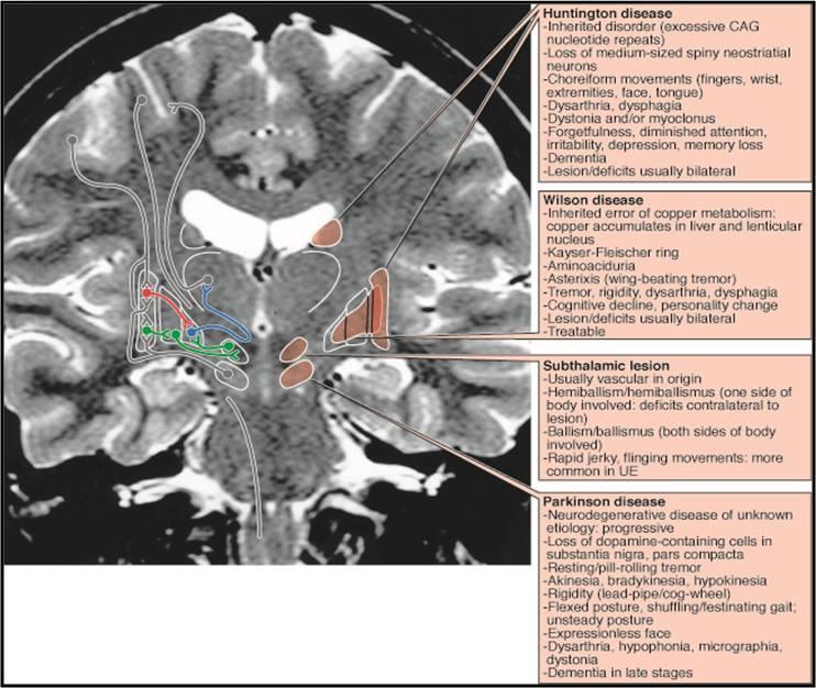
11 Treatment: - Patient lacks dopamine we give him L-Dopa - Loss of neurons Deep brain stimulation (in deep brain stimulation we either activate substantia nigra OR we inhibit the indirect pathway through inhibition of Subthalamic nucleus or Globus pallidus internal by electrode.(this is more common)) Nowadays, Internal circuits include Acetylcholine Anticholinergic drugs In some cases we mix dopamine and anticholinergic drugs for treatment. Antichloinergic drugs target AMPA (glutamate receptor) and A2 (alpha 2 receptor). (Commercially available) - Also basal ganglia contain substance P which is also targeted nowadays in treating Parkinson s. Hyperkinetic disorders : Disorder Huntington s chorea Dystonia Tardive dyskinesia DOPA-induced dyskinesia Hemiballismus Tourette s syndrome Causes Genetic (autosomal dominant) Genetic or idiopathic Chronic use of neuroleptic drugs Parkinson s therapy (antipsychotic therapy) Unilateral vascular accident, typically subthalamic nucleus Excessive D2-subtype DA receptor expression Genetic or idiopathic but mainly genetic Huntington s chorea: caused by degeneration of GABAergic neurons (atrophy of striatum), there would be excessive direct pathway and insufficient indirect pathway; it starts rostral and medial then caudal and lateral. Rostral part of basal ganglia (head of caudate) is affected first, accordingly early symptoms of Huntington s chorea are psychotic (depression) before motor, then motor is affected and it ends by the death of the patient within years. Rostral motor signs are: chorea (brief, involuntary movements) and dystonia (abnormal postures).
 If it is a bacterial infection specifically strep type A.")
12 Huntington s Normal degenerated head of caudate Dystonia: involuntary movement in one or more of the limbs (proximal and not distal unlike Athetosis). It s mostly idiopathic although it might be caused by trauma, ions concentration imbalance or infection (bacterial/viral) If it is a bacterial infection specifically strep type A. infection that is causing dystonia, then it s called Sydenham Chorea Sydenham chorea occurs in children, its symptoms appear 3-4 weeks after the infection, usually it resolves within 6 months but sometimes it doesn t. REMEMBER, If a lesion was found in: Head of caudate Huntington s chorea Lenticular nucleus Wilson disease (AKA hepatolenticular disorder) Subthalamic nucleus Hemiballismus Substantia Nigra Parkinson's disease
13
The motor regulator. 1) Basal ganglia/nucleus
 Basal ganglia/nucleus") The motor regulator 1) Basal ganglia/nucleus Neural structures involved in the control of movement Basal Ganglia - Components of the basal ganglia - Function of the basal ganglia - Connection and circuits
The motor regulator 1) Basal ganglia/nucleus Neural structures involved in the control of movement Basal Ganglia - Components of the basal ganglia - Function of the basal ganglia - Connection and circuits
神經解剖學 NEUROANATOMY BASAL NUCLEI 盧家鋒助理教授臺北醫學大學醫學系解剖學暨細胞生物學科臺北醫學大學醫學院轉譯影像研究中心.
 神經解剖學 NEUROANATOMY BASAL NUCLEI 盧家鋒助理教授臺北醫學大學醫學系解剖學暨細胞生物學科臺北醫學大學醫學院轉譯影像研究中心 http://www.ym.edu.tw/~cflu OUTLINE Components and Pathways of the Basal Nuclei Functions and Related Disorders of the Basal Nuclei
A. General features of the basal ganglia, one of our 3 major motor control centers:
 Reading: Waxman pp. 141-146 are not very helpful! Computer Resources: HyperBrain, Chapter 12 Dental Neuroanatomy Suzanne S. Stensaas, Ph.D. April 22, 2010 THE BASAL GANGLIA Objectives: 1. What are the
A. General features of the basal ganglia, one of our 3 major motor control centers:
 Reading: Waxman pp. 141-146 are not very helpful! Computer Resources: HyperBrain, Chapter 12 Dental Neuroanatomy Suzanne S. Stensaas, Ph.D. March 1, 2012 THE BASAL GANGLIA Objectives: 1. What are the main
Basal ganglia Sujata Sofat, class of 2009
 Basal ganglia Sujata Sofat, class of 2009 Basal ganglia Objectives Describe the function of the Basal Ganglia in movement Define the BG components and their locations Describe the motor loop of the BG
VL VA BASAL GANGLIA. FUNCTIONAl COMPONENTS. Function Component Deficits Start/initiation Basal Ganglia Spontan movements
 BASAL GANGLIA Chris Cohan, Ph.D. Dept. of Pathology/Anat Sci University at Buffalo I) Overview How do Basal Ganglia affect movement Basal ganglia enhance cortical motor activity and facilitate movement.
Damage on one side.. (Notes) Just remember: Unilateral damage to basal ganglia causes contralateral symptoms.
 Just remember: Unilateral damage to basal ganglia causes contralateral symptoms.") Lecture 20 - Basal Ganglia Basal Ganglia (Nolte 5 th Ed pp 464) Damage to the basal ganglia produces involuntary movements. Although the basal ganglia do not influence LMN directly (to cause this involuntary
Lecture 20 - Basal Ganglia Basal Ganglia (Nolte 5 th Ed pp 464) Damage to the basal ganglia produces involuntary movements. Although the basal ganglia do not influence LMN directly (to cause this involuntary
Anatomy of the basal ganglia. Dana Cohen Gonda Brain Research Center, room 410
 Anatomy of the basal ganglia Dana Cohen Gonda Brain Research Center, room 410 danacoh@gmail.com The basal ganglia The nuclei form a small minority of the brain s neuronal population. Little is known about
COGNITIVE SCIENCE 107A. Motor Systems: Basal Ganglia. Jaime A. Pineda, Ph.D.
 COGNITIVE SCIENCE 107A Motor Systems: Basal Ganglia Jaime A. Pineda, Ph.D. Two major descending s Pyramidal vs. extrapyramidal Motor cortex Pyramidal system Pathway for voluntary movement Most fibers originate
Basal Ganglia George R. Leichnetz, Ph.D.
 Basal Ganglia George R. Leichnetz, Ph.D. OBJECTIVES 1. To understand the brain structures which constitute the basal ganglia, and their interconnections 2. To understand the consequences (clinical manifestations)
Connections of basal ganglia
 Connections of basal ganglia Introduction The basal ganglia, or basal nuclei, are areas of subcortical grey matter that play a prominent role in modulating movement, as well as cognitive and emotional
Making Things Happen 2: Motor Disorders
 Making Things Happen 2: Motor Disorders How Your Brain Works Prof. Jan Schnupp wschnupp@cityu.edu.hk HowYourBrainWorks.net On the Menu in This Lecture In the previous lecture we saw how motor cortex and
Strick Lecture 4 March 29, 2006 Page 1
 Strick Lecture 4 March 29, 2006 Page 1 Basal Ganglia OUTLINE- I. Structures included in the basal ganglia II. III. IV. Skeleton diagram of Basal Ganglia Loops with cortex Similarity with Cerebellar Loops
Dr. Farah Nabil Abbas. MBChB, MSc, PhD
 Dr. Farah Nabil Abbas MBChB, MSc, PhD The Basal Ganglia *Functions in association with motor cortex and corticospinal pathways. *Regarded as accessory motor system besides cerebellum. *Receive most of
Teach-SHEET Basal Ganglia
 Teach-SHEET Basal Ganglia Purves D, et al. Neuroscience, 5 th Ed., Sinauer Associates, 2012 Common organizational principles Basic Circuits or Loops: Motor loop concerned with learned movements (scaling
UNIVERSITY OF JORDAN FACULTY OF MEDICINE DEPARTMENT OF PHYSIOLOGY & BIOCHEMISTRY NEUROPHYSIOLOGY (MEDICAL) Spring, 2014
 Spring, 2014") UNIVERSITY OF JORDAN FACULTY OF MEDICINE DEPARTMENT OF PHYSIOLOGY & BIOCHEMISTRY NEUROPHYSIOLOGY (MEDICAL) Spring, 2014 Textbook of Medical Physiology by: Guyton & Hall, 11 th edition 2006 Eman Al-Khateeb,
UNIVERSITY OF JORDAN FACULTY OF MEDICINE DEPARTMENT OF PHYSIOLOGY & BIOCHEMISTRY NEUROPHYSIOLOGY (MEDICAL) Spring, 2014 Textbook of Medical Physiology by: Guyton & Hall, 11 th edition 2006 Eman Al-Khateeb,
Outline of the next three lectures
 Outline of the next three lectures Lecture 35 Anatomy of the human cerebral cortex gross and microscopic cell types connections Vascular supply of the cerebral cortex Disorders involving the cerebral cortex
Basal Ganglia. Steven McLoon Department of Neuroscience University of Minnesota
 Basal Ganglia Steven McLoon Department of Neuroscience University of Minnesota 1 Course News Graduate School Discussion Wednesday, Nov 1, 11:00am MoosT 2-690 with Paul Mermelstein (invite your friends)
NS219: Basal Ganglia Anatomy
 NS219: Basal Ganglia Anatomy Human basal ganglia anatomy Analagous rodent basal ganglia nuclei Basal ganglia circuits: the classical model of direct and indirect pathways + Glutamate + - GABA - Gross anatomy
The Wonders of the Basal Ganglia
 Basal Ganglia The Wonders of the Basal Ganglia by Mackenzie Breton and Laura Strong /// https://kin450- neurophysiology.wikispaces.com/basal+ganglia Introduction The basal ganglia are a group of nuclei
PSY 315 Lecture 11 (2/23/2011) (Motor Control) Dr. Achtman PSY 215. Lecture 11 Topic: Motor System Chapter 8, pages
 (Motor Control) Dr. Achtman PSY 215. Lecture 11 Topic: Motor System Chapter 8, pages") Corrections: No Corrections Announcements: Exam #2 next Wednesday, March 2, 2011 Monday February 28, 2011 we will be going over the somatosensory system, and there will be time left in class to review
Corrections: No Corrections Announcements: Exam #2 next Wednesday, March 2, 2011 Monday February 28, 2011 we will be going over the somatosensory system, and there will be time left in class to review
For more information about how to cite these materials visit
 Author(s): Peter Hitchcock, PH.D., 2009 License: Unless otherwise noted, this material is made available under the terms of the Creative Commons Attribution Non-commercial Share Alike 3.0 License: http://creativecommons.org/licenses/by-nc-sa/3.0/
Movement Disorders. Psychology 372 Physiological Psychology. Background. Myasthenia Gravis. Many Types
 Background Movement Disorders Psychology 372 Physiological Psychology Steven E. Meier, Ph.D. Listen to the audio lecture while viewing these slides Early Studies Found some patients with progressive weakness
DISORDERS OF THE MOTOR SYSTEM. Jeanette J. Norden, Ph.D. Professor Emerita Vanderbilt University School of Medicine
 DISORDERS OF THE MOTOR SYSTEM Jeanette J. Norden, Ph.D. Professor Emerita Vanderbilt University School of Medicine THE MOTOR SYSTEM To understand disorders of the motor system, we need to review how a
Biological Bases of Behavior. 8: Control of Movement
 Biological Bases of Behavior 8: Control of Movement m d Skeletal Muscle Movements of our body are accomplished by contraction of the skeletal muscles Flexion: contraction of a flexor muscle draws in a
Neurodegenerative Disease. April 12, Cunningham. Department of Neurosciences
 Neurodegenerative Disease April 12, 2017 Cunningham Department of Neurosciences NEURODEGENERATIVE DISEASE Any of a group of hereditary and sporadic conditions characterized by progressive dysfunction,
Lecture XIII. Brain Diseases I - Parkinsonism! Brain Diseases I!
 Lecture XIII. Brain Diseases I - Parkinsonism! Bio 3411! Wednesday!! Lecture XIII. Brain Diseases - I.! 1! Brain Diseases I! NEUROSCIENCE 5 th ed! Page!!Figure!!Feature! 408 18.9 A!!Substantia Nigra in
Chapter 8. Control of movement
 Chapter 8 Control of movement 1st Type: Skeletal Muscle Skeletal Muscle: Ones that moves us Muscles contract, limb flex Flexion: a movement of a limb that tends to bend its joints, contraction of a flexor
GBME graduate course. Chapter 43. The Basal Ganglia
 GBME graduate course Chapter 43. The Basal Ganglia Basal ganglia in history Parkinson s disease Huntington s disease Parkinson s disease 1817 Parkinson's disease (PD) is a degenerative disorder of the
Voluntary Movement. Ch. 14: Supplemental Images
 Voluntary Movement Ch. 14: Supplemental Images Skeletal Motor Unit: The basics Upper motor neuron: Neurons that supply input to lower motor neurons. Lower motor neuron: neuron that innervates muscles,
Basal Ganglia. Introduction. Basal Ganglia at a Glance. Role of the BG
 Basal Ganglia Shepherd (2004) Chapter 9 Charles J. Wilson Instructor: Yoonsuck Choe; CPSC 644 Cortical Networks Introduction A set of nuclei in the forebrain and midbrain area in mammals, birds, and reptiles.
Functional Distinctions
 Functional Distinctions FUNCTION COMPONENT DEFICITS Start Basal Ganglia Spontaneous Movements Move UMN/LMN Cerebral Cortex Brainstem, Spinal cord Roots/peripheral nerves Plan Cerebellum Ataxia Adjust Cerebellum
skilled pathways: distal somatic muscles (fingers, hands) (brainstem, cortex) are giving excitatory signals to the descending pathway
 (brainstem, cortex) are giving excitatory signals to the descending pathway") L15 - Motor Cortex General - descending pathways: how we control our body - motor = somatic muscles and movement (it is a descending motor output pathway) - two types of movement: goal-driven/voluntary
L15 - Motor Cortex General - descending pathways: how we control our body - motor = somatic muscles and movement (it is a descending motor output pathway) - two types of movement: goal-driven/voluntary
PETER PAZMANY CATHOLIC UNIVERSITY Consortium members SEMMELWEIS UNIVERSITY, DIALOG CAMPUS PUBLISHER
 PETER PAZMANY CATHOLIC UNIVERSITY SEMMELWEIS UNIVERSITY Development of Complex Curricula for Molecular Bionics and Infobionics Programs within a consortial* framework** Consortium leader PETER PAZMANY
Basal Ganglia General Info
 Basal Ganglia General Info Neural clusters in peripheral nervous system are ganglia. In the central nervous system, they are called nuclei. Should be called Basal Nuclei but usually called Basal Ganglia.
The basal forebrain: Questions, chapter 29:
 The basal forebrain: Questions, chapter 29: 7) What is the "basal forebrain", and what is its involvement in Alzheimer' s Disease? The acetylcholine-containing neurons of the nucleus basalis of Meynart
MODULE 6: CEREBELLUM AND BASAL GANGLIA
 MODULE 6: CEREBELLUM AND BASAL GANGLIA This module will summarize the important neuroanatomical and key clinical concepts from Chapters 15 and 16 of the textbook for the course. The first part of this
Chapter 2: Studies of Human Learning and Memory. From Mechanisms of Memory, second edition By J. David Sweatt, Ph.D.
 Chapter 2: Studies of Human Learning and Memory From Mechanisms of Memory, second edition By J. David Sweatt, Ph.D. Medium Spiny Neuron A Current Conception of the major memory systems in the brain Figure
Systems Neuroscience Dan Kiper. Today: Wolfger von der Behrens
 Systems Neuroscience Dan Kiper Today: Wolfger von der Behrens wolfger@ini.ethz.ch 18.9.2018 Neurons Pyramidal neuron by Santiago Ramón y Cajal (1852-1934, Nobel prize with Camillo Golgi in 1906) Neurons
BASAL GANGLIA. Dr JAMILA EL MEDANY
 BASAL GANGLIA Dr JAMILA EL MEDANY OBJECTIVES At the end of the lecture, the student should be able to: Define basal ganglia and enumerate its components. Enumerate parts of Corpus Striatum and their important
Motor System Hierarchy
 Motor Pathways Lectures Objectives Define the terms upper and lower motor neurons with examples. Describe the corticospinal (pyramidal) tract and the direct motor pathways from the cortex to the trunk
Computational cognitive neuroscience: 8. Motor Control and Reinforcement Learning
 1 Computational cognitive neuroscience: 8. Motor Control and Reinforcement Learning Lubica Beňušková Centre for Cognitive Science, FMFI Comenius University in Bratislava 2 Sensory-motor loop The essence
Study Guide Unit 2 Psych 2022, Fall 2003
 Study Guide Unit 2 Psych 2022, Fall 2003 Subcortical Anatomy 1. Be able to locate the following structures and be able to indicate whether they are located in the forebrain, diencephalon, midbrain, pons,
Neocortex. Cortical Structures in the Brain. Neocortex Facts. Laminar Organization. Bark-like (cortical) structures: Shepherd (2004) Chapter 12
 structures: Shepherd (2004) Chapter 12") Neocortex Shepherd (2004) Chapter 12 Rodney Douglas, Henry Markram, and Kevan Martin Instructor: Yoonsuck Choe; CPSC 644 Cortical Networks Cortical Structures in the Brain Bark-like (cortical) structures:
Neocortex Shepherd (2004) Chapter 12 Rodney Douglas, Henry Markram, and Kevan Martin Instructor: Yoonsuck Choe; CPSC 644 Cortical Networks Cortical Structures in the Brain Bark-like (cortical) structures:
MOVEMENT OUTLINE. The Control of Movement: Muscles! Motor Reflexes Brain Mechanisms of Movement Mirror Neurons Disorders of Movement
 MOVEMENT 2 Dr. Steinmetz 3 OUTLINE The Control of Movement: Muscles! Motor Reflexes Brain Mechanisms of Movement Mirror Neurons Disorders of Movement Parkinson s Disease Huntington s Disease 1 4 TYPES
Exam 2 PSYC Fall (2 points) Match a brain structure that is located closest to the following portions of the ventricular system
 Match a brain structure that is located closest to the following portions of the ventricular system") Exam 2 PSYC 2022 Fall 1998 (2 points) What 2 nuclei are collectively called the striatum? (2 points) Match a brain structure that is located closest to the following portions of the ventricular system
Exam 2 PSYC 2022 Fall 1998 (2 points) What 2 nuclei are collectively called the striatum? (2 points) Match a brain structure that is located closest to the following portions of the ventricular system
Brain anatomy and artificial intelligence. L. Andrew Coward Australian National University, Canberra, ACT 0200, Australia
 Brain anatomy and artificial intelligence L. Andrew Coward Australian National University, Canberra, ACT 0200, Australia The Fourth Conference on Artificial General Intelligence August 2011 Architectures
CN V! touch! pain! Touch! P/T!
 CN V! touch! pain! Touch! P/T! Visual Pathways! L! R! B! A! C! D! LT! E! F! RT! G! hypothalamospinal! and! ALS! Vestibular Pathways! 1. Posture/Balance!!falling! 2. Head Position! 3. Eye-Head Movements
Extrapyramidal Motor System. Basal Ganglia or Striatum. Basal Ganglia or Striatum 3/3/2010
 Extrapyramidal Motor System Basal Ganglia or Striatum Descending extrapyramidal paths receive input from other parts of motor system: From the cerebellum From the basal ganglia or corpus striatum Caudate
nucleus accumbens septi hier-259 Nucleus+Accumbens birnlex_727
 Nucleus accumbens From Wikipedia, the free encyclopedia Brain: Nucleus accumbens Nucleus accumbens visible in red. Latin NeuroNames MeSH NeuroLex ID nucleus accumbens septi hier-259 Nucleus+Accumbens birnlex_727
Parkinsonism or Parkinson s Disease I. Symptoms: Main disorder of movement. Named after, an English physician who described the then known, in 1817.
 Parkinsonism or Parkinson s Disease I. Symptoms: Main disorder of movement. Named after, an English physician who described the then known, in 1817. Four (4) hallmark clinical signs: 1) Tremor: (Note -
Course Calendar - Neuroscience
 2006-2007 Course Calendar - Neuroscience Meeting Hours for entire semester: Monday - Friday 1:00-2:20 p.m. Room 1200, COM August 28 August 29 August 30 August 31 September 1 Course introduction, Neurocytology:
Basal nuclei, cerebellum and movement
 Basal nuclei, cerebellum and movement MSTN121 - Neurophysiology Session 9 Department of Myotherapy Basal Nuclei (Ganglia) Basal Nuclei (Ganglia) Role: Predict the effects of various actions, then make
Course Calendar
 Clinical Neuroscience BMS 6706C Charles, Ph.D., Course Director charles.ouimet@med.fsu.edu (850) 644-2271 2004 2005 Course Calendar Click here to return to the syllabus Meeting Hours for entire semester:
Movement disorders. Dr Rick Leventer Royal Children s Hospital
 Movement disorders Dr Rick Leventer Royal Children s Hospital richard.leventer@rch.org.au Movement disorders! conditions that affect the speed, fluency, quality, and ease of movement! usually affect TONE
STRUCTURE AND CIRCUITS OF THE BASAL GANGLIA
 STRUCTURE AND CIRCUITS OF THE BASAL GANGLIA Rastislav Druga Department of Anatomy, Second Faculty of Medicine 2017 Basal ganglia Nucleus caudatus, putamen, globus pallidus (medialis et lateralis), ncl.
1. The cerebellum coordinates fine movement through interactions with the following motor-associated areas:
 DENT/OBHS 131 2009 Take-home test 4 Week 6: Take-home test (2/11/09 close 2/18/09) 1. The cerebellum coordinates fine movement through interactions with the following motor-associated areas: Hypothalamus
1/2/2019. Basal Ganglia & Cerebellum a quick overview. Outcomes you want to accomplish. MHD-Neuroanatomy Neuroscience Block. Basal ganglia review
 This power point is made available as an educational resource or study aid for your use only. This presentation may not be duplicated for others and should not be redistributed or posted anywhere on the
A3.1.7 Motor Control. 10 November 2016 Institute of Psychiatry,Psychology and Neuroscience Marinela Vavla
 A3.1.7 Motor Control 10 November 2016 Institute of Psychiatry,Psychology and Neuroscience Marinela Vavla marinela.vavla@kcl.ac.uk Learning objectives Motor systems: components & organization Spinal cord
Department of Neurology, Rigshospitalet, 9 Blegdamsvej, PAULSON, O.B. Involuntary Movements. Tohoku J. Exp. Med., 1990, 161,
 Tohoku J. Exp. Med., 1990, 161, Suppl., 21-27 Involuntary Movements OLAF B. PAULSON Department of Neurology, Rigshospitalet, 9 Blegdamsvej, DK-2100, Copenhagen, Denmark PAULSON, O.B. Involuntary Movements.
Nsci 2100: Human Neuroanatomy 2017 Examination 3
 Name KEY Lab Section Nsci 2100: Human Neuroanatomy 2017 Examination 3 On this page, write your name and lab section. On your bubble answer sheet, enter your name (last name, space, first name), internet
Visualization and simulated animations of pathology and symptoms of Parkinson s disease
 Visualization and simulated animations of pathology and symptoms of Parkinson s disease Prof. Yifan HAN Email: bctycan@ust.hk 1. Introduction 2. Biochemistry of Parkinson s disease 3. Course Design 4.
Neuroanatomy. Dr. Maha ELBeltagy. Assistant Professor of Anatomy Faculty of Medicine The University of Jordan
 Neuroanatomy Dr. Maha ELBeltagy Assistant Professor of Anatomy Faculty of Medicine The University of Jordan 2018 Prof Yousry 10/15/17 Types of brain fibers THE WHITE MATTER OF THE BRAIN The white matter
Movement Disorders Will Garrett, M.D Assistant Professor of Neurology
 Movement Disorders Will Garrett, M.D Assistant Professor of Neurology I. The Basal Ganglia The basal ganglia are composed of several structures including the caudate and putamen (collectively called the
CNS MCQ 2 nd term. Select the best answer:
 Select the best answer: CNS MCQ 2 nd term 1) Vestibular apparatus: a) Represent the auditory part of the labyrinth. b) May help in initiating the voluntary movements. c) Contains receptors concerned with
2401 : Anatomy/Physiology
 Dr. Chris Doumen Week 7 2401 : Anatomy/Physiology The Cerebrum Central Nervous System TextBook Readings Pages 434-456 and 460-461 Make use of the figures in your textbook ; a picture is worth a thousand
Psyc 311A, fall 2008 Conference week 3 TA: Jürgen Germann
 Psyc 311A, fall 2008 Conference week 3 TA: Jürgen Germann e-mail: jurgen.germann@mcgill.ca Overview: 1. Meninges 2. Cerebral cortex-cytoarchitecture 3. Diencephalon (thalamus/hypothalamus) (this replaces
Basal Nuclei (Ganglia)
") Doctor said he will not go deep within these slides because we will take them in physiology, so he will explain the anatomical structures, and he will go faster in the functions sheet in yellow Basal Nuclei
Doctor said he will not go deep within these slides because we will take them in physiology, so he will explain the anatomical structures, and he will go faster in the functions sheet in yellow Basal Nuclei
10/3/2016. T1 Anatomical structures are clearly identified, white matter (which has a high fat content) appears bright.
 appears bright.") H2O -2 atoms of Hydrogen, 1 of Oxygen Hydrogen just has one single proton and orbited by one single electron Proton has a magnetic moment similar to the earths magnetic pole Also similar to earth in that
H2O -2 atoms of Hydrogen, 1 of Oxygen Hydrogen just has one single proton and orbited by one single electron Proton has a magnetic moment similar to the earths magnetic pole Also similar to earth in that
Cerebral Cortex 1. Sarah Heilbronner
 Cerebral Cortex 1 Sarah Heilbronner heilb028@umn.edu Want to meet? Coffee hour 10-11am Tuesday 11/27 Surdyk s Overview and organization of the cerebral cortex What is the cerebral cortex? Where is each
I: To describe the pyramidal and extrapyramidal tracts. II: To discuss the functions of the descending tracts.
 Descending Tracts I: To describe the pyramidal and extrapyramidal tracts. II: To discuss the functions of the descending tracts. III: To define the upper and the lower motor neurons. 1. The corticonuclear
Brain Stem and cortical control of motor function. Dr Z Akbari
 Brain Stem and cortical control of motor function Dr Z Akbari Brain stem control of movement BS nuclear groups give rise to descending motor tracts that influence motor neurons and their associated interneurons
KINE 4500 Neural Control of Movement. Lecture #1:Introduction to the Neural Control of Movement. Neural control of movement
 KINE 4500 Neural Control of Movement Lecture #1:Introduction to the Neural Control of Movement Neural control of movement Kinesiology: study of movement Here we re looking at the control system, and what
The Motor Systems. What s the motor system? Plan
 The Motor Systems What s the motor system? Parts of CNS and PNS specialized for control of limb, trunk, and eye movements Also holds us together From simple reflexes (knee jerk) to voluntary movements
Organization of the nervous system 2
 Organization of the nervous system 2 Raghav Rajan Bio 334 Neurobiology I August 22nd 2013 1 Orienting within the brain absolute axes and relative axes SUPERIOR (above) ANTERIOR (in front) Anterior/Posterior,
1. Which part of the brain is responsible for planning and initiating movements?
 Section: Chapter 10: Multiple Choice 1. Which part of the brain is responsible for planning and initiating movements? p.358 frontal lobe hippocampus basal ganglia cerebellum 2. The prefrontal cortex is
We are IntechOpen, the world s leading publisher of Open Access books Built by scientists, for scientists. International authors and editors
 We are IntechOpen, the world s leading publisher of Open Access books Built by scientists, for scientists 3,350 108,000 1.7 M Open access books available International authors and editors Downloads Our
Neural Basis of Motor Control. Chapter 4
 Neural Basis of Motor Control Chapter 4 Neurological Perspective A basic understanding of the physiology underlying the control of voluntary movement establishes a more comprehensive appreciation and awareness
The Cerebellum. Outline. Lu Chen, Ph.D. MCB, UC Berkeley. Overview Structure Micro-circuitry of the cerebellum The cerebellum and motor learning
 The Cerebellum Lu Chen, Ph.D. MCB, UC Berkeley 1 Outline Overview Structure Micro-circuitry of the cerebellum The cerebellum and motor learning 2 Overview Little brain 10% of the total volume of the brain,
Neural Basis of Motor Control
 Neural Basis of Motor Control Central Nervous System Skeletal muscles are controlled by the CNS which consists of the brain and spinal cord. Determines which muscles will contract When How fast To what
Degree of freedom problem
 KINE 4500 Neural Control of Movement Lecture #1:Introduction to the Neural Control of Movement Neural control of movement Kinesiology: study of movement Here we re looking at the control system, and what
Chapter 3. Structure and Function of the Nervous System. Copyright (c) Allyn and Bacon 2004
 Allyn and Bacon 2004") Chapter 3 Structure and Function of the Nervous System 1 Basic Features of the Nervous System Neuraxis: An imaginary line drawn through the center of the length of the central nervous system, from the
Chapter 3 Structure and Function of the Nervous System 1 Basic Features of the Nervous System Neuraxis: An imaginary line drawn through the center of the length of the central nervous system, from the
Brainstem. Steven McLoon Department of Neuroscience University of Minnesota
 Brainstem Steven McLoon Department of Neuroscience University of Minnesota 1 Course News Change in Lab Sequence Week of Oct 2 Lab 5 Week of Oct 9 Lab 4 2 Goal Today Know the regions of the brainstem. Know
Overview of Brain Structures
 First Overview of Brain Structures Psychology 470 Introduction to Chemical Additions Steven E. Meier, Ph.D. All parts are interrelated. You need all parts to function normally. Neurons = Nerve cells Listen
9.14 Class 32 Review. Limbic system
 9.14 Class 32 Review Limbic system 1 Lateral view Medial view Brainstem, sagittal section Sensory- Perceptual Motor Behavior Major functional modules of the CNS Motivation Courtesy of MIT Press. Used with
Motor Functions of Cerebral Cortex
 Motor Functions of Cerebral Cortex I: To list the functions of different cortical laminae II: To describe the four motor areas of the cerebral cortex. III: To discuss the functions and dysfunctions of
Introduction to the Central Nervous System: Internal Structure
 Introduction to the Central Nervous System: Internal Structure Objective To understand, in general terms, the internal organization of the brain and spinal cord. To understand the 3-dimensional organization
CHAPTER 16 LECTURE OUTLINE
 CHAPTER 16 LECTURE OUTLINE I. INTRODUCTION A. The components of the brain interact to receive sensory input, integrate and store the information, and transmit motor responses. B. To accomplish the primary
Cheyenne 11/28 Neurological Disorders II. Transmissible Spongiform Encephalopathy
 Cheyenne 11/28 Neurological Disorders II Transmissible Spongiform Encephalopathy -E.g Bovine4 Spongiform Encephalopathy (BSE= mad cow disease), Creutzfeldt-Jakob disease, scrapie (animal only) -Sporadic:
Prof. Saeed Abuel Makarem & Dr.Sanaa Alshaarawy
 Prof. Saeed Abuel Makarem & Dr.Sanaa Alshaarawy 1 Objectives By the end of the lecture, you should be able to: Describe the anatomy and main functions of the thalamus. Name and identify different nuclei
The Cerebral Cortex and Higher Intellectual Functions
 The Cerebral Cortex and Higher Intellectual Functions The Cerebral cortex consists of 2 cerebral hemisphere and each hemisphere consists of 5 lobes (frontal, parietal,temporal,occipital,insular lobe which
Chapter 14: Integration of Nervous System Functions I. Sensation.
 Chapter 14: Integration of Nervous System Functions I. Sensation A. General Organization 1. General senses have receptors a. The somatic senses provide information about & 1. Somatic senses include: a.
Embryological origin of thalamus
 diencephalon Embryological origin of thalamus The diencephalon gives rise to the: Thalamus Epithalamus (pineal gland, habenula, paraventricular n.) Hypothalamus Subthalamus (Subthalamic nuclei) The Thalamus:
BASAL GANGLIA: A "pit stop" that integrates the movement, cognition and emotion.
 BASAL GANGLIA: A "pit stop" that integrates the movement, cognition and emotion. Poster No.: C-0795 Congress: ECR 2011 Type: Educational Exhibit Authors: V. M. González Montaño, T. M. Zamorano Pozo, R.
Spinal Interneurons. Control of Movement
 Control of Movement Spinal Interneurons Proprioceptive afferents have a variety of termination patterns in the spinal cord. This can be seen by filling physiologically-identified fibers with HRP, so their
Thalamus: VA VM, MD S N. GPi Superior colliculus. compacta reticulata
 . Putamen & Caudate Putamen & Caudate GPe Neocortex Thalamus: VA VM, MD S N GPi Superior colliculus compacta reticulata Substantia Nigra Pedunculopontine nuc. of midbrain ret.form. Satellites of the corpus
Brainstem: Midbrain. 1. Midbrain gross external anatomy 2. Internal structure of the midbrain:
 Brainstem: Midbrain 1. Midbrain gross external anatomy 2. Internal structure of the midbrain: cerebral peduncles tegmentum tectum (guadrigeminal plate) Midbrain Midbrain general features location between
Anatomy and Physiology (Bio 220) The Brain Chapter 14 and select portions of Chapter 16
 The Brain Chapter 14 and select portions of Chapter 16") Anatomy and Physiology (Bio 220) The Brain Chapter 14 and select portions of Chapter 16 I. Introduction A. Appearance 1. physical 2. weight 3. relative weight B. Major parts of the brain 1. cerebrum 2.
Anatomy and Physiology (Bio 220) The Brain Chapter 14 and select portions of Chapter 16 I. Introduction A. Appearance 1. physical 2. weight 3. relative weight B. Major parts of the brain 1. cerebrum 2.
Unit VIII Problem 5 Physiology: Cerebellum
 Unit VIII Problem 5 Physiology: Cerebellum - The word cerebellum means: the small brain. Note that the cerebellum is not completely separated into 2 hemispheres (they are not clearly demarcated) the vermis
The Central Nervous System I. Chapter 12
 The Central Nervous System I Chapter 12 The Central Nervous System The Brain and Spinal Cord Contained within the Axial Skeleton Brain Regions and Organization Medical Scheme (4 regions) 1. Cerebral Hemispheres