Latent HSV-1 does not induce apoptosis in human trigeminal ganglia
|
|
|
- Ross Jordan
- 6 years ago
- Views:
Transcription
1 JVI Accepted Manuscript Posted Online 11 March 2015 J. Virol. doi: /jvi Copyright 2015, American Society for Microbiology. All Rights Reserved. 1 Latent HSV-1 does not induce apoptosis in human trigeminal ganglia Susanne Himmelein 1,2#, Anja Lindemann 1,2, Inga Sinicina 3, Michael Strupp 1,2, Thomas Brandt 2,5, Katharina Hüfner 1,2* Affiliations 1 Department of Neurology, Klinikum Grosshadern, Ludwig Maximilians University, Munich, Germany; 2 German Center for Vertigo and Balance Disorders, DSGZ, Ludwig Maximilians University, Munich, Germany; 3 Department of Legal Medicine, Ludwig Maximilians University, Munich, Germany; 5 Institute for Clinical Neurosciences, Klinikum Grosshadern, Ludwig Maximilians University, Munich, Germany; *Present address: Department of Biological Psychiatry, Medical University of Innsbruck, Innsbruck, Austria # Corresponding author: Dr. rer. nat. Susanne Himmelein, Department of Neurology, German Center for Vertigo and Balance Disorders, DSGZ, Klinikum Grosshadern, Ludwig Maximilians University, Feodor-Lynen Str. 19, Munich, Germany. Telephone: , Fax: and susanne.himmelein@med.uni-muenchen.de Running title: HSV-1 does not induce apoptosis Abstract in words: 74 Words in text:
2 Abstract Herpes-simplex-virus type 1 (HSV-1) can establish lifelong latency in human trigeminal ganglia. Latently infected ganglia contain CD8 + T cells which secrete granzyme B and are thus capable of inducing neuronal apoptosis. Using immunohistochemistry and single-cell RT-qPCR higher frequency and transcript levels of caspase-3 were found in HSV-1 negative compared to positive ganglia and neurons respectively. No TUNEL assay positive neurons were detected. The infiltrating T cells do not induce apoptosis in latently infected neurons Herpes-simplex-virus type 1 (HSV-1) can establish lifelong latency in sensory neurons of the trigeminal ganglia (TG). HSV-1 latency is characterized by expression of one viral RNA (the latency associated transcript (LAT)) in the absence of viral protein. LAT is believed to play a role in establishing latency (1, 2), in facilitating the process of reactivation (3-5), and at the same time promoting neuronal survival after HSV-1 infection by reducing apoptosis (6). In vitro, the anti-apoptotic effects of LAT are mediated by the inhibition of caspase-3-, -8- and -9-induced apoptosis (7, 8). In humans and animal models, CD8 + T cells are found in latently infected ganglia (9, 10). These CD8 + T cells have been shown to release lytic granules containing granzyme B (GrB) in humans (9, 11) and mice (12, 13). In the setting of HSV-1 latency, rather than inducing apoptosis, GrB cleaves infected-cell polypeptide 4 (ICP4)(13), an essential viral protein needed for viral gene expression, thereby preventing viral reactivation (14). It is a wellknown clinical phenomenon that even after multiple reactivations of HSV-1 in the TG, no sensory deficits occur. Here we investigate if this observation is mirrored by the pathoanatomical findings in human TG latently infected with HSV-1. 2
3 Human TG were obtained at autopsy at Ludwig Maximilians University (Munich, Germany) with the approval of the Ethics Committee of the Medical Faculty of the University (Supplementary Table S1). Whole ganglia including neurons projecting to all three branches were embedded directly after removal in Jung Tissue freezing medium (Leica Microsystems, Nussloch, Germany). Frozen sections of 10μm were cut for immunohistochemistry and ISH, while RNA and DNA were isolated from ten pooled 30μm sections. Immunohistochemistry was performed with antibodies against active caspase-3 (R&D Systems, Wiesbaden, Germany) and GrB (AbD Serotec, Puchheim, Germany), as described previously (15, 16). Staining was visualized using biotinconjugated secondary antibody (Dako, Hamburg, Germany), HRP-conjugated streptavidin (BioLegend, Fell, Germany), and 3-3 -diaminobenzidine (DAB; Dako) under an all-in-one fluorescence microscope (BZ-8100E, Keyence, Neu-Isenburg, Germany). Apoptosis was detected using a commercially available assay based on detecting terminal deoxynucleotidyl transferase (TdT)-mediated dutp nick end labeling, according to the manufacturer s instructions (Promega, Madison, USA). In situ hybridization for HSV-1 LAT (16, 17) was performed in combination with immunofluorescence against GrB or CD8 in subsequent staining steps. Appropriate fluorophore-conjugated probe or antibody was used for LAT and GrB or CD8 respectively (Dianova, Hamburg, Germany). Laser capture microdissection was done as described (17). Thirty LAT+ or LAT- neurons were marked electronically and microdissected. Single-cell RT-qPCR was performed using an Ambion kit (Life Technologies, Darmstadt, Germany) according to the manufacturer s instructions. Commercially and custom-made TaqMan Gene Expression 3
4 Assays were used (Life Technologies, Darmstadt, Germany). Statistical analyses were performed in Microsoft Excel and SPSS; p<0.05 was regarded as significant. Human TG sections from LAT+ and LAT- ganglia were investigated for signs of neuronal apoptosis: no TUNEL-positive neurons were found (Figure 1 A-C). Immunohistochemistry for the expression of active caspase-3 revealed neuronal staining in a limited number of cases (Table 1, Figure 1 D-F). A higher number of LAT- ganglia showed staining for active caspase-3 compared to LAT+ (Chi Square Test, p=0.043). Neurons positive for active caspase-3 did not, however, appear morphologically apoptotic. Caspase-3 expression was investigated using TaqMan RT-qPCR from the RNA of the cross-sectional area of the whole TG. No difference in the expression of caspase-3 between LAT+ and LAT- ganglia was observed (mean relative transcript number ( CI); mean ( CI), (Mann-Whitney U-Test p=0.545), Figure 2). No correlation between caspase-3 expression in whole TG and age or post-mortem delay was seen (Spearman correlation r=0.89, p=0.72; r= , p=0.87). When caspase-3 expression was assessed at a single-neuron level it was found to be higher in LAT- neurons vs LAT+ (mean relative transcript number ( CI); mean ( CI), (Mann-Whitney U-Test p=0.005), Figure 2). There was a negative correlation between the expression of LAT and caspase-3 on a single-cell level (Spearman correlation r=-0.552, p=0.009). Higher numbers of GrB positive cells were found in HSV-1 latently infected ganglia compared to non-infected (Mann-Whitney U-Test, p=0.05 (Table 1)). No indications that GrB positive cells surround more LAT+ or LAT- neurons were found (Figure 3). 4
5 In the current study we provide experimental evidence for the absence of apoptosis in sensory neurons latently infected with HSV-1. This finding could help to explain the clinical observation that sensory deficits do not occur even after recurrent HSV-1 reactivation. Elevated numbers of GrB-secreting T cells have been detected in HSV-1 latently infected ganglia. In most settings, GrB is known to cleave and activate caspase- 3, which in turn triggers the caspase cascade, leading to degradation of DNA and apoptosis (18, 19). The presence of CD8 + T cells expressing GrB in infected TG tissue suggests that these T cells are active and could induce apoptosis in infected neurons. However, the exact opposite seems to be the case. The TUNEL assays for apoptosis showed no positive neurons, and the detection of caspase-3 by immunohistochemistry was more frequent in individuals not infected with HSV-1. Active caspase-3 was found in some neurons; none of these neurons showed typical features of apoptosis in their cellular structure. This suggests that caspase-3 may play a role separate from that in the apoptotic caspase cascade. Functional studies have increasingly recognized that caspases have non-apoptotic functions in multiple cellular processes, such as inflammation, cell differentiation and proliferation (20, 21). In an animal model, active caspase-3 was also found in the nuclei of dorsal root ganglia neurons but these were not TUNEL-positive or morphologically apoptotic (22). From the current experiments we can only speculate about the mechanisms behind the inhibition of apoptosis in ganglia latently infected with HSV-1. As destruction of neurons is seen very rarely in mice (23, 24) and never in humans (11), infiltrating CD8 + T cells apparently do not release their full cytotoxic capacity. Knickelbein et al (12) describe a nonlethal mechanism of viral inactivation in which the lytic granule component, GrB, degrades the HSV-1 immediate 5
6 early protein, ICP4, which is essential for efficient viral transcription. Without inhibition of apoptosis, latently infected neurons could die in response to viral infection, thereby reducing the number of latent HSV-1 genomes through destruction of their host Acknowledgements This study was supported by a grant from the BMBF (German Ministry for Education and Research) IFB-01EO0901. We thank Katie Ogston and Sarah Flowerdew for copyediting the manuscript References 1. Thompson RL, Sawtell NM The herpes simplex virus type 1 latency-associated transcript gene regulates the establishment of latency. Journal of virology 71: Perng GC, Slanina SM, Yukht A, Ghiasi H, Nesburn AB, Wechsler SL The latencyassociated transcript gene enhances establishment of herpes simplex virus type 1 latency in rabbits. J.Virol. 74: Hill JM, Sedarati F, Javier RT, Wagner EK, Stevens JG Herpes simplex virus latent phase transcription facilitates in vivo reactivation. Virology 174: Perng GC, Dunkel EC, Geary PA, Slanina SM, Ghiasi H, Kaiwar R, Nesburn AB, Wechsler SL The latency-associated transcript gene of herpes simplex virus type 1 (HSV-1) is required for efficient in vivo spontaneous reactivation of HSV-1 from latency. Journal of virology 68: Thompson RL, Sawtell NM The herpes simplex virus type 1 latency associated transcript locus is required for the maintenance of reactivation competent latent infections. Journal of neurovirology 17: Perng GC, Jones C, Ciacci-Zanella J, Stone M, Henderson G, Yukht A, Slanina SM, Hofman FM, Ghiasi H, Nesburn AB, Wechsler SL Virus-induced neuronal apoptosis blocked by the herpes simplex virus latency-associated transcript. Science 287: Henderson G, Peng W, Jin L, Perng GC, Nesburn AB, Wechsler SL, Jones C Regulation of caspase 8- and caspase 9-induced apoptosis by the herpes simplex virus type 1 latencyassociated transcript. Journal of neurovirology 8 Suppl 2: Jiang X, Chentoufi AA, Hsiang C, Carpenter D, Osorio N, BenMohamed L, Fraser NW, Jones C, Wechsler SL The herpes simplex virus type 1 latency-associated transcript can protect neuron-derived C1300 and Neuro2A cells from granzyme B-induced apoptosis and CD8 T-cell killing. Journal of virology 85: Derfuss T, Segerer S, Herberger S, Sinicina I, Hufner K, Ebelt K, Knaus HG, Steiner I, Meinl E, Dornmair K, Arbusow V, Strupp M, Brandt T, Theil D Presence of HSV-1 immediate early 6
7 genes and clonally expanded T-cells with a memory effector phenotype in human trigeminal ganglia. Brain Pathol 17: Feldman LT, Ellison AR, Voytek CC, Yang L, Krause P, Margolis TP Spontaneous molecular reactivation of herpes simplex virus type 1 latency in mice. Proceedings of the National Academy of Sciences of the United States of America 99: Theil D, Derfuss T, Paripovic I, Herberger S, Meinl E, Schueler O, Strupp M, Arbusow V, Brandt T Latent herpesvirus infection in human trigeminal ganglia causes chronic immune response. The American journal of pathology 163: Knickelbein JE, Khanna KM, Yee MB, Baty CJ, Kinchington PR, Hendricks RL Noncytotoxic lytic granule-mediated CD8+ T cell inhibition of HSV-1 reactivation from neuronal latency. Science 322: Liu T, Tang Q, Hendricks RL Inflammatory infiltration of the trigeminal ganglion after herpes simplex virus type 1 corneal infection. Journal of virology 70: DeLuca NA, McCarthy AM, Schaffer PA Isolation and characterization of deletion mutants of herpes simplex virus type 1 in the gene encoding immediate-early regulatory protein ICP4. Journal of virology 56: Theil D, Arbusow V, Derfuss T, Strupp M, Pfeiffer M, Mascolo A, Brandt T Prevalence of HSV-1 LAT in human trigeminal, geniculate, and vestibular ganglia and its implication for cranial nerve syndromes. Brain Pathol 11: Flowerdew SE, Wick D, Himmelein S, Horn AK, Sinicina I, Strupp M, Brandt T, Theil D, Hufner K Characterization of neuronal populations in the human trigeminal ganglion and their association with latent herpes simplex virus-1 infection. PloS one 8:e Held K, Junker A, Dornmair K, Meinl E, Sinicina I, Brandt T, Theil D, Derfuss T Expression of herpes simplex virus 1-encoded micrornas in human trigeminal ganglia and their relation to local T-cell infiltrates. Journal of virology 85: Chowdhury D, Lieberman J Death by a thousand cuts: granzyme pathways of programmed cell death. Annual review of immunology 26: Pardo J, Aguilo JI, Anel A, Martin P, Joeckel L, Borner C, Wallich R, Mullbacher A, Froelich CJ, Simon MM The biology of cytotoxic cell granule exocytosis pathway: granzymes have evolved to induce cell death and inflammation. Microbes and infection / Institut Pasteur 11: Schwerk C, Schulze-Osthoff K Non-apoptotic functions of caspases in cellular proliferation and differentiation. Biochemical pharmacology 66: Wagner DC, Riegelsberger UM, Michalk S, Hartig W, Kranz A, Boltze J Cleaved caspase-3 expression after experimental stroke exhibits different phenotypes and is predominantly nonapoptotic. Brain research 1381: Cheng C, Zochodne DW Sensory neurons with activated caspase-3 survive long-term experimental diabetes. Diabetes 52: Decman V, Kinchington PR, Harvey SA, Hendricks RL Gamma interferon can block herpes simplex virus type 1 reactivation from latency, even in the presence of late gene expression. Journal of virology 79: Esaki S, Goshima F, Katsumi S, Watanabe D, Ozaki N, Murakami S, Nishiyama Y Apoptosis induction after herpes simplex virus infection differs according to cell type in vivo. Archives of virology 155:
8 Table 1: Total numbers and percentages of different markers analyzed Total neurons on caspase-3 stained slides Percentages of caspase-3 positive neurons Total neurons on GrB stained slides Percentages of GrB positive CD8 + T cells TG sections represent whole cross-sectional area with neurons projecting into all 3 branches LAT-, LAT+ individuals
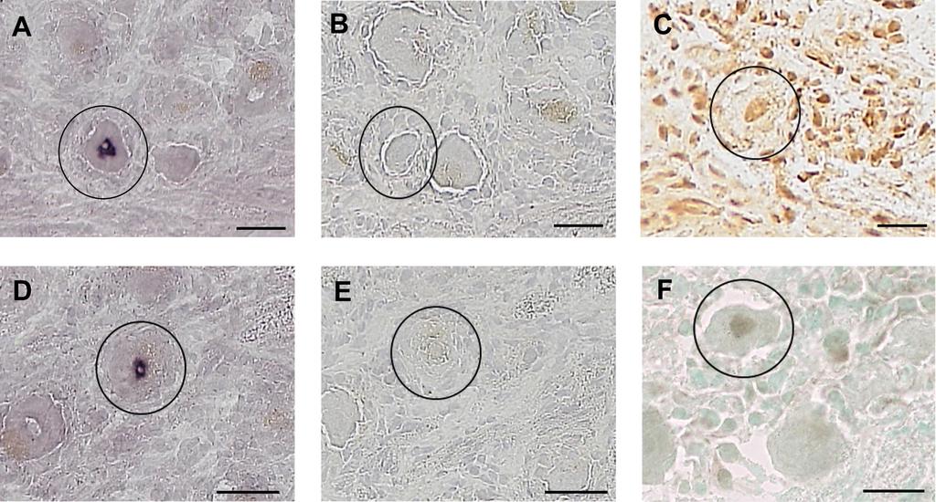
9 Figure legends Figure 1: (A, D) The micrograph shows human TG stained for LAT by ISH, circle indicates LAT positive neuron, (B) micrograph shows staining for TUNEL, circle indicates the same neuron as in (A) on a consecutive slide which is TUNEL negative. (C) Micrograph shows positive control for detection of DNA fragmentation (DNase I treatment), circle indicates TUNEL positive neuron. (E) Micrograph shows staining for active caspase-3, circle indicates the same neuron as in (D) on a consecutive slide which is active caspase-3 negative. (F) Micrograph shows active caspase-3 positive neuron, circle indicates active caspase-3 positive neuron. All tissues were counterstained with haematoxylin, except for (F), this tissue was counterstained with methylgreen. Scale bars represent 50µm Figure 2: Boxplot A shows the difference in the expression of caspase-3 in LAT+ and LAT- single neurons. Boxplot B shows the difference in the expression of caspase-3 in LAT+ and LAT- whole ganglia. Boxes denote interquartile ranges, lines denote medians, and whiskers denote 5th and 95th percentiles. Each measurement was done in duplicate. The results were normalized to the housekeeping gene Glyceraldehyde 3- phosphate dehydrogenase (GAPDH). Commercially available TaqMan Gene Expression Assays: Caspase-3: Assay ID: Hs _m1; GAPDH: Assay ID: Hs _g1. Custom-made TaqMan Gene Expression assay: LAT (F: CCCACGTACTCCAAGAAGGC; R: AGACCCAAGCATAGAGAGCCAG; Probe: CCCACCCCGCCTGTGTTTTTGTGT) (Life Technologies, Darmstadt)
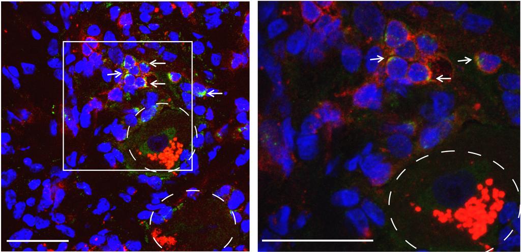
10 Figure 3: Fluorescence labeling of human TG stained for CD8 + T cells (red), GrB (green) and nucleus staining / DAPI (blue). Stained sections were analyzed by confocal imaging. The micrograph on the left was taken at 100x magnification for an overview, the one on the right at 400x for a more detailed view. Dashed circles indicate neurons with Lipofuscin in red, arrows indicate GrB-positive CD8 + T cells. Scale bars represent 50µm
11
12
13
14 Table 1: Total numbers and percentages of different markers analyzed Total neurons on caspase-3 stained slides Percentages of caspase-3 positive neurons Total neurons on GrB stained slides Percentages of GrB positive CD8 + T cells TG sections represent whole cross-sectional area with neurons projecting into all 3 branches LAT-, LAT+ individuals.
Latency of Herpes simplex virus type-1 in human geniculate and vestibular ganglia is associated with infiltration of CD8+ T-cells
 Latency of Herpes simplex virus type- in human geniculate and vestibular ganglia is associated with infiltration of CD+ T-cells Diethilde Theil To cite this version: Diethilde Theil. Latency of Herpes
HSV LAT AND NEURONAL SURVIVAL
 International Reviews of Immunology, 23: 187 198, 2004 Copyright # Taylor & Francis Inc. ISSN: 0883-0185 print/1563-5244 online DOI: 10.1080=08830180490265592 HSV LAT AND NEURONAL SURVIVAL DAVID C. BLOOM
Received 24 August 2010/Accepted 14 December 2010
 JOURNAL OF VIROLOGY, Mar. 2011, p. 2325 2332 Vol. 85, No. 5 0022-538X/11/$12.00 doi:10.1128/jvi.01791-10 Copyright 2011, American Society for Microbiology. All Rights Reserved. The Herpes Simplex Virus
Comparison of Herpes Simplex Virus Reactivation in Ganglia In Vivo and in Explants Demonstrates Quantitative and Qualitative Differences
 JOURNAL OF VIROLOGY, July 2004, p. 7784 7794 Vol. 78, No. 14 0022-538X/04/$08.00 0 DOI: 10.1128/JVI.78.14.7784 7794.2004 Copyright 2004, American Society for Microbiology. All Rights Reserved. Comparison
Restricted VZV transcription in human trigeminal ganglia
 JVI Accepts, published online ahead of print on 27 June 2012 J. Virol. doi:10.1128/jvi.01331-12 Copyright 2012, American Society for Microbiology. All Rights Reserved. 1 2 Restricted VZV transcription
A LAT-Associated Function Reduces Productive-Cycle Gene Expression during Acute Infection of Murine Sensory Neurons with Herpes Simplex Virus Type 1
 JOURNAL OF VIROLOGY, Aug. 1997, p. 5885 5893 Vol. 71, No. 8 0022-538X/97/$04.00 0 Copyright 1997, American Society for Microbiology A LAT-Associated Function Reduces Productive-Cycle Gene Expression during
ANALYSIS OF T CELL RESPONSES DURING ACTIVE VARICELLA ZOSTER VIRUS REACTIVATION IN HUMAN GANGLIA. and Allison Abendroth
 JVI Accepts, published online ahead of print on 18 December 2013 J. Virol. doi:10.1128/jvi.03445-13 Copyright 2013, American Society for Microbiology. All Rights Reserved. 1 2 ANALYSIS OF T CELL RESPONSES
Key issues in varicella-zoster virus latency
 Journal of NeuroVirology, 8(suppl. 2): 80 84, 2002 c 2002 Taylor & Francis ISSN 1355 0284/02 $12.00+.00 DOI: 10.1080/13550280290101058 Key issues in varicella-zoster virus latency Peter GE Kennedy Department
Acute lung injury in children : from viral infection and mechanical ventilation to inflammation and apoptosis Bern, R.A.
 UvA-DARE (Digital Academic Repository) Acute lung injury in children : from viral infection and mechanical ventilation to inflammation and apoptosis Bern, R.A. Link to publication Citation for published
(A) PCR primers (arrows) designed to distinguish wild type (P1+P2), targeted (P1+P2) and excised (P1+P3)14-
 PCR primers (arrows) designed to distinguish wild type (P1+P2), targeted (P1+P2) and excised (P1+P3)14-") 1 Supplemental Figure Legends Figure S1. Mammary tumors of ErbB2 KI mice with 14-3-3σ ablation have elevated ErbB2 transcript levels and cell proliferation (A) PCR primers (arrows) designed to distinguish
1 Supplemental Figure Legends Figure S1. Mammary tumors of ErbB2 KI mice with 14-3-3σ ablation have elevated ErbB2 transcript levels and cell proliferation (A) PCR primers (arrows) designed to distinguish
fl/+ KRas;Atg5 fl/+ KRas;Atg5 fl/fl KRas;Atg5 fl/fl KRas;Atg5 Supplementary Figure 1. Gene set enrichment analyses. (a) (b)
 (b)") KRas;At KRas;At KRas;At KRas;At a b Supplementary Figure 1. Gene set enrichment analyses. (a) GO gene sets (MSigDB v3. c5) enriched in KRas;Atg5 fl/+ as compared to KRas;Atg5 fl/fl tumors using gene set
KRas;At KRas;At KRas;At KRas;At a b Supplementary Figure 1. Gene set enrichment analyses. (a) GO gene sets (MSigDB v3. c5) enriched in KRas;Atg5 fl/+ as compared to KRas;Atg5 fl/fl tumors using gene set
The Latent Herpes Simplex Virus Type 1 Genome Copy Number in Individual Neurons Is Virus Strain Specific and Correlates with Reactivation
 JOURNAL OF VIROLOGY, July 1998, p. 5343 5350 Vol. 72, No. 7 0022-538X/98/$04.00 0 Copyright 1998, American Society for Microbiology. All Rights Reserved. The Latent Herpes Simplex Virus Type 1 Genome Copy
following ocular infection of naive mice with a recombinant HSV-1 expressing murine IL-4 Dhong Hyun Lee 1 and Homayon Ghiasi 1,*
 JVI Accepted Manuscript Posted Online 28 February 2018 J. Virol. doi:10.1128/jvi.00051-18 Copyright 2018 American Society for Microbiology. All Rights Reserved. 1 2 An M2 rather than T H 2 response contributes
Herpesviruses. Virion. Genome. Genes and proteins. Viruses and hosts. Diseases. Distinctive characteristics
 Herpesviruses Virion Genome Genes and proteins Viruses and hosts Diseases Distinctive characteristics Virion Enveloped icosahedral capsid (T=16), diameter 125 nm Diameter of enveloped virion 200 nm Capsid
Supplementary Figure 1. Nature Neuroscience: doi: /nn.4547
 Supplementary Figure 1 Characterization of the Microfetti mouse model. (a) Gating strategy for 8-color flow analysis of peripheral Ly-6C + monocytes from Microfetti mice 5-7 days after TAM treatment. Living
Susceptibility of sensory neurons to apoptosis following infection by bovine herpesvirus type 1
 University of Nebraska - Lincoln DigitalCommons@University of Nebraska - Lincoln Virology Papers Virology, Nebraska Center for April 2002 Susceptibility of sensory neurons to apoptosis following infection
Supplementary Information. Detection and delineation of oral cancer with a PARP1 targeted optical imaging agent
 Supplementary Information Detection and delineation of oral cancer with a PARP1 targeted optical imaging agent Authors: Susanne Kossatz a, Christian Brand a, Stanley Gutiontov b, Jonathan T.C. Liu c, Nancy
Genesis of cerebellar interneurons and the prevention of neural DNA damage require XRCC1.
 Genesis of cerebellar interneurons and the prevention of neural DNA damage require XRCC1. Youngsoo Lee, Sachin Katyal, Yang Li, Sherif F. El-Khamisy, Helen R. Russell, Keith W. Caldecott and Peter J. McKinnon.
Influenza virus exploits tunneling nanotubes for cell-to-cell spread
 Supplementary Information Influenza virus exploits tunneling nanotubes for cell-to-cell spread Amrita Kumar 1, Jin Hyang Kim 1, Priya Ranjan 1, Maureen G. Metcalfe 2, Weiping Cao 1, Margarita Mishina 1,
Determination of the temporal pattern and importance of BALF1 expression in Epstein-Barr viral infection
 Determination of the temporal pattern and importance of BALF1 expression in Epstein-Barr viral infection Melissa Mihelidakis May 6, 2004 7.340 Research Proposal Introduction Apoptosis, or programmed cell
Reactivation of herpes simplex virus type 1 in the mouse trigeminal ganglion: an in vivo study of virus antigen and immune cell infiltration
 Journal of General Virology (1996), 77, 2583-259. Printed in Great Britain Reactivation of herpes simplex virus type 1 in the mouse trigeminal ganglion: an in vivo study of virus antigen and immune cell
Restarting Lytic Gene Transcription at the Onset of Herpes Simplex Virus Reactivation.
 JVI Accepted Manuscript Posted Online 2 November 2016 J. Virol. doi:10.1128/jvi.01419-16 Copyright 2016, American Society for Microbiology. All Rights Reserved. 1 2 3 4 5 6 7 8 9 10 11 12 13 14 15 16 17
Nature Immunology: doi: /ni eee Supplementary Figure 1
 eee Supplementary Figure 1 Hyphae induce NET release, but yeast do not. (a) NET release by human peripheral neutrophils stimulated with a hgc1 yeast-locked C. albicans mutant (yeast) or pre-formed WT C.
Supplementary Information
 Supplementary Information Title Degeneration and impaired regeneration of gray matter oligodendrocytes in amyotrophic lateral sclerosis Authors Shin H. Kang, Ying Li, Masahiro Fukaya, Ileana Lorenzini,
Wednesday, October 19, 16. Viruses
 Viruses Image of an animal cell More realistic size of a virus compared to an animal cell Cells can fulfill all characteristics of life Viruses on their own can be considered lifeless chemicals, unless?
Spontaneous Regression Mechanisms of Lumbar Disc Herniation Role of apoptosis and macrophages during disc tissue resorption
 Spontaneous Regression Mechanisms of Lumbar Disc Herniation Role of apoptosis and macrophages during disc tissue resorption Shigeru Kobayashi, MD,PhD, 1 Riya Kosaka MD,PhD 2, Adam Meir, FRCS, 2 1 Dept.
Supplementary Figure 1
 Supplementary Figure 1 Supplementary Fig. 1: Quality assessment of formalin-fixed paraffin-embedded (FFPE)-derived DNA and nuclei. (a) Multiplex PCR analysis of unrepaired and repaired bulk FFPE gdna from
Regulation of the latency reactivation cycle by products encoded by the bovine herpesvirus 1 (BHV-1) latency-related gene
 latency-related gene") University of Nebraska - Lincoln DigitalCommons@University of Nebraska - Lincoln Papers in Veterinary and Biomedical Science Veterinary and Biomedical Sciences, Department of 2011 Regulation of the latency
University of Nebraska - Lincoln DigitalCommons@University of Nebraska - Lincoln Papers in Veterinary and Biomedical Science Veterinary and Biomedical Sciences, Department of 2011 Regulation of the latency
The toll-like receptor 4 ligands Mrp8 and Mrp14 play a critical role in the development of autoreactive CD8 + T cells
 1 SUPPLEMENTARY INFORMATION The toll-like receptor 4 ligands Mrp8 and Mrp14 play a critical role in the development of autoreactive CD8 + T cells Karin Loser 1,2,6, Thomas Vogl 2,3, Maik Voskort 1, Aloys
Supplemental Information. Otic Mesenchyme Cells Regulate. Spiral Ganglion Axon Fasciculation. through a Pou3f4/EphA4 Signaling Pathway
 Neuron, Volume 73 Supplemental Information Otic Mesenchyme Cells Regulate Spiral Ganglion Axon Fasciculation through a Pou3f4/EphA4 Signaling Pathway Thomas M. Coate, Steven Raft, Xiumei Zhao, Aimee K.
SUPPLEMENTARY INFORMATION
 DOI: 10.1038/ncb2607 Figure S1 Elf5 loss promotes EMT in mammary epithelium while Elf5 overexpression inhibits TGFβ induced EMT. (a, c) Different confocal slices through the Z stack image. (b, d) 3D rendering
TGF-β Signaling Regulates Neuronal C1q Expression and Developmental Synaptic Refinement
 Supplementary Information Title: TGF-β Signaling Regulates Neuronal C1q Expression and Developmental Synaptic Refinement Authors: Allison R. Bialas and Beth Stevens Supplemental Figure 1. In vitro characterization
Susceptibility of sensory neurons to apoptosis following infection by bovine herpesvirus type 1
 Journal of General Virology (2002), 83, 2257 2267. Printed in Great Britain... Susceptibility of sensory neurons to apoptosis following infection by bovine herpesvirus type 1 Gustavo A. Delhon, Marcelo
Explain the laboratory diagnosis of Rabies?
 Explain the laboratory diagnosis of Rabies? The standard test for rabies testing is dfa. This test has been thoroughly evaluated for more than 40 years, and is recognized as the most rapid and reliable
Persistence and Reactivation of Bovine Herpesvirus 1 in the Tonsils of Latently Infected Calves
 Persistence and Reactivation of Bovine Herpesvirus 1 in the Tonsils of Latently Infected Calves M. T. C. Winkler, A. Doster and C. Jones J. Virol. 2000, 74(11):5337. DOI: 10.1128/JVI.74.11.5337-5346.2000.
(A) RT-PCR for components of the Shh/Gli pathway in normal fetus cell (MRC-5) and a
 RT-PCR for components of the Shh/Gli pathway in normal fetus cell (MRC-5) and a") Supplementary figure legends Supplementary Figure 1. Expression of Shh signaling components in a panel of gastric cancer. (A) RT-PCR for components of the Shh/Gli pathway in normal fetus cell (MRC-5) and
Supplementary figure legends Supplementary Figure 1. Expression of Shh signaling components in a panel of gastric cancer. (A) RT-PCR for components of the Shh/Gli pathway in normal fetus cell (MRC-5) and
of HSV-1 infected rabbits
 _~~~~~~~~~~ 644 ORIGINAL ARTICLES Department of Pathology, the Medical School, University of Bristol, Bristol for latency, it C Lynas N J Maitland Correspondence to: Dr S D Cook, Bristol Eye Hospital,
The latency associated transcripts (LAT) of herpes simplex virus: still no end in sight
 of herpes simplex virus: still no end in sight") Review Journal of NeuroVirology (1997) 3, 313 ± 321 ã 1997 Journal of NeuroVirology, Inc. http://www.jneurovirol.com The latency associated transcripts (LAT) of herpes simplex virus: still no end in sight
Review Journal of NeuroVirology (1997) 3, 313 ± 321 ã 1997 Journal of NeuroVirology, Inc. http://www.jneurovirol.com The latency associated transcripts (LAT) of herpes simplex virus: still no end in sight
Supplementary Figure 1: Hsp60 / IEC mice are embryonically lethal (A) Light microscopic pictures show mouse embryos at developmental stage E12.
 Light microscopic pictures show mouse embryos at developmental stage E12.") Supplementary Figure 1: Hsp60 / IEC mice are embryonically lethal (A) Light microscopic pictures show mouse embryos at developmental stage E12.5 and E13.5 prepared from uteri of dams and subsequently genotyped.
Supplementary Figure 1: Hsp60 / IEC mice are embryonically lethal (A) Light microscopic pictures show mouse embryos at developmental stage E12.5 and E13.5 prepared from uteri of dams and subsequently genotyped.
Nature Neuroscience: doi: /nn Supplementary Figure 1
 Supplementary Figure 1 Atlas representations of the midcingulate (MCC) region targeted in this study compared against the anterior cingulate (ACC) region commonly reported. Coronal sections are shown on
INFLUENCE OF HERPES SIMPLEX VIRUS TYPE-1 GLYCOPROTEIN B
 INFLUENCE OF HERPES SIMPLEX VIRUS TYPE-1 GLYCOPROTEIN B EXPRESSION ON VIRAL PATHOGENECITY AND THE CD8 + T CELL RESPONSE by Srividya Ramachandran BSc. Biotechnology, Monash University, 2003 Submitted to
Stability determinants of Murine Cytomegalovirus long non-coding RNA7.2
 JVI Accepts, published online ahead of print on 23 July 2014 J. Virol. doi:10.1128/jvi.01695-14 Copyright 2014, American Society for Microbiology. All Rights Reserved. 1 2 3 Stability determinants of Murine
Herpes Simplex Virus Type 1 and Bovine Herpesvirus 1 Latency
 CLINICAL MICROBIOLOGY REVIEWS, Jan. 2003, p. 79 95 Vol. 16, No. 1 0893-8512/03/$08.00 0 DOI: 10.1128/CMR.16.1.79 95.2003 Copyright 2003, American Society for Microbiology. All Rights Reserved. Herpes Simplex
Transcription of the herpes simplex virus, type 1 genome during productive and quiescent. infection of neuronal and non-neuronal cells.
 JVI Accepts, published online ahead of print on 9 April 2014 J. Virol. doi:10.1128/jvi.00516-14 Copyright 2014, American Society for Microbiology. All Rights Reserved. 1 2 3 4 5 6 7 Transcription of the
Latent Infection with Herpes Simplex Virus Is Associated with Ongoing CD8 T-Cell Stimulation by Parenchymal Cells within Sensory Ganglia
 JOURNAL OF VIROLOGY, Dec. 2005, p. 14843 14851 Vol. 79, No. 23 0022-538X/05/$08.00 0 doi:10.1128/jvi.79.23.14843 14851.2005 Copyright 2005, American Society for Microbiology. All Rights Reserved. Latent
Molecular biology of herpes simplex virus latency
 J. Exp. Path. (1990) 71, I33-141 Current Status Review: Molecular biology of herpes simplex virus latency David S. Latchman Medical Molecular Biology Unit, Department of Biochemistry, University College
marker. DAPI labels nuclei. Flies were 20 days old. Scale bar is 5 µm. Ctrl is
 Supplementary Figure 1. (a) Nos is detected in glial cells in both control and GFAP R79H transgenic flies (arrows), but not in deletion mutant Nos Δ15 animals. Repo is a glial cell marker. DAPI labels
Supplementary Figure 1. Validation of astrocytes. Primary astrocytes were
 Supplementary Figure 1. Validation of astrocytes. Primary astrocytes were separated from the glial cultures using a mild trypsinization protocol. Anti-glial fibrillary acidic protein (GFAP) immunofluorescent
Supplementary Figure 1. EC-specific Deletion of Snail1 Does Not Affect EC Apoptosis. (a,b) Cryo-sections of WT (a) and Snail1 LOF (b) embryos at
 Cryo-sections of WT (a) and Snail1 LOF (b) embryos at") Supplementary Figure 1. EC-specific Deletion of Snail1 Does Not Affect EC Apoptosis. (a,b) Cryo-sections of WT (a) and Snail1 LOF (b) embryos at E10.5 were double-stained for TUNEL (red) and PECAM-1 (green).
Supplementary Figure 1. EC-specific Deletion of Snail1 Does Not Affect EC Apoptosis. (a,b) Cryo-sections of WT (a) and Snail1 LOF (b) embryos at E10.5 were double-stained for TUNEL (red) and PECAM-1 (green).
Supplementary Figure 1
 Supplementary Figure 1 The average sigmoid parametric curves of capillary dilation time courses and average time to 50% peak capillary diameter dilation computed from individual capillary responses averaged
Probe. Hind III Q,!?R'!! /0!!!!D1"?R'! vector. Homologous recombination
 Supple-Zhang Page 1 Wild-type locus Targeting construct Targeted allele Exon Exon3 Exon Probe P1 P P3 FRT FRT loxp loxp neo vector amh I Homologous recombination neo P1 P P3 FLPe recombination Q,!?R'!!
Received 29 May 2003/Accepted 24 November 2003
 JOURNAL OF VIROLOGY, Mar. 2004, p. 3184 3189 Vol. 78, No. 6 0022-538X/04/$08.00 0 DOI: 10.1128/JVI.78.6.3184 3189.2004 Copyright 2004, American Society for Microbiology. All Rights Reserved. A Mutation
Tissue speci c distribution of the herpes simplex virus type 1 latency-associated transcripts on polyribosomes during latent infection
 Short Communication Journal of NeuroVirology (1998) 4, 426 ± 432 ã 1998 Journal of NeuroVirology, Inc. http://www.jneurovirol.com Tissue speci c distribution of the herpes simplex virus type 1 latency-associated
Supplementary Figure 1: si-craf but not si-braf sensitizes tumor cells to radiation.
 Supplementary Figure 1: si-craf but not si-braf sensitizes tumor cells to radiation. (a) Embryonic fibroblasts isolated from wildtype (WT), BRAF -/-, or CRAF -/- mice were irradiated (6 Gy) and DNA damage
Nature Medicine: doi: /nm.4322
 1 2 3 4 5 6 7 8 9 10 11 Supplementary Figure 1. Predicted RNA structure of 3 UTR and sequence alignment of deleted nucleotides. (a) Predicted RNA secondary structure of ZIKV 3 UTR. The stem-loop structure
Supplementary Figure 1 Expression of Crb3 in mouse sciatic nerve: biochemical analysis (a) Schematic of Crb3 isoforms, ERLI and CLPI, indicating the
 Schematic of Crb3 isoforms, ERLI and CLPI, indicating the") Supplementary Figure 1 Expression of Crb3 in mouse sciatic nerve: biochemical analysis (a) Schematic of Crb3 isoforms, ERLI and CLPI, indicating the location of the transmembrane (TM), FRM binding (FB)
Supplementary Figure 1 Expression of Crb3 in mouse sciatic nerve: biochemical analysis (a) Schematic of Crb3 isoforms, ERLI and CLPI, indicating the location of the transmembrane (TM), FRM binding (FB)
Supplemental Figure 1. (A) The localization of Cre DNA recombinase in the testis of Cyp19a1-Cre mice was detected by immunohistchemical analyses
 The localization of Cre DNA recombinase in the testis of Cyp19a1-Cre mice was detected by immunohistchemical analyses") Supplemental Figure 1. (A) The localization of Cre DNA recombinase in the testis of Cyp19a1-Cre mice was detected by immunohistchemical analyses using an anti-cre antibody; testes at 1 week (left panel),
Supplemental Figure 1. (A) The localization of Cre DNA recombinase in the testis of Cyp19a1-Cre mice was detected by immunohistchemical analyses using an anti-cre antibody; testes at 1 week (left panel),
Supporting Online Material for
 www.sciencemag.org/cgi/content/full/1171320/dc1 Supporting Online Material for A Frazzled/DCC-Dependent Transcriptional Switch Regulates Midline Axon Guidance Long Yang, David S. Garbe, Greg J. Bashaw*
Supplementary Figure 1. Double-staining immunofluorescence analysis of invasive colon and breast cancers. Specimens from invasive ductal breast
 Supplementary Figure 1. Double-staining immunofluorescence analysis of invasive colon and breast cancers. Specimens from invasive ductal breast carcinoma (a) and colon adenocarcinoma (b) were staining
3 CHAPTER 3: RESULTS
 3 CHAPTER 3: RESULTS 3.1 Histopathology 3.1.1 Normal Squamous Epithelium The squamous epithelium that covers the ectocervix of the uterus is composed of different layers starting at the basement membrane
An Epstein-Barr virus-encoded microrna targets PUMA to promote host cell survival
 An Epstein-Barr virus-encoded microrna targets to promote host cell survival The Journal of Experimental Medicine 205(11): 2551-2560, 2008. 1 Elizabeth Yee-Wai Choy, Kam-Leung Siu, Kin-Hang Kok, Raymond
Received 12 June 2002/Accepted 13 September 2002
 JOURNAL OF VIROLOGY, Dec. 2002, p. 12758 12774 Vol. 76, No. 24 0022-538X/02/$04.00 0 DOI: 10.1128/JVI.76.24.12758 12774.2002 Copyright 2002, American Society for Microbiology. All Rights Reserved. General
Radioimmunoassay of Herpes Simplex Virus Antibody: Correlation with Ganglionic Infection
 J. gen. Virol. (I977), 3 6, ~ 371-375 Printed in Great Britain 371 Radioimmunoassay of Herpes Simplex Virus Antibody: Correlation with Ganglionic Infection By B. FORGHANI, TONI KLASSEN AND J. R. BARINGER
Chronic Viral Infections vs. Our Immune System: Revisiting our view of viruses as pathogens
 Chronic Viral Infections vs. Our Immune System: Revisiting our view of viruses as pathogens Tiffany A. Reese Assistant Professor Departments of Immunology and Microbiology Challenge your idea of classic
VZV, EBV, and HHV-6-8
 VZV, EBV, and HHV-6-8 Anne Gershon Common Features of Herpesviruses Morphology Basic mode of replication Primary infection followed by latency Ubiquitous Ability to cause recurrent infections (reactivation
Adaptive immune responses: T cell-mediated immunity
 MICR2209 Adaptive immune responses: T cell-mediated immunity Dr Allison Imrie allison.imrie@uwa.edu.au 1 Synopsis: In this lecture we will discuss the T-cell mediated immune response, how it is activated,
Departments of Ophthalmology and of Molecular Genetics and Biochemistry, School of Medicine, University of Pittsburgh, Pittsburgh, PA 15213
 [Frontiers in Bioscience 4, d200-211, February 15, 1999] LATENCY OF VARICELLA ZOSTER VIRUS; A PERSISTENTLY PERPLEXING STATE Paul R. Kinchington Departments of Ophthalmology and of Molecular Genetics and
Reactivation of Latent Herpes Simplex Virus After
 INFEcTION AND IMMUNITY, Apr. 1975, p. 635-639 Copyright 0 1975 American Society for Microbiology Vol. 11, No. 4 Printed in U.S.A. Reactivation of Latent Herpes Simplex Virus After Pneumococcal Pneumonia
Expression of acid base transporters in the kidney collecting duct in Slc2a7 -/-
 Supplemental Material Results. Expression of acid base transporters in the kidney collecting duct in Slc2a7 -/- and Slc2a7 -/- mice. The expression of AE1 in the kidney was examined in Slc26a7 KO mice.
Microarray profiling of gene expression after sleep deprivation and recovery sleep 03/05/2008
 Microarray profiling of gene expression after sleep deprivation and recovery sleep 03/05/2008 1. OVERVIEW...1 2. BEHAVIORAL CONDITIONS...1 3. LASER MICRODISSECTION/MICROARRAY...2 3.1. REGIONS OF INTEREST...
Oncolytic Immunotherapy: A Local and Systemic Antitumor Approach
 Oncolytic Immunotherapy: A Local and Systemic Antitumor Approach Oncolytic immunotherapy Oncolytic immunotherapy the use of a genetically modified virus to attack tumors and induce a systemic immune response
Case Report Relapsing Ipsilateral Vestibular Neuritis
 Hindawi Case Reports in Otolaryngology Volume 217, Article ID 362842, 6 pages https://doi.org/1.1155/217/362842 Case Report Relapsing Ipsilateral Vestibular Neuritis Duilio Emiliano De Schutter 1 and Nicolás
-738 proteins were found only in the ME subjects proteins were only found in PTLS samples proteins were only found in the normal controls.
 RECENT RESEARCH Currently there is much exciting research being published including the Schutzer et al. study that compared cerebrospinal fluid proteomes to differentiate ME and Post Treatment Lyme Syndrome
The dual role of gamma interferon during herpes simplex virus type 1 infection. Vilma Decman
 The dual role of gamma interferon during herpes simplex virus type 1 infection by Vilma Decman M.Sc., University of Zagreb, Faculty of Natural Sciences and Mathematics, Croatia 1999 Submitted to the Graduate
Human Herpesviruses. VZV, EBV, and HHV-6-8. The rash of VZV is vesicular. MID 34
 VZV, EBV, and HHV-6-8 Anne Gershon Human Herpesviruses Replication (lytic infection) occurs in a cascade Latency occurs when the cascade is interrupted Transcription of viral genome and protein synthesis
The pathogenesis of nervous distemper
 Veterinary Sciences Tomorrow - 2004 The pathogenesis of nervous distemper Marc Vandevelde Canine distemper is a highly contagious viral disease of dogs and of all animals in the Canidae, Mustellidae and
of Nebraska - Lincoln
 University of Nebraska - Lincoln DigitalCommons@University of Nebraska - Lincoln Papers in Veterinary and Biomedical Science Veterinary and Biomedical Sciences, Department of 2013 Bovine herpesvirus regulatory
WISP1 mediates IL-6-dependent proliferation in primary human lung fibroblasts
 WISP mediates IL-6-dependent proliferation in primary human lung fibroblasts Klee S, Lehmann M, Wagner DE, aarsma H, Königshoff M Comprehensive Pneumology Center, Helmholtz Zentrum München, Munich, Germany;
Longitudinal tracking of single live cancer cells to understand cell cycle effects of the
 Longitudinal tracking of single live cancer cells to understand cell cycle effects of the nuclear export inhibitor, selinexor Joshua M. Marcus 1, Russell T. Burke 1, John A. DeSisto 1, Yosef Landesman
SUPPLEMENTAL INFORMATIONS
 1 SUPPLEMENTAL INFORMATIONS Figure S1 Cumulative ZIKV production by testis explants over a 9 day-culture period. Viral titer values presented in Figure 1B (viral release over a 3 day-culture period measured
SHREE ET AL, SUPPLEMENTAL MATERIALS. (A) Workflow for tumor cell line derivation and orthotopic implantation.
 Workflow for tumor cell line derivation and orthotopic implantation.") SHREE ET AL, SUPPLEMENTAL MATERIALS SUPPLEMENTAL FIGURE AND TABLE LEGENDS Supplemental Figure 1. Derivation and characterization of TS1-TGL and TS2-TGL PyMT cell lines and development of an orthotopic
SHREE ET AL, SUPPLEMENTAL MATERIALS SUPPLEMENTAL FIGURE AND TABLE LEGENDS Supplemental Figure 1. Derivation and characterization of TS1-TGL and TS2-TGL PyMT cell lines and development of an orthotopic
Acute and Recurrent Herpes Simplex in Several Strains of Mice
 J. gen. Virol. (1981), 55, 31-40. Printed in Great Britain 31 Key words: herpes simplex~mice~latency~recurrence Acute and Recurrent Herpes Simplex in Several Strains of Mice By D. A. HARBOUR, T. J. HILL
Supplemental figure 1. PDGFRα is expressed dominantly by stromal cells surrounding mammary ducts and alveoli. A) IHC staining of PDGFRα in
 IHC staining of PDGFRα in") Supplemental figure 1. PDGFRα is expressed dominantly by stromal cells surrounding mammary ducts and alveoli. A) IHC staining of PDGFRα in nulliparous (left panel) and InvD6 mouse mammary glands (right
Supplemental figure 1. PDGFRα is expressed dominantly by stromal cells surrounding mammary ducts and alveoli. A) IHC staining of PDGFRα in nulliparous (left panel) and InvD6 mouse mammary glands (right
on November 21, 2018 by guest
 JOURNAL OF VIROLOGY, Oct. 1998, p. 7715 7721 Vol. 72, No. 10 0022-538X/98/$04.00 0 Copyright 1998, American Society for Microbiology. All Rights Reserved. Local Periocular Vaccination Protects against
Reactivation of latent herpes simplex virus from explanted dorsal root ganglia
 Journal of General Virology (1994), 75, 2017-2028. Printed in Great Britain 2017 Reactivation of latent herpes simplex virus from explanted dorsal root ganglia Marion Ecob-Prince* and Karen Hassan University
Programmed necrosis, not apoptosis, is a key mediator of cell loss and DAMP-mediated inflammation in dsrna-induced retinal degeneration
 Programmed necrosis, not apoptosis, is a key mediator of cell loss and DAMP-mediated inflammation in dsrna-induced retinal degeneration The Harvard community has made this article openly available. Please
Varicella-Zoster Virus Epithelial Keratitis in Herpes Zoster Ophthalmicus
 Varicella-Zoster Virus Epithelial Keratitis in Herpes Zoster Ophthalmicus Helena M. Tabery Varicella-Zoster Virus Epithelial Keratitis in Herpes Zoster Ophthalmicus In Vivo Morphology in the Human Cornea
Supplementary Information Titles Journal: Nature Medicine
 Supplementary Information Titles Journal: Nature Medicine Article Title: Corresponding Author: Supplementary Item & Number Supplementary Fig.1 Fig.2 Fig.3 Fig.4 Fig.5 Fig.6 Fig.7 Fig.8 Fig.9 Fig. Fig.11
Supplementary Materials for
 www.sciencesignaling.org/cgi/content/full/10/487/eaag2476/dc1 Supplementary Materials for Gene expression profiles of brain endothelial cells during embryonic development at bulk and single-cell levels
Kamal Mohan Khanna. B.S. in Biological Science, Youngstown State University, Submitted to the Graduate Faculty of
 Active Immunosurveillance by CD8 T Lymphocytes during Acute and Latent Herpes Simplex Virus-1 Infection by Kamal Mohan Khanna B.S. in Biological Science, Youngstown State University, 1997 Submitted to
Immunology - Lecture 2 Adaptive Immune System 1
 Immunology - Lecture 2 Adaptive Immune System 1 Book chapters: Molecules of the Adaptive Immunity 6 Adaptive Cells and Organs 7 Generation of Immune Diversity Lymphocyte Antigen Receptors - 8 CD markers
CELL BIOLOGY - CLUTCH CH THE IMMUNE SYSTEM.
 !! www.clutchprep.com CONCEPT: OVERVIEW OF HOST DEFENSES The human body contains three lines of against infectious agents (pathogens) 1. Mechanical and chemical boundaries (part of the innate immune system)
A Therapeutic Vaccine That Reduces Recurrent Herpes Simplex Virus Type 1 Corneal Disease
 A Therapeutic Vaccine That Reduces Recurrent Herpes Simplex Virus Type 1 Corneal Disease Anthony B. Nesburn, 1 ' 2 Rae Lyn Burke, 5 Homayon Ghiasi, 1 ' 2 Susan M. Slanina, 1 Steven L Wechsler 1 ' 2 and
effect on the upregulation of these cell surface markers. The mean peak fluorescence intensity
 SUPPLEMENTARY FIGURE 1 Supplementary Figure 1 ASIC1 disruption or blockade does not effect in vitro and in vivo antigen-presenting cell activation. (a) Flow cytometric analysis of cell surface molecules
Supplemental Figures:
 Supplemental Figures: Figure 1: Intracellular distribution of VWF by electron microscopy in human endothelial cells. a) Immunogold labeling of LC3 demonstrating an LC3-positive autophagosome (white arrow)
(a) Schematic diagram of the FS mutation of UVRAG in exon 8 containing the highly instable
 Schematic diagram of the FS mutation of UVRAG in exon 8 containing the highly instable") Supplementary Figure 1. Frameshift (FS) mutation in UVRAG. (a) Schematic diagram of the FS mutation of UVRAG in exon 8 containing the highly instable A 10 DNA repeat, generating a premature stop codon
Supplementary Figure 1. Frameshift (FS) mutation in UVRAG. (a) Schematic diagram of the FS mutation of UVRAG in exon 8 containing the highly instable A 10 DNA repeat, generating a premature stop codon
Conference Paper Programmed Cell Death Induced by Modulated Electrohyperthermia
 Conference Papers in Medicine, Article ID 187835, 3 pages http://dx.doi.org/10.1155/2013/187835 Conference Paper Programmed Cell Death Induced by Modulated Electrohyperthermia Meggyesházi Nóra, 1 Andócs
Supplemental Table 1. Primers used for RT-PCR analysis of inflammatory cytokines Gene Primer Sequence
 Supplemental Table 1. Primers used for RT-PCR analysis of inflammatory cytokines Gene Primer Sequence IL-1α Forward primer 5 -CAAGATGGCCAAAGTTCGTGAC-3' Reverse primer 5 -GTCTCATGAAGTGAGCCATAGC-3 IL-1β
EBV infection B cells and lymphomagenesis. Sridhar Chaganti
 EBV infection B cells and lymphomagenesis Sridhar Chaganti How EBV infects B-cells How viral genes influence the infected B cell Differences and similarities between in vitro and in vivo infection How
Supplementary Figures
 Supplementary Figures Supplementary Figure 1. nrg1 bns101/bns101 embryos develop a functional heart and survive to adulthood (a-b) Cartoon of Talen-induced nrg1 mutation with a 14-base-pair deletion in