Chapter 11. Lecture Outline. See separate PowerPoint slides for all figures and tables pre-inserted into PowerPoint without notes and animations.
|
|
|
- Egbert Higgins
- 5 years ago
- Views:
Transcription


1 Chapter 11 Lecture Outline See separate PowerPoint slides for all figures and tables pre-inserted into PowerPoint without notes and animations. Copyright The McGraw-Hill Companies, Inc. Permission required for reproduction or display. 11-1

2 Chapter 11 Functional Organization of Nervous Tissue 11-2
3 11.1 Functions of the Nervous System 1. Receiving sensory input. Monitor internal and external stimuli 2. Integrating information. Brain and spinal cord process sensory input and initiate responses 3. Controlling muscles and glands 4. Maintaining homeostasis. Regulate and coordinate physiology 5. Establishing and maintaining mental activity. Consciousness, thinking, memory, emotion 11-3
4 11.2 Divisions of the Nervous System Components Brain, spinal cord, nerves, sensory receptors Subdivisions Central nervous system (CNS): brain and spinal cord Peripheral nervous system (PNS): sensory receptors and nerves 11-4

5 Divisions of the Nervous System Copyright The McGraw-Hill Companies, Inc. Permission required for reproduction or display. Brain Cranial nerves Central nervous system Spinal cord Peripheral nervous system Spinal nerves 11-5
6 PNS Sensory receptors: ending of neurons or separate, specialized cells that detect such things as temperature, pain, touch, pressure, light, sound, odors Nerve: a bundle of axons and their sheaths that connects CNS to sensory receptors, muscles, and glands Cranial nerves: originate from the brain; 12 pairs Spinal nerves: originate from spinal cord; 31 pairs Ganglion: collection of neuron cell bodies outside CNS Plexus: extensive network of axons, and sometimes neuron cell bodies, located outside CNS 11-6
: transmits action potentials from receptors to CNS.")
7 Divisions of PNS Copyright The McGraw-Hill Companies, Inc. Permission required for reproduction or display. Dorsal root of spinal nerve Dorsal root ganglion Sensory neuron Sensory (afferent): transmits action potentials from receptors to CNS. Motor (efferent): transmits action potentials from CNS to effectors (muscles, glands) Spinal cord Spinal nerve (a) Sensory division Motor neuron Sensory receptor Spinal cord Ventral root of spinal nerve Spinal nerve (b) Somatic nervous system Skeletal muscle 11-7
8 Motor Division of PNS Somatic nervous system: from CNS to skeletal muscles. Voluntary. Single neuron system. Synapse: junction of a nerve cell with another cell. E.g., neuromuscular junction is a synapse between a neuron and skeletal muscle cell. Autonomic nervous system (ANS): from CNS to smooth muscle, cardiac muscle and certain glands. Subconscious or involuntary control. Two neuron system: first from CNS to ganglion; second from ganglion to effector. Divisions of ANS Sympathetic. Prepares body for physical activity. Parasympathetic. Regulates resting or vegetative functions such as digesting food or emptying of the urinary bladder. Enteric. plexuses within the wall of the digestive tract. Can control the digestive tract independently of the CNS, but still considered part of ANS because of the parasympathetic and sympathetic neurons that contribute to the plexi. 11-8
 (c) Autonomic nervous system Large intestine 11-9")
9 Autonomic Nervous System Copyright The McGraw-Hill Companies, Inc. Permission required for reproduction or display. Spinal nerve Autonomic ganglion Spinal cord First motor neuron Second motor neuron Effector organ (e.g., smooth muscle) (c) Autonomic nervous system Large intestine 11-9

: Brand X Pictures/PunchStock RF; (right): Royalty-Free/Corbis RF Receptor Sensory NS CNS")
10 Organization of the Nervous System Copyright The McGraw-Hill Companies, Inc. Permission required for reproduction or display. Sensory input Motor output Effectors: cardiac and smooth muscle; glands Effectors: Skeletal muscle Sympathetic division Parasympathetic division Autonomic nervous system Somatic nervous system Sensory division Motor division PNS Receptors, nerves, ganglia, plexuses Sensory Motor CNS Brain, spinal cord (left): Brand X Pictures/PunchStock RF; (right): Royalty-Free/Corbis RF Receptor Sensory NS CNS Motor NS Effector 11-10
11 11.3 Cells of Nervous System Neuroglia Support and protect neurons Neurons or nerve cells receive stimuli and transmit action potentials Organization Cell body or soma Dendrites: input Axons: output 11-11

12 Parts of the Neuron Neuron Cell Body. Nucleus, Nissl substance. Nissl substance = chromatophilic substance = rough E.R: primary site of protein synthesis. Dendrites: short, often highly branched. Dendritic spines: little protuberance where axons synapse with dendrite. Axons. Can branch to form collaterals. Axon hillock Initial segment: beginning of axon Trigger zone: site where action potentials are generated; axon hillock and part of axon nearest cell body Axoplasm Axolemma Presynaptic terminals (terminal boutons) Synaptic vesicles Copyright The McGraw-Hill Companies, Inc. Permission required for reproduction or display. Dendritic spine Mitochondrion Golgi apparatus Nucleolus Nucleus Nissl bodies Trigger zone Myelin sheath formed by Schwann cell Collateral axon Axon hillock Initial segment Axon Schwann cell Dendrites Neuron cell body Node of Ranvier Presynaptic terminals 11-12
13 Axonic Transport Mechanisms Axoplasm moved from cell body toward terminals. Supply for growth, repair, renewal. Can move cytoskeletal proteins, organelles away from cell body toward axon terminals. Into cell body: damaged organelles, recycled plasma membrane, and substances taken in by endocytosis can be transported up axon to cell body. Rabies and herpes virus can enter axons in damaged skin and be transported to CNS
14 Types of Neurons Functional classification Sensory or afferent: action potentials toward CNS Motor or efferent: action potentials away from CNS Interneurons or association neurons: within CNS from one neuron to another Structural classification Multipolar: most neurons in CNS; motor neurons Bipolar: sensory in retina of the eye and nose Unipolar: single process that divides into two branches. Part that extends to the periphery has dendrite-like sensory receptors Copyright The McGraw-Hill Companies, Inc. Permission required for reproduction or display. Dendrites Sensory receptors Dendrite Cell body Axon Cell body Axon Cell body Axon Axon branches function as a single axon (a) A multipolar neuron has many dendrites and an axon. (b) A bipolar neuron has a dendrite and an axon. (c) A pseudo-unipolar neuron appears to have an axon and no dendrites.
15 Neuroglia of the CNS: Astrocytes Copyright The McGraw-Hill Companies, Inc. Permission required for reproduction or display. Capillary Neuron Foot processes Astrocyte Processes form feet that cover the surfaces of neurons and blood vessels and the pia mater. Regulate what substances reach the CNS from the blood (bloodbrain barrier). Lots of microfilaments for support. Produce chemicals that promote tight junctions to form bloodbrain barrier Blood-brain barrier: protects neurons from toxic substances, allows the exchange of nutrients and waste products between neurons and blood, prevents fluctuations in the composition of the blood from affecting the functions of the brain. Regulate extracellular brain fluid composition 11-15
16 Neuroglia of the CNS: Ependymal Cells (a) (b) Copyright The McGraw-Hill Companies, Inc. Permission required for reproduction or display. Ependymal cells Ependymal cells Cilia Line brain ventricles and spinal cord central canal. Specialized versions of ependymal form choroid plexuses. Choroid plexus within certain regions of ventricles. Secrete cerebrospinal fluid. Cilia help move fluid thru the cavities of the brain. Have long processes on basal surface that extend within the brain tissue, may have astrocyte-like functions

17 Neuroglia of the CNS: Microglia and Oligodendrocytes Microglia: specialized macrophages. Respond to inflammation, phagocytize necrotic tissue, microorganisms, and foreign substances that invade the CNS. Oligodendrocytes: form myelin sheaths if surrounding axon. Single oligodendrocytes can form myelin sheaths around portions of several axons. Copyright The McGraw-Hill Companies, Inc. Permission required for reproduction or display. Nodeof Ranvier Microglial cell Oligodendrocyte Axon Myelin sheath Part of another oligodendrocyte 11-17
18 Neuroglia of the PNS Schwann cells or neurolemmocytes: wrap around portion of only one axon to form myelin sheath. Wrap around many times. During development, as cells grow around axon, cytoplasm is squeezed out and multiple layers of cell membrane wrap the axon. Cell membrane primarily phospholipid. Satellite cells: surround neuron cell bodies in sensory ganglia, provide support and nutrients 11-18


19 Copyright The McGraw-Hill Companies, Inc. Permission required for reproduction or display. TABLE 11.1 Types of Neuroglial Cells Neuroglial Cells Function Neuroglial Cells Function CNS Astrocytes Neuron Foot processes Astrocyte foot Processes cover the surfaces of neurons, blood vessels, and the pia mater membrane of the brain and spinal cord.the astrocytes provide structural support and play a role in regulating what substances from the blood reach the neurons. Microglia Microglial cell Microglia are phagocytic cells within the CNS. Capillary Astrocyte Oligodendrocytes Oligodendrocyte Extensions from oligodendrocytes form part of the myelin sheaths of several axons within the CNS. Ependymal cells (a) Ependymal cells Cilia Ependymal cells (a) Ciliatedependymal cells lining the ventricles of the brain and the central canal of the spinal cord help move cerebro spinal fluid. (b) Ependymal cells on the surface of the choroid plexus secrete cerebro spinal fluid. Nodeof Ranvier PNS Schwann cells and satellite cells Satellite cells Axon Myelin sheath Part of another oligodendrocyte Neuron cell bodies within ganglia are surrounded by satellite cells.schwann cells form the myelin sheath of an axon within the PNS. (b) Neuron cellbody Schwann cells Node of Ranvier Axon Myelinsheath 11-19
20 Myelinated and Unmyelinated Axons Myelinated axons Myelin protects and insulates axons from one another, speeds transmission, functions in repair of axons. Not continuous Nodes of Ranvier Completion of development of myelin sheaths at 1 yr. Degeneration of myelin sheaths occurs in multiple sclerosis and some cases of diabetes mellitus. Unmyelinated axons: rest in invaginations of Schwann cells or oligodendrocytes. Not wrapped around the axon; gray matter
 Nucleus Cytoplasm Axon Myelin sheath (a) Myelinated axon Nucleus Cytoplasm Axons")
21 Copyright The McGraw-Hill Companies, Inc. Permission required for reproduction or display. Node of Ranvier (no myelin sheath) Nucleus Cytoplasm Axon Myelin sheath (a) Myelinated axon Nucleus Cytoplasm Axons (b) Unmyelinated axons 11-21
 Muscle atrophies. Muscle undergoes hypertrophy.")
22 Copyright The McGraw-Hill Companies, Inc. Permission required for reproduction or display. Neuron cell body Axon Schwann cell Site of injury Muscle fiber (a) Muscle atrophies. Muscle undergoes hypertrophy. Axon Neuron cell body Two injured ends not in close proximity Muscle fiber (b) Muscle atrophies. Muscle remains atrophied
23 11.4 Organization of Nervous Tissue Gray matter: unmyelinated axons, cell bodies, dendrites, neuroglia. Integrative functions White matter: myelinated axons. Nerve tracts propagate actin potentials from one area in the CNS to another In brain: gray is outer cortex as well as inner nuclei; white is deeper. In spinal cord: white is outer, gray is deeper. PNS gray matter is groups of cell bodies called ganglia 11-23
24 11.5 Electrical Signals Cells produce electrical signals called action potentials Transfer of information from one part of body to another Electrical properties result from ionic concentration differences across plasma membrane and permeability of membrane 11-24

25 Concentration Differences Across the Plasma Membrane These ion concentrations are a result of two processes: the Na/K pump and membrane permeability. Note high concentration of Na and Cl ions outside and high concentration of K and proteins on inside. Note steep concentration gradient of Na and K, but in opposite directions
26 Permeability Characteristics of the Plasma Membrane Proteins: synthesized inside cell: Large, don't dissolve in phospholipids of membrane. Proteins are negatively charged. Cl - are repelled by proteins and they exit thru always-open nongated Cl - channels. Gated ion channels open and close because of some sort of stimulus. When they open, they change the permeability of the cell membrane. Ligand-gated: molecule that binds to a receptor; protein or glycoprotein 11-26
27 Leak Channels Many more of these for K + and Cl - than for Na +. So, at rest, more K + and Cl - are moving than Na +. How are they moving? Protein repels Cl -, they move out. K + are in higher concentration on inside than out, they move out. Always open and responsible for permeability when membrane is at rest. Specific for one type of ion although not absolute
 are closed.")
28 Leak Channels Copyright The McGraw-Hill Companies, Inc. Permission required for reproduction or display. Pr Pr 2 There are more K + leak channels than Na + leak channels. In the resting cell, only the leak channels are opened; the gated channels (not shown) are closed. Because of the ion concentration differences across the membrane, K + diffuses out of the cell down its concentration gradient and Na + diffuses into the cell down its concentration gradient. The tendency for K + to diffuse out of the cell is opposed by the tendency of the positively charged K + to be attracted back into the cell by the negatively charged proteins
29 Gated Ion Channels Gated ion channels. Gated ion channels open and close because of some sort of stimulus. When they open, they change the permeability of the cell membrane. Ligand-gated: open or close in response to ligand such as ACh binding to receptor protein. Receptor proteins are usually glycoproteins. E.g., acetylcholine binds to acetylcholine receptor on a Na + channel. Channel opens, Na + enters the cell
30 Voltage Gated Ion Channels Voltage-gated: open or close in response to small voltage changes across the cell membrane. At rest, membrane is negative on the inside relative to the outside. When cell is stimulated, that relative charge changes and voltage-gated ion channels either open or close. Most common voltage gated are Na + and K +. In cardiac and smooth muscle, Ca 2+ are important
31 Other Gated Ion Channels Touch receptors: respond to mechanical stimulation of the skin Temperature receptors: respond to temperature changes in the skin 11-31

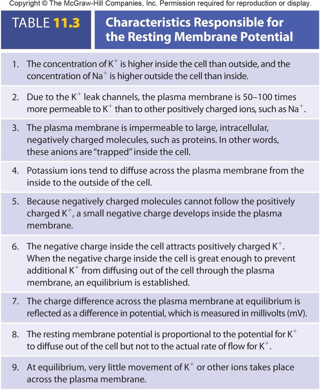
32 Establishing the (b) Resting Membrane Potential Number of charged Copyright The McGraw-Hill Companies, Inc. Permission required for reproduction or display. Neuron Oscilloscope 0 mv 70 Time molecules and ions inside and outside cell nearly equal Concentration of K + higher inside than outside cell, Na + higher outside than inside Potential difference: unequal distribution of charge exists between the immediate inside and immediate outside of the plasma membrane: -70 to -90 mv The resting membrane potential 11-32
33 Establishing the Resting Potential At equilibrium there is very little movement of K + or other ions across plasma membrane (Movement of K out through leakage channels = movement of ions is due to attraction to trapped proteins: N.B. leakage channels work in both directions. Movement of ions depends upon concentration gradient.) Na +, Cl -, and Ca 2+ do not have a great affect on resting potential since there are very few leakage channels for these ions. If leakage channels alone were responsible for resting membrane potential, in time Na + and K + ion concentrations would eventually equalize. But they are maintained by the Na/K pump. For each ATP that is consumed, three Na moved out, two K + moved in. Outside of plasma membrane slightly positive 11-33
 inside the cell membrane and a higher concentration of Na + (pink")
34 K + concentration gradient Na + concentration gradient Copyright The McGraw-Hill Companies, Inc. Permission required for reproduction or display. Na + K + K leak channel Pr Pr Na + leak channel Pr 1 In a resting cell, there is a higher concentration of K + (purple circles) inside the cell membrane and a higher concentration of Na + (pink circles) outside the cell membrane. Because the membrane is not permeable to negatively charged proteins (green) they are isolated to inside of the cell membrane. Pr Pr 2 There are more K + leak channels than Na + leak channels. In the resting cell, only the leak channels are opened; the gated channels (not shown) are closed. Because of the ion concentration differences across the membrane, K + diffuses out of the cell down its concentration gradient and Na + diffuses into the cell down its concentration gradient. The tendency for K + to diffuse out of the cell is opposed by the tendency of the positively charged K + to be attracted back into the cell by the negatively charged proteins. Sodiumpotassium pump Pr ATP ADP 3 ( a ) The sodium-potassium pump helps maintain the differential levels of Na+ and K + by pumping three Na + out of the cell in exchange for two K + into the cell. The pump is driven by ATP hydrolysis. The resting membrane potential is established when the movement of K + out of the cell is equal to the movement of K + into the cell
35 11-35

36 (mv) (mv) Changing the Resting Membrane Potential: K + Depolarization: Potential difference becomes smaller or less polar Hyperpolarization: Potential difference becomes greater or more polar K + concentration gradient alterations If extracellular concentration of K + increases: less gradient between inside and outside. Depolarization If extracellular ion concentration decreases: steeper gradient between inside and outside. Hyperpolarization K + membrane permeability changes. In resting membrane, K + in and out is equal through the leakage channels. But there are also gated K + channels in the membrane. If they open, more K + diffuses out but this is opposed by the negative charge that starts to develop as the K + diffuses out. Copyright The McGraw-Hill Companies, Inc. Permission required for reproduction or display. (a) Time Time Depolarization: movement of RMP toward zero Hyperpolarization: movement of RMP further away from zero (b) 11-36
37 Changes in Resting Membrane Potential: Na + Na + membrane permeability. Change the concentration of Na + inside or outside the cell, little effect because gates remain closed. But open gates (like when ACh attaches to receptors), Na + diffuses in, depolarizing the membrane
38 Changes in Resting Membrane Potential: Ca 2+ Voltage-gated Na + channels sensitive to changes in extracellular Ca 2+ concentrations If extracellular Ca 2+ concentration decreases- Na + gates open and membrane depolarizes. If extracellular concentration of Ca 2+ increasesgates close and membrane repolarizes or becomes hyperpolarized
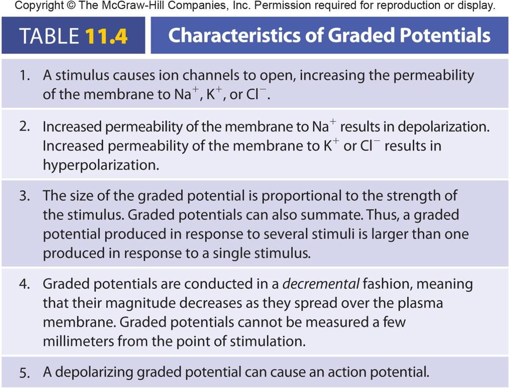
39 (mv) (mv) Graded Potentials Copyright The McGraw-Hill Companies, Inc. Permission required for reproduction or display. Result from Ligands binding to receptors Changes in charge across membrane Mechanical stimulation Temperature changes Spontaneous change in permeability Graded Magnitude varies from small to large depending on stimulus strength or frequency Can summate or add onto each other Spread (are conducted) over the plasma membrane in a decremental fashion: rapidly decrease in magnitude as they spread over the surface of the plasma membrane. Can cause generation of action potentials (a) Successively stronger stimuli of short duration from 1 4 Time 1 2 Two equal stimuli in short succession at 1 and 2 Time (b)
40 11-40
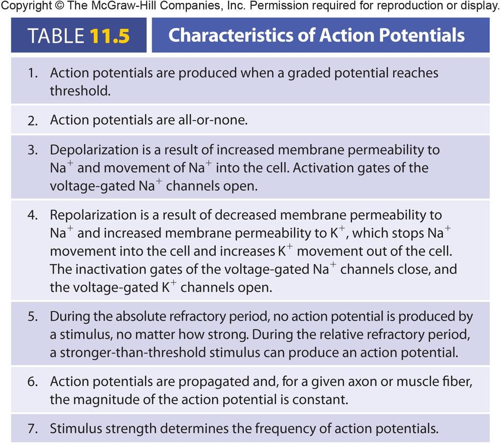
41 (mv) Action Potentials Depolarization phase followed by repolarization phase. Depolarization: more positive Repolarization: more negative (may get afterpotential [slight hyperpolarization]) Series of permeability changes when a graded potential causes depolarization of membrane. A large enough graded potential may cause the membrane to reach threshold. Then get action potential. All-or-none principle. No matter how strong the stimulus, as long as it is greater than threshold, then action potential will occur. Copyright The McGraw-Hill Companies, Inc. Permission required for reproduction or display Depolarization Graded potential Time (ms) Repolarization Threshold Afterpotential 11-41
42 Copyright The McGraw-Hill Companies, Inc. Permission required for reproduction or display. 1 Trigger zone Action potential propagation Action potentials in the 2 Action potentials are 3 communicating neuron propagated down the stimulate graded potentials in axon to the axon terminal. a receiving neuron that can summate at the trigger zone. Action potentials result in communication of the neuron with its target
43 11-43
 and most, but not all, K + channels (purple) are closed.")
 Threshold Resting membrane potential 2")
44 Membrane potential (mv) Membrane potential (mv) Membrane potential (mv) Membrane potential (mv) Membrane potential (mv) Operation of Gates: Action Potential Copyright The McGraw-Hill Companies, Inc. Permission required for reproduction or display. 1 Resting membrane potential. Na + channels (pink) and most, but not all, K + channels (purple) are closed. The outside of the plasma membrane is positively charged compared to the inside. Open inactivation gate Closed activation gate Na + Extracellular fluid Cytoplasm K + Voltage-gated Na + channel Voltage-gated K + channel T ime (ms) Threshold Resting membrane potential 2 Depolarization. Na + channels open. K + channels begin to open. Depolarization results because the inward movement of Na + makes the inside of the membrane more positive. Open activation gate Na + Na + Na + channels open. Na + diffuse into cell Depolarization Local potential 2 Resting membrane potential Time (ms) Threshold 3 Repolarization. Na + channels close and additional K + channels open. Na + movement into the cell stops, and K + movement out of the cell increases, causing repolarization. Closed activation gate K + diffuse out of cell Repolarization 3 Resting membrane potential Threshold Closed inactivation gate K + channels open. K + Na + channels close. K + Time (ms) 4 End of repolarization and afterpotential. Voltage-gated Na + channels are closed. Closure of the activation gates and opening of the inactivation gates reestablish the resting condition for Na + channels (see step 1). Diffusion of K + through voltage-gated channels produces the afterpotential. Activation gate closed Inactivation gate open K + channels open. Na + channel K + K + K + channels open Time (ms) Threshold 4 K + channels closed. Na + channel K + channels closed. 0 5 Resting membrane potential. The resting membrane potential is reestablished after the voltage-gated K + channels close. 70 Time (ms) Threshold
45 (mv) Copyright The McGraw-Hill Companies, Inc. Permission required for reproduction or display Absolute Refractory period Time (ms) Refractory Period Relative Threshold Sensitivity of area to further stimulation decreases for a time Parts Absolute Complete insensitivity exists to another stimulus From beginning of action potential until near end of repolarization. No matter how large the stimulus, a second action potential cannot be produced. Has consequences for function of muscle, particularly how often a.p.s can be produced. Relative A stronger-than-threshold stimulus can initiate another action potential 11-45
46 Action Potential Frequency Number of potentials produced per unit of time to a stimulus Threshold stimulus: causes a graded potential that is great enough to initiate an action potential. Subthreshold stimulus: does not cause a graded potential that is great enough to initiate an action potential. Maximal stimulus: just strong enough to produce a maximum frequency of action I potentials. Submaximal n stimulus: all stimuli between threshold and the maximal stimulus strength. s Supramaximal stimulus: any stimulus stronger than a maximal stimulus. These e stimuli cannot produce a greater frequency of action potentials than a maximal r stimulus
47 (mv) (mv) Copyright The McGraw-Hill Companies, Inc. Permission required for reproduction or display. Increasing frequency of action potentials Time(ms) 55 Stimulus 70 Threshold Subthreshold stimulus Threshold stimulus Submaximal stimulus Submaximal stimulus Maximal stimulus Supramaximal stimulus Increasing stimulus strength 11-47
48 Propagation of Action Potentials In an unmyelinated axon Threshold graded current at trigger zone causes action potential Action potential in one site causes action potential at the next location. Cannot go backwards because initial action potential site is depolarized yielding one-way conduction of impulse


49 Propagation of Action Potentials Copyright The McGraw-Hill Companies, Inc. Permission required for reproduction or display. Action potential propagation 1 Action potentials propagate in one direction along the axon. 2 An action potential (orange part of the membrane) generates local currents (black arrows) that tend to depolarize the membrane immediately adjacent to the action potential Outside of membrane becomes more negative as positive charges move away from it. Inside of membrane becomes more positive as positive charges move toward it Depolarization of the membrane adjacent to the site of action potential production 3 When depolarization caused by the local currents reaches threshold, a new action potential is produced adjacent to where the original action potential occurred Action potential propagation occurs in one direction because the absolute refractory period of the previous action potential prevents generation of an action potential in the reverse direction Absolute refractory period prevents another action potential. Site of next action potential 11-49
 at a node of Ranvier generates local currents (black arrows).")
50 Saltatory Conduction Copyright The McGraw-Hill Companies, Inc. Permission required for reproduction or display. 1 An action potential (orange) at a node of Ranvier generates local currents (black arrows). The local currents flow to the next node of Ranvier because the myelin sheath of the Schwann cell insulates the axon of the internode. Node of Ranvier + + Schwann cell Internode When the depolarization caused by the local currents reaches threshold at the next node of Ranvier, a new action potential is produced (orange) Action potential propagation is rapid in myelinated axons because the action potentials are produced at successive nodes of Ranvier (1 5) instead of at every part of the membrane along the axon Direction of action potential propagation 11-50
51 Speed of Conduction Faster in myelinated than in non-myelinated In myelinated axons, lipids act as insulation forcing ionic currents to jump from node to node In myelinated, speed is affected by thickness of myelin sheath Diameter of axons: large-diameter conduct more rapidly than small-diameter. Large have greater surface area and more voltage-gated Na + channels 11-51
52 Nerve Fiber Types Type A: large-diameter, myelinated. Conduct at m/s. Motor neurons supplying skeletal and most sensory neurons Type B: medium-diameter, lightly myelinated. Conduct at 3-15 m/s. Part of ANS Type C: small-diameter, unmyelinated. Conduct at 2 m/s or less. Part of ANS 11-52
53 11.6 The Synapse Junction between two cells Site where action potentials in one cell cause action potentials in another cell Types of cells in synapse Presynaptic Postsynaptic 11-53


54 Electrical Synapses Copyright The McGraw-Hill Companies, Inc. Permission required for reproduction or display. Electrical synapses connect cardiac muscle cells. An electrical synapse is a gap junction where the membranes of two cells are separated by a gap but connected by proteins called connexons. An action potential (orange arrow) in the plasma membrane generates local currents (black arrows) that flow to adjacent parts of the plasma membrane and through the gap junction. A local current stimulates the production of another action potential. Thus, the action potential propagates along the plasma membrane. A local current flows through a gap junction and stimulates the production of an action potential in the adjacent cardiac muscle cell. Thus, the action potential propagates to the adjacent cell. 2 Action Local potential currents Cardiac muscle cell 1 Electrical synapse Connexons Plasma membrane Gap junction Plasma membrane of an adjacent cell Gap junctions that allow graded current to flow between adjacent cells. Connexons: protein tubes in cell membrane. Found in cardiac muscle and many types of smooth muscle. Action potential of one cell causes action potential in next cell, almost as if the tissue were one cell. Important where contractile activity among a group of cells important
55 Chemical Synapses Components Presynaptic terminal Synaptic cleft Postsynaptic membrane Neurotransmitters released by action potentials in presynaptic terminal Synaptic vesicles: action potential causes Ca 2+ to enter cell that causes neurotransmitter to be released from vesicles Diffusion of neurotransmitter across synapse Postsynaptic membrane: when ACh binds to receptor, ligand-gated Na + channels open. If enough Na + diffuses into postsynaptic cell, it fires


56 Chemical Synapse Copyright The McGraw-Hill Companies, Inc. Permission required for reproduction or display. Action potential Axon Ca 2+ Voltage-gated Ca 2+ channel 2 1 Synaptic vesicle Presynaptic terminal Synaptic cleft Neurotransmitter 3 Postsynaptic membrane Na + Neurotransmitter bound to receptor site opens a ligand-gated 4 Na + channel Action potentials arriving at the presynaptic terminal cause voltage-gated Ca 2+ channels to open. Ca 2+ diffuse into the cell and cause synaptic vesicles to release neurotransmitter molecules. Neurotransmitter molecules diffuse from the presynaptic terminal across the synaptic cleft. Neurotransmitter molecules combine with their receptor sites and cause ligand-gated Na + channels to open. Na + diffuse into the cell (shown in illustration) or out of the cell (not shown) and cause a change in membrane potential
57 Neurotransmitter Removal Method depends on neurotransmitter/synapse. ACh: acetylcholinesterase splits ACh into acetic acid and choline. Choline recycled within presynaptic neuron. Norepinephrine: recycled within presynaptic neuron or diffuses away from synapse. Enzyme monoamine oxidase (MAO). Absorbed into circulation, broken down in liver
 Acetylcholine molecules bind to their receptors. Acetylcholine molecules unbind from their receptors.")


58 Removal of Neurotransmitter from Synaptic Cleft Copyright The McGraw-Hill Companies, Inc. Permission required for reproduction or display (a) Acetylcholine molecules bind to their receptors. Acetylcholine molecules unbind from their receptors. Acetylcholinesterase splits acetylcholine into choline and acetic acid, which prevents acetylcholine from again binding to its receptors. Choline is taken up by the presynaptic terminal. Choline is used to make new acetylcholine molecules that are packaged into synaptic vesicles. 1 2 Acetylcholinesterase Acetylcholine Choline Na + Acetyl- CoA 3 Choline Acetic acid 4 Acetylcholine CoA 1 Norepinephrine binds to its receptor. Norepinephrine Norepinephrine unbinds from its receptor. Norepinephrine is taken up by the presynaptic terminal, which prevents norepinephrine from again binding to its receptor. Norepinephrine is repackaged into synaptic vesicles or broken down by monoamine oxidase (MAO). 1 2 Na + 4 MAO Inactive metabolites 3 (b) Norepinephrine 11-58
59 Receptor Molecules in Synapses Neurotransmitter only "fits" in one receptor. Not all cells have receptors. Neurotransmitters are excitatory in some cells and inhibitory in others. Some neurotransmitters (norepinephrine) attach to the presynaptic terminal as well as postsynaptic and then inhibit the release of more neurotransmitter
60 Neuromodulators Chemicals produced by neurons that facilitate action potentials. Some of these act by increasing or decreasing the amount of neurotransmitter released by the presynaptic neuron. Act in axoaxonic synapses. Axon of one neuron synapses with axon of second neuron. Second neuron is actually presynaptic. This type of connection leads to release of neuromodulators in the synapse that can alter the amount of neurotransmitter produced by the second neuron
61 (mv) (mv) Postsynaptic Potentials Copyright The McGraw-Hill Companies, Inc. Permission required for reproduction or display Resting membrane potential Time (ms) (a) Excitatory postsynaptic potential (EPSP) Resting membrane potential Local depolarization (EPSP) Threshold Threshold Local hyperpolarization (IPSP) Excitatory postsynaptic potential (EPSP) Depolarization occurs and response stimulatory Depolarization might reach threshold producing an action potential and cell response Inhibitory postsynaptic potential (IPSP) Hyperpolarization and response inhibitory Decrease action potentials by moving membrane potential farther from threshold Time (ms) (b) Inhibitory postsynaptic potential (IPSP) 11-61

62 Presynaptic Inhibition and Facilitation Axoaxonic synapses: axon of one neuron synapses with the presynaptic terminal (axon) of another. Many of the synapses of CNS Presynaptic inhibition: reduction in amount of neurotransmitter released from presynaptic terminal. Endorphins can inhibit pain sensation Presynaptic facilitation: amount of neurotransmitter released from presynaptic terminal increases. Glutamate facilitating nitric oxide production Copyright The McGraw-Hill Companies, Inc. Permission required for reproduction or display. Action potential Inhibitory neuron (a) Presynaptic neuron Postsynaptic membrane Action potential Action potential (b) 11-62

63 Spatial Summation Copyright The McGraw-Hill Companies, Inc. Permission required for reproduction or display. Axon Action potential 1 0 mv Time Neuron cell body (a) Spatial summation. Action potentials 1 and 2 cause the production of graded potentials at two different dendrites. These graded potentials summate at the trigger zone to produce a graded potential that exceeds threshold, resulting in an action potential. Trigger zone Axon 0 Axon 0 mv Time 50 Action potential 2 mv 90 Time 11-63
64 Temporal Summation Copyright The McGraw-Hill Companies, Inc. Permission required for reproduction or display. (b) Temporal summation. Two action potentials arrive in close succession at the presynaptic membrane. The first action potential causes the production of a graded potential that does not reach threshold at the trigger zone. The second action potential results in the production of a second graded potential that summates with the first to reach threshold, resulting in the production of an action potential. Action potentials Trigger zone mv T ime 11-64
 (c) Combined spatial and temporal summation with both excitatory postsynaptic potentials (EPSPs) and inhibitory postsynaptic potentials (IPSPs).")
65 Combined Summation Copyright The McGraw-Hill Companies, Inc. Permission required for reproduction or display. Inhibitory Excitatory (with temporal summation) (c) Combined spatial and temporal summation with both excitatory postsynaptic potentials (EPSPs) and inhibitory postsynaptic potentials (IPSPs). An action potential is produced at the trigger zone when the graded potentials produced as a result of the EPSPs and IPSPs summate to reach Inhibitory Excitatory T rigger zone 0 50 mv 90 T ime Excitatory 11-65
66 11.7 Neuronal Pathways and Circuits Copyright The McGraw-Hill Companies, Inc. Permission required for reproduction or display. Input Input Input Output Outputs Outputs (after-discharges) (a) Convergent pathway (b) Divergent pathway (c) Reverberating circuit Organization of neurons in CNS varies in complexity Convergent pathways: many converge and synapse with smaller number of neurons. E.g., synthesis of data in brain. Divergent pathways: small number of presynaptic neurons synapse with large number of postsynaptic neurons. E.g., important information can be transmitted to many parts of the brain. Oscillating circuit: outputs cause reciprocal activation
Functions of the Nervous System
 Chapter 11 Functional Organization of Nervous Tissue 11-1 Functions of the Nervous System 1. Sensory input. Monitor internal and external stimuli 2. Integration. Brain and spinal cord process sensory input
Chapter 11: Functional Organization of Nervous Tissue
 Chapter 11: Functional Organization of Nervous Tissue I. Functions of the Nervous System A. List and describe the five major nervous system functions: 1. 2. 3. 4. 5. II. Divisions of the Nervous System
10.1: Introduction. Cell types in neural tissue: Neurons Neuroglial cells (also known as neuroglia, glia, and glial cells) Dendrites.
 Dendrites.") 10.1: Introduction Copyright The McGraw-Hill Companies, Inc. Permission required for reproduction or display. Cell types in neural tissue: Neurons Neuroglial cells (also known as neuroglia, glia, and glial
10.1: Introduction Copyright The McGraw-Hill Companies, Inc. Permission required for reproduction or display. Cell types in neural tissue: Neurons Neuroglial cells (also known as neuroglia, glia, and glial
Learning expectations for BIOL 131. Chapters 11, Nervous System Overview Read Chapter 11. You should be able to:
 NOTE The quiz will have question ONLY from the material we get through on Tuesday. The first midterm will cover all material from day one until the lecture before the second midterm. Learning expectations
Outline. Neuron Structure. Week 4 - Nervous System. The Nervous System: Neurons and Synapses
 Outline Week 4 - The Nervous System: Neurons and Synapses Neurons Neuron structures Types of neurons Electrical activity of neurons Depolarization, repolarization, hyperpolarization Synapses Release of
Chapter 11: Nervous System and Nervous Tissue
 Chapter 11: Nervous System and Nervous Tissue I. Functions and divisions of the nervous system A. Sensory input: monitor changes in internal and external environment B. Integrations: make decisions about
The Nervous System. Nervous System Functions 1. gather sensory input 2. integration- process and interpret sensory input 3. cause motor output
 The Nervous System Nervous System Functions 1. gather sensory input 2. integration- process and interpret sensory input 3. cause motor output The Nervous System 2 Parts of the Nervous System 1. central
Major Structures of the Nervous System. Brain, cranial nerves, spinal cord, spinal nerves, ganglia, enteric plexuses and sensory receptors
 Major Structures of the Nervous System Brain, cranial nerves, spinal cord, spinal nerves, ganglia, enteric plexuses and sensory receptors Nervous System Divisions Central Nervous System (CNS) consists
Nervous System. Master controlling and communicating system of the body. Secrete chemicals called neurotransmitters
 Nervous System Master controlling and communicating system of the body Interacts with the endocrine system to control and coordinate the body s responses to changes in its environment, as well as growth,
Unit Three. I. General Functions of the Nervous System. I. General Functions of the Nervous System
 10 Refer to the following URLs. It is a good idea to print them and bring them to class. Be sure to study these along with your book. http://www.sirinet.net/~jgjohnso/nervous.html http://faculty.washington.edu/chudler/ap.html
Biology 218 Human Anatomy
 Chapter 17 Adapted form Tortora 10 th ed. LECTURE OUTLINE A. Overview of the Nervous System (p. 537) 1. The nervous system and the endocrine system are the body s major control and integrating centers.
2/27/2019. Functions of the Nervous System. Nervous Tissue and Neuron Function. Fundamentals Of The Nervous System And Nervous Tissue
 Nervous Tissue and Neuron Function Fundamentals Of The Nervous System And Nervous Tissue Learn and Understand 1. Like muscle cells, neurons use membrane polarity upset (AP) as a signal therefore keeping
The Nervous System: Neural Tissue Pearson Education, Inc.
 13 The Nervous System: Neural Tissue Introduction Nervous System Characteristics Controls and adjust the activity of the body Provides swift but brief responses The nervous system includes: Central Nervous
Functions of Nervous System Neuron Structure
 Chapter 10 Nervous System I Divisions of the Nervous System Cell Types of Neural Tissue neurons neuroglial cells Central Nervous System brain spinal cord Peripheral Nervous System nerves cranial nerves
Hole s Human Anatomy and Physiology Eleventh Edition. Chapter 10
 PowerPoint Lecture Outlines to accompany Hole s Human Anatomy and Physiology Eleventh Edition Shier Butler Lewis Chapter 10 Copyright The McGraw-Hill Companies, Inc. Permission required for reproduction
Chapter 11 Introduction to the Nervous System and Nervous Tissue Chapter Outline
 Chapter 11 Introduction to the Nervous System and Nervous Tissue Chapter Outline Module 11.1 Overview of the Nervous System (Figures 11.1-11.3) A. The nervous system controls our perception and experience
The Nervous System 7PART A. PowerPoint Lecture Slide Presentation by Patty Bostwick-Taylor, Florence-Darlington Technical College
 PowerPoint Lecture Slide Presentation by Patty Bostwick-Taylor, Florence-Darlington Technical College The Nervous System 7PART A Functions of the Nervous System Sensory input gathering information To monitor
BI 232: Human Anatomy & Physiology
 BI 232: Human Anatomy & Physiology Roster Business Course Introduction and Syllabus Notecard Name E-mail Why you are taking the course Something interesting you did over break Lecture Tips Use the Study
BIOLOGY 2050 LECTURE NOTES ANATOMY & PHYSIOLOGY I (A. IMHOLTZ) FUNDAMENTALS OF THE NERVOUS SYSTEM AND NERVOUS TISSUE P1 OF 5
 FUNDAMENTALS OF THE NERVOUS SYSTEM AND NERVOUS TISSUE P1 OF 5") P1 OF 5 The nervous system controls/coordinates the activities of cells, tissues, & organs. The endocrine system also plays a role in control/coordination. The nervous system is more dominant. Its mechanisms
P1 OF 5 The nervous system controls/coordinates the activities of cells, tissues, & organs. The endocrine system also plays a role in control/coordination. The nervous system is more dominant. Its mechanisms
BIOL241 - Lecture 12a
 Cranial Nerves, source: training.seer.cancer.gov Nervous System Overview BIOL241 - Lecture 12a 1 Topics Divisions of the NS: CNS and PNS Structure and types of neurons Synapses Structure and function of
Chapter 4 Neuronal Physiology
 Chapter 4 Neuronal Physiology V edit. Pg. 99-131 VI edit. Pg. 85-113 VII edit. Pg. 87-113 Input Zone Dendrites and Cell body Nucleus Trigger Zone Axon hillock Conducting Zone Axon (may be from 1mm to more
Nervous tissue, charachteristics, neurons, glial cells
 Nervous tissue, charachteristics, neurons, glial cells Functional Organization of Nervous Tissue The Nervous System Components Brain, spinal cord, nerves, sensory receptors Responsible for Sensory perceptions,
Functional Organization of Nervous Tissue. Nervous tissue, charachteristics, neurons, glial cells. The Nervous System. The Nervous System 21/12/2010
 Nervous tissue, charachteristics, neurons, glial cells Functional Organization of Nervous Tissue The Nervous System Components Brain, spinal cord, nerves, sensory receptors Responsible for Sensory perceptions,
Neurophysiology scripts. Slide 2
 Neurophysiology scripts Slide 2 Nervous system and Endocrine system both maintain homeostasis in the body. Nervous system by nerve impulse and Endocrine system by hormones. Since the nerve impulse is an
The Nervous System PART A
 7 The Nervous System PART A PowerPoint Lecture Slide Presentation by Jerry L. Cook, Sam Houston University ESSENTIALS OF HUMAN ANATOMY & PHYSIOLOGY EIGHTH EDITION ELAINE N. MARIEB Structural Classification
Fundamentals of the Nervous System and Nervous Tissue. Nervous System. Basic Divisions of the Nervous System C H A P T E R 12.
 C H A P T E R 12 Fundamentals of the Nervous System and Nervous Tissue Nervous System Sensory input Integration Motor output Figure 12.1 Basic Divisions of the Nervous System Brain CNS Spinal cord Nerves
Hole s Human Anatomy and Physiology Tenth Edition. Chapter 10
 PowerPoint Lecture Outlines to accompany Hole s Human Anatomy and Physiology Tenth Edition Shier Butler Lewis Chapter 10 Copyright The McGraw-Hill Companies, Inc. Permission required for reproduction or
Chapter 17 Nervous System
 Chapter 17 Nervous System 1 The Nervous System Two Anatomical Divisions Central Nervous System (CNS) Brain and Spinal Cord Peripheral Nervous System (PNS) Two Types of Cells Neurons Transmit nerve impulses
BIOH111. o Cell Module o Tissue Module o Integumentary system o Skeletal system o Muscle system o Nervous system o Endocrine system
 BIOH111 o Cell Module o Tissue Module o Integumentary system o Skeletal system o Muscle system o Nervous system o Endocrine system Endeavour College of Natural Health endeavour.edu.au 1 TEXTBOOK AND REQUIRED/RECOMMENDED
Chapter 12 Nervous Tissue. Copyright 2009 John Wiley & Sons, Inc. 1
 Chapter 12 Nervous Tissue Copyright 2009 John Wiley & Sons, Inc. 1 Terms to Know CNS PNS Afferent division Efferent division Somatic nervous system Autonomic nervous system Sympathetic nervous system Parasympathetic
Chapter 9. Nervous System
 Chapter 9 Nervous System Central Nervous System (CNS) vs. Peripheral Nervous System(PNS) CNS Brain Spinal cord PNS Peripheral nerves connecting CNS to the body Cranial nerves Spinal nerves Neurons transmit
Nervous System. Electrical Signals.III Signal Transmission at Synapses Neurotransmitters.V Neural Circuits.VI
 Nervous System Overview.I Histology.II Electrical Signals.III Signal Transmission at Synapses Neurotransmitters.V Neural Circuits.VI Repairs.VII Pathology.VIII.IV 1 Controls and integrates all body activities
sensory input receptors integration Human Anatomy motor output Ch. 7 effectors Structural classification
 Human Anatomy Ch. 7 I. The Nervous System A. General characteristics 1. body s control & communication center a. 3 overlapping functions 1) sensory input: receptors monitor stimuli 2) integration: processes,
Collin County Community College BIOL Week 5. Nervous System. Nervous System
 Collin County Community College BIOL 2401 Week 5 Nervous System 1 Nervous System The process of homeostasis makes sure that the activities that occur in the body are maintained within normal physiological
Endocrine System Nervous System
 Cells Endocrine System Nervous System Tissues Controls Organs Nervous System vs Endocrine System Electrical signals (graded potentials and action potentials) and chemical signals (neurotransmitters) Fast
Endocrine System Nervous System
 Cells Endocrine System Nervous System Tissues Controls Organs Nervous System vs Endocrine System Electrical signals (graded potentials and action potentials) and chemical signals (neurotransmitters) Fast
Functions of the Nervous System. Fundamentals of the Nervous System & Nervous Tissue
 Fundamentals of the Nervous System & Nervous Tissue Overview Structure cell types & structures Neurophysiology membrane potential Synapse, neurotransmitters & receptors Functions of the Nervous System
MOLECULAR AND CELLULAR NEUROSCIENCE
 MOLECULAR AND CELLULAR NEUROSCIENCE BMP-218 November 4, 2014 DIVISIONS OF THE NERVOUS SYSTEM The nervous system is composed of two primary divisions: 1. CNS - Central Nervous System (Brain + Spinal Cord)
Warm-Up. Label the parts of the neuron below.
 Warm-Up Label the parts of the neuron below. A B C D E F G Warm-Up 1. One neuron transmits a nerve impulse at 40 m/s. Another conducts at the rate of 1 m/s. Which neuron has a myelinated axon? 2. List
Neurobiology. Cells of the nervous system
 Neurobiology Cells of the nervous system Anthony Heape 2010 1 The nervous system Central nervous system (CNS) Peripheral nervous system (PNS) 2 Enteric nervous system (digestive tract, gall bladder and
Introduction to Neurobiology
 Biology 240 General Zoology Introduction to Neurobiology Nervous System functions: communication of information via nerve signals integration and processing of information control of physiological and
THE NERVOUS SYSTEM. Neurons & Impulses
 THE NERVOUS SYSTEM Neurons & Impulses Organization of the Nervous System: Two Major Portions: The central nervous system (CNS) and the peripheral nervous system (PNS). CNS = Brain/Spinal Cord PNS = Nerves-provide
The Nervous System -The master controlling and communicating system of the body
 The Nervous System -The master controlling and communicating system of the body Functions: -Sensory input -Integration -Motor output Organization of the Nervous System Central nervous system (CNS) -Brain
A. Subdivisions of the Nervous System: 1. The two major subdivisions of the nervous system:
 BIO 211: ANATOMY & PHYSIOLOGY I 1 Ch 10 A Ch 10 B CHAPTER 10 NERVOUS SYSTEM 1 BASIC STRUCTURE and FUNCTION Dr. Lawrence G. Altman www.lawrencegaltman.com Some illustrations are courtesy of McGraw-Hill.
The Nervous System & Nervous tissue. Dr. Ali Ebneshahidi
 The Nervous System & Nervous tissue Dr. Ali Ebneshahidi Functions of the Nervous System 1. Nervous system and endocrine system are the chief control centers in maintaining body homeostasis. 2. Nervous
Nervous Tissue and Neurophysiology
 Nervous Tissue and Neurophysiology Objectives Describe the two major divisions of the nervous system and their characteristics. Identify the structures/functions of a typical neuron. Describe the location
Chapter 12 Nervous Tissue. Copyright 2009 John Wiley & Sons, Inc. 1
 Chapter 12 Nervous Tissue Copyright 2009 John Wiley & Sons, Inc. 1 Overview of the Nervous System The nervous system, along with the endocrine system, helps to keep controlled conditions within limits
Portions from Chapter 6 CHAPTER 7. The Nervous System: Neurons and Synapses. Chapter 7 Outline. and Supporting Cells
 CHAPTER 7 The Nervous System: Neurons and Synapses Chapter 7 Outline Neurons and Supporting Cells Activity in Axons The Synapse Acetylcholine as a Neurotransmitter Monoamines as Neurotransmitters Other
Nervous System Dr. Naim Kittana Department of Biomedical Sciences Faculty of Medicine & Health Sciences An-Najah National University
 Nervous System Department of Biomedical Sciences Faculty of Medicine & Health Sciences An-Najah National University Declaration The content and the figures of this seminar were directly adopted from the
Neurons, Synapses and Signaling. Chapter 48
 Neurons, Synapses and Signaling Chapter 48 Warm Up Exercise What types of cells can receive a nerve signal? Nervous Organization Neurons- nerve cells. Brain- organized into clusters of neurons, called
STRUCTURAL ELEMENTS OF THE NERVOUS SYSTEM
 STRUCTURAL ELEMENTS OF THE NERVOUS SYSTEM STRUCTURE AND MAINTENANCE OF NEURONS (a) (b) Dendrites Cell body Initial segment collateral terminals (a) Diagrammatic representation of a neuron. The break in
Chapter 12 Nervous Tissue. Copyright 2009 John Wiley & Sons, Inc. 1
 Chapter 12 Nervous Tissue Copyright 2009 John Wiley & Sons, Inc. 1 Overview of the Nervous System The nervous system, along with the endocrine system, helps to keep controlled conditions within limits
You can follow the path of the neural signal. The sensory neurons detect a stimulus in your finger and send that information to the CNS.
 1 Nervous system maintains coordination through the use of electrical and chemical processes. There are three aspects: sensory, motor, and integrative, which we will discuss throughout the system. The
anatomic divisions central nervous system peripheral nervous system Anatomic Divisions of the PNS afferent or sensory division
 Chapter 12 Functional Organization of the Nervous System I. Two anatomic divisions: CNS and PNS A. central nervous system (CNS) 1. consists of the brain and spinal cord and is encased in bone. 2. Surrounded
Nerve Cell Flashcards
 1. What does the word innervates mean? Refers to a nerve supplying a muscle or organ. For example, The phrenic nerve innervates the diaphragm muscle. 2. 3 parts of the Nervous System 1. Central Nervous
Nervous System. Chapter 9 Pages
 Nervous System Chapter 9 Pages 211-257 Chapter 9 Wordbytes 1. af- = toward 11. -ferrent = carried 2. arachn- = spider 12. gangli- = swelling 3. astro- = star 13. -glia = glue 4. auto- = self 14. mening-
DO NOW: ANSWER ON PG 73
 DO NOW: ANSWER ON PG 73 1. Name 1 neurotransmitter that we have learned about. 2. Draw a basic graph of a neuron action potential. Label resting potential, threshold, depolarization, and repolarization
Neurons, Synapses, and Signaling
 Chapter 48 Neurons, Synapses, and Signaling PowerPoint Lecture Presentations for Biology Eighth Edition Neil Campbell and Jane Reece Lectures by Chris Romero, updated by Erin Barley with contributions
Chapter 7. The Nervous System: Structure and Control of Movement
 Chapter 7 The Nervous System: Structure and Control of Movement Objectives Discuss the general organization of the nervous system Describe the structure & function of a nerve Draw and label the pathways
Chapter 7. Objectives
 Chapter 7 The Nervous System: Structure and Control of Movement Objectives Discuss the general organization of the nervous system Describe the structure & function of a nerve Draw and label the pathways
Neural Tissue. PowerPoint Lecture Presentations prepared by Jason LaPres. Lone Star College North Harris Pearson Education, Inc.
 12 Neural Tissue PowerPoint Lecture Presentations prepared by Jason LaPres Lone Star College North Harris An Introduction to the Nervous System Organs of the Nervous System Brain and spinal cord Sensory
Neurons, Synapses, and Signaling
 Neurons, Synapses, and Signaling The Neuron is the functional unit of the nervous system. Neurons are composed of a cell body, which contains the nucleus and organelles; Dendrites which are extensions
Ameen Alsaras. Ameen Alsaras. Mohd.Khatatbeh
 9 Ameen Alsaras Ameen Alsaras Mohd.Khatatbeh Nerve Cells (Neurons) *Remember: The neural cell consists of: 1-Cell body 2-Dendrites 3-Axon which ends as axon terminals. The conduction of impulse through
NERVOUS SYSTEM 1 CHAPTER 10 BIO 211: ANATOMY & PHYSIOLOGY I
 BIO 211: ANATOMY & PHYSIOLOGY I 1 Ch 10 A This set Ch 10 B CHAPTER 10 NERVOUS SYSTEM 1 BASIC STRUCTURE and FUNCTION Dr. Lawrence G. Altman www.lawrencegaltman.com Some illustrations are courtesy of McGraw-Hill.
Overview of the Nervous System A. Subdivisions of the Nervous System: 1. The two major subdivisions of the nervous system:
 BIO 211: ANATOMY & PHYSIOLOGY I 1 Ch 10 A This set Ch 10 B CHAPTER 10 NERVOUS SYSTEM 1 BASIC STRUCTURE and FUNCTION Dr. Lawrence G. Altman www.lawrencegaltman.com Some illustrations are courtesy of McGraw-Hill.
D) around, bypassing B) toward
 around, bypassing B) toward") Nervous System Practice Questions 1. Which of the following are the parts of neurons? A) brain, spinal cord, and vertebral column B) dendrite, axon, and cell body C) sensory and motor D) cortex, medulla
Nervous System Practice Questions 1. Which of the following are the parts of neurons? A) brain, spinal cord, and vertebral column B) dendrite, axon, and cell body C) sensory and motor D) cortex, medulla
NEURAL TISSUE (NEUROPHYSIOLOGY) PART I (A): NEURONS & NEUROGLIA
 PART I (A): NEURONS & NEUROGLIA") PART I (A): NEURONS & NEUROGLIA Neural Tissue Contains 2 kinds of cells: neurons: cells that send and receive signals neuroglia (glial cells): cells that support and protect neurons Neuron Types Sensory
PART I (A): NEURONS & NEUROGLIA Neural Tissue Contains 2 kinds of cells: neurons: cells that send and receive signals neuroglia (glial cells): cells that support and protect neurons Neuron Types Sensory
Fundamentals of the Nervous System and Nervous Tissue: Part A
 PowerPoint Lecture Slides prepared by Janice Meeking, Mount Royal College C H A P T E R 11 Fundamentals of the Nervous System and Nervous Tissue: Part A This is Your Brain on Music Assignment 1 With your
Study Guide Answer Key Nervous System
 Biology 12 Human Biology Textbook: BC Biology 12 Study Guide Answer Key Nervous System 1. Draw a neuron, label 3 parts and give the function of those parts. Dendrite: carry signals to the cell body Cell
FLASH CARDS. Kalat s Book Chapter 2 Alphabetical
 FLASH CARDS www.biologicalpsych.com Kalat s Book Chapter 2 Alphabetical absolute refractory period absolute refractory period Time when neuron will not re-fire no matter how much stimulus it gets. action
Nervous Tissue and Histology of CNS
 Nervous Tissue and Histology of CNS Functions of Nervous System Like the CPU of a computer, the nervous system is the master controlling system of the body. It is designed to constantly and rapidly adjust
Thursday, January 22, Nerve impulse
 Nerve impulse Transmembrane Potential caused by ions moving through cell membrane at different rates Two main ions of concern Na + - Sodium K + - potassium Cell membrane not freely permeable therefore
LECTURE STRUCTURE ASC171 NERVOUS SYSTEM PART 1: BACKGROUND 26/07/2015. Module 5
 LECTURE STRUCTURE PART 1: Background / Introduction PART 2: Structure of the NS, how it operates PART 3: CNS PART 4: PNS Why did the action potential cross the synaptic junction? To get to the other side
Outline. Animals: Nervous system. Neuron and connection of neurons. Key Concepts:
 Animals: Nervous system Neuron and connection of neurons Outline 1. Key concepts 2. An Overview and Evolution 3. Human Nervous System 4. The Neurons 5. The Electrical Signals 6. Communication between Neurons
2401 : Anatomy/Physiology
 Dr. Chris Doumen Week 5 2401 : Anatomy/Physiology Introduction Neural Tissue TextBook Readings Pages 388 through 397. Make use of the figures in your textbook ; a picture is worth a thousand words! Work
Overview of Nervous System
 Nervous Tissue Overview of the nervous system Nerve cells (neurons) Supportive cells (neuroglia) Electrophysiology of neurons Synapses Neural integration 12-1 Overview of Nervous System Endocrine and nervous
NERVOUS TISSUE. 1. Functional units of the nervous system; receive, process, store and transmit information to other neurons, muscle cells or glands.
 NERVOUS TISSUE LEARNING OBJECTIVES 1. Characterize and contrast the structure of neuronal cell bodies, dendrites and axons 2. List the classification of synapses and identify the basic structures of a
Nervous System. 2. Receives information from the environment from CNS to organs and glands. 1. Relays messages, processes info, analyzes data
 Nervous System 1. Relays messages, processes info, analyzes data 2. Receives information from the environment from CNS to organs and glands 3. Transmits impulses from CNS to muscles and glands 4. Transmits
ANATOMY AND PHYSIOLOGY OF NEURONS. AP Biology Chapter 48
 ANATOMY AND PHYSIOLOGY OF NEURONS AP Biology Chapter 48 Objectives Describe the different types of neurons Describe the structure and function of dendrites, axons, a synapse, types of ion channels, and
Chapter 11: Fundamentals of the Nervous System and Nervous Tissue
 Chapter 11: Fundamentals of the Nervous System and Nervous Tissue Objectives: 1. List the basic functions of the nervous system. 2. Explain the structural and functional divisions of the nervous system.
Chapter 7. The Nervous System
 Chapter 7 The Nervous System General overview of the nervous system functions Sensory input (info travels in along afferent pathways) Integration (information is processed) Sensory neurons Spinal cord
Meyers' A&P February 15, Unit 7. The Nervous System. I. Functions of the Nervous System. Monitors body's internal and external enviornments
 Unit 7 The Nervous System I. Functions of the Nervous System Monitors body's internal and external enviornments Integrates sensory information Coordinates voluntary & involuntary responses of many other
Neurons. Pyramidal neurons in mouse cerebral cortex expressing green fluorescent protein. The red staining indicates GABAergic interneurons.
 Neurons Pyramidal neurons in mouse cerebral cortex expressing green fluorescent protein. The red staining indicates GABAergic interneurons. MBL, Woods Hole R Cheung MSc Bioelectronics: PGEE11106 1 Neuron
浙江大学医学院基础医学整合课程 各论 III. The Nervous System. Dr. ZHANG Xiong Dept. of Physiology ZJU School of Medicine
 The Nervous System Dr. ZHANG Xiong Dept. of Physiology ZJU School of Medicine xiongzhang@zju.edu.cn http://10.202.77.12/ 1 Part 1. Summary of the nervous system 2 The Nervous System Central Nervous System
Biology Dr. Khalida Ibrahim Nervous system The nervous system is responsible for communication between different regions of the body, it is divided
 Biology Dr. Khalida Ibrahim Nervous system The nervous system is responsible for communication between different regions of the body, it is divided into: CNS (central nervous system) = brain + spinal cord
Neurophysiology. Corresponding textbook pages: ,
 Neurophysiology Corresponding textbook pages: 436-440, 442-455 Organization Helps maintain homeostasis in the body Nervous system and endocrine system Nervous system is faster due to nerve impulses 1 Fig.
Introduction to Nervous Tissue
 Introduction to Nervous Tissue Nervous Tissue Controls and integrates all body activities within limits that maintain life Three basic functions 1. sensing changes with sensory receptors 2. interpreting
NERVOUS SYSTEM. Somatic (SNS) Fibers - transmit impulses from CNS to control voluntary action of skeletal muscle
 Fibers - transmit impulses from CNS to control voluntary action of skeletal muscle") NERVOUS SYSTEM The master controlling and communicating system of the body --- cells communicate via electrical and chemical signals. Signals are rapid, specific and cause almost immediate responses. Functions
NERVOUS SYSTEM The master controlling and communicating system of the body --- cells communicate via electrical and chemical signals. Signals are rapid, specific and cause almost immediate responses. Functions
Neurons Chapter 7 2/19/2016. Learning Objectives. Cells of the Nervous System. Cells of the Nervous System. Cells of the Nervous System
 Learning Objectives Neurons Chapter 7 Identify and describe the functions of the two main divisions of the nervous system. Differentiate between a neuron and neuroglial cells in terms of structure and
Human Anatomy and Physiology - Problem Drill 11: Neural Tissue & The Nervous System
 Human Anatomy and Physiology - Problem Drill 11: Neural Tissue & The Nervous System Question No. 1 of 10 The human body contains different types of tissue. The tissue is formed into organs and organ systems.
Nervous Tissue. Dr. Heba Kalbouneh Associate Professor of Anatomy and Histology
 Nervous Tissue Dr. Heba Kalbouneh Associate Professor of Anatomy and Histology Controls and integrates all body activities within limits that maintain life Three basic functions 1. sensing changes with
The Nervous System. Dr. ZHANG Xiong Dept. of Physiology ZJU School of Medicine.
 The Nervous System Dr. ZHANG Xiong Dept. of Physiology ZJU School of Medicine Http://10.10.10.151 Part 1. Summary of the nervous system The Nervous System Central Nervous System Brain + Spinal Cord Peripheral
EE 791 Lecture 2 Jan 19, 2015
 EE 791 Lecture 2 Jan 19, 2015 Action Potential Conduction And Neural Organization EE 791-Lecture 2 1 Core-conductor model: In the core-conductor model we approximate an axon or a segment of a dendrite
Neurons, Synapses, and Signaling
 Overview: Lines of Communication Chapter 8 Neurons, Synapses, and Signaling Fig. 8- The cone snail kills prey with venom that disables neurons Neurons are nerve s that transfer information within the body
NERVOUS SYSTEM CELLS. a. afferent division CHAPTER 12 ORGANIZATION OF THE NERVOUS SYSTEM. Student Name
 Student Name CHAPTER 12 NERVOUS SYSTEM CELLS T he nervous system organizes and coordinates the millions of impulses received each day to make communication with and enjoyment of our environment possible.
Nervous system Overview ( The master communication system)
") Nervous system Overview ( The master communication system) Neuron process Cell body nucleus Neuroglia Nerve Tissue COMPOSITION OF NERVE TISSUE Two principal types of cells, neurons and supporting cells
Nervous system Overview ( The master communication system) Neuron process Cell body nucleus Neuroglia Nerve Tissue COMPOSITION OF NERVE TISSUE Two principal types of cells, neurons and supporting cells
Nervous System. Nervous System. Organization of the Nervous System. Histology of Nerve Tissue. Motor Division: Two Main Parts
 Nervous System The master controlling and communicating system of the body Functions: Sensory input monitoring stimuli occurring inside and outs1ide the body Nervous System Integration interpretation of
Axon Nerve impulse. Axoplasm Receptor. Axomembrane Stimuli. Schwann cell Effector. Myelin Cell body
 Nervous System Review 1. Explain a reflex arc. 2. Know the structure, function and location of a sensory neuron, interneuron, and motor neuron 3. What is (a) Neuron Axon Nerve impulse Axoplasm Receptor
Branches of the Nervous System
 The Nervous System Branches of the Nervous System There are 2 main branches of the nervous system Central Nervous System Brain Spinal Cord Peripheral Nervous System All nerves leading to rest of body Anatomy