NEURORADIOLOGY Part I
|
|
|
- Rudolph James Hood
- 5 years ago
- Views:
Transcription
1 NEURORADIOLOGY Part I Vörös Erika University of Szeged Department of Radiology SZEGED
2 DISEASES OF CNS BRAIN Developmental anomalies Cerebrovascular disorders Tumours Inflammatory diseases Trauma
3 DISEASES OF CNS BRAIN IMAGING METHODS Plain film radiography Ultrasonography (US) Computer tomography (CT) Magnetic resonance imaging (MRI) Digital subtraction angiography (DSA) (Cisternography, ventriculography)

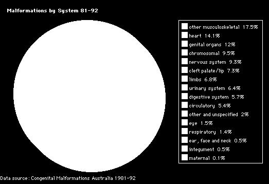
4 DISEASES OF CNS DEVELOPMENTAL ANOMALIES CNS 9.3%
5 DEVELOPMENTAL ANOMALIES:(due to some NOXA in the fetal life) Stage 1: Weeks 3-4 Dorsal Induction: Formation and Closure of the Neural Tube Three phases: Neurulation, canalization, retrogressive differentiation Failure: Anencephaly, Cephalocele, Chiari, Spinal dysraphism Stage 2: Weeks 5-10 Ventral Induction: Formation of the Brain Segments and Face Three vesicles (prosencephalon, mesencephalon, and rhombencephalon) form the cerebrum, mid-brain, cerebellum, and lower brain stem. Division into two hemispheres. Failure: Holoprosencephalies, Corpus callosum agenesis, Dandy Walker,Facial anomalies Stage 3: Months 2-5. Migration and Histogenesis Neuronal migration from germinal matrix to the cortex. Cortical organization. Disorders: Heterotopias, agyria-pachygyria, polymicrogyria, vascular malformations, teratomas, phakomatosis. Stage 4: 5-15 months; matures by 3 years. Myelination Inferior to superior; posterior to anterior. Failure: developmental delay, dysmyelinating disease


6 CONGENITAL ANOMALIES US, MRI, CT, conventional X-ray, DSA Chiari malformation + CC agenesis + meningocele + blockvertebra
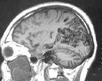
7 DISEASES OF CNS Cerebrovascular disorders - STROKE CT, CTA, MRA, DSA, US MRI Intracerebral lesions: ischaemic haemorrhagic Underlying vascular processes Developmental aneurysms vascular malformations Acquired stenoocclusive lesions thromboembolic processes


8 DISEASES OF CNS Cerebrovascular disorders Ischaemic infarcts Initial stage: CT, MR Hyperdense vessel sign Early ischaemic signs CT perfusion: CBFdefect-CBVdefect=penumbra Developing stage End stage CBF CBV


9 DISEASES OF CNS Cerebrovascular DWI PWI disorders Ischaemic infarcts Initial stage: CT, MR MR: PWI DWI=penumbra Developing stage End stage Mismatch


10 DISEASES OF CNS Cerebrovascular disorders Ischaemic infarcts Initial stage Developing stage: CT, MR Oedema -> mass effect Luxury perfusion End stage


11 DISEASES OF CNS Cerebrovascular disorders Ischaemic infarcts Initial stage Developing stage End stage: CT, MR Retraction -> CSF density/signal

12 DISEASES OF CNS Cerebrovascular disorders Haemorrhagic infarcts: CT Initial stage: method of choice Hyperdense, mass effect Developing stage: Density decreases End stage: Nearly CSF density Mass effect disappears


13 DISEASES OF CNS Cerebrovascular disorders Haemorrhagic infarcts: MR?? Initial stage: not sensitive Developing stage: Blood degradation products End stage

14 DISEASES OF CNS Cerebrovascular disorders Haemorrhagic infarcts : MR Initial stage Developing stage End stage: T2W sequences T2* GRE

 vascular")
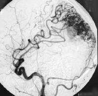
15 DISEASES OF CNS Cerebrovascular disorders Underlying processes Developmental lesions Aneurysms: Angiography (CTA/MRA, DSA) vascular malformations


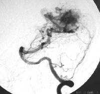

16 DISEASES OF CNS Cerebrovascular disorders Underlying processes Developmental lesions aneurysms vascular malformations CT,MR CTA, MRA, DSA


 US")
17 DISEASES OF CNS Cerebrovascular disorders Underlying processes Acquired vascular diseases stenoocclusive processes (arteriosclerosis) US angiography thromboembolic processes


18 DISEASES OF CNS Cerebrovascular disorders Underlying processes Acquired vascular diseases stenoocclusive processes (arteriosclerosis) thromboembolic processes: Arterial Extracranial - US Intracranial - CTA, MRA, DSA Venous


19 DISEASES OF CNS Cerebrovascular disorders Underlying processes Acquired vascular diseases stenoocclusive processes (arteriosclerosis) thromboembolic processes: Arterial Venous CT, MR CTA, MRA, (DSA)
20 DISEASES OF CNS INTRACRANIAL TUMOURS - classification Extraaxial (non-neurogenic) supratentorial infratentorial Intraaxial (neurogenic) supratentorial infratentorial
21 Imaging methods Plain film radiography Ultrasonography (US) Computertomography (CT): Conventional CT + contrast material CTA CT perfusion Magnetic resonance imaging (MRI): Conventional MRI: different sequences, contrast material MRA MRS DWI/PWI fmri Digital subtraction angiography (DSA)
22 Characteristics of tumours - imaging Abnormal tissue growth: CT, MRI Altered vascularisation: perfusion, angiography Changed metabolism: MRS Contrast enhancement: CT, MRI + iv. contrast material Perifocal edema: CT, MRI, MRS Space-occupying effect: CT, MRI Hydrocephalus: CT, MRI Bone lesions: CT, MRI Multiplicity: CT, MRI


23 Abnormal tissue growth Structure: homogeneity, calcification, cyst, necrosis haemorrhage Border: sharp blurred irregular e.g.


24 Changed vascularisation Gr. 3 astrocytoma Tumor vascularisation Increased CBV, CBF Dignity CBV correlation + Increased permeability: enhancement Angio: hypervascularisation Meningioma

25 Changed metabolism MR spectroscopy Normal brain Tumor Cho=cellularity, NAA=neuron integrity, Lac=anaerob metabolism
 Heterogeneity Biopsy (high grade parts) Effect of")
26 Changed metabolism MR spectroscopy: Tumor or not tumor Tumor diff. dg Grading Extension (focal, infiltration, enhancing and non-enhancing, over T2 parts) Heterogeneity Biopsy (high grade parts) Effect of therapy Consequencies of therapy (radiation necrosis or recidive tumor)

 Partly")


27 Contrast enhancement Non enhancing Enhancing (homogeneous, heterogeneous) Partly enhancing tumors Special signs: tail sign, closed-opened ring Malignity cm enhancement correlation in a histological group +
 Partly enhancing")

28 Contrast enhancement Non enhancing Enhancing (homogeneous, inhomogeneous) Partly enhancing Special signs: tail sign, closed or opened ring Malignity c.m. enhancement correlation in a histological group +


29 Perifocal edema Vasogenic type fingerlike involving only the white matter


30 Space-occupying effect Edema+tumor compression/dislocation herniations subfalcial, transtentorial, transforaminal


31 Hydrocephalus CSF-producing tumor: plexus papilloma Middle-line tumor plexus papilloma medulloblastoma ependymoma Meningial tumors: metastatic, haematological tu.


32 Bone changes Lytic Sclerotic Atrophic Destructive lesions

33 Multiplicity Metastases Glioblastoma Non-tumour diseases
34 INTRACRANIAL TUMOURS Neuroimaging = histological diagnosis
35 Diseases of CNS MRI INFLAMMATIONS Viral Bacterial Fungal Parasitic Immune-mediated Others Brain parenchyma encephalitis cerebritis, abscess multiple sclerosis Meningitis Vasculitis Osteomyelitis


36 Diseases of CNS INFLAMMATIONS Brain parenchyma encephalitis herpes encephalitis cerebritis, abscess multiple sclerosis Meningitis Vasculitis Osteomyelitis


37 Diseases of CNS INFLAMMATIONS Brain parenchyma encephalitis cerebritis, abscess multiple sclerosis Meningitis Vasculitis Osteomyelitis


38 Diseases of CNS INFLAMMATIONS Brain parenchyma encephalitis cerebritis, abscess multiple sclerosis Meningitis Vasculitis Osteomyelitis


39 Diseases of CNS INFLAMMATIONS Brain parenchyma encephalitis cerebritis, abscess multiple sclerosis Meningitis Vasculitis Osteomyelitis


40 Diseases of CNS INFLAMMATIONS Brain parenchyma encephalitis cerebritis, abscess multiple sclerosis Meningitis complications: hygroma abscess formation hydrocephalus thrombosis Vasculitis Osteomyelitis
41 Diseases of CNS TRAUMA Closed head trauma (intact dura) epidural haematoma CT Open head trauma (dural tear) impression fracture pneumatocele subdural haematoma foreign body contusion haematoma, edema edema secondary infection

 epidural")


42 Diseases of CNS TRAUMA Closed head trauma (intact dura mater) epidural haematoma subdural haematoma acute - hyperdense subacute - isodense chronic - hypodense contusion edema


43 Diseases of CNS TRAUMA Closed head trauma (intact dura mater) epidural haematoma subdural haematoma acute - hyperdense subacute - isodense chronic - hypodense contusion edema


44 Diseases of CNS TRAUMA Open head trauma (dural tear) impression fracture pneumatocele foreign body haematoma, edema secondary infection

45 Diseases of CNS TRAUMA Open head trauma (dural tear) impression fracture pneumatocele foreign body haematoma, edema infection
Spine and spinal cord
 NEURORADIOLOGY Spine and spinal cord Erika Vörös University of Szeged Department of Radiology SZEGED DISEASES OF SPINE AND SPINAL CORD I. Non-tumourous diseases developmental anomalies vascular disorders
NEURORADIOLOGY Part I
 NEURORADIOLOGY Part I Vörös Erika University of Szeged Department of Radiology SZEGED BRAIN IMAGING METHODS Plain film radiography Ultrasonography (US) Computer tomography (CT) Magnetic resonance imaging
Diagnostic modalities of the Central Nervous System
 NEURORADIOLOGY Kinga Karlinger, MD, PhD Associate Professor Semmelweis University, Budapest Diagnostic modalities of the Central Nervous System X-ray: screening is not used any more, x-ray images instead
NEURORADIOLOGY DIL part 5
 NEURORADIOLOGY DIL part 5 Masses and tumors K. Agyem MD, G. Hall MD, D. Palathinkal MD, Alexandre Menard March/April 2015 OVERVIEW Introduction to Neuroimaging - DIL part 1 Basic Brain Anatomy - DIL part
NEURORADIOLOGY. Part III. Angela Csomor University of Szeged Department of Radiology
 NEURORADIOLOGY Part III Angela Csomor University of Szeged Department of Radiology DISEASES OF SPINE AND SPINAL CORD I. Non-tumourous diseases developmental anomalies vascular disorders inflammatory processes
Index. aneurysm, 92 carotid occlusion, 94 ICA stenosis, 95 intracranial, 92 MCA, 94
 A ADC. See Apparent diffusion coefficient (ADC) Aneurysm cerebral artery aneurysm, 93 CT scan, 93 gadolinium, 93 Angiography, 13 Anoxic brain injury, 25 Apparent diffusion coefficient (ADC), 7 Arachnoid
Neuroradiology. J.Lisý
 Neuroradiology J.Lisý X-ray of skull/spine trauma (2 perpendicular projections) congenital developemental errors (scoliosis, spina bifida) Perimyelography (PMG) Lumbar puncture, isoosmolar iodine CM Dural
The central nervous system
 Sectc.qxd 29/06/99 09:42 Page 81 Section C The central nervous system CNS haemorrhage Subarachnoid haemorrhage Cerebral infarction Brain atrophy Ring enhancing lesions MRI of the pituitary Multiple sclerosis
An Introduction to Imaging the Brain. Dr Amy Davis
 An Introduction to Imaging the Brain Dr Amy Davis Common reasons for imaging: Clinical scenarios: - Trauma (NICE guidelines) - Stroke - Tumours - Seizure - Neurological degeneration memory, motor dysfunction,
NEURO IMAGING 2. Dr. Said Huwaijah Chairman of radiology Dep, Damascus Univercity
 NEURO IMAGING 2 Dr. Said Huwaijah Chairman of radiology Dep, Damascus Univercity I. EPIDURAL HEMATOMA (EDH) LOCATION Seventy to seventy-five percent occur in temporoparietal region. CAUSE Most likely caused
Cerebro-vascular stroke
 Cerebro-vascular stroke CT Terminology Hypodense lesion = lesion of lower density than the normal brain tissue Hyperdense lesion = lesion of higher density than normal brain tissue Isodense lesion = lesion
CT INTERPRETATION COURSE
 CT INTERPRETATION COURSE Refresher Course ASTRACAT October 2012 Stroke is a Clinical Diagnosis A clinical syndrome characterised by rapidly developing clinical symptoms and/or signs of focal loss of cerebral
Neuroimaging Core Curriculum
 Neuroimaging Core Curriculum Program Content The purpose of the training program is to prepare the physician for the independent practice of neuroimaging. Neuroimaging is the subspecialty of Neurology
Masses of the Corpus Callosum
 Masses of the Corpus Callosum Kesav Raghavan, HMS Year III Dr. Agenda Corpus Callosum Development and Anatomy Our Patient: Clinical Presentation Differential Diagnosis of Masses in the Corpus Callosum
Pathologic Analysis of CNS Surgical Specimens
 2015 Kenneth M. Earle Memorial Neuropathology Review Pathologic Analysis of CNS Surgical Specimens Peter C. Burger, MD Interdisciplinary Quality Control Familiarity with entities Use of diagnostic algorithm
Prenatal Prediction of The Neurologically Impaired Neonate By Ultrasound
 Prenatal Prediction of The Neurologically Impaired Neonate By Ultrasound Robert H. Debbs, D.O.,F.A.C.O.O.G. Professor of OB-GYN Perelman School of Medicine, University of Pennsylvania Director, Pennsylvania
CT INTERPRETATION COURSE
 CT INTERPRETATION COURSE Introductory Lecture on Basic Principles ASTRACAT 2012 Part One Stroke is a Clinical Diagnosis A clinical syndrome characterised by rapidly developing clinical symptoms and/or
CNS TUMORS. D r. Ali Eltayb ( U. of Omdurman. I ). M. Path (U. of Alexandria)
. M. Path (U. of Alexandria)") CNS TUMORS D r. Ali Eltayb ( U. of Omdurman. I ). M. Path (U. of Alexandria) CNS TUMORS The annual incidence of intracranial tumors of the CNS ISmore than intraspinal tumors May be Primary or Secondary
CNS TUMORS D r. Ali Eltayb ( U. of Omdurman. I ). M. Path (U. of Alexandria) CNS TUMORS The annual incidence of intracranial tumors of the CNS ISmore than intraspinal tumors May be Primary or Secondary
What Are We Going to Do? Fourth Year Meds Clinical Neuroanatomy. Hydrocephalus and Effects of Interruption of CSF Flow. Tube Blockage Doctrine
 Fourth Year Meds Clinical Neuroanatomy Ventricles, CSF, Brain Swelling etc. David A. Ramsay, Neuropathologist, LHSC What Are We Going to Do? Hydrocephalus and some effects of the interruption of CSF flow
Central Nervous System Congenital Abnormalities
 Central Nervous System Congenital Abnormalities Eva Brichtova, M.D., Ph.D., Department of Pediatric Sugery, Orthopaedics and Traumatology, University Hospital Brno Neural tube defects Dysraphism uncomplete
CT & MRI Evaluation of Brain Tumour & Tumour like Conditions
 CT & MRI Evaluation of Brain Tumour & Tumour like Conditions Dr. Anjana Trivedi 1, Dr. Jay Thakkar 2, Dr. Maulik Jethva 3, Dr. Ishita Virda 4 1 M.D. Radiology, Professor and Head, P.D.U. Medical College
Intracranial spontaneous hemorrhage mechanisms, imaging and management
 Intracranial spontaneous hemorrhage mechanisms, imaging and management Dora Zlatareva Department of Diagnostic Imaging Medical University, Sofia, Bulgaria Intracranial hemorrhage (ICH) ICH 15% of strokes
CNS Embryology 5th Menstrual Week (Dorsal View)
") Imaging of the Fetal Brain; Normal & Abnormal Alfred Abuhamad, M.D. Eastern Virginia Medical School CNS Embryology 5th Menstrual Week (Dorsal View) Day 20 from fertilization Neural plate formed in ectoderm
Imaging of the Fetal Brain; Normal & Abnormal Alfred Abuhamad, M.D. Eastern Virginia Medical School CNS Embryology 5th Menstrual Week (Dorsal View) Day 20 from fertilization Neural plate formed in ectoderm
HEAD AND NECK IMAGING. James Chen (MS IV)
") HEAD AND NECK IMAGING James Chen (MS IV) Anatomy Course Johns Hopkins School of Medicine Sept. 27, 2011 OBJECTIVES Introduce cross sectional imaging of head and neck Computed tomography (CT) Review head
HEAD AND NECK IMAGING James Chen (MS IV) Anatomy Course Johns Hopkins School of Medicine Sept. 27, 2011 OBJECTIVES Introduce cross sectional imaging of head and neck Computed tomography (CT) Review head
IMAGING OF INTRACRANIAL INFECTIONS
 IMAGING OF INTRACRANIAL INFECTIONS Dr Carolina Kachramanoglou LYSHOLM DEPARTMENT OF NEURORADIOLOGY NATIONAL HOSPITAL FOR NEUROLOGY AND NEUROSURGERY Plan Introduce MR sequences that are useful in the diagnosis
Benign brain lesions
 Benign brain lesions Diagnostic and Interventional Radiology Hung-Wen Kao Department of Radiology, Tri-Service General Hospital, National Defense Medical Center Computed tomography Hounsfield unit (HU)
Attenuation value in HU From -500 To HU From -10 To HU From 60 To 90 HU. From 200 HU and above
 Brain Imaging Common CT attenuation values Structure Air Fat Water Brain tissue Recent hematoma Calcifications Bone Brain edema and infarction Normal liver parenchyma Attenuation value in HU From -500
Imaging the Spinal Cord & Intradural Disease
 Department of Radiology University of California San Diego Imaging the Spinal Cord & Intradural Disease John R. Hesselink, M.D. Spinal Cord Diseases Tumors Syringohydromyelia Trauma Ischemia / Infarction
A Guide to the Radiologic Evaluation of Extra-Axial Hemorrhage
 July 2013 A Guide to the Radiologic Evaluation of Extra-Axial Hemorrhage John Dickson, Harvard Medical School Year III Agenda 1. Define extra-axial hemorrhage and introduce its subtypes 2. Review coup
Case Conference: Neuroradiology. Case 1: Tumor Case 1: 22yo F w/ HA and prior Seizures
 Case Conference: Neuroradiology Case 1: 22yo F w/ HA and prior Seizures David E. Rex, MD, PhD Stanford University Hospital Department of Radiology Case 1: Tumor Most likely gangiloglioma, oligodendroglioma,
Malformations of the Nervous System November 10, Dr. Peter Ostrow
 Malformations of the Nervous System November 10, 2016 Dr. Peter Ostrow Malformations of the Nervous System 1. Abnormal closure of the neural tube 1. Disorders of forebrain formation 1. Cortical anomalies
Spine. Neuroradiology. Spine. Spine Pathology. Distribution of fractures. Radiological algorithm. Role of radiology 18/11/2015
 Spine Neuroradiology Spine Prof.Dr.Nail Bulakbaşı X Ray: AP/L/Oblique Vertebra & disc spaces CT & CTA Vertebra, discs, vessels MRI & MRA Vertebra, disc, vessels, meninges Spinal cord & nerves Myelography
STROKE - IMAGING. Dr RAJASEKHAR REDDY 2nd Yr P.G. RADIODIAGNOSIS KIMS,Narkatpalli.
 STROKE - IMAGING Dr RAJASEKHAR REDDY 2nd Yr P.G. RADIODIAGNOSIS KIMS,Narkatpalli. STROKE Describes a clinical event that consists of sudden onset of neurological symptoms Types Infarction - occlusion of
The Brain: Prenatal and Postnatal Effects of Congenital Heart Disease. Dianna M. E. Bardo, M D Swedish Cherry Hill Radia, Inc.
 The Brain: Prenatal and Postnatal Effects of Congenital Heart Disease Dianna M. E. Bardo, M D Swedish Cherry Hill Radia, Inc. Seattle, WA embryology We recognize the VACTERL association and frequency of
secondary effects and sequelae of head trauma.
 Neuroimaging of vascular/secondary secondary effects and sequelae of head trauma. Andrès Server Alonso Department of Neuroradiology Division of Radiology Ullevål University Hospital Oslo, Norway. Guidelines
Adult - Cerebrovascular. Adult - Cranio-Cervical Junction. Adult - Epilepsy. Adult - Hydrocephalus
 list for SET and IMG Neurosurgery Adult - Cerebrovascular Aneurysm - Clipping: Anterior circulation Aneurysm - Clipping: Posterior circulation AVM excision Carotid endarterectomy Carotid trapping Cavernoma
NEURORADIOLOGY Angela Lignelli, MD
 Neuroradiology NEURORADIOLOGY Angela Lignelli, MD Plain radiographs CT MRI Cerebral Angiogram Myelograms Neuroradiology Computerized Axial Tomography (CT) CT without and with contrast CTA CT angiogram
NEURORADIOLOGY Angela Lignelli, MD
 NEURORADIOLOGY Angela Lignelli, MD Neuroradiology Plain radiographs CT MRI Cerebral Angiogram Myelograms 1 Neuroradiology Computerized Axial Tomography (CT) CT without and with contrast CTA CT angiogram
NEURORADIOLOGY Angela Lignelli, MD
 Neuroradiology NEURORADIOLOGY Angela Lignelli, MD Plain radiographs CT MRI Cerebral Angiogram Myelograms Neuroradiology Computerized Axial Tomography (CT) CT without and with contrast CTA CT angiogram
Applicable Neuroradiology
 For the Clinical Neurology Clerkship LSU Medical School New Orleans Amy W Voigt, MD Clerkship Director Introduction The field of Radiology first developed following the discovery of X-Rays by Wilhelm Roentgen
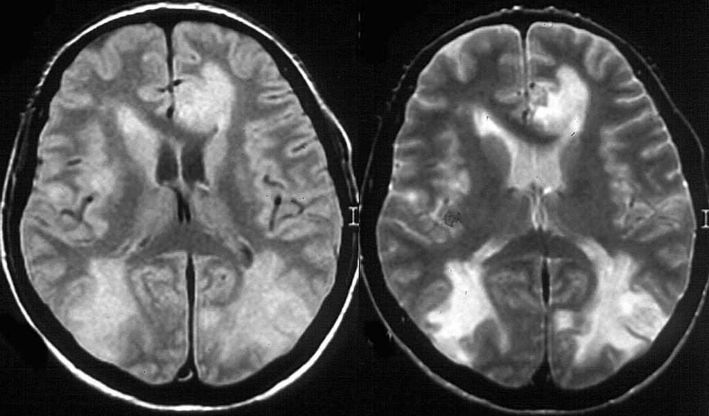
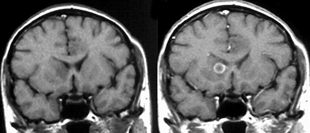
RING ENCHANCING LESION BY M.S. HEMHNATH
 RING ENCHANCING LESION BY M.S. HEMHNATH A 21 YRS FEMALE CAME WITH H/O HEADACHE AND SEIZURE FOR THE PAST ONE MONTH. NO OTHER FOCAL NEUROLOGICAL DEFICIT. DIFFERENTIAL DIAGNOSIS For this case are Neurocysticerosis
Cerebrovascular diseases-2
 Cerebrovascular diseases-2 Primary angiitis of CNS - Other causes of infarction i. Hypercoagulable states ii. Drug-abuse such as amphetamine, heroin and cocain Note - The venous side of the circulation
Neuroimaging. spine / spinal cord
 Neuroimaging spine / spinal cord Spine & spinal cord imaging methodology Plain x-ray of spine Computed tomography CT - traditional ( normal CT) - reconstructions - myelo-ct Magnetic resonance MR - standard
Infections. Meningitis- leptomenings (usually bakterial)
") Neuropathology Neuropathology Cerebral Edema, raised intracranial pressure, Herniatio Malformations, Developmental Diseases Perinatal Brain Injury Trauma Cerebrovascular Diseases Infections Transmissible
Neuropathology Neuropathology Cerebral Edema, raised intracranial pressure, Herniatio Malformations, Developmental Diseases Perinatal Brain Injury Trauma Cerebrovascular Diseases Infections Transmissible
UPSTATE Comprehensive Stroke Center. Neurosurgical Interventions Satish Krishnamurthy MD, MCh
 UPSTATE Comprehensive Stroke Center Neurosurgical Interventions Satish Krishnamurthy MD, MCh Regional cerebral blood flow is important Some essential facts Neurons are obligatory glucose users Under anerobic
RINGS N THINGS: Imaging Patterns in Differential Diagnosis. Anne G. Osborn, M.D.
 RINGS N THINGS: Imaging Patterns in Differential Diagnosis Anne G. Osborn, M.D. ExpDDxs: Intra-axial (Parenchymal) Lesions Ring-enhancing lesions, solitary 1 Ring-enhancing lesion crossing corpus callosum
Neuroanatomy. Assistant Professor of Anatomy Faculty of Medicine The University of Jordan Dr Maha ELBeltagy
 Neuroanatomy Dr. Maha ELBeltagy Assistant Professor of Anatomy Faculty of Medicine The University of Jordan 2018 Development of the Central Nervous System Development of the nervous system Development
AAP ZIKA ECHO (EXTENSION FOR COMMUNITY HEALTHCARE OUTCOMES)
") AAP ZIKA ECHO (EXTENSION FOR COMMUNITY HEALTHCARE OUTCOMES) HOUSEKEEPING ITEMS For educational and quality improvement purposes, this ECHO session will be recorded Project ECHO collects participation data
AAP ZIKA ECHO (EXTENSION FOR COMMUNITY HEALTHCARE OUTCOMES) HOUSEKEEPING ITEMS For educational and quality improvement purposes, this ECHO session will be recorded Project ECHO collects participation data
Vascular Pattern in Tumours
 Acta Radiologica ISSN: 0001-6926 (Print) (Online) Journal homepage: https://www.tandfonline.com/loi/iaro20 Vascular Pattern in Tumours To cite this article: (1957) Vascular Pattern in Tumours, Acta Radiologica,
Non-Traumatic Neuro Emergencies
 Department of Radiology University of California San Diego Non-Traumatic Neuro Emergencies John R. Hesselink, M.D. Nontraumatic Neuroemergencies 1. Acute focal neurological deficit 2. Worst headache of
Intracranial Lesions: MRI Signs for Localization
 Intracranial Lesions: MRI Signs for Localization Poster No.: C-1574 Congress: ECR 2017 Type: Educational Exhibit Authors: M. Cucos, A. Puiu, S. Manole ; Cluj-Napoca/RO, Cluj napoca/ RO Keywords: Cerebrospinal
In-Training Examination for Diagnostic Radiology Residents Rationales
 28th Annual In-Training Examination for Diagnostic Radiology Residents Rationales Sponsored by: Commission on Education Committee on Residency Training in Diagnostic Radiology February 3, 2005 The American
Magnetic Resonance Imaging for Neurological Conditions. Lawrance Yip Department of Radiology Queen Mary Hospital
 Magnetic Resonance Imaging for Neurological Conditions Lawrance Yip Department of Radiology Queen Mary Hospital Outline Strength and limitations of MRI for neurological conditions MR Imaging techniques
Supplementary Online Content
 Supplementary Online Content Honein MA, Dawson AL, Petersen E, et al; US Zika Pregnancy Registry Collaboration. Birth Defects Among Fetuses and Infants of US Women With Laboratory Evidence of Possible
EEG IN FOCAL ENCEPHALOPATHIES: CEREBROVASCULAR DISEASE, NEOPLASMS, AND INFECTIONS
 246 Figure 8.7: FIRDA. The patient has a history of nonspecific cognitive decline and multiple small WM changes on imaging. oligodendrocytic tumors of the cerebral hemispheres (11,12). Electroencephalogram
Laura Tormoehlen, M.D. Neurology and EM-Toxicology Indiana University
 Laura Tormoehlen, M.D. Neurology and EM-Toxicology Indiana University Disclosures! No conflicts of interest to disclose Neuroimaging 101! Plain films! Computed tomography " Angiography " Perfusion! Magnetic
intracranial anomalies
 Chapter 5: Fetal Central Nervous System 84 intracranial anomalies Hydrocephaly Dilatation of ventricular system secondary to an increase in the amount of CSF. Effects of hydrocephalus include flattening
Introduction to Neuroimaging Aaron S. Field, MD, PhD Assistant Professor of Radiology Neuroradiology Section University of Wisconsin Madison
 Introduction to Neuroimaging Aaron S. Field, MD, PhD Assistant Professor of Radiology Neuroradiology Section University of Wisconsin Madison Updated 7/17/07 Neuroimaging Modalities Radiography (X-Ray)
Introduction to Neuroimaging spine. John J. McCormick MD
 Introduction to Neuroimaging spine John J. McCormick MD Neuroanatomy Netter drawings Radiographic Anatomy Cervical Spine Cervical Spine Oblique View Cervical Spine Dens View Thoracic Spine Lumbar Spine
Brain AVM with Accompanying Venous Aneurysm with Intracerebral and Intraventricular Hemorrhage
 Cronicon OPEN ACCESS EC PAEDIATRICS Case Report Brain AVM with Accompanying Venous Aneurysm with Intracerebral and Intraventricular Hemorrhage Dimitrios Panagopoulos* Neurosurgical Department, University
Tutorials. By Dr Sharon Truter
 Tutorials By Dr Sharon Truter To the Tutorials By Dr Sharon Truter What to expect from the Tutorials What to expect from these tutorials Outlines, structure, guided reading, explanations, mnemonics Begin
CNS pathology Third year medical students. Dr Heyam Awad 2018 Lecture 5: disturbed fluid balance and increased intracranial pressure
 CNS pathology Third year medical students Dr Heyam Awad 2018 Lecture 5: disturbed fluid balance and increased intracranial pressure ILOs Understand causes and symptoms of increased intracranial pressure.
Head Trauma Inservice (October)
") John Tramell - Head Trauma Inservice, October 2005.doc Page 1 Head Trauma Inservice (October) Head trauma is the leading cause of death in trauma patients. Having a basic understanding of the anatomy and
John Tramell - Head Trauma Inservice, October 2005.doc Page 1 Head Trauma Inservice (October) Head trauma is the leading cause of death in trauma patients. Having a basic understanding of the anatomy and
M555 Medical Neuroscience Blood Flow in CNS Meninges Blood Brain Barrier CSF
 M555 Medical Neuroscience Blood Flow in CNS Meninges Blood Brain Barrier CSF Arterial Blood Flow to CNS approximately % of what goes wrong within the skull that produces neurological deficits is vascular
Stroke imaging. Why image stroke patients? Stroke. Treatment of infarct. Methods for infarct diagnosis. Treatment of infarct.
 Stroke imaging Stroke Infarct: -Arterial thrombosis/embolus -Hypoxic/ischemic -Venous thrombosis Non-traumatic hemorrhage: -Intracerebral -Subarachnoid Johan Wikström MD PhD Associate Professor of Radiology
ISCHEMIC STROKE IMAGING
 ISCHEMIC STROKE IMAGING ผศ.พญ พญ.จ ร ร ตน ธรรมโรจน ภาคว ชาร งส ว ทยา คณะแพทยศาสตร มหาว ทยาล ยขอนแก น A case of acute hemiplegia Which side is the abnormality, right or left? Early Right MCA infarction
Diffusion-weighted imaging and ADC mapping in the differentiation of intraventricular brain tumors
 Diffusion-weighted imaging and ADC mapping in the differentiation of intraventricular brain tumors Poster No.: C-2652 Congress: ECR 2010 Type: Educational Exhibit Topic: Neuro Authors: M. Gavrilov, T.
Disclosure. + Outline. Case-based approach to neurological emergencies that might present to the ED
 Kathleen R. Fink, MD University of Washington 5 th Nordic Emergency Radiology Course May 21, 2015 Disclosure My spouse receives research salary support from: Bracco BayerHealthcare Guerbet Outline Case-based
PA SYLLABUS. Syllabus for students of the FACULTY OF MEDICINE No.2
 Approved At the meeting of the Faculty Council Medicine No. of Approved At the meeting of the chair of Neurosurgery No. of Dean of the Faculty Medicine No.2 PhD, associate professor M. Betiu Head of the
JMSCR Vol 04 Issue 12 Page December 2016
 www.jmscr.igmpublication.org Impact Factor 5.244 Index Copernicus Value: 83.27 ISSN (e)-2347-176x ISSN (p) 2455-0450 DOI: https://dx.doi.org/10.18535/jmscr/v4i12.113 Role of Computed Tomography in Evaluation
THE ROLE OF IMAGING IN DIAGNOSIS OF SUBDURAL HEMATOMA: REVIEW ARTICLE
 THE ROLE OF IMAGING IN DIAGNOSIS OF SUBDURAL HEMATOMA: REVIEW ARTICLE * Dr. Sumendra Raj Pandey, Prof. Dr. Liu Pei WU, Dr. Sohan Kumar Sah, Dr. Lalu Yadav, Md. Sadam Husen Haque and Rajan KR. Chaurasiya
ARTERIOVENOUS MALFORMATION OR CONTUSION : A DIAGNOSTIC DILEMMA. Yong Pei Yee, Ibrahim Lutfi Shuaib, Jafri Malin Abdullah*
 Malaysian Journal of Medical Sciences, Vol. 8, No. 2, July 2001 (47-51) CASE REPORT ARTERIOVENOUS MALFORMATION OR CONTUSION : A DIAGNOSTIC DILEMMA Yong Pei Yee, Ibrahim Lutfi Shuaib, Jafri Malin Abdullah*
Neuroradiology: Imaging and Stroke
 Neuroradiology: Imaging and Stroke Stroke 2017 William Gallmann January 28, 2017 Stroke Arterial ischemia/infarct accounts for ~85% Cerebral venous occlusions - 0.5-1% Spontaneous intracranial hemorrhage
CT - Brain Examination
 CT - Brain Examination Submitted by: Felemban 1 CT - Brain Examination The clinical indication of CT brain are: a) Chronic cases (e.g. headache - tumor - abscess) b) ER cases (e.g. trauma - RTA - child
JMSCR Vol 05 Issue 08 Page August 2017
 www.jmscr.igmpublication.org Impact Factor 5.84 Index Copernicus Value: 83.27 ISSN (e)-2347-176x ISSN (p) 2455-0450 DOI: https://dx.doi.org/10.18535/jmscr/v5i8.19 Magnetic Resonance Imaging in Evaluation
Vascular Disorders. Nervous System Disorders (Part B-1) Module 8 -Chapter 14. Cerebrovascular disease S/S 1/9/2013
 Module 8 -Chapter 14. Cerebrovascular disease S/S 1/9/2013") Nervous System Disorders (Part B-1) Module 8 -Chapter 14 Overview ACUTE NEUROLOGIC DISORDERS Vascular Disorders Infections/Inflammation/Toxins Metabolic, Endocrinologic, Nutritional, Toxic Neoplastic Traumatic
Nervous System Disorders (Part B-1) Module 8 -Chapter 14 Overview ACUTE NEUROLOGIC DISORDERS Vascular Disorders Infections/Inflammation/Toxins Metabolic, Endocrinologic, Nutritional, Toxic Neoplastic Traumatic
Imaging of Acute Cerebral Trauma
 July, 2005 Imaging of Acute Cerebral Trauma Louis Rivera, Harvard Medical School, Year III 46 y/o Female s/p Trauma - Unrestrained? MVC requiring Med Flight - Facial bruising/swelling - DEEP COMA - SEIZURES
Meninges and Ventricles
 Meninges and Ventricles Irene Yu, class of 2019 LEARNING OBJECTIVES Describe the meningeal layers, the dural infolds, and the spaces they create. Name the contents of the subarachnoid space. Describe the
A Case of Naso-Ethmoidal Meningoencephalocele
 A Case of Naso-Ethmoidal Meningoencephalocele Divyanshu Dubey, Sonjjay Pande, Pradeep Dubey, Anshudha Sawhney Vol. 3 No. 8 (August 2011) International Journal of Collaborative Research on Internal Medicine
SURGICAL MANAGEMENT OF BRAIN TUMORS
 SURGICAL MANAGEMENT OF BRAIN TUMORS LIGIA TATARANU, MD, Ph D NEUROSURGICAL CLINIC, BAGDASAR ARSENI CLINICAL HOSPITAL BUCHAREST, ROMANIA SURGICAL INDICATIONS CONFIRMING HISTOLOGIC DIAGNOSIS REDUCING TUMOR
IMAGING OF A CASE OF SPINAL MENINGIOMA- A CASE REPORT
 IMAGING OF A CASE OF SPINAL MENINGIOMA- A CASE REPORT Ramneet Wadi 1, Anil Kumar Shukla 2, Seetha Pramila V. V 3, Sabyasachi Basu 4, Sonam Sanjay 5 1Postgraduate Student, Department of Radiodiagnosis,
Imaging Acute Stroke and Cerebral Ischemia
 Department of Radiology University of California San Diego Imaging Acute Stroke and Cerebral Ischemia John R. Hesselink, M.D. Causes of Stroke Arterial stenosis Thrombosis Embolism Dissection Hypotension
Keep Imaging Simple: An Introduction To Neuroimaging
 Keep Imaging Simple: An Introduction To Neuroimaging Meghan Elkins, OD, FAAO Please silence all mobile devices and remove items from chairs so others can sit. Unauthorized recording of this session is
CT and MR findings of systemic lupus erythematosus involving the brain: Differential diagnosis based on lesion distribution
 CT and MR findings of systemic lupus erythematosus involving the brain: Differential diagnosis based on lesion distribution Poster No.: C-2723 Congress: ECR 2010 Type: Educational Exhibit Topic: Neuro
ORIGINAL ARTICLE COMPUTED TOMOGRAPHY EVALUATION OF SPONTANEOUS INTRACRANIAL HAEMORRHAGE
 Available online at www.journalijmrr.com INTERNATIONAL JOURNAL OF MODERN RESEARCH AND REVIEWS IJMRR ISSN: 2347-8314 Int. J. Modn. Res. Revs. Volume 3, Issue 4, pp 647-652, April, 2015 ORIGINAL ARTICLE
Automated Identification of Neoplasia in Diagnostic Imaging text reports
 Automated Identification of Neoplasia in Diagnostic Imaging text reports "This work has been funded in whole or in part with Federal funds from the National Cancer Institute, National Institutes of Health,
Symposium: OB/GY US (Room B) CNS Anomalies
 CNS Anomalies") 82 Symposium: OB/GY US (Room B) 11 : 50 1 2 : 10 CNS Anomalies Brain area Midline structure S u p r a t e n t o r i a l ventricular system Cerebral hemisphere Posterior fossa Head size and shape Image
82 Symposium: OB/GY US (Room B) 11 : 50 1 2 : 10 CNS Anomalies Brain area Midline structure S u p r a t e n t o r i a l ventricular system Cerebral hemisphere Posterior fossa Head size and shape Image
BRAIN STEM AND CEREBELLUM..
 Lecture Title: BRAIN STEM AND CEREBELLUM.. (CNS Block, Radiology) Dr. Hamdy Hassan Ass.Prof. Consultant Radiology Department KKHU King Saud University Lecture Objectives.. Students at the end of the lecture
Pathology with CT Sectional Anatomy Correlation, Part I. Tom Haller, RT (R)(CT)(MR)
(CT)(MR)") Pathology with CT Sectional Anatomy Correlation, Part I Tom Haller, RT (R)(CT)(MR) Objectives Upon completion of the course, the participant will be able to: 1. State pathologies that commonly require
Pathology with CT Sectional Anatomy Correlation, Part I Tom Haller, RT (R)(CT)(MR) Objectives Upon completion of the course, the participant will be able to: 1. State pathologies that commonly require
Pediatric Spine Tumors (and other masses)
") Pediatric Spine Tumors (and other masses) Francisco A Perez, MD, PhD Assistant Professor Neuroradiology and Pediatric Radiology Seattle Children s Hospital University of Washington, Seattle Commercial
Pediatric Spine Tumors (and other masses) Francisco A Perez, MD, PhD Assistant Professor Neuroradiology and Pediatric Radiology Seattle Children s Hospital University of Washington, Seattle Commercial
Ventricles, CSF & Meninges. Steven McLoon Department of Neuroscience University of Minnesota
 Ventricles, CSF & Meninges Steven McLoon Department of Neuroscience University of Minnesota 1 Coffee Hour Thursday (Sept 14) 8:30-9:30am Surdyk s Café in Northrop Auditorium Stop by for a minute or an
Imaging of Cerebrovascular Disease
 Imaging of Cerebrovascular Disease A Practical Guide Val M. Runge, MD Editor-in-Chief of Investigative Radiology Institute for Diagnostic, Interventional, and Pediatric Radiology Inselspital, University
Head CT Scan Interpretation: A Five-Step Approach to Seeing Inside the Head Lawrence B. Stack, MD
 Head CT Scan Interpretation: A Five-Step Approach to Seeing Inside the Head Lawrence B. Stack, MD Five Step Approach 1. Adequate study 2. Bone windows 3. Ventricles 4. Quadrigeminal cistern 5. Parenchyma
ATTENDING PHYSICIAN'S STATEMENT STROKE / BRAIN ANEURYSM SURGERY OR CEREBRAL SHUNT INSERTION / CAROTID ARTERY SURGERY
 ATTENDING PHYSICIAN'S STATEMENT STROKE / BRAIN ANEURYSM SURGERY OR CEREBRAL SHUNT INSERTION / CAROTID ARTERY SURGERY A) Patient s Particulars Name of Patient Gender NRIC/FIN or Passport No. Date of Birth
Department of Radiology, University of Szeged. Imaging of the skeleton
 Imaging of the skeleton Methods of examination: plain x-ray (radiography, densitometry) x-ray with contrast material (fistulography, angiography) ultrasound (b-mode, Doppler, color, duplex) computed tomography
CNS pathology Third year medical students. Dr Heyam Awad 2018 Lecture 12: CNS tumours 2/3
 CNS pathology Third year medical students Dr Heyam Awad 2018 Lecture 12: CNS tumours 2/3 Pilocytic astrocytoma Relatively benign ( WHO grade 1) Occurs in children and young adults Mostly: in the cerebellum
Advanced Neuroimaging for Acute Stroke
 Advanced Neuroimaging for Acute Stroke E. Bradshaw Bunney, MD, FACEP Professor Department Of Emergency Medicine University of Illinois at Chicago Swedish American Belvidere Hospital Disclosures FERNE Board
Bacterial, viral, protoozal and fungal infections of the CNS
 Bacterial, viral, protoozal and fungal infections of the CNS Prof. Isidro Ferrer, Institut Neuropatologia, Servei Anatomia Patològica, IDIBELL-Hospital Universitari de Bellvitge, Universitat de Barcelona,
Sample page. Radiology. Cross Coder. Essential links from CPT codes to ICD-10-CM and HCPCS
 Cross Coder 2018 Radiology Essential links from CPT codes to ICD-10-CM and HCPCS POWER UP YOUR CODING with Optum360, your trusted coding partner for 32 years. Visit optum360coding.com. Contents Introduction...
Case Report Intracranial Capillary Hemangioma in the Posterior Fossa of an Adult Male
 Case Reports in Radiology Volume 2016, Article ID 6434623, 4 pages http://dx.doi.org/10.1155/2016/6434623 Case Report Intracranial Capillary Hemangioma in the Posterior Fossa of an Adult Male Jordan Nepute,