What is DTI, its use in research and clinical practice, and its future potential
|
|
|
- Maximilian Simon
- 5 years ago
- Views:
Transcription
1 NA-MIC What is DTI, its use in research and clinical practice, and its future potential Martin Styner with thanks to many, many people Departments of Computer Science and Psychiatry UNC Neurodevelopmental Disorders Research Center UNC Neuro Image Research and Analysis Lab
2 TOC Interpretation/validation of DTI properties!rat spinal cord studies Validation of tractography What can DTI be used for? Many different applications!main part of the talk Future of DTI: looks bright 2


3 DTI Properties (again) Fractional Anisotropy (Tensor shape), 0..1 Mean Diffusion (Tensor volume) WM/GM CSF Axial Diffusion In WM: Diffusion parallel to axon! 1 " Radial Diffusion! 2 " = λ 1 = λ (λ 2 + λ 3 ) =! 3 " = λ 2 In WM: Diffusion orthogonal to axon


4 Spinal Cord Degeneration DTI (AD/RD) & immunohistochemistry of Wallerian degeneration Unilateral L2 L4 dorsal axotomy in rat spine column DTI revealed dorsal lesion extending from lumbar to cervical cord 3D AD RD 4


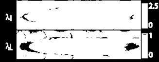

5 Spinal Cord Degeneration AD RD Lesion at Day 3 Day 30 AD RD 5


6 Immunohistochemistry LFB: Myelin SMI : neurofilaments! 31: hyper-phosphylated! 32: hypo-phosphylated 6
7 Spinal Cord Degeneration Day 3 (as compared to unlesioned side)! DTI: significantly reduced AD and increased RD.! Immuno: Reduced phosphorylated, increased nonphosphorylated neurofilaments, swollen axons, myelin ovoids, no loss of myelin. Day 30 (as compared to day 3)! DTI: no reduction in AD but increase in RD! Immuno: Gradual clearance of myelin, no changes in neurofilament Conclusion:! DTI, AD/RD sensitive to axon degeneration! FA captures all effects, but cannot differentiate! Correlation of RD with myelin degeneration! Correlation of AD with loss of phosphorylated neurofilaments 7


8 Demyelinating Lesions Rat model of autoimmune encephalomyelitis/ms Injection of cytokines (TNF-!, IFN-") or lipopolysaccharides => spinal cord lesions DTI & Immunohistochemistry FA, AD and RD correlate with axon counts and degenerating axon counts FA and T2-w intensity correspond to changes in myelin loss and axon phosphorylation 8

9 Validation of Tractography Are results of DTI tractography anatomically correct? Yes and No Many studies using synthetic ground truth & MRI phantoms show convincingly positive results In/Ex vivo: stimulation mapping, manganese imaging, tracer studies! Several performed in primates, Dauguet 2007 (NeuroImage) Tracer 9 DTI Tracts
10 Validation of Tractography Good agreement for major fiber bundles Sensitivities to fiber crossings and small fiber bundles! Higher models of diffusion (Qball, DSI)! Anatomical knowledge via source and target selection! Novel tractography on DTI: multi-tensor or probabilistic tracking Overall convincing evidence for DTI tractography! Major fiber tracts are valid! #fibers highly variable!! Size of tracts variable! Number of fibers Scans' Comparison:Fibers (ROI: essai3-th69) Second Method without Interpolation Second Method with Interpolation Scan 1 Scan 2 Scan 3 Scan 4 Scan 5 Scan 6 Average

11 Intermezzo PhDcomics: Brain on a stick 11
12 Applications of DTI General:! Atlases! Parcellation of striatum, thalamus! Segmentation of MS lesions Neoplasm, preoperative planning Demyelinating and neurodegenerative diseases Normal brain development and aging Congenital anomalies and diseases of white matter Traumatic brain injury Ischemia and stroke Epilepsy Dementia, schizophrenia, depression, autism 12


13 DTI is Translational Humans Large Variability Translational Research Mouse, Rat Genetic control, small variability Monkey Reduced complexity and variability 13


14 DTI Population Atlases Definition of standard space SNR increase Better tractography Neonate 1 year Rhesus (15mo) 2 year Adult


15 Fiber tracts in Atlas fornix and uncinate genu, splenium, motor uncinate 15

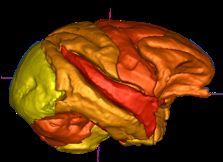
16 Brain Evolution Arcuate fasciculus, associated with language/ expression! Temporal lobe projection absent/smaller in nonhuman primates Rilling, 2008 Nature Neuroscience Probabilistic tractography 16


17 DTI based Segmentation Tuch 2003 DTI based clustering of thalamus Striatal subdivision Via cortical connectivity Draganski, 08 J Neuroscience Ziyan and Westin, MICCAI 08 17
18 DTI: Cerebral Neoplasms Deviated Infiltrated Edematous Destroyed 18 Courtesy of AL Alexander


19 T2W Pilocytic Astrocytoma Preop ADC T2W Postop ADC FA FA # $ 1 FA FA # $ 1 Courtesy of AL Alexander


20 Tract Infiltration T2W ADC T2W ADC Oligo Edema FA FA # $ 1 FA FA # $ 1 Courtesy of AL Alexander


21 Cerebral Palsy Courtesy of Susumu Mori


22 Multiple Sclerosis


23 Fetal Alcohol Syndrome Control Rodent model of FAS 3D MRI reconstruction DTI & Histology Fiber Tracking Acute EtOH GD7 Absent fornix columns 23 Thickened, dysplastic fimbria


24 Normal Brain Development Courtesy P Hueppi Univ Geneva Courtesy J. Neil Washington Univ., St. Louis


25 WM Anisotropy Changes with Age frontal pericallosal genu FA!.8".7".6".5".4" Anterior Genu" Left pericallosal" Right pericallosal" centrum semiovale.3" splenium FA! parietal pericallosal 20" 30" 40" 50" 60" 70" 80" Age!.8".7".6".5".4".3" Posterior 20" 30" 40" 50" 60" 70" 80" Splenium" Left pericallosal" Right pericallosal".48".46".44".42".40".38".36" Centrum Semiovale.34" 20" 30" 40" 50" 60" 70" 80" Age! Courtesy K. Lim, Univ. Minn.

 Adult Corpus")

26 Corpus Callosum Tracts: Study of Early Development Neonate (2 wks) Infant (1 year) Adult Corpus callosum: Commissural bundles, color coding of FA (0=blue, 1=red)
 Myelination and axon")

27 Early postnatal development of white matter on neonates Analysis of white matter in healthy controls (N=47) Myelination and axon elimination: FA center >> peripheral FA splenium > genu MD splenium & genu > intcaps T1w splenium & genu < intcaps!"#$%&'()**+(,-./(


28 1-2 year old: CC Tracts 28 Goodlett 2009 NeuroImage


29 Left Motor Tract 29 Goodlett 2009 NeuroImage

, abuse")

30 Monkey Brain Studies Harlow Primate UWisc / Emory Studies: Intrauterine exposure (Flu, LPS), abuse Understanding brain development & environment Regression with age 30


31 DTI Comparison %FA vs %MD Y 1 Y %Fractional Anisotropy %Mean Diffusivity 31

32 How the brain of a toddler really looks like 32

33 Krabbe Leukodystrophy Rare, lethal genetic leukodystrophy! Autosomal recessive pattern (not X-linked)! Worldwide: 1 in 80,000 births.! Isolated communities: 6 per 1,000 births Deficiency in galactosylceramidase enzyme! Buildup of undigested fats affects myelin sheath! Imperfect growth and development of myelin! Severe degeneration of mental and motor skills Lorenzo s Oil featured similar leukodystrophy Normal at birth, symptoms usually start 2-6 mts Fever, uncontrollable crying, seizures, vomiting, spasticity, paralysis, blind, finally death within 2y Juvenile- and adult-onset cases rare Escolar 2009 AJNR
34 Krabbe: Treatment Therapy (Maria Escolar, UNC), Duke! Myeloablative chemotherapy followed by stem cell transplantation from umbilical-cord blood! Treatment at Birth, no effect at symptomatic stage! Treated kids show differences in motor abilities! Survival rate depends on survival of therapy (15 of 17 ~ 88%) New Krabbe s screening with enzyme test! New York started August 2006! Parents often wait DTI: Assessing damage at birth via DTI! Illustration of damage to parents? Diagnosis?! Prediction of developmental outcome for motor abilities Here: Prelim data of project

35 Motor Related Fibertracts Prefrontal cortex Premotor cortex Motor cortex VL thalamus superior cerebellar peduncle corona radiata internal capsule cerebral peduncle Pontine nuclei Deep cerebellar nuc-dentate Cerebellar cortex middle cerebellar peduncle Courtesy of Jim Fallon Left and right hemispheric Cortico-spinal tracts


36 Controls - Left Internal Capsule Tracts Krabbe s - Left Internal Capsule Tracts P1 P2 P3 P4 Worst motor development FA = 0 FA = 1
37 FA Statistics along Fibers FA along Left CorticoSpinal Tract Healthy Krabbe P FA Statistics over 6 Krabbe, 53 Healthy neonate babies
38 FA Stats Center Region Left Corticospinal Tract Left FA Controls Krabbe Left FA Ratio Controls Krabbe FA Value FA ratio (actual FA/Expected FA) FA Distance Along the Corticospinal Tract Gestational Age at Scan (days) Gestational Age at Scan (days) Center region selection => Mean FA computation FA ratio = FA divided by expected FA given gestational age at birth, at scan, birth weight, gender

 Fine")
39 Outcome Correlation FA Ratio at Baseline Cognition Developmental Age Gross motor Calendar Age (months) Fine Motor
40 Conclusion Krabbe Correlation of DTI with outcome after treatment Current investigation:! Natural history of development with DTI! Can DTI predict, when symptoms will arise if untreated? 40
41 Future of DTI Highly valuable MR based modality! Many applications! Considerable validation (though more is needed) What s next?! Higher order of diffusion representation! Improved tractography algorithms! Network analyses! Need for automatic, blackbox processing 41


42 Higher Order Diffusion Representations Active field of research since 2003! Qball, Tuch! DSI, Van Wedeen No real clinical tools yet! Next evolutionary stage for DTI? 42
 Main issue: stability, clinical application Hagmann 2008 PLOS")
43 Network Analyses Structural network using diffusion spectral imaging Combination with functional imaging (resting state, event driven) Main issue: stability, clinical application Hagmann 2008 PLOS Biology 43
44 Blackbox Processing DTI property images (FA, MD, AD, RD) clinically useful But tractography application lag behind in clinical use Current processing is! Mostly interactive! Significant training in DTI necessary Need for automatic blackbox tools! No technical training needed! Adequate in presence of pathology! Includes analysis framework 44
45 The last slide We love DTI! And there are many reasons why, as shown in this talk Thanks! 45
Diffusion Tensor Imaging in Psychiatry
 2003 KHBM DTI in Psychiatry Diffusion Tensor Imaging in Psychiatry KHBM 2003. 11. 21. 서울대학교 의과대학 정신과학교실 권준수 Neuropsychiatric conditions DTI has been studied in Alzheimer s disease Schizophrenia Alcoholism
Modeling of early-infant brain growth using longitudinal data from diffusion tensor imaging.
 Modeling of early-infant brain growth using longitudinal data from diffusion tensor imaging. Guido Gerig, Neda Sadeghi, PhD, Marcel Prastawa, Tom Fletcher, Clement Vachet Scientific Computing and Imaging
Is DTI Increasing the Connectivity Between the Magnet Suite and the Clinic?
 Current Literature In Clinical Science Is DTI Increasing the Connectivity Between the Magnet Suite and the Clinic? Spatial Patterns of Water Diffusion Along White Matter Tracts in Temporal Lobe Epilepsy.
Diffusion Tensor Imaging 12/06/2013
 12/06/2013 Beate Diehl, MD PhD FRCP University College London National Hospital for Neurology and Neurosurgery Queen Square London, UK American Epilepsy Society Annual Meeting Disclosure None Learning
Leah Militello, class of 2018
 Leah Militello, class of 2018 Objectives 1. Describe the general organization of cerebral hemispheres. 2. Describe the locations and features of the different functional areas of cortex. 3. Understand
P. Hitchcock, Ph.D. Department of Cell and Developmental Biology Kellogg Eye Center. Wednesday, 16 March 2009, 1:00p.m. 2:00p.m.
 Normal CNS, Special Senses, Head and Neck TOPIC: CEREBRAL HEMISPHERES FACULTY: LECTURE: READING: P. Hitchcock, Ph.D. Department of Cell and Developmental Biology Kellogg Eye Center Wednesday, 16 March
Diffusion Tensor Imaging in brain tumours
 Diffusion Tensor Imaging in brain tumours @MarionSmits, MD PhD Associate Professor of Neuroradiology Dept. of Radiology, Erasmus MC, Rotterdam (NL) Honorary Consultant and Reader UCLH National Hospital
FUNCTIONAL MAGNETIC RESONANCE IMAGING IN FOLLOW-UP OF CEREBRAL GLIAL TUMORS
 Anvita Bieza FUNCTIONAL MAGNETIC RESONANCE IMAGING IN FOLLOW-UP OF CEREBRAL GLIAL TUMORS Summary of Doctoral Thesis to obtain PhD degree in medicine Specialty Diagnostic Radiology Riga, 2013 Doctoral thesis
Fig.1: A, Sagittal 110x110 mm subimage close to the midline, passing through the cingulum. Note that the fibers of the corpus callosum run at a
 Fig.1 E Fig.1:, Sagittal 110x110 mm subimage close to the midline, passing through the cingulum. Note that the fibers of the corpus callosum run at a slight angle are through the plane (blue dots with
Diffusion tensor imaging of the infant brain: From technical problems to neuroscientific breakthroughs Jessica Dubois
 Diffusion tensor imaging of the infant brain: From technical problems to neuroscientific breakthroughs Jessica Dubois L. Hertz-Pannier, G. Dehaene-Lambertz, J.F. Mangin, D. Le Bihan Inserm U56, U663; NeuroSpin
Cerebral Cortex 1. Sarah Heilbronner
 Cerebral Cortex 1 Sarah Heilbronner heilb028@umn.edu Want to meet? Coffee hour 10-11am Tuesday 11/27 Surdyk s Overview and organization of the cerebral cortex What is the cerebral cortex? Where is each
Stereotactic Diffusion Tensor Tractography For Gamma Knife Stereotactic Radiosurgery
 Disclosures The authors of this study declare that they have no commercial or other interests in the presentation of this study. This study does not contain any use of offlabel devices or treatments. Stereotactic
Chronology of normal brain myelination in newborns with MR imaging
 Chronology of normal brain myelination in newborns with MR imaging Poster No.: C-0577 Congress: ECR 2012 Type: Authors: Keywords: DOI: Scientific Exhibit F. Fernandez Usagre; Sevilla/ES Neuroradiology
Telencephalon (Cerebral Hemisphere)
") Telencephalon (Cerebral Hemisphere) OUTLINE The Cortex - Lobes, Sulci & Gyri - Functional Subdivisions - Limbic Lobe & Limbic System The Subcortex - Basal Ganglia - White Matter (Internal Capsule) - Relations
Telencephalon (Cerebral Hemisphere) OUTLINE The Cortex - Lobes, Sulci & Gyri - Functional Subdivisions - Limbic Lobe & Limbic System The Subcortex - Basal Ganglia - White Matter (Internal Capsule) - Relations
NeuroImage 61 (2012) Contents lists available at SciVerse ScienceDirect. NeuroImage. journal homepage:
 Contents lists available at SciVerse ScienceDirect. NeuroImage. journal homepage:") NeuroImage 61 (2012) 542 557 Contents lists available at SciVerse ScienceDirect NeuroImage journal homepage: www.elsevier.com/locate/ynimg Quantitative tract-based white matter development from birth to
NeuroImage 61 (2012) 542 557 Contents lists available at SciVerse ScienceDirect NeuroImage journal homepage: www.elsevier.com/locate/ynimg Quantitative tract-based white matter development from birth to
Introduction to the Central Nervous System: Internal Structure
 Introduction to the Central Nervous System: Internal Structure Objective To understand, in general terms, the internal organization of the brain and spinal cord. To understand the 3-dimensional organization
Pearls and Pitfalls of MR Diffusion in Clinical Neurology
 Pearls and Pitfalls of MR Diffusion in Clinical Neurology Dr. Alberto Bizzi Neuroradiology Unit Fondazione IRCCS Istituto Neurologico Carlo Besta Milan, Italy Email: alberto_bizzi@fastwebnet.it Diffusion
PRETERM BIRTH RESULTS IN ALTERATIONS IN NEURAL CONNECTIVITY AT AGE 16 YEARS
 Yale University EliScholar A Digital Platform for Scholarly Publishing at Yale Yale Medicine Thesis Digital Library School of Medicine 8-17-2010 PRETERM BIRTH RESULTS IN ALTERATIONS IN NEURAL CONNECTIVITY
Post Stroke Brain Plasticity
 Post Stroke Brain Plasticity François CHOLLET MD, PhD Neurology Department: Stroke Unit Toulouse University Hospital (CHU) Neurosciences Institute of Toulouse CNRS, INSERM, University, CHU Versailles le
An Atlas of Brainstem Connectomes from HCP Data
 An Atlas of Brainstem Connectomes from HCP Data Presented During: Poster Session Tuesday, June 27, 2017: 12:45 PM - 02:45 PM Stand-By Time Tuesday, June 27, 2017: 12:45 PM - 2:45 PM Submission No: 1653
Cerebellum. Steven McLoon Department of Neuroscience University of Minnesota
 Cerebellum Steven McLoon Department of Neuroscience University of Minnesota 1 Anatomy of the Cerebellum The cerebellum has approximately half of all the neurons in the central nervous system. The cerebellum
CEREBRUM Dr. Jamila Elmedany Dr. Essam Eldin Salama
 CEREBRUM Dr. Jamila Elmedany Dr. Essam Eldin Salama Objectives At the end of the lecture, the student should be able to: List the parts of the cerebral hemisphere (cortex, medulla, basal nuclei, lateral
Diffusion-Weighted and Conventional MR Imaging Findings of Neuroaxonal Dystrophy
 AJNR Am J Neuroradiol 25:1269 1273, August 2004 Diffusion-Weighted and Conventional MR Imaging Findings of Neuroaxonal Dystrophy R. Nuri Sener BACKGROUND AND PURPOSE: Neuroaxonal dystrophy is a rare progressive
10/3/2016. T1 Anatomical structures are clearly identified, white matter (which has a high fat content) appears bright.
 appears bright.") H2O -2 atoms of Hydrogen, 1 of Oxygen Hydrogen just has one single proton and orbited by one single electron Proton has a magnetic moment similar to the earths magnetic pole Also similar to earth in that
H2O -2 atoms of Hydrogen, 1 of Oxygen Hydrogen just has one single proton and orbited by one single electron Proton has a magnetic moment similar to the earths magnetic pole Also similar to earth in that
Medial View of Cerebellum
 Meds 5371 System Neuroscience D. L. Oliver CEREBELLUM Anterior lobe (spinal) Posterior lobe (cerebral) Flocculonodular lobe (vestibular) Medial View of Cerebellum 1 Ventral View of Cerebellum Flocculus
Lecturer. Prof. Dr. Ali K. Al-Shalchy MBChB/ FIBMS/ MRCS/ FRCS 2014
 Lecturer Prof. Dr. Ali K. Al-Shalchy MBChB/ FIBMS/ MRCS/ FRCS 2014 Dorsal root: The dorsal root carries both myelinated and unmyelinated afferent fibers to the spinal cord. Posterior gray column: Long
Advanced magnetic resonance imaging for monitoring brain development and injury
 Advanced magnetic resonance imaging for monitoring brain development and injury Stéphane Sizonenko, MD-PhD Division of Development and Growth Department of Child and Adolescent Medicine Geneva University
Prof. Saeed Abuel Makarem & Dr.Sanaa Alshaarawy
 Prof. Saeed Abuel Makarem & Dr.Sanaa Alshaarawy 1 Objectives By the end of the lecture, you should be able to: Describe the anatomy and main functions of the thalamus. Name and identify different nuclei
Outline of the next three lectures
 Outline of the next three lectures Lecture 35 Anatomy of the human cerebral cortex gross and microscopic cell types connections Vascular supply of the cerebral cortex Disorders involving the cerebral cortex
DWI assessment of ischemic changes in the fetal brain
 DWI assessment of ischemic changes in the fetal brain Dafi Bergman, 4 th year Medical student in the 4-year program, Sackler school of medicine B.Sc Life and Medical Sciences, Tel Aviv University Supervised
Research Article Corticospinal Tract Change during Motor Recovery in Patients with Medulla Infarct: A Diffusion Tensor Imaging Study
 BioMed Research International, Article ID 524096, 5 pages http://dx.doi.org/10.1155/2014/524096 Research Article Corticospinal Tract Change during Motor Recovery in Patients with Medulla Infarct: A Diffusion
Heidi M. Feldman, MD, PhD,* Jason D. Yeatman, BA, Eliana S. Lee, BS,* Laura H. F. Barde, PhD,* Shayna Gaman-Bean, MD*
 Review Article Diffusion Tensor Imaging: A Review for Pediatric Researchers and Clinicians Heidi M. Feldman, MD, PhD,* Jason D. Yeatman, BA, Eliana S. Lee, BS,* Laura H. F. Barde, PhD,* Shayna Gaman-Bean,
Lab 2. we will look into several angled horizontal sections ( orbitomeatal plane ) i.e passing from the orbit into the ear
 i.e passing from the orbit into the ear") we will look into several angled horizontal sections ( orbitomeatal plane ) i.e passing from the orbit into the ear Figure I page 76 : looking at the key on the left side this section passed through the
we will look into several angled horizontal sections ( orbitomeatal plane ) i.e passing from the orbit into the ear Figure I page 76 : looking at the key on the left side this section passed through the
Regional and Lobe Parcellation Rhesus Monkey Brain Atlas. Manual Tracing for Parcellation Template
 Regional and Lobe Parcellation Rhesus Monkey Brain Atlas Manual Tracing for Parcellation Template Overview of Tracing Guidelines A) Traces are performed in a systematic order they, allowing the more easily
Introduction to Diffusion Tractography
 Introduction to Diffusion Tractography In vivo tracer Manganese Tractography ex vivo Tim B. Dyrby, PhD Danish Research Centre for Magnetic Resonance (DRCMR) Copenhagen University Hospital Hvidovre Denmark
Surgery Within and Around Critical White Matter Tracts
 Surgery Within and Around Critical White Matter Tracts Kaisorn L. Chaichana, M.D. Assistant Professor of Neurosurgery, Oncology, and Otolaryngology-Head & Neck Surgery Mayo Clinic Florida, Jacksonville,
Online supplement data :
 Online supplement data : ROI ROI name MNI coord (mm) HC>SZ HC>S ZR HC>P BP HC>P BPR Heritabil ity h^2 Heritability p-value MCP Middle cerebellar peduncle 0-41 -44 2.05 2.12 1.63 0.69 0.38 0.004473 PCT
The Central Nervous System I. Chapter 12
 The Central Nervous System I Chapter 12 The Central Nervous System The Brain and Spinal Cord Contained within the Axial Skeleton Brain Regions and Organization Medical Scheme (4 regions) 1. Cerebral Hemispheres
Overview. Fundamentals of functional MRI. Task related versus resting state functional imaging for sensorimotor mapping
 Functional MRI and the Sensorimotor System in MS Nancy Sicotte, MD, FAAN Professor and Vice Chair Director, Multiple Sclerosis Program Director, Neurology Residency Program Cedars-Sinai Medical Center
General Sensory Pathways of the Trunk and Limbs
 General Sensory Pathways of the Trunk and Limbs Lecture Objectives Describe gracile and cuneate tracts and pathways for conscious proprioception, touch, pressure and vibration from the limbs and trunk.
Visualization strategies for major white matter tracts identified by diffusion tensor imaging for intraoperative use
 International Congress Series 1281 (2005) 793 797 www.ics-elsevier.com Visualization strategies for major white matter tracts identified by diffusion tensor imaging for intraoperative use Ch. Nimsky a,b,
Language comprehension in young people with severe cerebral palsy in relation to language tracts: a diffusion tensor imaging study
 Language comprehension in young people with severe cerebral palsy in relation to language tracts: a diffusion tensor imaging study Laurike Harlaar, Petra J. Pouwels, Joke J. Geytenbeek, Kim J. Oostrom,
Shape Modeling of the Corpus Callosum for Neuroimaging Studies of the Brain (Part I) Dongqing Chen, Ph.D.
 Dongqing Chen, Ph.D.") The University of Louisville CVIP Lab Shape Modeling of the Corpus Callosum for Neuroimaging Studies of the Brain (Part I) Dongqing Chen, Ph.D. Computer Vision & Image Processing (CVIP) Laboratory Department
The University of Louisville CVIP Lab Shape Modeling of the Corpus Callosum for Neuroimaging Studies of the Brain (Part I) Dongqing Chen, Ph.D. Computer Vision & Image Processing (CVIP) Laboratory Department
Stroke School for Internists Part 1
 Stroke School for Internists Part 1 November 4, 2017 Dr. Albert Jin Dr. Gurpreet Jaswal Disclosures I receive a stipend for my role as Medical Director of the Stroke Network of SEO I have no commercial
 Spinal Cord Tracts DESCENDING SPINAL TRACTS: Are concerned with somatic motor function, modification of ms. tone, visceral innervation, segmental reflexes. Main tracts arise form cerebral cortex and others
MR Assessment of Myelination in Infants and Children: Usefulness of Marker Sites
 731 MR ssessment of Myelination in Infants and Children: Usefulness of Marker Sites C. Roger ird 1 Mary Hedberg urton P. Drayer Paul J. Keller Richard. Flom John. Hodak retrospective study was made of
Cortical Control of Movement
 Strick Lecture 2 March 24, 2006 Page 1 Cortical Control of Movement Four parts of this lecture: I) Anatomical Framework, II) Physiological Framework, III) Primary Motor Cortex Function and IV) Premotor
Patologie infiammatorie encefaliche e midollari
 Patologie infiammatorie encefaliche e midollari Maria Laura Stromillo Department of Medicine, Surgery and Neuroscience Inflammatory disorders of the CNS NMOSD ADEM Multiple Sclerosis Neuro-Myelitis Optica
doi: /brain/awq175 Brain 2010: 133;
 doi:10.1093/brain/awq175 Brain 2010: 133; 2348 2364 2348 BRAIN A JOURNAL OF NEUROLOGY The structural plasticity of white matter networks following anterior temporal lobe resection Mahinda Yogarajah, 1
MRI-Based Classification Techniques of Autistic vs. Typically Developing Brain
 MRI-Based Classification Techniques of Autistic vs. Typically Developing Brain Presented by: Rachid Fahmi 1 2 Collaborators: Ayman Elbaz, Aly A. Farag 1, Hossam Hassan 1, and Manuel F. Casanova3 1Computer
Anatomy Lab (1) Theoretical Part. Page (2 A) Page (2B)
 Theoretical Part. Page (2 A) Page (2B)") Anatomy Lab (1) This sheet only includes the extra notes for the lab handout regarding the theoretical part, as for the practical part it includes everything the doctor mentioned. Theoretical Part Page
Anatomy Lab (1) This sheet only includes the extra notes for the lab handout regarding the theoretical part, as for the practical part it includes everything the doctor mentioned. Theoretical Part Page
Normal myelination: a practical pictorial review
 Normal myelination: a practical pictorial review Poster No.: C-1486 Congress: ECR 2016 Type: Authors: Keywords: DOI: Educational Exhibit M. Gaha, N. Mama, N. Arifa, H. Jemni, K. Tlili Graiess; Sousse/TN
Gross Organization I The Brain. Reading: BCP Chapter 7
 Gross Organization I The Brain Reading: BCP Chapter 7 Layout of the Nervous System Central Nervous System (CNS) Located inside of bone Includes the brain (in the skull) and the spinal cord (in the backbone)
MORNING COFFEE
 Final on-site schedule for 5. October MICCAI 2015 BrainLes: Brain-lesion workshop (BrainLes) Ischemic Stroke Lesion Segmentation Challenge (ISLES) Brain Tumor Image Segmentation Challenge (BRATS) MORNING
Lecture VIII. The Spinal Cord, Reflexes and Brain Pathways!
 Reflexes and Brain Bio 3411! Monday!! 1! Readings! NEUROSCIENCE 5 th ed: Review Chapter 1 pp. 11-21;!!Read Chapter 9 pp. 189-194, 198! THE BRAIN ATLAS 3 rd ed:! Read pp. 4-17 on class web site! Look at
Diffusion Tensor Imaging Assessment of Brain White Matter Maturation During the First Postnatal Year
 DTI of White Matter in infants Neuroradiology Original Research James M. Provenzale 1 Luxia Liang 1,2 David DeLong 1 Leonard E. White 3 Provenzale JM, Liang L, DeLong D, White LE Keywords: anisotropy,
I: To describe the pyramidal and extrapyramidal tracts. II: To discuss the functions of the descending tracts.
 Descending Tracts I: To describe the pyramidal and extrapyramidal tracts. II: To discuss the functions of the descending tracts. III: To define the upper and the lower motor neurons. 1. The corticonuclear
CISC 3250 Systems Neuroscience
 CISC 3250 Systems Neuroscience Levels of organization Central Nervous System 1m 10 11 neurons Neural systems and neuroanatomy Systems 10cm Networks 1mm Neurons 100μm 10 8 neurons Professor Daniel Leeds
DEGREE (if applicable)
") NAME: Sheng-Kwei (Victor) Song OMB No. 0925-0001 and 0925-0002 (Rev. 10/15 Approved Through 10/31/2018) BIOGRAPHICAL SKETCH Provide the following information for the Senior/key personnel and other significant
NAME: Sheng-Kwei (Victor) Song OMB No. 0925-0001 and 0925-0002 (Rev. 10/15 Approved Through 10/31/2018) BIOGRAPHICAL SKETCH Provide the following information for the Senior/key personnel and other significant
CEREBRUM & CEREBRAL CORTEX
 CEREBRUM & CEREBRAL CORTEX Seonghan Kim Dept. of Anatomy Inje University, College of Medicine THE BRAIN ANATOMICAL REGIONS A. Cerebrum B. Diencephalon Thalamus Hypothalamus C. Brain Stem Midbrain Pons
Strick Lecture 3 March 22, 2017 Page 1
 Strick Lecture 3 March 22, 2017 Page 1 Cerebellum OUTLINE I. External structure- Inputs and Outputs Cerebellum - (summary diagram) 2 components (cortex and deep nuclei)- (diagram) 3 Sagittal zones (vermal,
Attenuation value in HU From -500 To HU From -10 To HU From 60 To 90 HU. From 200 HU and above
 Brain Imaging Common CT attenuation values Structure Air Fat Water Brain tissue Recent hematoma Calcifications Bone Brain edema and infarction Normal liver parenchyma Attenuation value in HU From -500
IMAGING OF HYPOXIC ISCHEMIC INJURY IN A NEONATE FN3 STATE MEETING NEMOURS CHILDREN'S HOSPITAL ORLANDO,FL 08/04/18
 IMAGING OF HYPOXIC ISCHEMIC INJURY IN A NEONATE FN3 STATE MEETING NEMOURS CHILDREN'S HOSPITAL ORLANDO,FL 08/04/18 Dhanashree Rajderkar,MD Assistant Professor Department of Radiology University of Florida
CEREBRUM. Dr. Jamila EL Medany
 CEREBRUM Dr. Jamila EL Medany Objectives At the end of the lecture, the student should be able to: List the parts of the cerebral hemisphere (cortex, medulla, basal nuclei, lateral ventricle). Describe
Student Lab #: Date. Lab: Gross Anatomy of Brain Sheep Brain Dissection Organ System: Nervous Subdivision: CNS (Central Nervous System)
") Lab: Gross Anatomy of Brain Sheep Brain Dissection Organ System: Nervous Subdivision: CNS (Central Nervous System) Student Lab #: Date 1 Objectives: 1. Learn the main components making up a motor neuron.
Lab: Gross Anatomy of Brain Sheep Brain Dissection Organ System: Nervous Subdivision: CNS (Central Nervous System) Student Lab #: Date 1 Objectives: 1. Learn the main components making up a motor neuron.
Ch 13: Central Nervous System Part 1: The Brain p 374
 Ch 13: Central Nervous System Part 1: The Brain p 374 Discuss the organization of the brain, including the major structures and how they relate to one another! Review the meninges of the spinal cord and
Functional MRI and Diffusion Tensor Imaging
 Functional MRI and Diffusion Tensor Imaging Andrew Steven March 23, 2018 Ochsner Neuroscience Symposium None Disclosure 1 Objectives Review basic principles of BOLD fmri and DTI. Discuss indications and
fmri and Tractography in Preoperative Neurosurgical Planning Ronald L. Wolf, M.D., Ph.D. University of Pennsylvania Medical Center
 fmri and Tractography in Preoperative Neurosurgical Planning Ronald L. Wolf, M.D., Ph.D. University of Pennsylvania Medical Center Acknowledgements/Disclosures Radiology Ragini Verma Birkan Tunc Sumei
Auditory and Vestibular Systems
 Auditory and Vestibular Systems Objective To learn the functional organization of the auditory and vestibular systems To understand how one can use changes in auditory function following injury to localize
Visual system invades the endbrain: pathways to striatum and cortex (continued) Why this happened in evolution
 Why this happened in evolution") Visual system invades the endbrain: pathways to striatum and cortex (continued) Why this happened in evolution What were the adaptive advantages? Visual information reaching the striatum directly: Advantages
Visual system invades the endbrain: pathways to striatum and cortex (continued) Why this happened in evolution What were the adaptive advantages? Visual information reaching the striatum directly: Advantages
Note: Waxman is very sketchy on today s pathways and nonexistent on the Trigeminal.
 Dental Neuroanatomy Thursday, February 3, 2011 Suzanne Stensaas, PhD Note: Waxman is very sketchy on today s pathways and nonexistent on the Trigeminal. Resources: Pathway Quiz for HyperBrain Ch. 5 and
Diffusion Tensor Tractography in the Presurgical Assessment of Cerebral Gliomas
 The Neuroradiology Journal 27: 75-84, 2014 - doi: 10.15274/NRJ-2014-10008 www.centauro.it Diffusion Tensor Tractography in the Presurgical Assessment of Cerebral Gliomas ZAHRA FARSHIDFAR 1, FARIBORZ FAEGHI
SENSORY (ASCENDING) SPINAL TRACTS
 SPINAL TRACTS") SENSORY (ASCENDING) SPINAL TRACTS Dr. Jamila El-Medany Dr. Essam Eldin Salama OBJECTIVES By the end of the lecture, the student will be able to: Define the meaning of a tract. Distinguish between the different
SENSORY (ASCENDING) SPINAL TRACTS Dr. Jamila El-Medany Dr. Essam Eldin Salama OBJECTIVES By the end of the lecture, the student will be able to: Define the meaning of a tract. Distinguish between the different
THE BEAUTIFUL BRAIN (how it may help us to understand complex pain conditions)
") THE BEAUTIFUL BRAIN (how it may help us to understand complex pain conditions) Dr Gary S Lee Consultant Clinical Psychologist Pain Management Alistair Day Superintendent Radiographer MRI Dr Stephan Voigt
THE BEAUTIFUL BRAIN (how it may help us to understand complex pain conditions) Dr Gary S Lee Consultant Clinical Psychologist Pain Management Alistair Day Superintendent Radiographer MRI Dr Stephan Voigt
PETER PAZMANY CATHOLIC UNIVERSITY Consortium members SEMMELWEIS UNIVERSITY, DIALOG CAMPUS PUBLISHER
 PETER PAZMANY CATHOLIC UNIVERSITY SEMMELWEIS UNIVERSITY Development of Complex Curricula for Molecular Bionics and Infobionics Programs within a consortial* framework** Consortium leader PETER PAZMANY
Nature Neuroscience doi: /nn Supplementary Figure 1. Characterization of viral injections.
 Supplementary Figure 1 Characterization of viral injections. (a) Dorsal view of a mouse brain (dashed white outline) after receiving a large, unilateral thalamic injection (~100 nl); demonstrating that
Advances in Clinical Neuroimaging
 Advances in Clinical Neuroimaging Joseph I. Tracy 1, PhD, ABPP/CN; Gaelle Doucet 2, PhD; Xaiosong He 2, PhD; Dorian Pustina 2, PhD; Karol Osipowicz 2, PhD 1 Department of Radiology, Thomas Jefferson University,
A3.1.7 Motor Control. 10 November 2016 Institute of Psychiatry,Psychology and Neuroscience Marinela Vavla
 A3.1.7 Motor Control 10 November 2016 Institute of Psychiatry,Psychology and Neuroscience Marinela Vavla marinela.vavla@kcl.ac.uk Learning objectives Motor systems: components & organization Spinal cord
Neurophysiology of systems
 Neurophysiology of systems Motor cortex (voluntary movements) Dana Cohen, Room 410, tel: 7138 danacoh@gmail.com Voluntary movements vs. reflexes Same stimulus yields a different movement depending on context
Dentate imaging: methods and applications
 Dentate imaging: methods and applications Dagmar Timmann Department of Neurology University of Duisburg-Essen, Germany Cerebellar output via deep nuclei Folie 2 Brodal 2010 Cerebellar nuclei Fastigial
Brainstem. Steven McLoon Department of Neuroscience University of Minnesota
 Brainstem Steven McLoon Department of Neuroscience University of Minnesota 1 Course News Change in Lab Sequence Week of Oct 2 Lab 5 Week of Oct 9 Lab 4 2 Goal Today Know the regions of the brainstem. Know
Human Cognitive Developmental Neuroscience. Jan 27
 Human Cognitive Developmental Neuroscience Jan 27 Wiki Definition Developmental cognitive neuroscience is an interdisciplinary scientific field that is situated at the boundaries of Neuroscience Psychology
Cerebellum: little brain. Cerebellum. gross divisions
 Cerebellum The anatomy of the cerebellum and its gross divisions Its principal input and output pathways The organization of the cerebellar cortex Role of climbing vs. mossy fibre input The parallel-fibre/
ADULT-ONSET (INFRATENTORIAL) LEUKOENCEPHALOPATHY as PRESENTING MANIFESTATION of ERDHEIM-CHESTER DISEASE
 LEUKOENCEPHALOPATHY as PRESENTING MANIFESTATION of ERDHEIM-CHESTER DISEASE") ADULT-ONSET (INFRATENTORIAL) LEUKOENCEPHALOPATHY as PRESENTING MANIFESTATION of ERDHEIM-CHESTER SEASE GIULIO CAVALLI, M.D. INTERNAL MECINE AND CLINICAL IMMUNOLOGY IRCCS SAN RAFFAELE HOSPITAL VITA-SALUTE
ADULT-ONSET (INFRATENTORIAL) LEUKOENCEPHALOPATHY as PRESENTING MANIFESTATION of ERDHEIM-CHESTER SEASE GIULIO CAVALLI, M.D. INTERNAL MECINE AND CLINICAL IMMUNOLOGY IRCCS SAN RAFFAELE HOSPITAL VITA-SALUTE
Imaging in Pediatric `neurohiv Dr Jackie Hoare Head of Liaison Psychiatry Groote Schuur Hospital, UCT
 Imaging in Pediatric `neurohiv Dr Jackie Hoare Head of Liaison Psychiatry Groote Schuur Hospital, UCT ? Spectrum of Neurocognitive disorders The adult literature on HIV related CNS damage supports a spectrum
Disclosure Information AACPDM 68 th Annual Meeting September 10-13, 2014 Diffusion Tensor Imaging: Analysis options in pediatric neuroimaging research
 Disclosure Information AACPDM 68 th Annual Meeting September 10-13, 2014 Diffusion Tensor Imaging: Analysis options in pediatric neuroimaging research Andrea Poretti, MD Research Associate Section of Pediatric
Unit VIII Problem 5 Physiology: Cerebellum
 Unit VIII Problem 5 Physiology: Cerebellum - The word cerebellum means: the small brain. Note that the cerebellum is not completely separated into 2 hemispheres (they are not clearly demarcated) the vermis
The Effect of Local Fiber Model On Population Studies
 The Effect of Local Fiber Model On Population Studies James G. Malcolm Marek Kubicki Martha E. Shenton Yogesh Rathi Psychiatry Neuroimaging Laboratory, Harvard Medical School, Boston, MA VA Boston Healthcare
Normal Myelination of Anatomic Nerve Fiber Bundles: MR Analysis
 AJNR Am J Neuroradiol 19:1129 1136, June 1998 Normal Myelination of Anatomic Nerve Fiber Bundles: MR Analysis Hiroyuki Nakagawa, Satoru Iwasaki, Kimihiko Kichikawa, Akio Fukusumi, Toshiaki Taoka, Hajime
Diffusion-Tensor Imaging Assessment of White Matter Maturation in Childhood and Adolescence
 Neuroradiology/Head and Neck Imaging Original Research Moon et al. White Matter Diffusion-Tensor Imaging Neuroradiology/Head and Neck Imaging Original Research Won-Jin Moon 1 James M. Provenzale 2,3 Basar
Publication for the Philips MRI Community Issue 39 December 2009
 FieldStrength Publication for the Philips MRI Community Issue 39 December 2009 32-channel coil boosts 3.0T neuro imaging at Kennedy Krieger Kennedy Krieger Institute sees significantly better fmri, DTI,
Anatomy and Physiology (Bio 220) The Brain Chapter 14 and select portions of Chapter 16
 The Brain Chapter 14 and select portions of Chapter 16") Anatomy and Physiology (Bio 220) The Brain Chapter 14 and select portions of Chapter 16 I. Introduction A. Appearance 1. physical 2. weight 3. relative weight B. Major parts of the brain 1. cerebrum 2.
Anatomy and Physiology (Bio 220) The Brain Chapter 14 and select portions of Chapter 16 I. Introduction A. Appearance 1. physical 2. weight 3. relative weight B. Major parts of the brain 1. cerebrum 2.
Lecture - Chapter 13: Central Nervous System
 Lecture - Chapter 13: Central Nervous System 1. Describe the following structures of the brain, what is the general function of each: a. Cerebrum b. Diencephalon c. Brain Stem d. Cerebellum 2. What structures
Visualization and Quantification of the Striato pallidonigral Fibers in Parkinson's Disease Using Diffusion Tensor Imaging
 Visualization and Quantification of the Striato pallidonigral Fibers in Parkinson's Disease Using Diffusion Tensor Imaging Yu Zhang, Katherine Wu, Shannon Buckley, Norbert Schuff On behalf of the Parkinson
Stuttering Research. Vincent Gracco, PhD Haskins Laboratories
 Stuttering Research Vincent Gracco, PhD Haskins Laboratories Stuttering Developmental disorder occurs in 5% of children Spontaneous remission in approximately 70% of cases Approximately 1% of adults with
Fibre orientation dispersion in the corpus callosum relates to interhemispheric functional connectivity
 Fibre orientation dispersion in the corpus callosum relates to interhemispheric functional connectivity ISMRM 2017: http://submissions.mirasmart.com/ismrm2017/viewsubmissionpublic.aspx?sei=8t1bikppq Jeroen
For more information about how to cite these materials visit
 Author(s): Peter Hitchcock, PH.D., 2009 License: Unless otherwise noted, this material is made available under the terms of the Creative Commons Attribution Non-commercial Share Alike 3.0 License: http://creativecommons.org/licenses/by-nc-sa/3.0/
Cerebellum: little brain. Cerebellum. gross divisions
 Cerebellum The anatomy of the cerebellum and its gross divisions Its principal input and output pathways The organization of the cerebellar cortex Role of climbing vs. mossy fibre input The parallel-fibre/
The Nervous System. Functions of the Nervous System input gathering To monitor occurring inside and outside the body Changes =
 The Nervous System Functions of the Nervous System input gathering To monitor occurring inside and outside the body Changes = To process and sensory input and decide if is needed output A response to integrated
Structural And Functional Integration: Why all imaging requires you to be a structural imager. David H. Salat
 Structural And Functional Integration: Why all imaging requires you to be a structural imager David H. Salat salat@nmr.mgh.harvard.edu Salat:StructFunct:HST.583:2015 Structural Information is Critical