PRETERM BIRTH RESULTS IN ALTERATIONS IN NEURAL CONNECTIVITY AT AGE 16 YEARS
|
|
|
- Joel Bruce
- 5 years ago
- Views:
Transcription
1 Yale University EliScholar A Digital Platform for Scholarly Publishing at Yale Yale Medicine Thesis Digital Library School of Medicine PRETERM BIRTH RESULTS IN ALTERATIONS IN NEURAL CONNECTIVITY AT AGE 16 YEARS Katherine Mullen Follow this and additional works at: Recommended Citation Mullen, Katherine, "PRETERM BIRTH RESULTS IN ALTERATIONS IN NEURAL CONNECTIVITY AT AGE 16 YEARS" (2010). Yale Medicine Thesis Digital Library This Open Access Thesis is brought to you for free and open access by the School of Medicine at EliScholar A Digital Platform for Scholarly Publishing at Yale. It has been accepted for inclusion in Yale Medicine Thesis Digital Library by an authorized administrator of EliScholar A Digital Platform for Scholarly Publishing at Yale. For more information, please contact elischolar@yale.edu.
2 PRETERM BIRTH RESULTS IN ALTERATIONS IN NEURAL CONNECTIVITY AT AGE 16 YEARS A Thesis Submitted to the Yale University School of Medicine in Partial Fulfillment of the Requirements for the Degree of Doctor of Medicine by Katherine Marie Mullen 2010
3 Abstract: Preterm Birth Results in Alterations in Neural Connectivity at Age 16 Years Katherine M. Mullen 1,2, Betty R. Vohr 3, Karol H. Katz 1,4, Karen C. Schneider 3, Cheryl Lacadie 2, Michelle Hampson 2, R. Todd Constable 2 and Laura R. Ment 1,5. 1 Pediatrics, Yale School of Medicine, New Haven, CT, United States; 2 Diagnostic Radiology, Yale University, New Haven, CT, United States; 3 Pediatrics, Warren Alpert Medical School of Brown University, Providence, RI, United States;; 4 Epidemiology and Public Health, Yale School of Medicine, New Haven, CT, United States and 5 Neurology, Yale School of Medicine, New Haven, CT, United States. Very low birth weight preterm (PT) children are at high risk for brain injury. This study investigates microstructural differences in the brains of PT adolescents relative to term control subjects using diffusion tensor imaging (DTI), as well as studying their neurodevelopmental outcomes. Forty-four PT subjects ( grams birth weight) without neonatal brain injury and 41 term controls were evaluated at age 16 years with DTI, the Wechsler Intelligence Scale for Children - III (WISC), the Peabody Picture Vocabulary Test - Revised (PPVT), and the Comprehensive Test of Phonological Processing (CTOPP). PT subjects scored lower than term subjects on WISC full scale (p = 0.002), verbal (p = 0.027), and performance IQ tests (p = 0.001), as well as CTOPP phonological awareness (p = 0.005), but scored comparably to term subjects on PPVT and CTOPP Rapid Naming tests. PT subjects had lower fractional anisotropy (FA) values, suggestive of white matter disorganization, in multiple regions including bilateral uncinate fasciculi (left: p = 0.004; right: p = 0.002), bilateral external capsules (left: p < ; right: p = 0.001), the splenium of the corpus callosum (p = 0.014), and white matter serving the inferior frontal gyrus bilaterally (left: p < ; right: p = 0.005). FA values in both the left and right uncinate fasciculi correlated with PPVT scores (a semantic language task) in the PT subjects (left: R = 0.314, p = 0.038; right: R = 0.336, p = 0.026). FA values in the left and right arcuate fasciculi correlated with CTOPP Rapid Naming scores (a phonologic task) in the PT subjects (left: R = 0.424, p = 0.004; right: R = 0.301, p = 0.047). These data support for the first time that the recently proposed concept of dual pathways underlying language function are present in PT adolescents. These include a left-sided dorsal pathway associated with phonological and articulatory processing (arcuate fasciculus), and a bilateral ventral pathway for semantic, receptive language processing (uncinate fasciculus). The striking bilateral dorsal correlations for the PT group suggest that prematurely born subjects rely more heavily on the right hemisphere than typically developing adolescents for performance of phonological language tasks. These findings may represent either a delay in maturation or the engagement of alternative neural pathways for language in the developing PT brain.
4 Acknowledgements This work was supported by NS and NCRR CTSA-T32 Medical Student Research Fellowship from the National Institutes of Health. I am greatly appreciative to my mentors, Dr. Laura Ment and Dr. Todd Constable, for the opportunity to work on this project and for their support and help in its completion. I would also like to thank all those whose support enabled me to finish this thesis, particularly my friends for their cheer and my family for their love. Above all, I am deeply grateful to Dave Gorin, whose love and friendship humbles me and makes all worthwhile.
5 Table of Contents 1. Introduction 1 2. Statement of Purpose Methods Results.. 16 Table Table Table Figure Table Table Figure Discussion References. 46
6 1 Introduction Consequences of preterm birth in the brain Premature birth is a pressing public health matter, as nearly 13% of infants in the United States are born preterm and infants weighing under 1500 grams at birth comprise 1.5% of births (1). Premature infants suffer a high risk of perinatal brain damage compared to term infants (2-4). Though survival of infants weighing grams at birth has increased to 85% (5), these children face a range of developmental disabilities ranging from cerebral palsy to learning disabilities (4, 6). The brains of infants born prematurely face both increased challenge and increased susceptibility to a number of factors resulting in brain injury and secondary disturbance of growth. Active migration, axonal growth, proliferation and maturation of oligodendrocytes, and development of synaptic connections typically occur during the third trimester of fetal development, and all may be disrupted by premature birth. Severe brain injuries such as intraventricular hemorrhage, periventricular hemorrhagic infarction, and ventricular dilatation are recognized as potentially devastating complications of preterm birth. Apart from these three catastrophic but rare complications, preterm neonates may suffer a characteristic pattern of brain injury known as encephalopathy of prematurity (7). Typically, these infants suffer injury to cerebral white matter known as periventricular leukomalacia (PVL). PVL has multiple forms, and may include either localized or diffuse changes. The pathology of localized PVL is characterized by necrosis of all cell types on a microscopic or macroscopic scale. Macroscopic foci of necrosis lead to cystic structures observable by ultrasound, leading to the characterization of this subgroup as cystic PVL, which is the subtype most likely
7 2 to result in severe developmental abnormalities such as cerebral palsy (7). Non-cystic PVL, on the other hand, is characterized by microscopic necrotic foci, which lead to nonspecific glial scars which may not be detected by ultrasound. Preterm birth damages the development of the brain through multiple mechanisms. Ischemia and inflammation are initiating factors which potentiate each other, with excitotoxicity and free-radical attack as downstream effectors of this damage. Pathological correlates of diffuse injury include the disruption of premyelinating oligodendrocytes, which are particularly susceptible to injury, with subsequent reactive proliferation of oligodendrocyte precursors which unfortunately have impaired ability to myelinate axons (7). There is evidence in an animal model that the pattern of injury in preterm birth may be influenced by the distribution of immature oligodendrocytes in the brain (8, 9). In addition to glial disruption, diffuse disruption of axons has been reported in the brains of preterm infants (10). Deep gray matter is also commonly affected, including the thalamus, basal ganglia, and cerebellum, though it is unknown whether this is a primary injury or secondary trophic disturbance (7). Investigation of the neuropathology of preterm birth has utilized post-mortem pathological analysis and study of animal models of prematurity, including subjecting animals to hypoxia, hypoxia in conjunction with ischemia through ligation of the carotid artery, or premature delivery (11). However, neuroimaging of premature infants presents a vital non-invasive method for studying changes within the still-developing brain. Longterm effects of preterm birth on cognitive ability
8 3 In addition to feared neurological complications of preterm birth such as cerebral palsy, a range of developmental deficits in cognitive and motor function in preterm children have been observed to persist until early adulthood, indicating long-term disruption of brain function (6, 12). Preterm children have the greatest risk of neurodevelopmental deficits with severe brain injury such as intraventricular hemorrhage, periventricular hemorrhagic infarction, periventricular leukomalacia, or severe ventriculomegaly; however, even preterm children without these forms of brain injury are more likely than control term children to have lower IQ scores and require more support in reading, writing, and mathematics (13) at 12 years of age. Male infants are at particular risk for poor outcome after premature birth (14-17). The mechanism for these differences is as yet unknown. Despite the prevalence of these difficulties, the brains of infants and children exhibit remarkable plasticity, and many catch up over time to children born at term in terms of both developmental and neuroimaging parameters (18, 19). Some studies have proposed that this compensation relates to utilization of alternative pathways in order to bypass injured white matter structures (20-22). Neuroimaging and principles of diffusion tensor imaging Multiple neuroimaging techniques have been used to better identify and describe the sequelae of preterm birth in the brains of these infants (23-25). While ultrasound and computed tomography have been used for some time in the identification of intraventricular hemorrhage and periventricular leukomalacia, the advent of magnetic resonance imaging (MRI) became crucial to identifying more subtle changes in the brains
9 4 of preterm infants (26). Subtle changes such as delays in gray-white differentiation, hyperintensity of white matter, smaller corpus callosum, and ventriculomegaly were appreciated with much greater sensitivity with MRI (2, 27). Methods such as voxel-based morphometry, providing volumetric analysis of white and gray matter in the developing brain, have identified relative decreases in volume of widespread white matter regions in preterm infants, suggestive of neuronal loss (15, 28-31). Diffusion tensor imaging (DTI) is a relatively recent magnetic resonance imaging technique which provides a means of assessing the integrity of white matter tracts at a microstructural level. It is more sensitive than conventional magnetic resonance imaging for detecting subtle abnormalities (32-38). As part of DTI analyses, fractional anisotropy (FA) values indicate the degree to which water diffusion is restricted along one axis relative to all others. Within cerebral white matter, water preferentially diffuses along axons, with diffusion perpendicular to this axis restricted by structural barriers including cell membranes. Higher FA values serve as a marker for the coherence of white matter tracts, as the constraints of the tissue organization into axon bundles within well-formed tracts limit the direction of water flow. Alterations in FA may result from changes in fiber organization, axonal size (39), or activity-dependent changes in myelination (18). DTI has become a valuable tool in assessing white matter integrity on a microstructural level in the developing preterm brain (40-42). FA values in white matter tracts tend to increase both over the course of fetal development and after birth. DTI conducted on fetuses in utero confirms increases in FA as the fetuses progress closer to term, particularly in the corticospinal tract and corpus callosum (43).FA values in multiple white matter tracts including the splenium and
10 5 posterior body of the corpus callosum, the posterior limb of the internal capsule, the left frontal white matter, and the left inferior longitudinal white matter at term equivalent age demonstrate a linear correlation with gestational age at birth (44). Lower birthweight has also been associated with lower FA values. FA values in the corpus callosum at 11 years of age have been shown to be correlated with birthweight (45). Indeed, in studies of healthy infants born between 34 and 41 weeks gestation, FA values are higher in subcortical white matter tracts such as the corticospinal tract, callosal radiations, and thalamic radiations in infants born closer to term (46). One study has suggested that this increase in FA in early development may be accelerated by preterm birth, as evidenced by higher FA values in preterm infants at term equivalent than in control term infants; this difference is hypothesized to be due to the increased stimulation associated with premature birth (47). The implications of this intriguing finding for later development have not been fully explored; studies comparing FA in older preterm children and adolescents to term controls have more commonly found lower FA values in the preterm subjects, as will be discussed later. In addition to the insult of prematurity, multiple other confounding factors may impact FA values and brain development in these infants. Independent of factors including age at scan and degree of prematurity, lung disease has been shown to be associated with white matter abnormalities in preterm children (44). In this study using tract-based spatial statistics, a method based in DTI data, infants with greater than two days of mechanical ventilation showed reduced FA in the genu of the corpus callosum at term equivalent age, and infants who developed chronic lung disease showed reduced FA in the left inferior longitudinal fasciculus (44). In addition, postnatal infections and
11 6 hypotension have also been indicated to confer an increased risk of white matter injury, as measured by lower FA on DTI and lower N-acetylaspartate/choline on MR spectroscopy (48). Brain areas found to be affected by preterm birth Multiple previous studies have shown deficits in FA values in white matter tracts in premature infants at term-equivalent age as compared to term infants (41, 42, 44, 47, 49-55). For example, in a study using tract-based spatial statistics, a DTI-based method, reductions in FA were founds in term-equivalent preterm infants in regions including the centrum semiovale, frontal white matter, and genu of the corpus callosum (52). In this study, infants born at 28 weeks of gestation or less were found to have lower FA in the external capsule and portions of the posterior limb of the internal capsule and body of the corpus callosum than infants born at greater gestational ages (52). Studies have also been conducted to study changes in neuroimaging parameters persisting to late childhood and adolescence (14, 29, 30, 56-63). Lower FA values in the external capsules, posterior corpus callosum, and fornix have also been reported in adolescents born preterm (64), and in the internal and external capsule, corpus callosum, and superior, middle superior, and inferior fasciculi of 15 year olds born with very low birthweight (60). In a study of 12 year old children born preterm (30), multiple areas of decreased FA were found in preterm children compared to term children, including bilateral anterior portions of the uncinate fasciculi, the splenium of the corpus callosum, and the right inferior fronto-occipital fasciculus.
12 7 The relationship between FA and developmental outcome, however, is complex; not all studies demonstrate globally lower FA values in preterm children than term children. For example, significantly higher FA values were found in the corticospinal tracts of preterm infants at term equivalent age than term controls, though within the same cohort FA values were reduced in the splenium of the corpus callosum in the preterm group (54). This may represent a rearrangement of white matter tracts compensating for white matter injury in preterm infants, effects of differences in water concentration, or the presence of crossing fibers, and calls attention to the complex tissue properties that may affect FA values. Neurodevelopmental testing correlates with FA values FA values have been shown to correlate with performance on multiple measures of neurodevelopmental function (18, 60, 65-72). In school age preterm children, wholebrain FA was an independent variable affecting full scale IQ after adjusting for birthweight, gestational age, and gender (59). Other studies have correlated cognitive scores with FA in specific regions of the brain. DTI of low birthweight preterm infants showed lower FA values in the posterior limb of the internal capsule in infants with cerebral palsy or other neurological deficits compared to neurologically intact infants (67, 70). In older children (mean 5 years of age), those with cerebral palsy also showed lower FA in thalamocortical radiations, correlating with reduced contralateral touch threshold, proprioception, and motor deficits (73).
13 8 DTI data has also been used to correlate FA values in the optic radiations, defined using probabilistic diffusion tractography (71) or quantitative fiber tracking analysis (72), with measures of visual function at term equivalent age (71) or earlier (72). Preterm children studied with DTI and the Griffiths Mental Development Scale at 2 years of corrected age showed linear relations of developmental quotient to FA values in the corpus callosum and right cingulum, correlations between performance sub-scores and the corpus callosum and right cingulum, and correlations between eye-hand coordination scores to FA in the cingulum, fornix, anterior commissure, corpus callosum, and right uncinate fasciculus (61). Reading performance scores have been found to positively correlate with FA values in the genu and body of the corpus callosum in a group of 11 year old preterm children (45). In 12 year old children born preterm, correlations were found between the left anterior uncinate fasciculus with WISC verbal IQ and full scale IQ, in addition to PPVT scores (30). In adolescents at 15 years of age with birthweight less than 1500 grams, correlations were found between low IQ and low FA in the external capsule and inferior and middle superior fasciculus, as well as between low FA in the external capsule, posterior internal capsule, and inferior fasciculus with visual motor and visual perceptual deficits (60). In a cohort of young adults born preterm, increases in mean diffusivity in the genu of the corpus callosum were correlated with lower performance IQ (63). Multiple studies, therefore, indicate a link between DTI/FA findings in the preterm brain and neurodevelopmental and cognitive outcome. It is important to note, however, that statements of causation cannot be made. It is not clear whether changes in
14 9 white matter microstructure, as measured by differences in FA in specific pathways, are the cause or the result of poor test performance. Language processing: lateralization and the dual pathway system Understanding of the brain structures underlying language function has become more complex since initial pathologic-deficit correlations identifying Wernicke s area, important for semantically appropriate language comprehension and production, and Broca s area, important for production of speech sounds. Recent investigations have proposed dual systems of language processing, analogous to dual pathways identified in visual processing (74-76). In the visual system, a ventral pathway carries what information for object recognition, and a dorsal pathway carries how information for spatial and sensorimotor processing. In the language processing model, a ventral pathway processes comprehension of speech, with mapping of sounds to semantic representations, while a dorsal pathway is involved in matching speech signals to phonological and articulatory representations. The prototypical task calling upon the dorsal pathway is repetition, while the ventral pathway is vital in understanding meaningful speech. Further studies have studied the white matter tract correlates of these theoretical pathways. Each pathway involves fibers traveling from the superior temporal gyrus. The superior longitudinal fasciculus (arcuate fasciculus) has been identified as the primary component of the dorsal pathway, while the ventral pathways are likely comprised of fibers traveling through the extreme capsule (76). The uncinate fasciculus, a ventral pathway connecting the temporal and frontal lobes which runs close to the projected ventral pathway through the extreme capsule, has also been implicated in language
15 10 processing (77, 78). The ventral pathway is thought to be bilateral, while the dorsal pathway tends to be strongly left-dominant (74, 77). Of note, the above investigations of the dual pathway language systems have taken place in adult subjects. In fact, in young children (5 years of age), there appears to be less specialization of semantic vs. syntactic tasks than in adults (79). Though the developmental timing of specialization of these pathways is not fully understood, fmri studies in older children (10-12 years of age) have described differential patterns of activation in response to semantic and phonologic tasks (21, 81-82). These studies have not yet elucidated the white matter correlates of this functional separation in developing children. Further, preterm children may develop language pathways differently than normally developing term children (78). In an fmri task analyzing passive listening to language, children born preterm were shown to preferentially engage areas involved in phonological processing, while children born at term were more likely to activate semantic processing systems (21, 80). Functional connectivity analyses performed with fmri techniques have shown stronger connections between Wernicke s area and rightsided cortical regions in preterm children than in term children, implying changes in lateralization of language processing (78). Summary In summary, children born preterm are at significant risk of brain injury and developmental disability. Neuroimaging measures such as DTI are valuable in investigating microstructural changes in the preterm brain, and in multiple previous
16 11 studies have shown longstanding alterations in white matter microstructure in preterm children correlating with cognitive and developmental performance. For language tasks in particular, preterm children may engage alternative pathways, including increased utilization of the right hemisphere. These findings may represent a delay in maturation compared to term control subjects or they may be employed by the preterm group to compensate underlying changes in glio- and/or neurogenesis.
17 12 Statement of Purpose Overall, this project will examine the long-term impact of preterm birth on neural connectivity, using DTI data as an indicator of the microstructural integrity of white matter tracts and cognitive testing from preterm and term 16 year old subjects. First, FA values in a variety of regions of interest will be statistically compared by group and gender. We hypothesize that FA values will be lower in preterm than term children at 16 years of age, indicating more disorganization of white matter tracts, in regions such as the uncinate fasciculus, which is the major ventral pathway involved in semantic language processing. Since previous studies have found differences in male and female subsets, we hypothesize that gender will influence differences between preterm and term adolescents. Second, we will statistically compare cognitive testing scores, taking group and gender into account. We hypothesize that preterm children will suffer deficits in cognitive scores, particularly on verbal testing, compared to term children. Finally, we hypothesize that scores on language subsets of cognitive tests will positively correlate with FA values in white matter regions known to be important in language processing, such as the arcuate and uncinate fasciculi. We further hypothesize that these correlations will exist in both hemispheres of preterm children, given previous fmri research showing activation of bilateral language networks in preterm children (78).
18 13 Methods This study was performed at the Yale University School of Medicine, New Haven, CT and Brown Medical School, Providence, RI. The protocols were reviewed and approved by institutional review boards at each location. Children provided written assent; parent(s) provided written consent for the study. All scans were obtained and analyzed at Yale University. Subjects The preterm cohort consisted of 44 children with no evidence for intraventricular hemorrhage (IVH), periventricular leukomalacia and/or low pressure ventriculomegaly. Subjects had normal neurologic findings and total ventricular CSF volume within 2 SD of the mean ventricular volume of term control subjects at 12 years of age and no contraindications to MRI. All preterm subjects enrolled in the follow-up component of the Multicenter Randomized Indomethacin IVH Prevention Trial (83, 84) were sequentially recruited for the MRI study when they reached 16 years of age. These children are representative of the cohort of subjects with no evidence of neonatal brain injury from which they were selected with respect to gender, handedness, FSIQ scores, minority status, and maternal education. Forty-one healthy term children, aged 16 years, were recruited from the local community and group-matched with the PT group by age, sex and minority status. The assessments of neonatal health status and neurologic outcome have been previously described (66). Blinded assessment of intelligence was performed using the Wechsler Intelligence Scale for Children-III (WISC) (85). Children also received the
19 14 Peabody Picture Vocabulary Test Revised (PPVT), and the Developmental Test of Visual Motor Integration (VMI), the Comprehensive Test of Phonological Processing (CTOPP), and the Total Word Reading Efficiency test (TOWRE). Diffusion Tensor Imaging Imaging was performed on a Siemens Sonata 1.5 T scanner. DTI data were obtained using a double spin echo EPI sequence with 32 directions, 1 b values (1000s/mm2) and 1 average with TE=87, TR=6200, 128x128 acquisition matrix, Bandwidth 1630, Flip Angle 90, FOV=20x20cm, with 40 slices, 3mm thick, skip 0mm. Thirty-two separate acquisitions were averaged and the diffusion tensor computed from these data. FA values were calculated by KM from the tensor data using BioImageSuite software (Yale University) and nonlinearly registered to a single subject FA map selected from the control group of children. Both groups of subjects were registered by KM to this single subject template to form composite maps. An average tensor across subjects was also computed by KM after nonlinear registration of all subjects to a reference FA map, and the control group tensor was used to create a composite tricolor directionality map. This tricolor directionality map from the control group allows fiber bundles to be delineated according to the direction of diffusion along the fibers, and it was used by KM to manually define anatomical regions of interest (ROIs) based on fiber bundle location with reference to a previously published DTI atlas of white matter tracts(86). Since all of the subjects are registered in the same composite space, these ROIs were directly applied by KM to each single subject and to group FA
20 15 maps to generate individual FA values for each ROI for each subject for second level statistical analysis. Fiber Tracking ROIs defining the splenium of the corpus callosum and bilateral external capsules were defined using fiber tracking. We used fiber tracking on the tensor data of each subject to extract and define these regions as customized individual ROIs. Statistical Methods Demographic and cognitive data were analyzed using standard chi-squared statistics for categorical data. Continuous-valued data were analyzed using analysis of covariance (ANCOVA) including the terms group, gender, and group-by-gender interaction. For the DTI data, the ROI-based FA values were entered into an ANCOVA model to examine main effects of group and gender, and an interaction term for groupby-gender. General Linear Models were used for evaluating associations between selected cognitive scores and FA in specific ROIs adjusting for prognostic factors.
21 16 Results Table 1. Neonatal data Neonatal characteristics of the preterm population are shown in Table 1. The included preterm subjects weighed between 600 and 1250 grams at birth, with an average birthweight of 994 grams ± 184 grams. The average gestational age of preterm subjects was 28.3 weeks ± 1.9 weeks. No subjects had evidence of intraventricular hemorrhage, periventricular leukomalacia, or ventriculomegaly by ultrasound as neonates. One quarter of the subjects (26%) developed bronchopulmonary dysplasia.
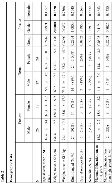
22 17 Table 2. Demographic data Demographic data of the term and preterm cohorts is presented in Table 2. Notably, there was a higher percentage of male subjects in the preterm cohort (59%) than in the term cohort (41%). There was no significant difference in the preterm and term cohorts in age at scan. There was a trend for higher weight in males than females among both preterm and term groups, with no difference between preterm and term cohorts. There was a significant gender effect on height, with males taller than females in both preterm and term groups. In addition, there was a significant group by gender interaction in height (p=0.0320), such that preterm males were slightly taller than term males (170.4 ± 9.2 cm vs ± 9.8 cm) while preterm females on average lagged notably behind term females (156.0 ± 9.9 cm vs ± 5.6 cm). Among the preterm subjects, 92% of males and 78% of females were righthanded, while 88% of male term subjects and 100% of female term subjects were righthanded, but these differences did not show significant group or gender effects. There were no significant differences in percentage of non-white subjects or percentage of subjects who had received special services as children. There was a trend for higher levels of maternal education in the term cohort compared to the preterm cohort. While the mothers of preterm male and female subjects had respectively 13.2 ± 2.2 and 13.4 ± 2.2 years of education, the mothers of term male and female subjects had 14.1 ± 3.0 and 14.8 ± 2.9 years of education respectively. There was not a significant gender interaction. There were not a significantly different number of mothers with less than a high school education in the preterm or term cohorts.
23 18
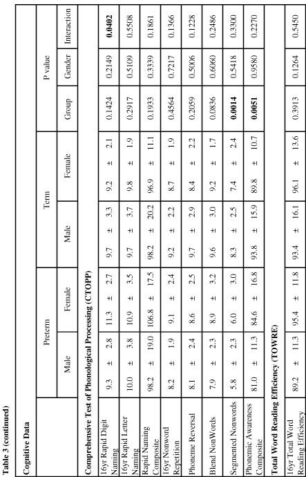
24 19 Table 3. Cognitive testing data by group and gender Results of cognitive testing of all 16 year old subjects are presented in Table 3, including Wechsler Intelligence Scale for Children-III (WISC-III), Peabody Picture Vocabulary Test- Revised (PPVT-R), Visual Motor Integration (VMI), Comprehensive Test of Phonological Processing (CTOPP), and Test of Word Reading Efficiency (TOWRE). For the WISC-III full scale intelligence quotient (IQ), male preterm subjects on average scored 95.3 ± 12.6 and female preterm subjects scored 92.9 ± 16.6, while male term subjects on average scored ± 15.6 and female term subjects scored ±16.8. There was a significant difference between term and preterm cohorts (p=0.0020). The WISC-III Verbal IQ testing showed that male preterm subjects scored on average 98.5 ± 14.7, and female preterms scored 93.6 ± 16.1, while male term subjects on average scored ± 15.1 and female term subjects scored ± This represented a significant difference between preterm and term groups (p=0.0269). The WISC-III Verbal Comprehension IQ testing showed a trend for differences between the groups (p=0.0587), with average scores of 99.2 ± 14.4 in male preterms, 95.6 ± 16.7 in female preterms, ± 13.5 in male terms, and ± 16.0 in female terms. The WISC-III Performance IQ statistics demonstrate significant group differences between term and preterm subjects, with subjects achieving average scores of 92.9 ± 14.9 for male preterms, 93.4 ± 16.1 for female preterms, ± 15.8 for male terms, and ± 18.4 for female terms.
25 20
26 21
27 22 There were no significant differences between groups or genders in PPVT scores; averages in male and female preterm and term groups ranged between 99 and 108 with standard deviations of 19 to 22. Preterm subjects on average scored significantly lower than term subjects on VMI testing (p=0.0015), and there was a trend for male subjects scoring better than female subjects (p=0.0935). Preterm males scored on average 78.8 ± 13.8 on this test, while preterm females scored 75.9 ± 11.1; meanwhile, term males scored 89.8 ± 11.7 and term females scored 83.2 ± While some subsets of CTOPP testing revealed differences between term and preterm cohorts, other scores were indistinguishable. Rapid Naming composite scores, which are composed of Rapid Digit Naming and Rapid Letter Naming tasks, revealed no significant differences between the groups. Within the Rapid Digit Naming subset, however, there was a significant group-by-gender effect (p=0.0402) such that preterm females scored higher than preterm males (11.3 ± 2.7 vs. 9.3 ± 2.8), while term males scored higher than preterm females (9.7 ± 3.3 vs. 9.2 ± 2.1). Preterm subjects also performed comparably to term subjects on Non-word Repetition and Phoneme Reversal tasks. The Phonemic Awareness Composite score, made up of tasks involving Blended Non-words and Segmented Non-words, showed a significant difference (p=0.0051) between term and preterm cohorts. While preterm males achieved an average score of 81.0 ± 11.3 and preterm females scored 84.6 ± 16.8, term males scored 93.8 ± 15.9 and term females scored 89.8 ± The subsets revealed significantly higher scores in terms than preterms in the Segmented Non-words task but no significant differences in the Blended Non-words task.
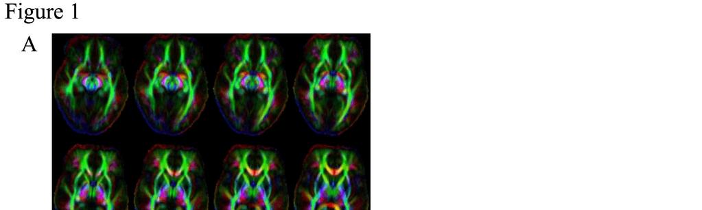
28 23 TOWRE 16 year old standard scores revealed no significant differences among the term and preterm cohorts. Figure 1. Mapping regions of interest Axial slices through a representative diffusion tensor image of a control brain, showing directional fractional anisotropy values, are shown in Figure 1A. Red represents left-right orientation of fibers, blue represents superior-inferior orientation of fibers, and green represents anterior-posterior orientation of fibers. Greater intensity of color represents higher values of fractional anisotropy. In Figures 1B-1C, representative ROIs are shown in blue overlying a grayscale fractional anisotropy map of a control brain, in which high levels of FA are white and low FA regions are black. Figure 1B shows the mapped region of the left arcuate fasciculus in blue in sagittal, coronal, and axial projections over a control brain. Figure 1C shows the left uncinate fasciculus in blue in sagittal, coronal, and axial planes over a control brain.


29 24
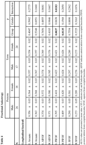
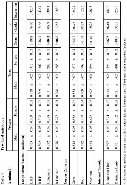
30 25 Table 4. Fractional anisotropy in regions of interest by group and gender Quantitative fractional anisotropy values were obtained in each brain within a variety of regions of interest representing white matter tracts, and statistics regarding effects of group and gender were calculated for each cohort. No significant differences were seen within the groups for multiple regions of interest, including left and right arcuate fasciculi, left and right AIFOF, left and right SFOF, left and right ILF, posterior limbs of the left and right internal capsules, left and right cingulum, left and right fornices, and left and right forceps major. Term subjects had significantly higher fractional anisotropy values than preterm subjects in left and right PIFOF regions (p= and p= respectively). In addition, there was a trend in the left PIFOF for a gender effect (p=0.0638) and a significant group-by-gender interaction (p=0.0183), with lower fractional anisotropy values in male preterm subjects (0.395 ± 0.03) compared to female preterm subjects (0.418 ± 0.02), while male and female term subjects had very similar values (0.423 ± 0.02 vs ± 0.02). The left and right uncinate fasciculi each had significantly higher FA values in the term group than the preterm group (left: p=0.0042, right: p=0.002), and there was a trend for an effect of gender in the left uncinate fasciculus (p=0.0639) such that males had higher FA values than females. In the left uncinate fasciculus, male and female preterms had FA values of ± 0.02 and ± 0.02 respectively, while male and female terms had respective FA values of ± 0.02 and ± In the right uncinate fasciculus, male and female preterm subjects had FA values of ± 0.02 and ±
31 respectively, while male and female term subjects had FA values of ± 0.02 and ± 0.02 respectively. In the corpus callosum, there was a significant effect of gender in the genu (p=0.0377) and a trend in the body (p=0.0573) such that females had higher FA values in both term and preterm cohorts. In the splenium there was not a notable effect of gender, but the preterm group (male: ± 0.05 and female: ± 0.06) had significantly lower FA values than the term group (male ± 0.04 and female ± 0.03), with p= The anterior limb of the left internal capsule demonstrated a significant effect of gender (p=0.0219) such that male had higher FA values than females (0.397 ± 0.02 vs ± 0.02 in the preterm group, and ± 0.02 vs ± 0.02 in the term cohort). These values showed trends for effects of group (p=0.0623) and group by gender (p=0.0842). There were no significant effects in the anterior limb of the right internal capsule. Preterm children on average had significantly lower FA values in both left and right external capsules than term children (p< and p= respectively). On the left, the average FA value in male preterms was ± 0.02 and in female preterms was ± 0.01, while term children had average values of ± 0.03 in males and ± 0.03 in females. On the right, average FA values in preterms were ± 0.03 in males and ± 0.02 in females, while averages in term subjects were ± 0.03 and ± 0.02 in males and females respectively. In addition to long white matter tracts, segments of subcortical white matter deep to important gyri were analyzed. Preterm children had lower FA values than term
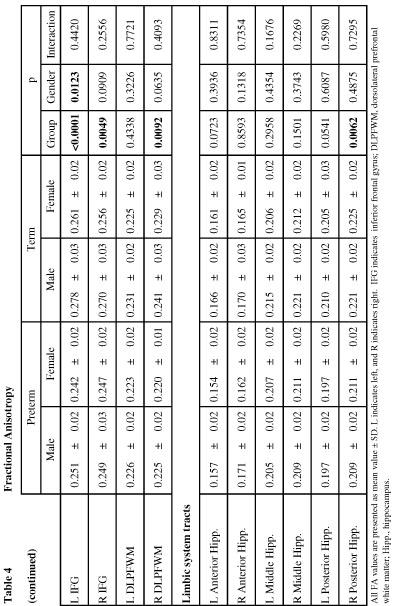
32 27 children in white matter serving both left and right inferior precentral gyri (p= and p= respectively). There was a trend towards higher FA in preterms than terms in the white matter serving the posterior portion of the left superior temporal gyrus (p=0.0926). The average FA values in this region were ± 0.03 and ± 0.03 for male and female preterm subjects respectively, and in term subjects the average values were ± 0.02 for males and ± 0.02 for females. There was no significant difference between the groups in the right posterior STG. The white matter serving the inferior frontal gyrus showed significant effects of both group and gender, such that males had higher average FA values than females and the term subjects had higher FA than preterm subjects. On the left, average FA values were ± 0.02 and ± 0.02 in male and female preterms respectively, while term subjects had average values of ± 0.03 in males and ± 0.02 in females. Both group and gender differences were significant (p< and p= respectively). On the right, average values in preterms were ± 0.03 and ± 0.02 in males and females respectively, while male term subjects had average FA of ± 0.03 and female term subjects had average FA of ± This difference between groups was significant (p=0.0049) while there was a trend for a gender effect (p=0.0909). The hippocampus was analyzed in anterior, middle, and posterior divisions. On the left, there were trends for higher FA in term subjects than preterm subjects in the anterior and posterior segments, but no difference in the middle portion. On the right, anterior and middle divisions showed no significant differences, but the posterior third showed a significantly higher average FA values in terms than preterms (p=0.0062), with
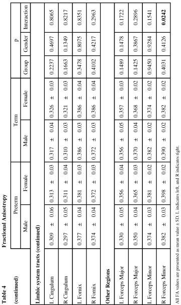
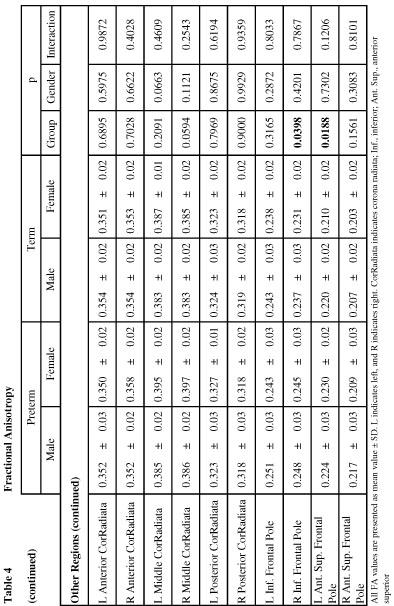
33 28 values of ± 0.02 and ± 0.02 in male and female preterms respectively, while terms had average values of ± 0.02 in males and ± 0.02 in females. In the forceps minor, which is the frontal radiation of the corpus callosum, there was a significant group-by-gender effect (p=0.0240) such that female preterms had greater FA values than male preterms (0.398 ± 0.02 vs ± 0.03 respectively) while female term subjects had lower FA values than male term subjects (0.382 ± 0.02 vs ± 0.02 respectively). Just inferior to this region, the right inferior frontal pole showed a significantly higher FA values in preterm subjects than terms (p=0.0398), with FA values of ± 0.03 and ± 0.03 in male and female subjects, compared to values of ± 0.03 and ± 0.02 in male and female term subjects. There were no significant differences in the left inferior frontal pole. Superior to this region, the left anterior superior frontal pole also showed higher FA values in preterm than term subjects (p=0.0188), with values of ± 0.03 and ± 0.02 in male and female preterm subjects, while term subjects had average values of ± 0.02 in males and ± 0.02 in females. There was no significant effect in the right anterior superior frontal pole. While there were no significant differences in the anterior or posterior segments of the corona radiata, there were trends in the middle segment, such that in the left middle corona radiata, females tended to have higher FA values than males (p=0.0663), while in the right corona radiata, preterms tended to have higher FA values than terms (p=0.0594).
34 29
35 30
36 31
37 32
38 33
39 34
40 35 Table 5. Correlations between FA in specific ROIs and cognitive testing Correlations between FA in specific ROIs and selected cognitive tests are presented in Table 5. Scores on PPVT, a measure of semantic processing, are correlated with left and right uncinate fasciculi, left and right external capsules, and white matter serving the left and right inferior frontal gyri. Rapid Naming Composite scores, a subset of CTOPP testing highlighting phonological processing, are correlated with left and right arcuate fasciculi. In Table 5A, these correlations are presented for all subjects (n=85), with statistical modeling taking into account age at scan, preterm vs. term status, and gender. In this analysis, there are significant positive correlations between PPVT scores and bilateral uncinate fasciculi: on the left, R 2 =0.1109, with p=0.0107, and on the right, R 2 = with p= Trends also exist for positive correlations between Rapid Naming Composite scores and bilateral arcuate fasciculi: on the left, R 2 =0.1077, with p=0.0774, while on the right, R 2 =0.1116, with p= There are no significant associations between PPVT scores and left and right external capsules or white matter deep to the IFG. In Table 5B, the data from the preterm group alone is analyzed with statistical consideration of birthweight and the presence of bronchopulmonary dysplasia in addition to age at scan and gender. With this consideration, there remains a significant positive association between FA values in the right uncinate fasciculus and PPVT scores in the preterm group (R 2 =0.1712, p=0.0273) and a similar positive trend between these scores and values in the left uncinate (R 2 =0.1401, p=0.0605). The positive correlation between PPVT scores and the right uncinate fasciculus is now a trend (R 2 =0.1269, p=0.0848). FA

41 36 values in both left and right arcuate fasciculi again show significant positive correlations with CTOPP Rapid Naming Composite scores in the preterm group. On the left, correlations with Rapid Naming scores show R 2 = and p=0.0051, while the right arcuate correlates with these scores such that R 2 = and p= Because previous studies in this cohort at age 12 demonstrated gender effects, we tested the effect of gender in the models for the correlations between uncinate and arcuate to language testing. FA in the right uncinate contributed to PPVT for the preterm males (R 2 =0.2559, p=0.0186), while FA in the left and right arcuate each contributed to CTOPP Rapid Naming scores in the preterm females (left: R 2 =0.5582, p=0.0167; right: R 2 =0.5364, p=0.0229). Finally, there were no significant correlations in the term population.
42 37 Figure 2. Correlations between FA in specific ROIs and cognitive testing Graphic representations of significant correlations between FA values in select ROIs and scores on specific cognitive tests are presented in Figure 2, with dots representing each subject, a solid line representing the R value for the regression model, and dotted lines representing a 95% confidence interval for the correlation. Each graph represents data seen in Table 5. In Figure 2A, correlations between PPVT scores and FA of the left uncinate for all subjects (n=85) are presented. Figure 2B shows the correlations between all subjects PPVT scores and right uncinate FA. Figure 2C focuses on preterm subjects correlation between PPVT scores and FA in the left uncinate. In Figure 2D, PPVT scores of preterm subjects are correlated with the FA values in the right uncinate fasciculus. Figure 2E shows the Rapid Naming Composite scores of all subjects correlating with FA in the left arcuate. In Figure 2F, all subjects Rapid Naming Composite scores correlate with FA in the right arcuate. Figure 2G shows the Rapid Naming Composite scores of preterm subjects correlating with FA of the left arcuate. Finally, Figure 2H demonstrates preterm subjects Rapid Naming Composite scores correlating with FA of the right arcuate.
43 38
44 39 Discussion Neonatal data The group of infants participating in this study did not show evidence of severe brain injury as infants, but one quarter did develop bronchopulmonary dysplasia. Chronic lung disease has been shown to have an effect on FA measures in the brain independent of variables in preterm subject such as gestational age (52). The presence of BPD, along with birthweight, was taken into account in statistical modeling of the correlation between cognitive scoring and FA values in Tables 5c and 5d. Birthweight, which also has been shown to impact FA measures (45), was also taken into account in Tables 5c and 5d. Demographic data While males outnumbered females in the preterm group, there were more females than males in the control term group. Given previously reported differences in outcomes between male and female preterm subjects (14), gender was taken into account in statistical models for comparison of cognitive data, fractional anisotropy values, and correlations between the two, and average scores and values are presented separately for males and females in each table. Potential factors which could influence cognitive performance or brain structure include levels of maternal education, which is likely to affect performance on cognitive exams, and percentage of right-handed subjects, which impacts laterality of language within the brain. There were higher numbers of left-handed subjects and a trend for lower
45 40 levels of maternal education in the preterm cohort than in the term group, though neither was significant. Cognitive data Preterm subjects showed impairment relative to term control subjects on multiple tests, including WISC full scale IQ, verbal IQ, and performance IQ, VMI, and the CTOPP Phonological Awareness Composite score, with particular difficulties with Segmented Non-Words. Notably, while preterm subjects had lower average scores on multiple IQ measures than term controls, their scores were comparable to term subjects on certain tests. Specifically, preterms performed well on PPVT, TOWRE, CTOPP Rapid Naming Composite scores, Non-Word Repetition, and Phoneme Reversal. Good performance on CTOPP components including repetition may indicate preserved phonological processing ability, PPVT measures semantic understanding through testing of receptive vocabulary, and TOWRE is a measure of reading ability. These preserved abilities span a range of different subsystems of linguistic processing and ability. Fractional anisotropy Our data show lower FA values, implying white matter microstructural disorganization, in multiple white matter areas in preterm subjects without evidence of severe neonatal brain injury. These alterations in white matter were seen in long intrahemispheric association tracts, interhemispheric connections, limbic structures, and frontal lobe white matter areas. The splenium, which has multiple cross-hemispheric connections and is the last part of the corpus callosum to develop, shows particular
46 41 deficits in FA in preterm subjects compared to term subjects; this is consistent with reports from multiple cohorts (30, 87-89). Deficits in FA in the white matter of the hippocampus may indicate barriers to working memory, and is consistent with evidence from an animal model showing anisotropy decreases in hippocampi along with difficulties in spatial memory (90). Previous reports in children have also suggested that preterm birth impacts the hippocampi, showing volume losses in the hippocampi of preterm children compared to controls (91). The posterior segment of the inferior frontooccipital fasciculus, a bundle of intrahemispheric connection fibers, again shows bilateral deficits in FA in preterm subjects relative to term subjects, consistent with the finding at 12 years of age (30). There are significant differences between terms and preterms in the areas implicated in the ventral pathway of language processing. Bilateral uncinate fasciculi show decreased FA in preterm subjects. In addition, bilateral external capsules, areas which include fibers in the extreme capsule (since the resolution of the study did not permit separation of the two tracts), show decreased FA in preterms. The white matter serving the IFG, which is contiguous with the uncinate fasciculus, also shows bilateral deficits in FA in the preterm group. Of note, the arcuate fasciculus, the primary component of the dorsal, phonological language processing pathway, shows no significant FA differences between terms and preterms. While previous reports have noted FA deficits in the posterior limb of the internal capsule in preterm subjects, no such deficit was noted in our cohort. These previous studies included higher percentage of subjects with severe brain injury at birth leading to cerebral palsy and severe neurological deficits. White matter deep to the inferior portion
Insults to the Developing Brain & Effect on Neurodevelopmental Outcomes
 Insults to the Developing Brain & Effect on Neurodevelopmental Outcomes Ira Adams-Chapman, MD Assistant Professor of Pediatrics Director, Developmental Progress Clinic Emory University School of Medicine
P. Hitchcock, Ph.D. Department of Cell and Developmental Biology Kellogg Eye Center. Wednesday, 16 March 2009, 1:00p.m. 2:00p.m.
 Normal CNS, Special Senses, Head and Neck TOPIC: CEREBRAL HEMISPHERES FACULTY: LECTURE: READING: P. Hitchcock, Ph.D. Department of Cell and Developmental Biology Kellogg Eye Center Wednesday, 16 March
Is DTI Increasing the Connectivity Between the Magnet Suite and the Clinic?
 Current Literature In Clinical Science Is DTI Increasing the Connectivity Between the Magnet Suite and the Clinic? Spatial Patterns of Water Diffusion Along White Matter Tracts in Temporal Lobe Epilepsy.
Supplementary appendix
 Supplementary appendix This appendix formed part of the original submission and has been peer reviewed. We post it as supplied by the authors. Supplement to: Schiller R, IJsselstijn H, Hoskote A, et al.
Fig.1: A, Sagittal 110x110 mm subimage close to the midline, passing through the cingulum. Note that the fibers of the corpus callosum run at a
 Fig.1 E Fig.1:, Sagittal 110x110 mm subimage close to the midline, passing through the cingulum. Note that the fibers of the corpus callosum run at a slight angle are through the plane (blue dots with
Visualization strategies for major white matter tracts identified by diffusion tensor imaging for intraoperative use
 International Congress Series 1281 (2005) 793 797 www.ics-elsevier.com Visualization strategies for major white matter tracts identified by diffusion tensor imaging for intraoperative use Ch. Nimsky a,b,
Heidi M. Feldman, MD, PhD,* Jason D. Yeatman, BA, Eliana S. Lee, BS,* Laura H. F. Barde, PhD,* Shayna Gaman-Bean, MD*
 Review Article Diffusion Tensor Imaging: A Review for Pediatric Researchers and Clinicians Heidi M. Feldman, MD, PhD,* Jason D. Yeatman, BA, Eliana S. Lee, BS,* Laura H. F. Barde, PhD,* Shayna Gaman-Bean,
CEREBRUM Dr. Jamila Elmedany Dr. Essam Eldin Salama
 CEREBRUM Dr. Jamila Elmedany Dr. Essam Eldin Salama Objectives At the end of the lecture, the student should be able to: List the parts of the cerebral hemisphere (cortex, medulla, basal nuclei, lateral
Telencephalon (Cerebral Hemisphere)
") Telencephalon (Cerebral Hemisphere) OUTLINE The Cortex - Lobes, Sulci & Gyri - Functional Subdivisions - Limbic Lobe & Limbic System The Subcortex - Basal Ganglia - White Matter (Internal Capsule) - Relations
Telencephalon (Cerebral Hemisphere) OUTLINE The Cortex - Lobes, Sulci & Gyri - Functional Subdivisions - Limbic Lobe & Limbic System The Subcortex - Basal Ganglia - White Matter (Internal Capsule) - Relations
Advanced magnetic resonance imaging for monitoring brain development and injury
 Advanced magnetic resonance imaging for monitoring brain development and injury Stéphane Sizonenko, MD-PhD Division of Development and Growth Department of Child and Adolescent Medicine Geneva University
Gross Organization I The Brain. Reading: BCP Chapter 7
 Gross Organization I The Brain Reading: BCP Chapter 7 Layout of the Nervous System Central Nervous System (CNS) Located inside of bone Includes the brain (in the skull) and the spinal cord (in the backbone)
DWI assessment of ischemic changes in the fetal brain
 DWI assessment of ischemic changes in the fetal brain Dafi Bergman, 4 th year Medical student in the 4-year program, Sackler school of medicine B.Sc Life and Medical Sciences, Tel Aviv University Supervised
Diffusion Tensor Imaging in Psychiatry
 2003 KHBM DTI in Psychiatry Diffusion Tensor Imaging in Psychiatry KHBM 2003. 11. 21. 서울대학교 의과대학 정신과학교실 권준수 Neuropsychiatric conditions DTI has been studied in Alzheimer s disease Schizophrenia Alcoholism
Cerebral Cortex 1. Sarah Heilbronner
 Cerebral Cortex 1 Sarah Heilbronner heilb028@umn.edu Want to meet? Coffee hour 10-11am Tuesday 11/27 Surdyk s Overview and organization of the cerebral cortex What is the cerebral cortex? Where is each
Diffusion tensor imaging of the infant brain: From technical problems to neuroscientific breakthroughs Jessica Dubois
 Diffusion tensor imaging of the infant brain: From technical problems to neuroscientific breakthroughs Jessica Dubois L. Hertz-Pannier, G. Dehaene-Lambertz, J.F. Mangin, D. Le Bihan Inserm U56, U663; NeuroSpin
Surgery Within and Around Critical White Matter Tracts
 Surgery Within and Around Critical White Matter Tracts Kaisorn L. Chaichana, M.D. Assistant Professor of Neurosurgery, Oncology, and Otolaryngology-Head & Neck Surgery Mayo Clinic Florida, Jacksonville,
Title:Atypical language organization in temporal lobe epilepsy revealed by a passive semantic paradigm
 Author's response to reviews Title:Atypical language organization in temporal lobe epilepsy revealed by a passive semantic paradigm Authors: Julia Miro (juliamirollado@gmail.com) Pablo Ripollès (pablo.ripolles.vidal@gmail.com)
Announcement. Danny to schedule a time if you are interested.
 Announcement If you need more experiments to participate in, contact Danny Sanchez (dsanchez@ucsd.edu) make sure to tell him that you are from LIGN171, so he will let me know about your credit (1 point).
Perinatal cerebral white matter injuries influence early communication and language development
 Perinatal cerebral white matter injuries influence early communication and language development Blazenka Brozovic University of Zagreb Department of Speech and Language Pathology Developmental Neurolinguistic
Diffusion Tensor Imaging 12/06/2013
 12/06/2013 Beate Diehl, MD PhD FRCP University College London National Hospital for Neurology and Neurosurgery Queen Square London, UK American Epilepsy Society Annual Meeting Disclosure None Learning
Normal myelination: a practical pictorial review
 Normal myelination: a practical pictorial review Poster No.: C-1486 Congress: ECR 2016 Type: Authors: Keywords: DOI: Educational Exhibit M. Gaha, N. Mama, N. Arifa, H. Jemni, K. Tlili Graiess; Sousse/TN
Stuttering Research. Vincent Gracco, PhD Haskins Laboratories
 Stuttering Research Vincent Gracco, PhD Haskins Laboratories Stuttering Developmental disorder occurs in 5% of children Spontaneous remission in approximately 70% of cases Approximately 1% of adults with
Leah Militello, class of 2018
 Leah Militello, class of 2018 Objectives 1. Describe the general organization of cerebral hemispheres. 2. Describe the locations and features of the different functional areas of cortex. 3. Understand
Myers Psychology for AP*
 Myers Psychology for AP* David G. Myers PowerPoint Presentation Slides by Kent Korek Germantown High School Worth Publishers, 2010 *AP is a trademark registered and/or owned by the College Board, which
Supplementary Information Methods Subjects The study was comprised of 84 chronic pain patients with either chronic back pain (CBP) or osteoarthritis
 or osteoarthritis") Supplementary Information Methods Subjects The study was comprised of 84 chronic pain patients with either chronic back pain (CBP) or osteoarthritis (OA). All subjects provided informed consent to procedures
Supplementary Information Methods Subjects The study was comprised of 84 chronic pain patients with either chronic back pain (CBP) or osteoarthritis (OA). All subjects provided informed consent to procedures
Define functional MRI. Briefly describe fmri image acquisition. Discuss relative functional neuroanatomy. Review clinical applications.
 Dr. Peter J. Fiester November 14, 2012 Define functional MRI. Briefly describe fmri image acquisition. Discuss relative functional neuroanatomy. Review clinical applications. Briefly discuss a few examples
NIH Public Access Author Manuscript J Child Neurol. Author manuscript; available in PMC 2014 December 28.
 NIH Public Access Author Manuscript Published in final edited form as: J Child Neurol. 2013 June ; 28(6): 774 780. doi:10.1177/0883073812449693. Case Series: Fractional Anisotropy Along the Trajectory
Web Training Modules Module 13: MR Imaging for Brain Injuries
 Web Training Modules Module 13: MR Imaging for Brain Injuries 1 Introduction... 4 Mild Traumatic Brain Injuries... 5 Structural MRI... 5 Diffusion Tensor Imaging... 6 Diffusion Kurtosis Imaging... 11 Functional
CEREBRUM & CEREBRAL CORTEX
 CEREBRUM & CEREBRAL CORTEX Seonghan Kim Dept. of Anatomy Inje University, College of Medicine THE BRAIN ANATOMICAL REGIONS A. Cerebrum B. Diencephalon Thalamus Hypothalamus C. Brain Stem Midbrain Pons
Research Article Corticospinal Tract Change during Motor Recovery in Patients with Medulla Infarct: A Diffusion Tensor Imaging Study
 BioMed Research International, Article ID 524096, 5 pages http://dx.doi.org/10.1155/2014/524096 Research Article Corticospinal Tract Change during Motor Recovery in Patients with Medulla Infarct: A Diffusion
Imaging in Pediatric `neurohiv Dr Jackie Hoare Head of Liaison Psychiatry Groote Schuur Hospital, UCT
 Imaging in Pediatric `neurohiv Dr Jackie Hoare Head of Liaison Psychiatry Groote Schuur Hospital, UCT ? Spectrum of Neurocognitive disorders The adult literature on HIV related CNS damage supports a spectrum
Pediatric MS MRI Study Methodology
 General Pediatric MS MRI Study Methodology SCAN PREPARATION axial T2-weighted scans and/or axial FLAIR scans were obtained for all subjects when available, both T2 and FLAIR scans were scored. In order
Advances in Clinical Neuroimaging
 Advances in Clinical Neuroimaging Joseph I. Tracy 1, PhD, ABPP/CN; Gaelle Doucet 2, PhD; Xaiosong He 2, PhD; Dorian Pustina 2, PhD; Karol Osipowicz 2, PhD 1 Department of Radiology, Thomas Jefferson University,
fmri (functional MRI)
") Lesion fmri (functional MRI) Electroencephalogram (EEG) Brainstem CT (computed tomography) Scan Medulla PET (positron emission tomography) Scan Reticular Formation MRI (magnetic resonance imaging) Thalamus
Lesion fmri (functional MRI) Electroencephalogram (EEG) Brainstem CT (computed tomography) Scan Medulla PET (positron emission tomography) Scan Reticular Formation MRI (magnetic resonance imaging) Thalamus
Diffusion tensor imaging: serial quantitation of white matter tract maturity in premature newborns $
 Diffusion tensor imaging: serial quantitation of white matter tract maturity in premature newborns $ Savannah C. Partridge, a Pratik Mukherjee, a Roland G. Henry, a Steven P. Miller, b,c Jeffrey I. Berman,
THE ESSENTIAL BRAIN INJURY GUIDE
 THE ESSENTIAL BRAIN INJURY GUIDE Neuroanatomy & Neuroplasticity Section 2 Contributors Erin D. Bigler, PhD Michael R. Hoane, PhD Stephanie Kolakowsky-Hayner, PhD, CBIST, FACRM Dorothy A. Kozlowski, PhD
CEREBRUM. Dr. Jamila EL Medany
 CEREBRUM Dr. Jamila EL Medany Objectives At the end of the lecture, the student should be able to: List the parts of the cerebral hemisphere (cortex, medulla, basal nuclei, lateral ventricle). Describe
Stereotactic Diffusion Tensor Tractography For Gamma Knife Stereotactic Radiosurgery
 Disclosures The authors of this study declare that they have no commercial or other interests in the presentation of this study. This study does not contain any use of offlabel devices or treatments. Stereotactic
IMAGING OF HYPOXIC ISCHEMIC INJURY IN A NEONATE FN3 STATE MEETING NEMOURS CHILDREN'S HOSPITAL ORLANDO,FL 08/04/18
 IMAGING OF HYPOXIC ISCHEMIC INJURY IN A NEONATE FN3 STATE MEETING NEMOURS CHILDREN'S HOSPITAL ORLANDO,FL 08/04/18 Dhanashree Rajderkar,MD Assistant Professor Department of Radiology University of Florida
Fetal CNS MRI. Daniela Prayer. Division of Neuroradiology And Musculoskeletal Radiology. Medical University of Vienna, AUSTRIA
 Fetal CNS MRI Daniela Prayer Division of Neuroradiology And Musculoskeletal Radiology Medical University of Vienna, AUSTRIA Methods Normal development Malformations Acquired pathology MR- methods for assessment
Brain anatomy tutorial. Dr. Michal Ben-Shachar 459 Neurolinguistics
 Brain anatomy tutorial Dr. Michal Ben-Shachar 459 Neurolinguistics The human brain Left hemisphere Right hemisphere http://www.brainmuseum.org/ Zoom out Zoom in Types of Brain Tissue Gray Matter: Cell
doi: /brain/awq347 Brain 2011: 134; White matter damage and cognitive impairment after traumatic brain injury
 doi:10.1093/brain/awq347 Brain 2011: 134; 449 463 449 BRAIN A JOURNAL OF NEUROLOGY White matter damage and cognitive impairment after traumatic brain injury Kirsi Maria Kinnunen, 1 Richard Greenwood, 2
The neurolinguistic toolbox Jonathan R. Brennan. Introduction to Neurolinguistics, LSA2017 1
 The neurolinguistic toolbox Jonathan R. Brennan Introduction to Neurolinguistics, LSA2017 1 Psycholinguistics / Neurolinguistics Happy Hour!!! Tuesdays 7/11, 7/18, 7/25 5:30-6:30 PM @ the Boone Center
FUNCTIONAL MAGNETIC RESONANCE IMAGING IN FOLLOW-UP OF CEREBRAL GLIAL TUMORS
 Anvita Bieza FUNCTIONAL MAGNETIC RESONANCE IMAGING IN FOLLOW-UP OF CEREBRAL GLIAL TUMORS Summary of Doctoral Thesis to obtain PhD degree in medicine Specialty Diagnostic Radiology Riga, 2013 Doctoral thesis
Fibre orientation dispersion in the corpus callosum relates to interhemispheric functional connectivity
 Fibre orientation dispersion in the corpus callosum relates to interhemispheric functional connectivity ISMRM 2017: http://submissions.mirasmart.com/ismrm2017/viewsubmissionpublic.aspx?sei=8t1bikppq Jeroen
doi: /brain/aws276 Brain 2012: 135;
 doi:10.1093/brain/aws276 Brain 2012: 135; 3781 3798 3781 BRAIN A JOURNAL OF NEUROLOGY Interhemispheric temporal lobe connectivity predicts language impairment in adolescents born preterm Gemma B. Northam,
MR Assessment of Myelination in Infants and Children: Usefulness of Marker Sites
 731 MR ssessment of Myelination in Infants and Children: Usefulness of Marker Sites C. Roger ird 1 Mary Hedberg urton P. Drayer Paul J. Keller Richard. Flom John. Hodak retrospective study was made of
Language comprehension in young people with severe cerebral palsy in relation to language tracts: a diffusion tensor imaging study
 Language comprehension in young people with severe cerebral palsy in relation to language tracts: a diffusion tensor imaging study Laurike Harlaar, Petra J. Pouwels, Joke J. Geytenbeek, Kim J. Oostrom,
Human Paleoneurology and the Evolution of the Parietal Cortex
 PARIETAL LOBE The Parietal Lobes develop at about the age of 5 years. They function to give the individual perspective and to help them understand space, touch, and volume. The location of the parietal
10/3/2016. T1 Anatomical structures are clearly identified, white matter (which has a high fat content) appears bright.
 appears bright.") H2O -2 atoms of Hydrogen, 1 of Oxygen Hydrogen just has one single proton and orbited by one single electron Proton has a magnetic moment similar to the earths magnetic pole Also similar to earth in that
H2O -2 atoms of Hydrogen, 1 of Oxygen Hydrogen just has one single proton and orbited by one single electron Proton has a magnetic moment similar to the earths magnetic pole Also similar to earth in that
Periventricular white matter injury, that is, periventricular
 ORIGINAL RESEARCH S. Yoshida K. Hayakawa A. Yamamoto T. Kanda Y. Yamori Pontine Hypoplasia in Children with Periventricular Leukomalacia BACKGROUND AND PURPOSE: The brain stem in patients with periventricular
Post Stroke Brain Plasticity
 Post Stroke Brain Plasticity François CHOLLET MD, PhD Neurology Department: Stroke Unit Toulouse University Hospital (CHU) Neurosciences Institute of Toulouse CNRS, INSERM, University, CHU Versailles le
Overview. Fundamentals of functional MRI. Task related versus resting state functional imaging for sensorimotor mapping
 Functional MRI and the Sensorimotor System in MS Nancy Sicotte, MD, FAAN Professor and Vice Chair Director, Multiple Sclerosis Program Director, Neurology Residency Program Cedars-Sinai Medical Center
Use of Multimodal Neuroimaging Techniques to Examine Age, Sex, and Alcohol-Related Changes in Brain Structure Through Adolescence and Young Adulthood
 American Psychiatric Association San Diego, CA 24 May 2017 Use of Multimodal Neuroimaging Techniques to Examine Age, Sex, and Alcohol-Related Changes in Brain Structure Through Adolescence and Young Adulthood
Functional MRI and Diffusion Tensor Imaging
 Functional MRI and Diffusion Tensor Imaging Andrew Steven March 23, 2018 Ochsner Neuroscience Symposium None Disclosure 1 Objectives Review basic principles of BOLD fmri and DTI. Discuss indications and
Introduction to the Central Nervous System: Internal Structure
 Introduction to the Central Nervous System: Internal Structure Objective To understand, in general terms, the internal organization of the brain and spinal cord. To understand the 3-dimensional organization
High spatial resolution reveals excellent detail in pediatric neuro imaging
 Publication for the Philips MRI Community Issue 46 2012/2 High spatial resolution reveals excellent detail in pediatric neuro imaging Achieva 3.0T with 32-channel SENSE Head coil has become the system
Brain Structure and Function in Nephropathic Cystinosis
 Brain Structure and Function in Nephropathic Cystinosis Doris A. Trauner M.D. Professor, Depts. of Neurosciences and Pediatrics University of California San Diego School of Medicine La Jolla, CA USA Cystinosis
Summary of findings from the previous meta-analyses of DTI studies in MDD patients. SDM (39) 221 Left superior longitudinal
 221 Left superior longitudinal") Supplemental Data Table S1 Summary of findings from the previous meta-analyses of DTI studies in MDD patients Study Analysis Method Included studies, n MDD (medicated) HC Results (MDDHC)
Supplemental Data Table S1 Summary of findings from the previous meta-analyses of DTI studies in MDD patients Study Analysis Method Included studies, n MDD (medicated) HC Results (MDDHC)
Chronology of normal brain myelination in newborns with MR imaging
 Chronology of normal brain myelination in newborns with MR imaging Poster No.: C-0577 Congress: ECR 2012 Type: Authors: Keywords: DOI: Scientific Exhibit F. Fernandez Usagre; Sevilla/ES Neuroradiology
White matter integrity of the cerebellar peduncles as a mediator of effects of prenatal alcohol exposure on eyeblink conditioning
 White matter integrity of the cerebellar peduncles as a mediator of effects of prenatal alcohol exposure on eyeblink conditioning Jia Fan 1,2, Sandra W. Jacobson 2-3,5, Christopher D. Molteno 3, Bruce
Homework Week 2. PreLab 2 HW #2 Synapses (Page 1 in the HW Section)
") Homework Week 2 Due in Lab PreLab 2 HW #2 Synapses (Page 1 in the HW Section) Reminders No class next Monday Quiz 1 is @ 5:30pm on Tuesday, 1/22/13 Study guide posted under Study Aids section of website
Homework Week 2 Due in Lab PreLab 2 HW #2 Synapses (Page 1 in the HW Section) Reminders No class next Monday Quiz 1 is @ 5:30pm on Tuesday, 1/22/13 Study guide posted under Study Aids section of website
Extremely preterm infants are at high risk of brain injury
 ORIGINAL RESEARCH J.L.Y. Cheong D.K. Thompson H.X. Wang R.W. Hunt P.J. Anderson T.E. Inder L.W. Doyle Abnormal White Matter Signal on MR Imaging Is Related to Abnormal Tissue Microstructure BACKGROUND
Seamless pre-surgical fmri and DTI mapping
 Seamless pre-surgical fmri and DTI mapping Newest release Achieva 3.0T X-series and Eloquence enable efficient, real-time fmri for brain activity mapping in clinical practice at Nebraska Medical Center
Chapter 14, Part 2! Chapter 14 Part 2 Brain/Cranial Nerves! The Cerebrum and Cranial Nerves! pp !
 Chapter 14, Part 2! The Cerebrum and Cranial pp. 482 505! SECTION 14-9! The cerebrum, the largest region of the brain, contains motor, sensory, and association areas! 2! White Matter of the Cerebrum! 1.
Cognitive Neuroscience Cortical Hemispheres Attention Language
 Cognitive Neuroscience Cortical Hemispheres Attention Language Based on: Chapter 18 and 19, Breedlove, Watson, Rosenzweig, 6e/7e. Cerebral Cortex Brain s most complex area with billions of neurons and
Chapter 14, Part 2! The Cerebrum and Cranial Nerves! pp !
 Chapter 14, Part 2! The Cerebrum and Cranial pp. 482 505! SECTION 14-9! The cerebrum, the largest region of the brain, contains motor, sensory, and association areas! 2! 1! ! Chapter 14 Part 2 Brain/Cranial
Auditory and Vestibular Systems
 Auditory and Vestibular Systems Objective To learn the functional organization of the auditory and vestibular systems To understand how one can use changes in auditory function following injury to localize
shows syntax in his language. has a large neocortex, which explains his language abilities. shows remarkable cognitive abilities. all of the above.
 Section: Chapter 14: Multiple Choice 1. Alex the parrot: pp.529-530 shows syntax in his language. has a large neocortex, which explains his language abilities. shows remarkable cognitive abilities. all
PSYC& 100: Biological Psychology (Lilienfeld Chap 3) 1
 1") PSYC& 100: Biological Psychology (Lilienfeld Chap 3) 1 1 What is a neuron? 2 Name and describe the functions of the three main parts of the neuron. 3 What do glial cells do? 4 Describe the three basic
PSYC& 100: Biological Psychology (Lilienfeld Chap 3) 1 1 What is a neuron? 2 Name and describe the functions of the three main parts of the neuron. 3 What do glial cells do? 4 Describe the three basic
Normal Myelination of Anatomic Nerve Fiber Bundles: MR Analysis
 AJNR Am J Neuroradiol 19:1129 1136, June 1998 Normal Myelination of Anatomic Nerve Fiber Bundles: MR Analysis Hiroyuki Nakagawa, Satoru Iwasaki, Kimihiko Kichikawa, Akio Fukusumi, Toshiaki Taoka, Hajime
Diffusion-Weighted and Conventional MR Imaging Findings of Neuroaxonal Dystrophy
 AJNR Am J Neuroradiol 25:1269 1273, August 2004 Diffusion-Weighted and Conventional MR Imaging Findings of Neuroaxonal Dystrophy R. Nuri Sener BACKGROUND AND PURPOSE: Neuroaxonal dystrophy is a rare progressive
Table 1. Summary of PET and fmri Methods. What is imaged PET fmri BOLD (T2*) Regional brain activation. Blood flow ( 15 O) Arterial spin tagging (AST)
 Regional brain activation. Blood flow ( 15 O) Arterial spin tagging (AST)") Table 1 Summary of PET and fmri Methods What is imaged PET fmri Brain structure Regional brain activation Anatomical connectivity Receptor binding and regional chemical distribution Blood flow ( 15 O)
Table 1 Summary of PET and fmri Methods What is imaged PET fmri Brain structure Regional brain activation Anatomical connectivity Receptor binding and regional chemical distribution Blood flow ( 15 O)
Analysis between clinical and MRI findings of childhood and teenages with epilepsy after hypoxic-ischemic encephalopathy in neonates periods
 Analysis between clinical and MRI findings of childhood and teenages with epilepsy after hypoxic-ischemic encephalopathy in neonates periods Poster No.: C-0401 Congress: ECR 2015 Type: Scientific Exhibit
doi: /brain/awr099 Brain 2011: 134; White matter damage in primary progressive aphasias: a diffusion tensor tractography study
 doi:10.1093/brain/awr099 Brain 2011: 134; 3011 3029 3011 BRAIN A JOURNAL OF NEUROLOGY White matter damage in primary progressive aphasias: a diffusion tensor tractography study Sebastiano Galantucci, 1,2
doi: /brain/awq175 Brain 2010: 133;
 doi:10.1093/brain/awq175 Brain 2010: 133; 2348 2364 2348 BRAIN A JOURNAL OF NEUROLOGY The structural plasticity of white matter networks following anterior temporal lobe resection Mahinda Yogarajah, 1
Brain tissue and white matter lesion volume analysis in diabetes mellitus type 2
 Brain tissue and white matter lesion volume analysis in diabetes mellitus type 2 C. Jongen J. van der Grond L.J. Kappelle G.J. Biessels M.A. Viergever J.P.W. Pluim On behalf of the Utrecht Diabetic Encephalopathy
MRI and M/EEG studies of the White Matter Development in Human Fetuses and Infants: Review and Opinion
 Brain Plasticity 2 (2016) 49 69 DOI 10.3233/BPL-160031 IOS Press Review 49 MRI and M/EEG studies of the White Matter Development in Human Fetuses and Infants: Review and Opinion Jessica Dubois a,, Parvaneh
Cerebrum-Cerebral Hemispheres. Cuneyt Mirzanli Istanbul Gelisim University
 Cerebrum-Cerebral Hemispheres Cuneyt Mirzanli Istanbul Gelisim University The largest part of the brain. Ovoid shape. Two incompletely separated cerebral hemispheres. The outer surface of the cerebral
Hypoxic ischemic brain injury in neonates - early MR imaging findings
 Hypoxic ischemic brain injury in neonates - early MR imaging findings Poster No.: C-1208 Congress: ECR 2015 Type: Authors: Keywords: DOI: Educational Exhibit E.-M. Heursen, R. Reina Cubero, T. Guijo Hernandez,
Modeling of early-infant brain growth using longitudinal data from diffusion tensor imaging.
 Modeling of early-infant brain growth using longitudinal data from diffusion tensor imaging. Guido Gerig, Neda Sadeghi, PhD, Marcel Prastawa, Tom Fletcher, Clement Vachet Scientific Computing and Imaging
Alterations in the inferior longitudinal fasciculus in autism and associations with visual processing: a diffusion-weighted MRI study
 Boets et al. Molecular Autism (2018) 9:10 DOI 10.1186/s13229-018-0188-6 RESEARCH Open Access Alterations in the inferior longitudinal fasciculus in autism and associations with visual processing: a diffusion-weighted
The Central Nervous System I. Chapter 12
 The Central Nervous System I Chapter 12 The Central Nervous System The Brain and Spinal Cord Contained within the Axial Skeleton Brain Regions and Organization Medical Scheme (4 regions) 1. Cerebral Hemispheres
Differences in brain structure and function between the sexes has been a topic of
 Introduction Differences in brain structure and function between the sexes has been a topic of scientific inquiry for over 100 years. In particular, this topic has had significant interest in the past
The association of children s mathematic abilities with both adults cognitive abilities
 Supplemental material for The association of children s mathematic abilities with both adults cognitive abilities and intrinsic fronto-parietal networks is altered in preterm born individuals by Bäuml,
Online supplement data :
 Online supplement data : ROI ROI name MNI coord (mm) HC>SZ HC>S ZR HC>P BP HC>P BPR Heritabil ity h^2 Heritability p-value MCP Middle cerebellar peduncle 0-41 -44 2.05 2.12 1.63 0.69 0.38 0.004473 PCT
With improvements in neonatal care, the mortality rate of
 ORIGINAL RESEARCH B.J.M. van Kooij L.S. de Vries G. Ball I.C. van Haastert M.J.N.L. Benders F. Groenendaal S.J. Counsell Neonatal Tract-Based Spatial Statistics Findings and Outcome in Preterm Infants
Gross Morphology of the Brain
 Gross Morphology of the Brain Done by : Marah Marahleh & Razan Krishan *slides in bold Principal Parts of the Brain Cerebrum : largest part of the brain Diencephalon Thalamus & hypothalamus Cerebellum
Procedia - Social and Behavioral Sciences 159 ( 2014 ) WCPCG 2014
 WCPCG 2014") Available online at www.sciencedirect.com ScienceDirect Procedia - Social and Behavioral Sciences 159 ( 2014 ) 743 748 WCPCG 2014 Differences in Visuospatial Cognition Performance and Regional Brain Activation
Available online at www.sciencedirect.com ScienceDirect Procedia - Social and Behavioral Sciences 159 ( 2014 ) 743 748 WCPCG 2014 Differences in Visuospatial Cognition Performance and Regional Brain Activation
Newborn Hypoxic Ischemic Brain Injury. Hisham Dahmoush, MBBCh FRCR Lucile Packard Children s Hospital at Stanford
 Newborn Hypoxic Ischemic Brain Injury Hisham Dahmoush, MBBCh FRCR Lucile Packard Children s Hospital at Stanford NO DISCLOSURES INTRODUCTION Neonatal hypoxic-ischemic encephalopathy (HIE) is a major cause
Shape Modeling of the Corpus Callosum for Neuroimaging Studies of the Brain (Part I) Dongqing Chen, Ph.D.
 Dongqing Chen, Ph.D.") The University of Louisville CVIP Lab Shape Modeling of the Corpus Callosum for Neuroimaging Studies of the Brain (Part I) Dongqing Chen, Ph.D. Computer Vision & Image Processing (CVIP) Laboratory Department
The University of Louisville CVIP Lab Shape Modeling of the Corpus Callosum for Neuroimaging Studies of the Brain (Part I) Dongqing Chen, Ph.D. Computer Vision & Image Processing (CVIP) Laboratory Department
1. Processes nutrients and provides energy for the neuron to function; contains the cell's nucleus; also called the soma.
 1. Base of brainstem; controls heartbeat and breathing 2. tissue destruction; a brain lesion is a naturally or experimentally caused destruction of brain tissue 3. A thick band of axons that connects the
Classification and Statistical Analysis of Auditory FMRI Data Using Linear Discriminative Analysis and Quadratic Discriminative Analysis
 International Journal of Innovative Research in Computer Science & Technology (IJIRCST) ISSN: 2347-5552, Volume-2, Issue-6, November-2014 Classification and Statistical Analysis of Auditory FMRI Data Using
Imaging the Premature Brain- New Knowledge
 Imaging the Premature Brain- New Knowledge Stein Magnus Aukland Haukeland University Hospital University of Bergen NORWAY No disclosure Imaging modalities O Skull X-ray O Computer Tomography O Cerebral
Text to brain: predicting the spatial distribution of neuroimaging observations from text reports (submitted to MICCAI 2018)
") 1 / 22 Text to brain: predicting the spatial distribution of neuroimaging observations from text reports (submitted to MICCAI 2018) Jérôme Dockès, ussel Poldrack, Demian Wassermann, Fabian Suchanek, Bertrand
1 / 22 Text to brain: predicting the spatial distribution of neuroimaging observations from text reports (submitted to MICCAI 2018) Jérôme Dockès, ussel Poldrack, Demian Wassermann, Fabian Suchanek, Bertrand
Quantitative Neuroimaging- Gray and white matter Alteration in Multiple Sclerosis. Lior Or-Bach Instructors: Prof. Anat Achiron Dr.
 Quantitative Neuroimaging- Gray and white matter Alteration in Multiple Sclerosis Lior Or-Bach Instructors: Prof. Anat Achiron Dr. Shmulik Miron INTRODUCTION Multiple Sclerosis general background Gray
Stroke School for Internists Part 1
 Stroke School for Internists Part 1 November 4, 2017 Dr. Albert Jin Dr. Gurpreet Jaswal Disclosures I receive a stipend for my role as Medical Director of the Stroke Network of SEO I have no commercial
doi: /brain/awr335 Brain 2012: 135; Neural systems for speech and song in autism
 doi:10.1093/brain/awr335 Brain 2012: 135; 961 975 961 BRAIN A JOURNAL OF NEUROLOGY Neural systems for speech and song in autism Grace Lai, 1,2 Spiro P. Pantazatos, 2,3 Harry Schneider 2 and Joy Hirsch
The human brain weighs roughly 1.5 kg and has an average volume of 1130 cm 3. A sheep s brain weighs in however at kg.
 Sheep Brain Dissection Objectives: 1. List and describe the principal structures of the sheep brain 2. Identify important parts of the sheep brain in a preserved specimen Materials: Dissection tools, lab
Pearls and Pitfalls of MR Diffusion in Clinical Neurology
 Pearls and Pitfalls of MR Diffusion in Clinical Neurology Dr. Alberto Bizzi Neuroradiology Unit Fondazione IRCCS Istituto Neurologico Carlo Besta Milan, Italy Email: alberto_bizzi@fastwebnet.it Diffusion