Diffusion tensor imaging of the infant brain: From technical problems to neuroscientific breakthroughs Jessica Dubois
|
|
|
- Tobias Newton
- 5 years ago
- Views:
Transcription

; Hôpital Necker-Enfants")
1 Diffusion tensor imaging of the infant brain: From technical problems to neuroscientific breakthroughs Jessica Dubois L. Hertz-Pannier, G. Dehaene-Lambertz, J.F. Mangin, D. Le Bihan Inserm U56, U663; NeuroSpin (CEA); Hôpital Necker-Enfants malades Saclay, Paris (France)


2 Brain development during infancy Sensori-motor and cognitive advances Birth 3 months 6 months 1 year Post-mortem myelin staining adulte Cycles of myelination In vivo MRI: T1-w images Fleschig, 190 Paus et al, 001 Baumann and Pham-Dinh, 001 From Yakovlev and Lecours, 1967
 to study the infant white")

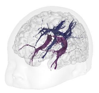
3 Brain development and diffusion MRI Potential of diffusion MRI and diffusion tensor imaging (DTI) to study the infant white matter development: spatial organization of fiber fascicles fascicles maturation Hüppi et al, 001 Neil et al, 1998, 00; Mukherjee et al, 001
4 Diffusion tensor imaging (DTI) Diffusion of water molecules Oriented organization
5 Diffusion tensor imaging (DTI) 3 1 // Diffusion tensor D D Diffusion of water molecules Oriented organization 1/ D D D FA Mean diffusivity Fractional anisotropy DTI indices


6 Diffusion tensor imaging (DTI) Diffusion of water molecules Oriented organization Diffusion tensor 1 D 0 0 // DTI indices White matter fibers Mean diffusivity D <D> map.10-3 mm.s -1 Fractional anisotropy 3 FA D D D 1 FA map / RGB map R-L A-P I-S
7 Methodological objectives: Objectives Develop a DTI methodology dedicated to the infant brain Evaluate the spatial organization of immature white matter fascicles Quantify the fascicles maturation with DTI indices Neuroscientific objectives: Classify the differential maturation of fascicles Evaluate inter-hemispherical asymmetries of the developing white matter Correlate structural maturation with functional maturation for the developing visual system
8 Methodological objectives: Objectives Develop a DTI methodology dedicated to the infant brain Evaluate the spatial organization of immature white matter fascicles Quantify the fascicles maturation with DTI indices Neuroscientific objectives: Classify the differential maturation of fascicles Evaluate inter-hemispherical asymmetries of the developing white matter Correlate structural maturation with functional maturation for the developing visual system


9 Difficulties in imaging infants brain T1-w MRI adult -m old infant Constraints in MR images: - contrast: high water content, tissues immaturity - spatial resolution: structures size, partial volume effects DTI-RGB map Constraints in MR acquisition: - time - scanner noise - wake up - motion Only a few MRI studies in healthy infants without sedation
10 Optimization of diffusion orientations sets E Methodological objective: Optimize DTI acquisitions in function of the possible babies wakening Electrostatical model i j 0 E ij E 0 ij 1 r r i Orientation number SNR + spatial estimation BUT acquisition time Compromise between orientation number and acquisition time? OR Outsmart temporal order of orientations within a set j 1 r r i j
11 Optimization of diffusion orientations sets E Methodological objective: Optimize DTI acquisitions in function of the possible babies wakening Electrostatical model E i j i j 0 E ij E 0 ije ij 0 ij 1 r r i Orientation number SNR + spatial estimation BUT acquisition time Compromise between orientation number and acquisition time? OR Outsmart temporal order of orientations within a set j 1 r r i j (Dubois et al, MAGMA 006)


12 Optimization of diffusion orientations sets Methodological objective: Optimize DTI acquisitions in function of the possible babies wakening Orientation number SNR + spatial estimation BUT acquisition time Compromise between orientation number and acquisition time? OR Outsmart temporal order of orientations within a set Electrostatical model E E i j i j 0 E ij E 0 ije ij 0 ij 1 r r i j 1 r r i j Acquisition time 1min min 3min (Dubois et al, MAGMA 006)
![Subjects Subjects and DTI acquisition 3 healhty infants (age : 10.3 ± 3.8 weeks, [3.9-18.4] w) 6 healhty adults (age : 5.7 ± 1.4 years) Acquisition 1.](/docs-images/83/87241411/images/13-0.jpg "5T Signa LX GEMS Birdcage head coil Protection against noise, no sedation DTI imaging b = 700s.mm - 15 orientations of diffusion gradients Spatial resolution 1.9 x")
13 Subjects Subjects and DTI acquisition 3 healhty infants (age : 10.3 ± 3.8 weeks, [ ] w) 6 healhty adults (age : 5.7 ± 1.4 years) Acquisition 1.5T Signa LX GEMS Birdcage head coil Protection against noise, no sedation DTI imaging b = 700s.mm - 15 orientations of diffusion gradients Spatial resolution 1.9 x 1.9 x.5mm 3 30 axial slices covering the whole brain Acquisition time 3min40s
14 Data post-processing Softwares: Anatomist (Rivière et al, 000) BrainVISA (Cointepas et al, 003) SPM (Functional Imaging Laboratory, Londres) Estimation of diffusion tensor parameters Reconstruction of fiber fascicles by tractography
15 Reconstruction of fiber fascicles Non linear tractography Regularization of the particles trajectory (Perrin, Mangin et al, 005) Local diffusion field i v dif v i1 v i v i Particles speed 1 v v i i1 i dif i index of anisotropy
16 Reconstruction of fiber fascicles Non linear tractography Regularization of the particles trajectory (Perrin, Mangin et al, 005) Local diffusion field i v dif v i1 v i v i Particles speed 1 v v i i1 i dif i index of anisotropy Infants white matter <D> <.10-3 mm.s -1 ; FA > < maximal curvature angle < 45 a priori hypothesis on the fasciculus trajectory (Catani et al, 00)
17 Data post-processing Softwares: Anatomist (Rivière et al, 000) BrainVISA (Cointepas et al, 003) SPM (Functional Imaging Laboratory, Londres) Estimation of diffusion tensor parameters Reconstruction of fiber fascicles by tractography Normalization of DTI maps through a FA template


18 Normalization of DTI maps Step 1 For each infant T w anatomical image non-linear normalization T w anatomical template coregistration DTI b=0 image FA map normalized FA map FA template Average over all infants


19 Normalization of DTI maps Step FA map For each infant non-linear normalization FA template Normalized FA map <D> map Fasciculus mask Fasciculus probability map Normalized <D> map Normalized fasciculus mask Average over all infants
20 Methodological objectives: Objectives Develop a DTI methodology dedicated to the infant brain Evaluate the spatial organization of immature white matter fascicles Quantify the fascicles maturation with DTI indices Neuroscientific objectives: Classify the differential maturation of fascicles Evaluate inter-hemispherical asymmetries of the developing white matter Correlate structural maturation with functional maturation for the developing visual system
21 Corpus callosum RGB map tractography FA template + probability map ccg ccs 0.7 ccb 0.1


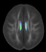
22 Cortico-spinal and spino-thalamic tracts RGB map tractography FA template + probability map cst stt cst stt 0.7 cst 0.1
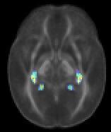
23 Optic radiations and internal capsule (anterior limb) RGB map tractography FA template + probability map or 0.7 alic alic 0.1 or


24 Uncinate fasciculus and external capsule RGB map tractography FA template + probability map unc ec unc 0.7 ec 0.1


25 Inferior longitudinal and arcuate fascicles RGB map tractography FA template + probability map ilf af ilf af

26 Fornix and cingulum RGB map tractography FA template + probability map cg fx cg fx 0.7 cg 0.1
27 Organization of infants fascicles Early identification of white matter fascicles despite immaturity All kinds of fascicles: commissures, projection, association and limbic fascicles Methodological limitations: spatial resolution / partial volume tensor model / fiber crossings tractography / anatomical hypotheses
28 Methodological objectives: Objectives Develop a DTI methodology dedicated to the infant brain Evaluate the spatial organization of immature white matter fascicles Quantify the fascicles maturation with DTI indices Neuroscientific objectives: Classify the differential maturation of fascicles Evaluate inter-hemispherical asymmetries of the developing white matter Correlate structural maturation with functional maturation for the developing visual system


29 Quantification of DTI indices Averaging approach over volumes of interest - sections of tracts >> regions of interest (Dubois et al, NeuroImage 006) - individual volumes >> group volumes Voxel-by-voxel approch Approach along the tract
30 Methodological objectives: Objectives Develop a DTI methodology dedicated to the infant brain Evaluate the spatial organization of immature white matter fascicles Quantify the fascicles maturation with DTI indices Neuroscientific objectives: Classify the differential maturation of fascicles Evaluate inter-hemispherical asymmetries of the developing white matter Correlate structural maturation with functional maturation for the developing visual system

31 Differential maturation of fascicles Spatio-temporal pattern of maturation across the brain, according to the development of functional systems Focus on eleven fascicles of interest Relevance of DTI indices (Dubois et al, Hum Brain Map 008)
32 Classification model of fascicles maturation For each fasciculus (f) and each DTI index (I) Maturation state and speed - State: median over babies % median over adults + fasciculus % "average" - Speed: age-related change over babies % adults and "average" state speed bb ad I f M I f ad aver M aver, M f I M 1 bb I bb ad I f M I f ad aver M aver f, I 1 bb I I I
33 Classification model of fascicles maturation For each fasciculus (f) and each DTI index (I) Maturation state and speed - State: median over babies % median over adults + fasciculus % "average" - Speed: age-related change over babies % adults and "average" Maturation stage state speed bb ad I f M I f ad aver M aver, M f I M 1 bb I bb ad I f M I f ad aver M aver f, I 1 bb I I I
34 Maturation processes and DTI indices Complementarity of DTI indices in white matter 1- to 4-months old infants Possibility to detect maturation earlier with <D> than with FA?

35 Classification model of fascicles maturation For each fasciculus (f) Maturation phase <D> stage % FA stage In vivo validation: infants DTI data f more mature than others f more immature than others (Dubois et al, Hum Brain Map 008)
36 Methodological objectives: Objectives Develop a DTI methodology dedicated to the infant brain Evaluate the spatial organization of immature white matter fascicles Quantify the fascicles maturation with DTI indices Neuroscientific objectives: Classify the differential maturation of fascicles Evaluate inter-hemispherical asymmetries of the developing white matter Correlate structural maturation with functional maturation for the developing visual system


37 Functional asymmetries during development Language: left hemispherical predominance in the infant brain mv + L R L R Dehaene-Lambertz, 000 Dehaene-Lambertz et al, 00 Somato-sensorial response: lateralization in the newborn brain Erberich et al, 005
,")
: asymmetries")


38 Anatomical asymmetries during development Planum temporale (left), Heschl gyrus (left), superior temporal sulcus (right): asymmetries in the fœtal, newborn and child brain (Witelson et al, 1973; Chi et al, 1977) What about white matter? DTI in the adult brain Buchel et al, 004 Parker et al, 005

39 White matter asymmetries in infants Voxel-by-voxel analysis of FA map Asymmetries of the temporal arcuate fasciculus and the inferior cortico-spinal tract favouring the left hemisphere (Dubois et al, Cereb Cortex 009)

40 White matter asymmetries in infants Analysis of the fascicles macroscopic geometry The temporal arcuate fasciculus is larger and more posterior on the left side (Dubois et al, Cereb Cortex 009)
41 White matter asymmetries in infants Analysis of DTI indices over the tracts sections FA is higher on the left side in the parietal arcuate fasciculus and the inferior cortico-spinal tract (Dubois et al, Cereb Cortex 009)
42 White matter asymmetries in infants Arcuate fasciculus temporal: asymmetry in macroscopic geometry (larger volume on the left side) parietal: asymmetry in microscopic organization (higher compacity on the left side) Cortico-spinal tract inferior: asymmetry in maturation (advance on the left side) Structural asymmetries are observed early on in the infant brain, in immature white matter networks implicated in language perception and manuality.
43 Methodological objectives: Objectives Develop a DTI methodology dedicated to the infant brain Evaluate the spatial organization of immature white matter fascicles Quantify the fascicles maturation with DTI indices Neuroscientific objectives: Classify the differential maturation of fascicles Evaluate inter-hemispherical asymmetries of the developing white matter Correlate structural maturation with functional maturation for the developing visual system

44 Visual system development Visual pathways: early anatomical maturation Vision: one of the first developing functions after birth Cycles of myelination Baumann and Pham-Dinh, 001 From Yakovlev and Lecours, 1967
45 Visual system development Visual pathways: early anatomical maturation Vision: one of the first developing functions after birth Correlations? Structural maturation of visual pathways: DTI imaging Functional development of vision: event-related potentials (ERP) Decrease of P 1 latency with age Subjects: 16 / 3 infants (age: [ ] w.) McCulloch et al, 1999
46 Functional maturation of the visual system Visual ERP Recording system: 64 electrodes, 4ms Visual stimulations (faces): 1.5s Averaging of events signals latency of P1 conduction speed of visual information
: 1.")
 and age in the")

47 Functional maturation of the visual system Visual ERP Recording system: 64 electrodes, 4ms Visual stimulations (faces): 1.5s Averaging of events signals latency of P1 conduction speed of visual information Model ERP / DTI Speed depends on DTI indices (FA, ) and age in the optic radiations Fibers myelination First correlation between structural and functional brain maturations in infants speed visual ERP dis tan ce P1 index DTI R =0.86 (Dubois et al, J Neurosci 008) age
 cortex (abs 1.0) 1 1 1 Myelination of geniculo-cortical (1) and cortico-geniculate () fibers?")
48 Structural maturation of the optic radiations 3 sections of the tract Anterior region: maturation advance Posterior region: acute changes Along the tract anterior region middle region posterior region FA LGN (abs 0.0) cortex (abs 1.0) Myelination of geniculo-cortical (1) and cortico-geniculate () fibers? (McCart et al, 1994) (Dubois et al, J Neurosci 008)
49 Conclusion DTI study of the organization and maturation of white matter fiber fascicles in healthy infants Dedicated methodology % technical problems Potential of tractography with DTI indices quantification Neuroscientific breakthroughs Future directions Subjects: developmental period (preterm newborns, infants > 4-m old), pathological development (epilepsies, etc.) Acquisition: 3T + parallel imaging (spatial resolution, time), orientation number Post-processing: diffusion model, tracts clustering, TBSS registration, etc. Analyses: diffusion indices % relaxation times (T1, T), white matter % cortex Anatomo-functional applications: auditory / tactile stimulations

50 Many thanks! Inserm U56 / U663 CEA NeuroSpin L. Hertz-Pannier G. Dehaene-Lambertz D. Le Bihan J.F. Mangin C. Poupon M. Perrin Y. Cointepas D. Rivière E. Duchesnay A. Cachia F. Lethimonnier F. Leroy V. Santoro C. Chiron F. Brunelle C. Soarès T. Gliga etc. All the babies and parents And thank you for your attention!
MRI and M/EEG studies of the White Matter Development in Human Fetuses and Infants: Review and Opinion
 Brain Plasticity 2 (2016) 49 69 DOI 10.3233/BPL-160031 IOS Press Review 49 MRI and M/EEG studies of the White Matter Development in Human Fetuses and Infants: Review and Opinion Jessica Dubois a,, Parvaneh
Diffusion Tensor Imaging 12/06/2013
 12/06/2013 Beate Diehl, MD PhD FRCP University College London National Hospital for Neurology and Neurosurgery Queen Square London, UK American Epilepsy Society Annual Meeting Disclosure None Learning
Fig.1: A, Sagittal 110x110 mm subimage close to the midline, passing through the cingulum. Note that the fibers of the corpus callosum run at a
 Fig.1 E Fig.1:, Sagittal 110x110 mm subimage close to the midline, passing through the cingulum. Note that the fibers of the corpus callosum run at a slight angle are through the plane (blue dots with
NeuroImage 61 (2012) Contents lists available at SciVerse ScienceDirect. NeuroImage. journal homepage:
 Contents lists available at SciVerse ScienceDirect. NeuroImage. journal homepage:") NeuroImage 61 (2012) 542 557 Contents lists available at SciVerse ScienceDirect NeuroImage journal homepage: www.elsevier.com/locate/ynimg Quantitative tract-based white matter development from birth to
NeuroImage 61 (2012) 542 557 Contents lists available at SciVerse ScienceDirect NeuroImage journal homepage: www.elsevier.com/locate/ynimg Quantitative tract-based white matter development from birth to
Summary of findings from the previous meta-analyses of DTI studies in MDD patients. SDM (39) 221 Left superior longitudinal
 221 Left superior longitudinal") Supplemental Data Table S1 Summary of findings from the previous meta-analyses of DTI studies in MDD patients Study Analysis Method Included studies, n MDD (medicated) HC Results (MDDHC)
Supplemental Data Table S1 Summary of findings from the previous meta-analyses of DTI studies in MDD patients Study Analysis Method Included studies, n MDD (medicated) HC Results (MDDHC)
Functional MRI and Diffusion Tensor Imaging
 Functional MRI and Diffusion Tensor Imaging Andrew Steven March 23, 2018 Ochsner Neuroscience Symposium None Disclosure 1 Objectives Review basic principles of BOLD fmri and DTI. Discuss indications and
Diffusion Tensor Imaging in Psychiatry
 2003 KHBM DTI in Psychiatry Diffusion Tensor Imaging in Psychiatry KHBM 2003. 11. 21. 서울대학교 의과대학 정신과학교실 권준수 Neuropsychiatric conditions DTI has been studied in Alzheimer s disease Schizophrenia Alcoholism
Telencephalon (Cerebral Hemisphere)
") Telencephalon (Cerebral Hemisphere) OUTLINE The Cortex - Lobes, Sulci & Gyri - Functional Subdivisions - Limbic Lobe & Limbic System The Subcortex - Basal Ganglia - White Matter (Internal Capsule) - Relations
Telencephalon (Cerebral Hemisphere) OUTLINE The Cortex - Lobes, Sulci & Gyri - Functional Subdivisions - Limbic Lobe & Limbic System The Subcortex - Basal Ganglia - White Matter (Internal Capsule) - Relations
Cortical Folding and Connectivity in the developing brain
 The Rise and Fall of the Brain ISMRM Monday April20th Page 1 Cortical Folding and Connectivity in the developing brain Petra S. Hüppi, M.D. Jessica Dubois, Ph.D. Department of Pediatrics, Children s Hospital,
Fibre orientation dispersion in the corpus callosum relates to interhemispheric functional connectivity
 Fibre orientation dispersion in the corpus callosum relates to interhemispheric functional connectivity ISMRM 2017: http://submissions.mirasmart.com/ismrm2017/viewsubmissionpublic.aspx?sei=8t1bikppq Jeroen
Diffusion tensor imaging: serial quantitation of white matter tract maturity in premature newborns $
 Diffusion tensor imaging: serial quantitation of white matter tract maturity in premature newborns $ Savannah C. Partridge, a Pratik Mukherjee, a Roland G. Henry, a Steven P. Miller, b,c Jeffrey I. Berman,
Modeling of early-infant brain growth using longitudinal data from diffusion tensor imaging.
 Modeling of early-infant brain growth using longitudinal data from diffusion tensor imaging. Guido Gerig, Neda Sadeghi, PhD, Marcel Prastawa, Tom Fletcher, Clement Vachet Scientific Computing and Imaging
Heidi M. Feldman, MD, PhD,* Jason D. Yeatman, BA, Eliana S. Lee, BS,* Laura H. F. Barde, PhD,* Shayna Gaman-Bean, MD*
 Review Article Diffusion Tensor Imaging: A Review for Pediatric Researchers and Clinicians Heidi M. Feldman, MD, PhD,* Jason D. Yeatman, BA, Eliana S. Lee, BS,* Laura H. F. Barde, PhD,* Shayna Gaman-Bean,
Visualization strategies for major white matter tracts identified by diffusion tensor imaging for intraoperative use
 International Congress Series 1281 (2005) 793 797 www.ics-elsevier.com Visualization strategies for major white matter tracts identified by diffusion tensor imaging for intraoperative use Ch. Nimsky a,b,
P. Hitchcock, Ph.D. Department of Cell and Developmental Biology Kellogg Eye Center. Wednesday, 16 March 2009, 1:00p.m. 2:00p.m.
 Normal CNS, Special Senses, Head and Neck TOPIC: CEREBRAL HEMISPHERES FACULTY: LECTURE: READING: P. Hitchcock, Ph.D. Department of Cell and Developmental Biology Kellogg Eye Center Wednesday, 16 March
Is DTI Increasing the Connectivity Between the Magnet Suite and the Clinic?
 Current Literature In Clinical Science Is DTI Increasing the Connectivity Between the Magnet Suite and the Clinic? Spatial Patterns of Water Diffusion Along White Matter Tracts in Temporal Lobe Epilepsy.
Diffusion Tensor Imaging in brain tumours
 Diffusion Tensor Imaging in brain tumours @MarionSmits, MD PhD Associate Professor of Neuroradiology Dept. of Radiology, Erasmus MC, Rotterdam (NL) Honorary Consultant and Reader UCLH National Hospital
Advanced magnetic resonance imaging for monitoring brain development and injury
 Advanced magnetic resonance imaging for monitoring brain development and injury Stéphane Sizonenko, MD-PhD Division of Development and Growth Department of Child and Adolescent Medicine Geneva University
Cerebral Cortex 1. Sarah Heilbronner
 Cerebral Cortex 1 Sarah Heilbronner heilb028@umn.edu Want to meet? Coffee hour 10-11am Tuesday 11/27 Surdyk s Overview and organization of the cerebral cortex What is the cerebral cortex? Where is each
Auditory and Vestibular Systems
 Auditory and Vestibular Systems Objective To learn the functional organization of the auditory and vestibular systems To understand how one can use changes in auditory function following injury to localize
Seamless pre-surgical fmri and DTI mapping
 Seamless pre-surgical fmri and DTI mapping Newest release Achieva 3.0T X-series and Eloquence enable efficient, real-time fmri for brain activity mapping in clinical practice at Nebraska Medical Center
Post Stroke Brain Plasticity
 Post Stroke Brain Plasticity François CHOLLET MD, PhD Neurology Department: Stroke Unit Toulouse University Hospital (CHU) Neurosciences Institute of Toulouse CNRS, INSERM, University, CHU Versailles le
Online supplement data :
 Online supplement data : ROI ROI name MNI coord (mm) HC>SZ HC>S ZR HC>P BP HC>P BPR Heritabil ity h^2 Heritability p-value MCP Middle cerebellar peduncle 0-41 -44 2.05 2.12 1.63 0.69 0.38 0.004473 PCT
CISC 3250 Systems Neuroscience
 CISC 3250 Systems Neuroscience Levels of organization Central Nervous System 1m 10 11 neurons Neural systems and neuroanatomy Systems 10cm Networks 1mm Neurons 100μm 10 8 neurons Professor Daniel Leeds
Language comprehension in young people with severe cerebral palsy in relation to language tracts: a diffusion tensor imaging study
 Language comprehension in young people with severe cerebral palsy in relation to language tracts: a diffusion tensor imaging study Laurike Harlaar, Petra J. Pouwels, Joke J. Geytenbeek, Kim J. Oostrom,
Stuttering Research. Vincent Gracco, PhD Haskins Laboratories
 Stuttering Research Vincent Gracco, PhD Haskins Laboratories Stuttering Developmental disorder occurs in 5% of children Spontaneous remission in approximately 70% of cases Approximately 1% of adults with
Leah Militello, class of 2018
 Leah Militello, class of 2018 Objectives 1. Describe the general organization of cerebral hemispheres. 2. Describe the locations and features of the different functional areas of cortex. 3. Understand
Stereotactic Diffusion Tensor Tractography For Gamma Knife Stereotactic Radiosurgery
 Disclosures The authors of this study declare that they have no commercial or other interests in the presentation of this study. This study does not contain any use of offlabel devices or treatments. Stereotactic
PRETERM BIRTH RESULTS IN ALTERATIONS IN NEURAL CONNECTIVITY AT AGE 16 YEARS
 Yale University EliScholar A Digital Platform for Scholarly Publishing at Yale Yale Medicine Thesis Digital Library School of Medicine 8-17-2010 PRETERM BIRTH RESULTS IN ALTERATIONS IN NEURAL CONNECTIVITY
APPLICATION OF PHOTOGRAMMETRY TO BRAIN ANATOMY
 http://medifitbiologicals.com/central-nervous-system-cns/ 25/06/2017 PSBB17 ISPRS International Workshop APPLICATION OF PHOTOGRAMMETRY TO BRAIN ANATOMY E. Nocerino, F. Menna, F. Remondino, S. Sarubbo,
doi: /brain/awr335 Brain 2012: 135; Neural systems for speech and song in autism
 doi:10.1093/brain/awr335 Brain 2012: 135; 961 975 961 BRAIN A JOURNAL OF NEUROLOGY Neural systems for speech and song in autism Grace Lai, 1,2 Spiro P. Pantazatos, 2,3 Harry Schneider 2 and Joy Hirsch
November/December 2008 The Brain Unveiled A new imaging method offers a spectacular view of neural structures. By Emily Singer
 November/December 2008 The Brain Unveiled A new imaging method offers a spectacular view of neural structures. By Emily Singer A new imaging method that offers an unprecedented view of complex neural structures
Tracking the language pathways in edema patients: Preliminary results.
 Tracking the language pathways in edema patients: Preliminary results. Sarah M. E. Gierhan 1,2, Peter Rhone 3,4, Alfred Anwander 1, Isabel Jost 3, Clara Frydrychowicz 3, Karl-Titus Hoffmann 4, Jürgen Meixensberger
MRI-Based Classification Techniques of Autistic vs. Typically Developing Brain
 MRI-Based Classification Techniques of Autistic vs. Typically Developing Brain Presented by: Rachid Fahmi 1 2 Collaborators: Ayman Elbaz, Aly A. Farag 1, Hossam Hassan 1, and Manuel F. Casanova3 1Computer
Investigation of brain structure in the 1-month infant
 https://doi.org/10.1007/s00429-017-1600-2 ORIGINAL ARTICLE Investigation of brain structure in the 1-month infant Douglas C. Dean III 1,2 E. M. Planalp 1,3 W. Wooten 2 C. K. Schmidt 1,2 S. R. Kecskemeti
doi: /brain/awq175 Brain 2010: 133;
 doi:10.1093/brain/awq175 Brain 2010: 133; 2348 2364 2348 BRAIN A JOURNAL OF NEUROLOGY The structural plasticity of white matter networks following anterior temporal lobe resection Mahinda Yogarajah, 1
10/3/2016. T1 Anatomical structures are clearly identified, white matter (which has a high fat content) appears bright.
 appears bright.") H2O -2 atoms of Hydrogen, 1 of Oxygen Hydrogen just has one single proton and orbited by one single electron Proton has a magnetic moment similar to the earths magnetic pole Also similar to earth in that
H2O -2 atoms of Hydrogen, 1 of Oxygen Hydrogen just has one single proton and orbited by one single electron Proton has a magnetic moment similar to the earths magnetic pole Also similar to earth in that
Early Maturation of the Linguistic Dorsal Pathway in Human Infants
 1500 The Journal of Neuroscience, January 26, 2011 31(4):1500 1506 Behavioral/Systems/Cognitive Early Maturation of the Linguistic Dorsal Pathway in Human Infants François Leroy, 1,2,3 Hervé Glasel, 1,2,3
Alterations in the inferior longitudinal fasciculus in autism and associations with visual processing: a diffusion-weighted MRI study
 Boets et al. Molecular Autism (2018) 9:10 DOI 10.1186/s13229-018-0188-6 RESEARCH Open Access Alterations in the inferior longitudinal fasciculus in autism and associations with visual processing: a diffusion-weighted
Use of Multimodal Neuroimaging Techniques to Examine Age, Sex, and Alcohol-Related Changes in Brain Structure Through Adolescence and Young Adulthood
 American Psychiatric Association San Diego, CA 24 May 2017 Use of Multimodal Neuroimaging Techniques to Examine Age, Sex, and Alcohol-Related Changes in Brain Structure Through Adolescence and Young Adulthood
Advances in Clinical Neuroimaging
 Advances in Clinical Neuroimaging Joseph I. Tracy 1, PhD, ABPP/CN; Gaelle Doucet 2, PhD; Xaiosong He 2, PhD; Dorian Pustina 2, PhD; Karol Osipowicz 2, PhD 1 Department of Radiology, Thomas Jefferson University,
CEREBRUM Dr. Jamila Elmedany Dr. Essam Eldin Salama
 CEREBRUM Dr. Jamila Elmedany Dr. Essam Eldin Salama Objectives At the end of the lecture, the student should be able to: List the parts of the cerebral hemisphere (cortex, medulla, basal nuclei, lateral
Normal Myelination of Anatomic Nerve Fiber Bundles: MR Analysis
 AJNR Am J Neuroradiol 19:1129 1136, June 1998 Normal Myelination of Anatomic Nerve Fiber Bundles: MR Analysis Hiroyuki Nakagawa, Satoru Iwasaki, Kimihiko Kichikawa, Akio Fukusumi, Toshiaki Taoka, Hajime
CEREBRUM. Dr. Jamila EL Medany
 CEREBRUM Dr. Jamila EL Medany Objectives At the end of the lecture, the student should be able to: List the parts of the cerebral hemisphere (cortex, medulla, basal nuclei, lateral ventricle). Describe
The neurolinguistic toolbox Jonathan R. Brennan. Introduction to Neurolinguistics, LSA2017 1
 The neurolinguistic toolbox Jonathan R. Brennan Introduction to Neurolinguistics, LSA2017 1 Psycholinguistics / Neurolinguistics Happy Hour!!! Tuesdays 7/11, 7/18, 7/25 5:30-6:30 PM @ the Boone Center
10/10/2011. Tools for Visualization and Labeling. SPM MRIcron MRIcroGL xjview MARINA FreeSurfer Talairach Daemon SPM Anatomy Toolbox WFU Pickatlas
 Tools for Visualization and Labeling Visualization and Labeling SPM MRIcron MRIcroGL xjview MARINA FreeSurfer Talairach Daemon SPM Anatomy Toolbox WFU Pickatlas Gesture imitation with variable memory load
Diffusion Tensor Imaging Assessment of Brain White Matter Maturation During the First Postnatal Year
 DTI of White Matter in infants Neuroradiology Original Research James M. Provenzale 1 Luxia Liang 1,2 David DeLong 1 Leonard E. White 3 Provenzale JM, Liang L, DeLong D, White LE Keywords: anisotropy,
3/1/18. Overview of the Talk. Important Aspects of Neuroimaging Technology
 3/1/18 Considerations for the Use of Neuroimaging for Predicting Recovery of Speech and Language in Aphasia Linda I. Shuster, Ph.D., CCC-SLP Overview of the Talk Important aspects of neuroimaging technology
Overview. Fundamentals of functional MRI. Task related versus resting state functional imaging for sensorimotor mapping
 Functional MRI and the Sensorimotor System in MS Nancy Sicotte, MD, FAAN Professor and Vice Chair Director, Multiple Sclerosis Program Director, Neurology Residency Program Cedars-Sinai Medical Center
Diffusion-Tensor Imaging Assessment of White Matter Maturation in Childhood and Adolescence
 Neuroradiology/Head and Neck Imaging Original Research Moon et al. White Matter Diffusion-Tensor Imaging Neuroradiology/Head and Neck Imaging Original Research Won-Jin Moon 1 James M. Provenzale 2,3 Basar
doi: /brain/awq347 Brain 2011: 134; White matter damage and cognitive impairment after traumatic brain injury
 doi:10.1093/brain/awq347 Brain 2011: 134; 449 463 449 BRAIN A JOURNAL OF NEUROLOGY White matter damage and cognitive impairment after traumatic brain injury Kirsi Maria Kinnunen, 1 Richard Greenwood, 2
Define functional MRI. Briefly describe fmri image acquisition. Discuss relative functional neuroanatomy. Review clinical applications.
 Dr. Peter J. Fiester November 14, 2012 Define functional MRI. Briefly describe fmri image acquisition. Discuss relative functional neuroanatomy. Review clinical applications. Briefly discuss a few examples
Introduction to Diffusion Tractography
 Introduction to Diffusion Tractography In vivo tracer Manganese Tractography ex vivo Tim B. Dyrby, PhD Danish Research Centre for Magnetic Resonance (DRCMR) Copenhagen University Hospital Hvidovre Denmark
Classification and Statistical Analysis of Auditory FMRI Data Using Linear Discriminative Analysis and Quadratic Discriminative Analysis
 International Journal of Innovative Research in Computer Science & Technology (IJIRCST) ISSN: 2347-5552, Volume-2, Issue-6, November-2014 Classification and Statistical Analysis of Auditory FMRI Data Using
Fetal CNS MRI. Daniela Prayer. Division of Neuroradiology And Musculoskeletal Radiology. Medical University of Vienna, AUSTRIA
 Fetal CNS MRI Daniela Prayer Division of Neuroradiology And Musculoskeletal Radiology Medical University of Vienna, AUSTRIA Methods Normal development Malformations Acquired pathology MR- methods for assessment
Brain diffusion tensor imaging changes in cerebrotendinous xanthomatosis reversed with
 Brain diffusion tensor imaging changes in cerebrotendinous xanthomatosis reversed with treatment Claudia B. Catarino, MD, PhD, 1*, Christian Vollmar, MD, PhD, 2,3* Clemens Küpper, MD, 1,4 Klaus Seelos,
Effects Of Attention And Perceptual Uncertainty On Cerebellar Activity During Visual Motion Perception
 Effects Of Attention And Perceptual Uncertainty On Cerebellar Activity During Visual Motion Perception Oliver Baumann & Jason Mattingley Queensland Brain Institute The University of Queensland The Queensland
Structural Network Analysis of Brain Development in Young Preterm Neonates
 Structural Network Analysis of Brain Development in Young Preterm Neonates Colin J Brown a,, Steven P Miller b, Brian G Booth a, Shawn Andrews a, Vann Chau b, Kenneth J Poskitt c, Ghassan Hamarneh a a
Vision II. Steven McLoon Department of Neuroscience University of Minnesota
 Vision II Steven McLoon Department of Neuroscience University of Minnesota 1 Ganglion Cells The axons of the retinal ganglion cells form the optic nerve and carry visual information into the brain. 2 Optic
doi: /brain/awr099 Brain 2011: 134; White matter damage in primary progressive aphasias: a diffusion tensor tractography study
 doi:10.1093/brain/awr099 Brain 2011: 134; 3011 3029 3011 BRAIN A JOURNAL OF NEUROLOGY White matter damage in primary progressive aphasias: a diffusion tensor tractography study Sebastiano Galantucci, 1,2
Research Article Corticospinal Tract Change during Motor Recovery in Patients with Medulla Infarct: A Diffusion Tensor Imaging Study
 BioMed Research International, Article ID 524096, 5 pages http://dx.doi.org/10.1155/2014/524096 Research Article Corticospinal Tract Change during Motor Recovery in Patients with Medulla Infarct: A Diffusion
Gross Organization I The Brain. Reading: BCP Chapter 7
 Gross Organization I The Brain Reading: BCP Chapter 7 Layout of the Nervous System Central Nervous System (CNS) Located inside of bone Includes the brain (in the skull) and the spinal cord (in the backbone)
What is DTI, its use in research and clinical practice, and its future potential
 NA-MIC What is DTI, its use in research and clinical practice, and its future potential Martin Styner with thanks to many, many people Departments of Computer Science and Psychiatry UNC Neurodevelopmental
CEREBRUM & CEREBRAL CORTEX
 CEREBRUM & CEREBRAL CORTEX Seonghan Kim Dept. of Anatomy Inje University, College of Medicine THE BRAIN ANATOMICAL REGIONS A. Cerebrum B. Diencephalon Thalamus Hypothalamus C. Brain Stem Midbrain Pons
The Central Nervous System
 The Central Nervous System Cellular Basis. Neural Communication. Major Structures. Principles & Methods. Principles of Neural Organization Big Question #1: Representation. How is the external world coded
Characterizing Connectivity in Brain Networks Using Magnetic Resonance Imaging
 University of Pennsylvania ScholarlyCommons Publicly Accessible Penn Dissertations Summer 8-13-2010 Characterizing Connectivity in Brain Networks Using Magnetic Resonance Imaging Jeffrey T. Duda University
White matter integrity of the cerebellar peduncles as a mediator of effects of prenatal alcohol exposure on eyeblink conditioning
 White matter integrity of the cerebellar peduncles as a mediator of effects of prenatal alcohol exposure on eyeblink conditioning Jia Fan 1,2, Sandra W. Jacobson 2-3,5, Christopher D. Molteno 3, Bruce
A review of diffusion tensor imaging studies in schizophrenia
 Journal of Psychiatric Research xxx (2005) xxx xxx Review A review of diffusion tensor imaging studies in schizophrenia Marek Kubicki a,b, Robert McCarley a, *, Carl-Fredrik Westin b, Hae-Jeong Park a,b,d,
Shape Modeling of the Corpus Callosum for Neuroimaging Studies of the Brain (Part I) Dongqing Chen, Ph.D.
 Dongqing Chen, Ph.D.") The University of Louisville CVIP Lab Shape Modeling of the Corpus Callosum for Neuroimaging Studies of the Brain (Part I) Dongqing Chen, Ph.D. Computer Vision & Image Processing (CVIP) Laboratory Department
The University of Louisville CVIP Lab Shape Modeling of the Corpus Callosum for Neuroimaging Studies of the Brain (Part I) Dongqing Chen, Ph.D. Computer Vision & Image Processing (CVIP) Laboratory Department
Neuroimaging biomarkers and predictors of motor recovery: implications for PTs
 Neuroimaging biomarkers and predictors of motor recovery: implications for PTs 2018 Combined Sections Meeting of the American Physical Therapy Association New Orleans, LA February 21-24, 2018 Presenters:
An Atlas of Brainstem Connectomes from HCP Data
 An Atlas of Brainstem Connectomes from HCP Data Presented During: Poster Session Tuesday, June 27, 2017: 12:45 PM - 02:45 PM Stand-By Time Tuesday, June 27, 2017: 12:45 PM - 2:45 PM Submission No: 1653
Title:Atypical language organization in temporal lobe epilepsy revealed by a passive semantic paradigm
 Author's response to reviews Title:Atypical language organization in temporal lobe epilepsy revealed by a passive semantic paradigm Authors: Julia Miro (juliamirollado@gmail.com) Pablo Ripollès (pablo.ripolles.vidal@gmail.com)
Spurious group differences due to head motion in a diffusion MRI study
 Spurious group differences due to head motion in a diffusion MRI study The MIT Faculty has made this article openly available. Please share how this access benefits you. Your story matters. Citation As
Disclosure Information AACPDM 68 th Annual Meeting September 10-13, 2014 Diffusion Tensor Imaging: Analysis options in pediatric neuroimaging research
 Disclosure Information AACPDM 68 th Annual Meeting September 10-13, 2014 Diffusion Tensor Imaging: Analysis options in pediatric neuroimaging research Andrea Poretti, MD Research Associate Section of Pediatric
HAL author manuscript
 HAL author manuscript 1 Journal of Neurology Neurosurgery & Psychiatry / Journal of Neurology Neurosurgery an Diffusion tensor imaging and voxel-based morphometry study in amyotrophic lateral sclerosis:
Speed, Comfort and Quality with NeuroDrive
 Speed, Comfort and Quality with NeuroDrive Echelon Oval provides a broad range of capabilities supporting fast, accurate diagnosis of brain conditions and injuries. From anatomical depiction to vascular
fmri and Tractography in Preoperative Neurosurgical Planning Ronald L. Wolf, M.D., Ph.D. University of Pennsylvania Medical Center
 fmri and Tractography in Preoperative Neurosurgical Planning Ronald L. Wolf, M.D., Ph.D. University of Pennsylvania Medical Center Acknowledgements/Disclosures Radiology Ragini Verma Birkan Tunc Sumei
Cognitive Neuroscience Cortical Hemispheres Attention Language
 Cognitive Neuroscience Cortical Hemispheres Attention Language Based on: Chapter 18 and 19, Breedlove, Watson, Rosenzweig, 6e/7e. Cerebral Cortex Brain s most complex area with billions of neurons and
Pathway from the eye to the cortex
 Vision: CNS 2017 Pathway from the eye to the cortex Themes of this lecture Visual information is analyzed in more complicated ways than in the retina. One major pathway from the eye leads to the striate
Outline of the next three lectures
 Outline of the next three lectures Lecture 35 Anatomy of the human cerebral cortex gross and microscopic cell types connections Vascular supply of the cerebral cortex Disorders involving the cerebral cortex
Enlarged Virchow-Robin spaces in a young man: a constrained spherical deconvolution tractography study
 Acta Biomed 2017; Vol. 88, N. 3: 319-324 DOI: 10.23750/abm.v%vi%i.5181 Mattioli 1885 Case report Enlarged Virchow-Robin spaces in a young man: a constrained spherical deconvolution tractography study Alberto
Chronology of normal brain myelination in newborns with MR imaging
 Chronology of normal brain myelination in newborns with MR imaging Poster No.: C-0577 Congress: ECR 2012 Type: Authors: Keywords: DOI: Scientific Exhibit F. Fernandez Usagre; Sevilla/ES Neuroradiology
Case Report. Annals of Rehabilitation Medicine INTRODUCTION
 Case Report Ann Rehabil Med 2014;38(6):871-875 pissn: 2234-0645 eissn: 2234-0653 http://dx.doi.org/10.5535/arm.2014.38.6.871 Annals of Rehabilitation Medicine Intracranial Hemorrhage in the Corpus Callosum
Homework Week 2. PreLab 2 HW #2 Synapses (Page 1 in the HW Section)
") Homework Week 2 Due in Lab PreLab 2 HW #2 Synapses (Page 1 in the HW Section) Reminders No class next Monday Quiz 1 is @ 5:30pm on Tuesday, 1/22/13 Study guide posted under Study Aids section of website
Homework Week 2 Due in Lab PreLab 2 HW #2 Synapses (Page 1 in the HW Section) Reminders No class next Monday Quiz 1 is @ 5:30pm on Tuesday, 1/22/13 Study guide posted under Study Aids section of website
LEC 1B ANATOMY OF THE NERVOUS SYSTEM. Cogs 17 * UCSD
 LEC 1B ANATOMY OF THE NERVOUS SYSTEM Cogs 17 * UCSD Cerebral Cortex A 6-layer sheet of cells, unfolded = < 1 m square X 3 mm thick Cortex 6 layers of cells Nissl Stain for Cell Bodies Info projected to
SECOND ANNUAL 3D ADVANCED FIBER DISSECTION COURSE: ACQUIRING THE MENTAL IMAGERY NECESSARY TO OPERATE THE BRAIN Santander, 26 th and 27 th October
 SECOND ANNUAL 3D ADVANCED FIBER DISSECTION COURSE: ACQUIRING THE MENTAL IMAGERY NECESSARY TO OPERATE THE BRAIN Santander, 26 th and 27 th October 2018 The white matter of the cerebrum underlies the outer
Human Paleoneurology and the Evolution of the Parietal Cortex
 PARIETAL LOBE The Parietal Lobes develop at about the age of 5 years. They function to give the individual perspective and to help them understand space, touch, and volume. The location of the parietal
Introduction to the Central Nervous System: Internal Structure
 Introduction to the Central Nervous System: Internal Structure Objective To understand, in general terms, the internal organization of the brain and spinal cord. To understand the 3-dimensional organization
With improvements in neonatal care, the mortality rate of
 ORIGINAL RESEARCH B.J.M. van Kooij L.S. de Vries G. Ball I.C. van Haastert M.J.N.L. Benders F. Groenendaal S.J. Counsell Neonatal Tract-Based Spatial Statistics Findings and Outcome in Preterm Infants
A Comparative Evaluation of Voxel-based Spatial Mapping in Diffusion Tensor Imaging
 A Comparative Evaluation of Voel-based Spatial Mapping in Diffusion Tensor Imaging Ryan P. Cabeen 1, Mark E. Bastin 2, David H. Laidlaw 1 1 Department of Computer Science, Brown University, Providence,
Regional and Lobe Parcellation Rhesus Monkey Brain Atlas. Manual Tracing for Parcellation Template
 Regional and Lobe Parcellation Rhesus Monkey Brain Atlas Manual Tracing for Parcellation Template Overview of Tracing Guidelines A) Traces are performed in a systematic order they, allowing the more easily
White matter connectivity in children with autism spectrum disorders: a tract-based spatial statistics study
 Billeci et al. BMC Neurology 2012, 12:148 RESEARCH ARTICLE Open Access White matter connectivity in children with autism spectrum disorders: a tract-based spatial statistics study Lucia Billeci 1, Sara
FUNCTIONAL MAGNETIC RESONANCE IMAGING IN FOLLOW-UP OF CEREBRAL GLIAL TUMORS
 Anvita Bieza FUNCTIONAL MAGNETIC RESONANCE IMAGING IN FOLLOW-UP OF CEREBRAL GLIAL TUMORS Summary of Doctoral Thesis to obtain PhD degree in medicine Specialty Diagnostic Radiology Riga, 2013 Doctoral thesis
Advanced imaging techniques to study brain development in nutritional interventions
 Advanced imaging techniques to study brain development in nutritional interventions Stéphane V. Sizonenko, MD-PhD, PD Division of Development and Growth Department of Child and Adolescent School of Medicine
A few notions of brain anatomy
 A few notions of brain anatomy Christophe Pallier CNRS, INSERM 562, Orsay, France Note some slides were taken from lectures available from the excellent web site 'fmri for dummies' by Jody Culham. Drawing
NIH Public Access Author Manuscript J Child Neurol. Author manuscript; available in PMC 2014 December 28.
 NIH Public Access Author Manuscript Published in final edited form as: J Child Neurol. 2013 June ; 28(6): 774 780. doi:10.1177/0883073812449693. Case Series: Fractional Anisotropy Along the Trajectory
PSY 302: CHAPTER 3 NOTES THE BRAIN (PART II) - 9/5/17. By: Joseline
 - 9/5/17. By: Joseline") PSY 302: CHAPTER 3 NOTES THE BRAIN (PART II) - 9/5/17 By: Joseline Left 3 MAJOR FISSURES : 2HEMISPHERES Right Lateral Ventricle Central Fissure Third Ventricle Sulcus Lateral Fissure Gyros Fissure- Fissures
PSY 302: CHAPTER 3 NOTES THE BRAIN (PART II) - 9/5/17 By: Joseline Left 3 MAJOR FISSURES : 2HEMISPHERES Right Lateral Ventricle Central Fissure Third Ventricle Sulcus Lateral Fissure Gyros Fissure- Fissures
Patterns of Brain Tumor Recurrence Predicted From DTI Tractography
 Patterns of Brain Tumor Recurrence Predicted From DTI Tractography Anitha Priya Krishnan 1, Isaac Asher 2, Dave Fuller 2, Delphine Davis 3, Paul Okunieff 2, Walter O Dell 1,2 Department of Biomedical Engineering
Brain anatomy tutorial. Dr. Michal Ben-Shachar 459 Neurolinguistics
 Brain anatomy tutorial Dr. Michal Ben-Shachar 459 Neurolinguistics The human brain Left hemisphere Right hemisphere http://www.brainmuseum.org/ Zoom out Zoom in Types of Brain Tissue Gray Matter: Cell
Quantitative Investigation of Connections of the Prefrontal Cortex in the Human and Macaque using Probabilistic Diffusion Tractography
 8854 The Journal of Neuroscience, September 28, 2005 25(39):8854 8866 Behavioral/Systems/Cognitive Quantitative Investigation of Connections of the Prefrontal Cortex in the Human and Macaque using Probabilistic