SECOND ANNUAL 3D ADVANCED FIBER DISSECTION COURSE: ACQUIRING THE MENTAL IMAGERY NECESSARY TO OPERATE THE BRAIN Santander, 26 th and 27 th October
|
|
|
- Junior Long
- 5 years ago
- Views:
Transcription

1 SECOND ANNUAL 3D ADVANCED FIBER DISSECTION COURSE: ACQUIRING THE MENTAL IMAGERY NECESSARY TO OPERATE THE BRAIN Santander, 26 th and 27 th October 2018

2
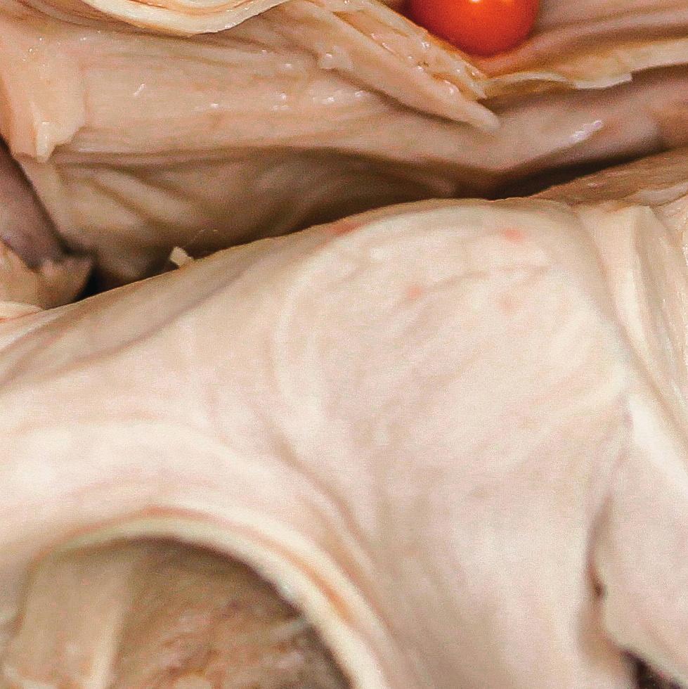
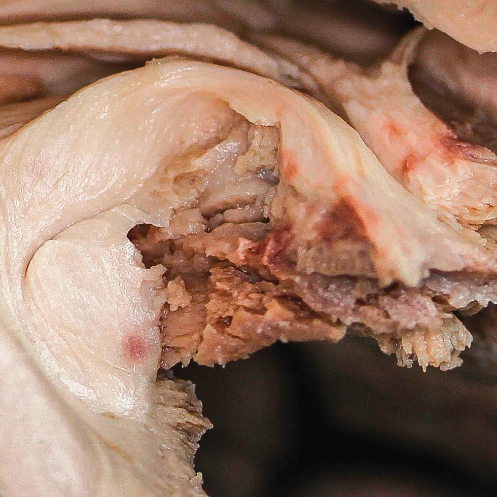
3 The white matter of the cerebrum underlies the outer cortex of gray matter, and is composed of densely packed axons that are organized in fascicles or fiber tracts. These tracts have a complex three-dimensional (3D) configuration within the hemispheres, the brainstem and the spinal cord. A detailed knowledge of the architectural anatomy of the white matter tracts is paramount, for strategically planning for surgical management of parenchymal brain lesions, such as gliomas. Neuroanatomical laboratory training is very valuable to study and understand the anatomy of white matter fibers. In particular, cortex-sparing fiber dissection facilitates knowledge of this complex anatomy. None of the recently developed surgical guides such as neuronavigation, intraoperative magnetic resonance imaging or ultrasonography can provide a similar comprehensive understanding of the 3D fiber pathways organization. In the present course, the participants will learn the technique of cortexsparing fiber dissection in order to acquire the mental imagery of the main white matter tracts. We wanted to give a practical perspective to the course; therefore, in the second day, the participants will directly apply the knowledge acquired to practice surgical approaches in the laboratory. The congress will be held in the prestigious School of Medicine at the University of Cantabria. We look forward to welcoming you in Santander. Juan Martino The Scientific Secretariat


4 COURSE DIRECTOR Professor Juan Martino Mail: SCIENTIFIC COMMITTEE Dr. Emmanuel Mandonnet Hospital Lariboisiere. Paris. France CO-DIRECTORS Professor Juan A. García-Porrero Department of Anatomy and Cellular Biology. Cantabria University. Santander. Spain Dr. Alejandro Fernández-Coello Hospital Universitari de Bellvitge. Barcelona. Spain Professor Rubén Martin-Láez Dr. Pablo González-López Hospital General de Alicante. Alicante. Spain HONORED GUEST Professor Hugues Duffau Centre Hospitalier Universitaire de Montpellier. Montpellier. France Dr. David Mato


5 Dr. Cristian de Quintana Schmidt Hospital de la Santa Creu i Sant Pau. Barcelona. Spain Dr. Carlos Santos Dr. Carlos Velasquez Dra. Patricia López Dr. Guillermo García-Catalán. Monserrat Fernández-Calderón Department of Anatomy and Cellular Biology. Cantabria University. Santander. Spain Dr. Jesús Esteban
6
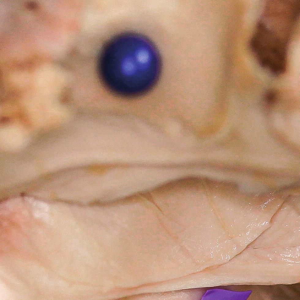
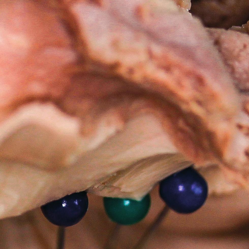
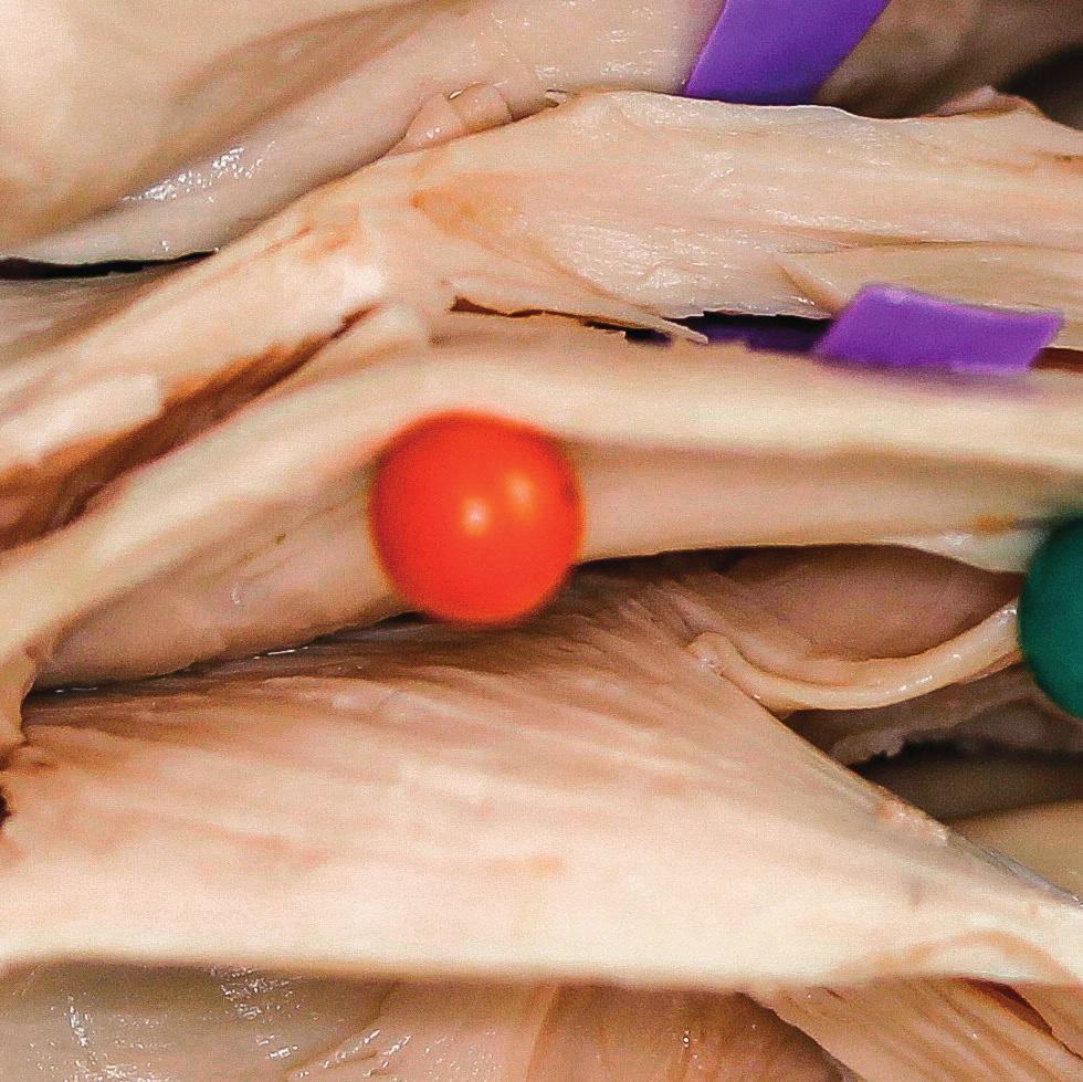
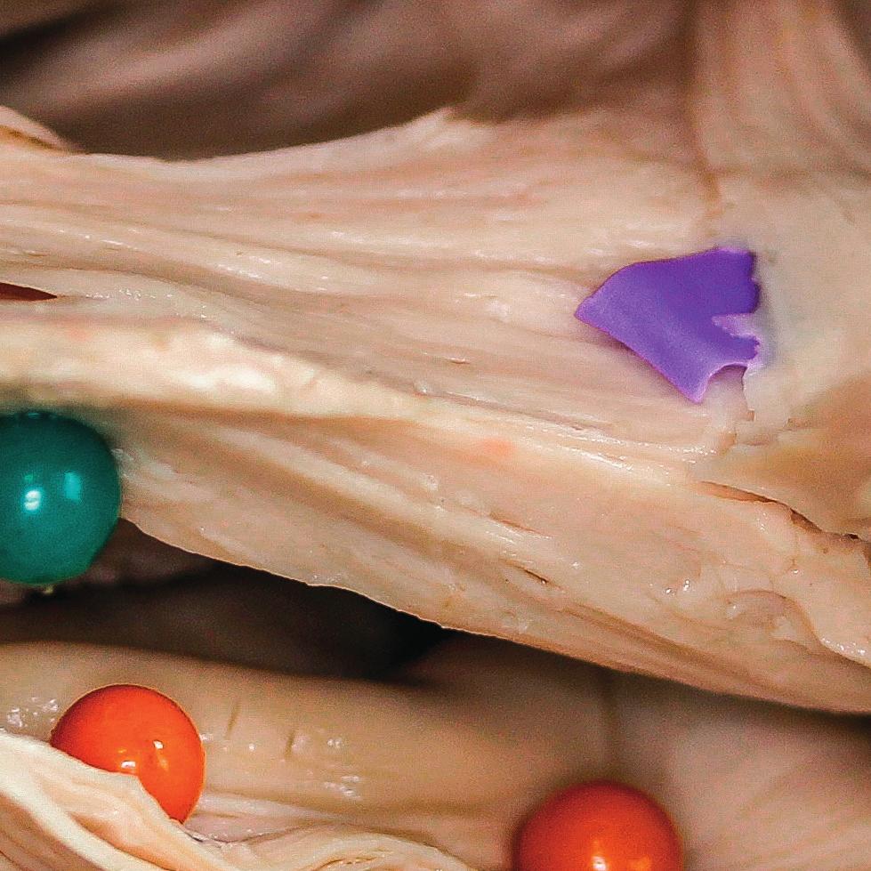
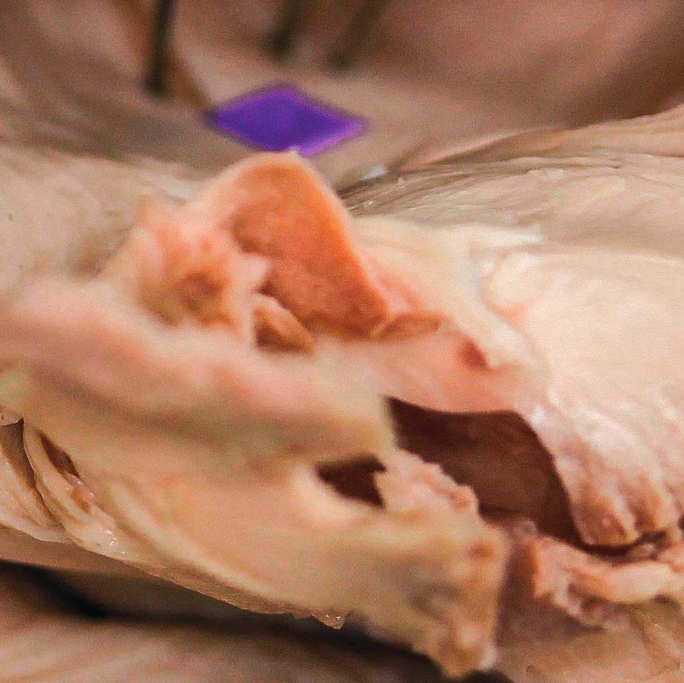
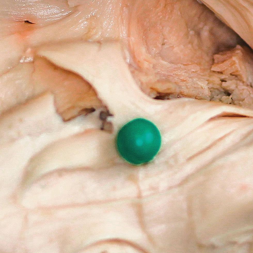
7 Maximal number of participants per course, with full hands-on registration: 8 Maximal number of participants per course, with limited registration: 20 Course dates: 26 th and 27 th October 2018 Course equipment and facilities: Anatomy laboratory at the University of Cantabria. Two cerebral hemispheres for each participant. The specimens were previously selected to ensure the quality for dissection. The specimen s vessels were injected with red and blue colorants for greater similarity with a real brain. Microscope Kinevo 900 Carl Zeiss: one for the course. Microscopes (techno-scopes) Carl Zeiss: one for each participant. Ultrasonic aspirators CUSA (Integra): one for each participant. 3D television (75 inches, Full HD): one for the course. Neuronavigation system: one for the course. Video camera: one for the course. 3D Glasses: one for each participant. Instruments for dissection for each participant. Course venue: Anatomy Laboratory. Department of Anatomy and Cellular Biology. School of Medicine. Cantabria University. Av. Herrera Oria, s/n Santander (Cantabria). Spain. Registration fees: Full hands-on registration: VAT. Includes lectures attendance, lunch and refreshments breaks, and course dinner on Friday. It also includes the hands-on part: dissection of cerebral hemispheres and simulation of surgical approaches. Limited registration: VAT. Includes lectures attendance, lunch and refreshments breaks, and course dinner on Friday. However, it does NOT include the hands-on part: dissection of cerebral hemispheres and simulation of surgical approaches. The participants with limited registration will follow the dissections and surgical approaches performed by the professors. Technical Secretariat: AFORO CONGRESOS Pasaje de Peña 2, 3º C. Edificio Simeón Santander. Spain Phone: zulema@aforocongresos.com Target Audience and Objectives: This activity was designed for Neurosurgeons, Neurologists, Neuroradiologists, Residents/Fellows in these specialties, and Neuro-nurses. After the conclusion of this activity, participants will be able to: Identify the anatomy of the white matter fiber tracts. Comprehensive understanding of the 3D anatomical relationships between the white matter connections. Evaluate surgical approaches to challenging areas: the dominant insular lobe and the supplementary motor area. Discuss surgical cases and analyze different treatment options of tumors located within eloquent areas.
8 PROGRAM. Friday, 26 th of October :00 Registration. 08:10 Opening. Dr. Juan A. García-Porrero, Dr. Juan Martino 08:15 How to prepare the brains for fiber dissection. Dr. David Mato 08:30 3D LECTURE: anatomy of the dorsal associative tracts of the brain: superior longitudinal fasciculus, arcuate fasciculus, middle longitudinal fasciculus. Dr. Juan Martino 09:00 Functional roles of the dorsal associative tracts of the brain. Dr. Alejandro Fernández-Coello 09:15 Hands-on session: fiber dissection of the dorsal associative tracts. Each participant will have one hemisphere to dissect. The participants will learn how to remove the arachnoid membranes and the cortex without damaging the underling white matter. The participants will dissect the dorsal associative tracts: the subcomponents of the superior longitudinal fasciculus, arcuate fasciculus, and middle longitudinal fasciculus. Professor Hugues Duffau, Dr. Juan Martino, Dr. Alejandro Fernández-Coello, Dr. Emmanuel Mandonnet, Dr. Pablo González-López, Dr. David Mato, Dr. Carlos Velasquez, Dr. Carlos Santos, Dr. Guillermo García-Catalán, Dr. Jesús Esteban, Dra. Patricia López, Monserrat Fernández-Calderón 13:00 Lunch.
9 14:00 3D LECTURE: anatomy of the ventral associative tracts and the fascicles related to the insula: inferior longitudinal fasciculus, inferior fronto-occipital fasciculus and uncinate fasciculus. Dr. Juan Martino 14:30 Functional roles of the ventral associative tracts and the fascicles related to the insula. Dr. Alejandro Fernández-Coello 14:45 Intraoperative electrical stimulation mapping of associative fiber pathways. Professor Hugues Duffau 15:15 3D LECTURE: limbic and paralimbic tumors. Anatomy and related surgical approaches. Dr. Pablo González-López 15:45 Hands-on session: fiber dissection of the ventral associative tracts and the fascicles related to the insula. Each participant will have one hemisphere to dissect. The participants will dissect the ventral associative tracts and the tracts related to the insula region: inferior longitudinal fasciculus, inferior fronto-occipital fasciculus and uncinate fasciculus. Professor Hugues Duffau, Dr. Juan Martino, Dr. Alejandro Fernández-Coello, Dr. Emmanuel Mandonnet, Dr. Pablo González-López, Dr. David Mato, Dr. Carlos Velasquez, Dr. Carlos Santos, Dr. Guillermo García-Catalán, Dr. Jesús Esteban, Dra. Patricia López, Monserrat Fernández-Calderón 21:00 Course dinner.
10 PROGRAM. Saturday, 27 th of October :00 Insula glioma surgery: the transopercular approach. Professor Hugues Duffau 08:30 How to simulate a surgical approach to insular gliomas in the laboratory. Dr. Emmanuel Mandonnet 09:00 Presentation of the insular surgical case. Dr. Juan Martino 09:15 Hands-on session: transopercular approach to the insula. Professor Hugues Duffau will perform a step by step transopercular approach to the insula in the specimen. Simultaneously, each participant will perform the approach in the specimens. The participants will use a real magnetic resonance image (MRI) of a frontotemporo-insular glioma to guide the resection. We will have a unique opportunity to ask Professor Duffau many questions about the challenges of this approach: how to preserve the deep functional connections (inferior fronto-occipital fasciculus, uncinate fasciculus, pyramidal pathway, etc.), and vascular structures (lenticulostriate arteries). Professor Hugues Duffau, Dr. Emmanuel Mandonnet, Dr. Juan Martino, Dr. Alejandro Fernández-Coello, Dr. Pablo González-López, Dr. David Mato, Dr. Carlos Velasquez, Dr. Carlos Santos, Dr. Guillermo García-Catalán, Dr. Jesús Esteban, Dra. Patricia López, Monserrat Fernández-Calderón 13:00 Lunch.
11 14:00 Diffusion tensor imaging tractography reconstruction of the white matter tracts. Dr. Cristian de Quintana Schmidt 14:20 Hands-on session: approach to the supplementary motor area. Professor Hugues Duffau will perform a step by step frontal approach to the supplementary motor area. Simultaneously, each participant will perform the approach in the specimens. The participant will use a real MRI of a glioma infiltrating the supplementary motor area to guide the resection. In this approach, the deep functional connections are the superior longitudinal fasciculus, subcallosal fasciculus, frontal aslant tract, and pyramidal pathway. Professor Hugues Duffau, Dr. Emmanuel Mandonnet, Dr. Juan Martino, Dr. Alejandro Fernández-Coello, Dr. Pablo González-López, Dr. David Mato, Dr. Carlos Velasquez, Dr. Carlos Santos, Dr. Guillermo García-Catalán, Dr. Jesús Esteban, Dra. Patricia López, Monserrat Fernández-Calderón 15:45 Hands-on session: fiber dissection of the central core and the medial surface of the hemisphere. Each participant will have one hemisphere to dissect. The participants will dissect the central core (basal ganglia and internal capsule) and the tracts related to the medial surface of the hemisphere (cingulum, pyramidal pathway, and subcallosal fasciculus). Professor Hugues Duffau, Dr. Emmanuel Mandonnet, Dr. Juan Martino, Dr. Alejandro Fernández-Coello, Dr. Pablo González-López, Dr. David Mato, Dr. Carlos Velasquez, Dr. Carlos Santos, Dr. Guillermo García-Catalán, Dr. Jesús Esteban, Dra. Patricia López, Monserrat Fernández-Calderón 16:45 Closing remarks.


12 SECOND ANNUAL 3D ADVANCED FIBER DISSECTION COURSE: ACQUIRING THE MENTAL IMAGERY NECESSARY TO OPERATE THE BRAIN Santander, 26 th and 27 th October MK039 V.3 06/18 DISEÑADO Y PRODUCIDO POR MBA SURGICAL EMPOWERMENT Technical Secretariat: AFORO CONGRESOS Pasaje de Peña 2, 3º C. Edificio Simeón Santander. Spain Phone: zulema@aforocongresos.com Sponsoring companies:
APPLICATION OF PHOTOGRAMMETRY TO BRAIN ANATOMY
 http://medifitbiologicals.com/central-nervous-system-cns/ 25/06/2017 PSBB17 ISPRS International Workshop APPLICATION OF PHOTOGRAMMETRY TO BRAIN ANATOMY E. Nocerino, F. Menna, F. Remondino, S. Sarubbo,
P. Hitchcock, Ph.D. Department of Cell and Developmental Biology Kellogg Eye Center. Wednesday, 16 March 2009, 1:00p.m. 2:00p.m.
 Normal CNS, Special Senses, Head and Neck TOPIC: CEREBRAL HEMISPHERES FACULTY: LECTURE: READING: P. Hitchcock, Ph.D. Department of Cell and Developmental Biology Kellogg Eye Center Wednesday, 16 March
Leah Militello, class of 2018
 Leah Militello, class of 2018 Objectives 1. Describe the general organization of cerebral hemispheres. 2. Describe the locations and features of the different functional areas of cortex. 3. Understand
Cerebral Cortex 1. Sarah Heilbronner
 Cerebral Cortex 1 Sarah Heilbronner heilb028@umn.edu Want to meet? Coffee hour 10-11am Tuesday 11/27 Surdyk s Overview and organization of the cerebral cortex What is the cerebral cortex? Where is each
Fig.1: A, Sagittal 110x110 mm subimage close to the midline, passing through the cingulum. Note that the fibers of the corpus callosum run at a
 Fig.1 E Fig.1:, Sagittal 110x110 mm subimage close to the midline, passing through the cingulum. Note that the fibers of the corpus callosum run at a slight angle are through the plane (blue dots with
SURGICAL MANAGEMENT OF BRAIN TUMORS
 SURGICAL MANAGEMENT OF BRAIN TUMORS LIGIA TATARANU, MD, Ph D NEUROSURGICAL CLINIC, BAGDASAR ARSENI CLINICAL HOSPITAL BUCHAREST, ROMANIA SURGICAL INDICATIONS CONFIRMING HISTOLOGIC DIAGNOSIS REDUCING TUMOR
Telencephalon (Cerebral Hemisphere)
") Telencephalon (Cerebral Hemisphere) OUTLINE The Cortex - Lobes, Sulci & Gyri - Functional Subdivisions - Limbic Lobe & Limbic System The Subcortex - Basal Ganglia - White Matter (Internal Capsule) - Relations
Telencephalon (Cerebral Hemisphere) OUTLINE The Cortex - Lobes, Sulci & Gyri - Functional Subdivisions - Limbic Lobe & Limbic System The Subcortex - Basal Ganglia - White Matter (Internal Capsule) - Relations
10/3/2016. T1 Anatomical structures are clearly identified, white matter (which has a high fat content) appears bright.
 appears bright.") H2O -2 atoms of Hydrogen, 1 of Oxygen Hydrogen just has one single proton and orbited by one single electron Proton has a magnetic moment similar to the earths magnetic pole Also similar to earth in that
H2O -2 atoms of Hydrogen, 1 of Oxygen Hydrogen just has one single proton and orbited by one single electron Proton has a magnetic moment similar to the earths magnetic pole Also similar to earth in that
Surgery Within and Around Critical White Matter Tracts
 Surgery Within and Around Critical White Matter Tracts Kaisorn L. Chaichana, M.D. Assistant Professor of Neurosurgery, Oncology, and Otolaryngology-Head & Neck Surgery Mayo Clinic Florida, Jacksonville,
Pearls and Pitfalls of MR Diffusion in Clinical Neurology
 Pearls and Pitfalls of MR Diffusion in Clinical Neurology Dr. Alberto Bizzi Neuroradiology Unit Fondazione IRCCS Istituto Neurologico Carlo Besta Milan, Italy Email: alberto_bizzi@fastwebnet.it Diffusion
Student Lab #: Date. Lab: Gross Anatomy of Brain Sheep Brain Dissection Organ System: Nervous Subdivision: CNS (Central Nervous System)
") Lab: Gross Anatomy of Brain Sheep Brain Dissection Organ System: Nervous Subdivision: CNS (Central Nervous System) Student Lab #: Date 1 Objectives: 1. Learn the main components making up a motor neuron.
Lab: Gross Anatomy of Brain Sheep Brain Dissection Organ System: Nervous Subdivision: CNS (Central Nervous System) Student Lab #: Date 1 Objectives: 1. Learn the main components making up a motor neuron.
Visualization strategies for major white matter tracts identified by diffusion tensor imaging for intraoperative use
 International Congress Series 1281 (2005) 793 797 www.ics-elsevier.com Visualization strategies for major white matter tracts identified by diffusion tensor imaging for intraoperative use Ch. Nimsky a,b,
CEREBRUM & CEREBRAL CORTEX
 CEREBRUM & CEREBRAL CORTEX Seonghan Kim Dept. of Anatomy Inje University, College of Medicine THE BRAIN ANATOMICAL REGIONS A. Cerebrum B. Diencephalon Thalamus Hypothalamus C. Brain Stem Midbrain Pons
Homework Week 2. PreLab 2 HW #2 Synapses (Page 1 in the HW Section)
") Homework Week 2 Due in Lab PreLab 2 HW #2 Synapses (Page 1 in the HW Section) Reminders No class next Monday Quiz 1 is @ 5:30pm on Tuesday, 1/22/13 Study guide posted under Study Aids section of website
Homework Week 2 Due in Lab PreLab 2 HW #2 Synapses (Page 1 in the HW Section) Reminders No class next Monday Quiz 1 is @ 5:30pm on Tuesday, 1/22/13 Study guide posted under Study Aids section of website
November/December 2008 The Brain Unveiled A new imaging method offers a spectacular view of neural structures. By Emily Singer
 November/December 2008 The Brain Unveiled A new imaging method offers a spectacular view of neural structures. By Emily Singer A new imaging method that offers an unprecedented view of complex neural structures
Seamless pre-surgical fmri and DTI mapping
 Seamless pre-surgical fmri and DTI mapping Newest release Achieva 3.0T X-series and Eloquence enable efficient, real-time fmri for brain activity mapping in clinical practice at Nebraska Medical Center
NED-MNAZI MMOJA INSTITUE BRAIN & SPINE CENTER KITENDO CHA UPASUAJI WA MGONGO NA VICHWA
 NED-MNAZI MMOJA INSTITUE BRAIN & SPINE CENTER KITENDO CHA UPASUAJI WA MGONGO NA VICHWA 3DN NED Foundation Neuroanatomy Training Project under the auspices Neurosurgery COSECSA Program Program Objectives
Biological Bases of Behavior. 3: Structure of the Nervous System
 Biological Bases of Behavior 3: Structure of the Nervous System Neuroanatomy Terms The neuraxis is an imaginary line drawn through the spinal cord up to the front of the brain Anatomical directions are
b. The groove between the two crests is called 2. The neural folds move toward each other & the fuse to create a
 Chapter 13: Brain and Cranial Nerves I. Development of the CNS A. The CNS begins as a flat plate called the B. The process proceeds as: 1. The lateral sides of the become elevated as waves called a. The
CEREBRUM. Dr. Jamila EL Medany
 CEREBRUM Dr. Jamila EL Medany Objectives At the end of the lecture, the student should be able to: List the parts of the cerebral hemisphere (cortex, medulla, basal nuclei, lateral ventricle). Describe
Organization of The Nervous System PROF. MOUSAED ALFAYEZ & DR. SANAA ALSHAARAWY
 Organization of The Nervous System PROF. MOUSAED ALFAYEZ & DR. SANAA ALSHAARAWY Objectives At the end of the lecture, the students should be able to: List the parts of the nervous system. List the function
3rd Annual EANS Young Neurosurgeons CME Meeting 2013
 Programme 3rd Annual EANS Young Neurosurgeons CME Meeting 2013 Thursday 4th to Sunday 7th April 2013 The Royal College of Surgeons of England, London, UK Course Chairmen Mr Andreas Demetriades Consultant
Dissection of the Sheep Brain
 Dissection of the Sheep Brain Laboratory Objectives After completing this lab, you should be able to: 1. Identify the main structures in the sheep brain and to compare them with those of the human brain.
The Central Nervous System I. Chapter 12
 The Central Nervous System I Chapter 12 The Central Nervous System The Brain and Spinal Cord Contained within the Axial Skeleton Brain Regions and Organization Medical Scheme (4 regions) 1. Cerebral Hemispheres
CEREBRUM Dr. Jamila Elmedany Dr. Essam Eldin Salama
 CEREBRUM Dr. Jamila Elmedany Dr. Essam Eldin Salama Objectives At the end of the lecture, the student should be able to: List the parts of the cerebral hemisphere (cortex, medulla, basal nuclei, lateral
The human brain weighs roughly 1.5 kg and has an average volume of 1130 cm 3. A sheep s brain weighs in however at kg.
 Sheep Brain Dissection Objectives: 1. List and describe the principal structures of the sheep brain 2. Identify important parts of the sheep brain in a preserved specimen Materials: Dissection tools, lab
Lecture 4 The BRAINSTEM Medulla Oblongata
 Lecture 4 The BRAINSTEM Medulla Oblongata Introduction to brainstem 1- Medulla oblongata 2- Pons 3- Midbrain - - - occupies the posterior cranial fossa of the skull. connects the narrow spinal cord
Lecture - Chapter 13: Central Nervous System
 Lecture - Chapter 13: Central Nervous System 1. Describe the following structures of the brain, what is the general function of each: a. Cerebrum b. Diencephalon c. Brain Stem d. Cerebellum 2. What structures
BIOL Dissection of the Sheep and Human Brain
 BIOL 2401 Dissection of the Sheep and Human Brain Laboratory Objectives After completing this lab, you should be able to: Identify the main structures in the sheep brain and to compare them with those
2 ND Baltic Sea International Hands-on Course on FULL HD Endoscopic Neurosurgery May, 2010
 2 ND Baltic Sea 2010 International Hands-on Course on FULL HD Endoscopic Neurosurgery 27 29 May, 2010 Departments of Neurosurgery and Anatomy Greifswald, Germany Invitation Dear colleagues, It is a great
The heterogeneous space distribution of Superior Longitudinal Fascicle in human telencephalon. Neuronal imaging and forensic implications
 Rom J Leg Med [0] 69-78 [0] DOI: 0./rjlm.0.69 0 Romanian Society of Legal Medicine The heterogeneous space distribution of Superior Longitudinal Fascicle in human telencephalon. Neuronal imaging and forensic
The Nervous System: Sensory and Motor Tracts of the Spinal Cord
 15 The Nervous System: Sensory and Motor Tracts of the Spinal Cord PowerPoint Lecture Presentations prepared by Steven Bassett Southeast Community College Lincoln, Nebraska Introduction Millions of sensory
Gross Organization I The Brain. Reading: BCP Chapter 7
 Gross Organization I The Brain Reading: BCP Chapter 7 Layout of the Nervous System Central Nervous System (CNS) Located inside of bone Includes the brain (in the skull) and the spinal cord (in the backbone)
ANATOMY & PHYSIOLOGY DISSECTION OF THE SHEEP BRAIN LAB GROUP:
 ANATOMY & PHYSIOLOGY DISSECTION OF THE SHEEP BRAIN LAB GROUP: Introduction The purpose of the sheep brain dissection is to familiarize you with the three dimensional structure of the brain and teach you
Tracking the language pathways in edema patients: Preliminary results.
 Tracking the language pathways in edema patients: Preliminary results. Sarah M. E. Gierhan 1,2, Peter Rhone 3,4, Alfred Anwander 1, Isabel Jost 3, Clara Frydrychowicz 3, Karl-Titus Hoffmann 4, Jürgen Meixensberger
Cerebellum 1/20/2016. Outcomes you need to be able to demonstrate. MHD Neuroanatomy Module
 This power point is made available as an educational resource or study aid for your use only. This presentation may not be duplicated for others and should not be redistributed or posted anywhere on the
Diffusion Tensor Imaging in Psychiatry
 2003 KHBM DTI in Psychiatry Diffusion Tensor Imaging in Psychiatry KHBM 2003. 11. 21. 서울대학교 의과대학 정신과학교실 권준수 Neuropsychiatric conditions DTI has been studied in Alzheimer s disease Schizophrenia Alcoholism
Histology of the CNS
 Histology of the CNS Lecture Objectives Describe the histology of the cerebral cortex layers. Describe the histological features of the cerebellum; layers and cells of cerebellar cortex. Describe the elements
Organization of The Nervous System PROF. SAEED ABUEL MAKAREM
 Organization of The Nervous System PROF. SAEED ABUEL MAKAREM Objectives By the end of the lecture, you should be able to: List the parts of the nervous system. List the function of the nervous system.
The Nervous System PART B
 7 The Nervous System PART B PowerPoint Lecture Slide Presentation by Jerry L. Cook, Sam Houston University ESSENTIALS OF HUMAN ANATOMY & PHYSIOLOGY EIGHTH EDITION ELAINE N. MARIEB The Reflex Arc Reflex
June 4 th - 6 th 2014 Milazzo, Italy. 3 rd Workshop on Synaptic Plasticity: from bench to bedside
 June 4 th - 6 th 2014 Milazzo, Italy 3 rd Workshop on Synaptic Plasticity: from bench to bedside WELCOME Dear Colleagues and Friends, We welcome you to the 3rd Workshop on Synaptic Plasticity: from bench
1. The basic anatomy of the Central Nervous System (CNS)
") Psyc 311A, fall 2008 Conference week 1 Sept 9 th to 11 th TA: Jürgen Germann; e-mail: jurgen.germann@mcgill.ca Overview: 1. The basic anatomy of the Central Nervous System (CNS) 2. Cells of the CNS 3.
Psyc 311A, fall 2008 Conference week 1 Sept 9 th to 11 th TA: Jürgen Germann; e-mail: jurgen.germann@mcgill.ca Overview: 1. The basic anatomy of the Central Nervous System (CNS) 2. Cells of the CNS 3.
Essentials of Human Anatomy & Physiology. Seventh Edition. The Nervous System. Copyright 2003 Pearson Education, Inc. publishing as Benjamin Cummings
 Essentials of Human Anatomy & Physiology Seventh Edition The Nervous System Copyright 2003 Pearson Education, Inc. publishing as Benjamin Cummings Functions of the Nervous System 1. Sensory input gathering
The 6 Pillar Approach for Comprehensive Management of Subcortical Lesions
 The 6 Pillar Approach for Comprehensive Management of Subcortical Lesions March 6-7, 2015 Sheraton Atlanta Hotel 165 Courtland Street NE Atlanta, Georgia March 6, 2015 from 7:00 am 5:15 pm March 7, 2015
Cerebrum-Cerebral Hemispheres. Cuneyt Mirzanli Istanbul Gelisim University
 Cerebrum-Cerebral Hemispheres Cuneyt Mirzanli Istanbul Gelisim University The largest part of the brain. Ovoid shape. Two incompletely separated cerebral hemispheres. The outer surface of the cerebral
Cerebral Bypass and Advanced Neuroendovascular Course 2018
 Mayo Clinic Cerebral Bypass and Advanced Neuroendovascular Course 2018 Co-directed by: Giuseppe Lanzino MD and Leonardo Rangel-Castilla MD Honored Guest: Luca Regli MD, Zurich, Switzerland Thursday, November
Diffusion Tensor Imaging in brain tumours
 Diffusion Tensor Imaging in brain tumours @MarionSmits, MD PhD Associate Professor of Neuroradiology Dept. of Radiology, Erasmus MC, Rotterdam (NL) Honorary Consultant and Reader UCLH National Hospital
Medical Neuroscience Tutorial Notes
 Medical Neuroscience Tutorial Notes Finding the Central Sulcus MAP TO NEUROSCIENCE CORE CONCEPTS 1 NCC1. The brain is the body's most complex organ. LEARNING OBJECTIVES After study of the assigned learning
Medical Neuroscience Tutorial Notes
 Medical Neuroscience Tutorial Notes Lateral Surface of the Brain MAP TO NEUROSCIENCE CORE CONCEPTS 1 NCC1. The brain is the body's most complex organ. LEARNING OBJECTIVES After study of the assigned learning
Gross Morphology of the Brain
 Gross Morphology of the Brain Done by : Marah Marahleh & Razan Krishan *slides in bold Principal Parts of the Brain Cerebrum : largest part of the brain Diencephalon Thalamus & hypothalamus Cerebellum
LEC 1B ANATOMY OF THE NERVOUS SYSTEM. Cogs 17 * UCSD
 LEC 1B ANATOMY OF THE NERVOUS SYSTEM Cogs 17 * UCSD Cerebral Cortex A 6-layer sheet of cells, unfolded = < 1 m square X 3 mm thick Cortex 6 layers of cells Nissl Stain for Cell Bodies Info projected to
UPDATE CLINICAL NEUROLOGY V SPECIAL MINISYMPOSIA ON HEAD TRAUMA AND THE SURGICAL AND MEDICAL TREATMENTS FOR EPILEPSY
 PENNSYLVANIA NEUROLOGICAL SOCIETY UPDATE CLINICAL NEUROLOGY V SPECIAL MINISYMPOSIA ON HEAD TRAUMA AND THE SURGICAL AND MEDICAL TREATMENTS FOR EPILEPSY Sponsored jointly with Stroke Update 2011, UPMC /
DEVELOPMENT OF BRAIN
 Ahmed Fathalla OBJECTIVES At the end of the lecture, students should: List the components of brain stem. Describe the site of brain stem. Describe the relations between components of brain stem & their
BALTIC SEA International Hands-on Course on FULL HD Endoscopic Neurosurgery Department of Neurosurgery Greifswald, Germany
 BALTIC SEA 2009 International Hands-on Course on FULL HD Endoscopic Neurosurgery 01. 03.10.2009 Department of Neurosurgery Greifswald, Germany INVITATION Dear colleagues, It is a great pleasure to invite
The 2018 Northwestern Medicine Neuro-Oncology CME Symposium
 The 2018 Northwestern Medicine Neuro-Oncology CME Symposium Friday, April 20 Northwestern Memorial Hospital 251 East Huron Street, Feinberg Pavilion Third Floor, Pritzker Auditorium Chicago, IL 7:15 am
Skull Base Course Surgical Approaches to Cerebral Aneurysms
 Skull Base Course Surgical Approaches to Cerebral Aneurysms September 2-4, 2015 Department of Neurosurgery SkillsLab, Erasmus MC, Rotterdam, NL Hands-on Course pterional, pretemporal, transcavernous and
Dendrites Receive impulse from the axon of other neurons through synaptic connection. Conduct impulse towards the cell body Axon
 Dendrites Receive impulse from the axon of other neurons through synaptic connection. Conduct impulse towards the cell body Axon Page 22 of 237 Conduct impulses away from cell body Impulses arise from
Sincerely, Ms. Paoloni and Mrs. Whitney
 Dear Students, Welcome to AP Psychology! We will begin our course of study focusing on the nervous system with a particular emphasis on how the brain and neurotransmitters influence our behaviors. In preparation
Medical Neuroscience Tutorial Notes
 Medical Neuroscience Tutorial Notes Blood Supply to the Brain MAP TO NEUROSCIENCE CORE CONCEPTS 1 NCC1. The brain is the body's most complex organ. LEARNING OBJECTIVES After study of the assigned learning
Cerebellum. Steven McLoon Department of Neuroscience University of Minnesota
 Cerebellum Steven McLoon Department of Neuroscience University of Minnesota 1 Anatomy of the Cerebellum The cerebellum has approximately half of all the neurons in the central nervous system. The cerebellum
Anatomy and Physiology (Bio 220) The Brain Chapter 14 and select portions of Chapter 16
 The Brain Chapter 14 and select portions of Chapter 16") Anatomy and Physiology (Bio 220) The Brain Chapter 14 and select portions of Chapter 16 I. Introduction A. Appearance 1. physical 2. weight 3. relative weight B. Major parts of the brain 1. cerebrum 2.
Anatomy and Physiology (Bio 220) The Brain Chapter 14 and select portions of Chapter 16 I. Introduction A. Appearance 1. physical 2. weight 3. relative weight B. Major parts of the brain 1. cerebrum 2.
9th INTERNATIONAL TRAINING COURSE.
 9th INTERNATIONAL TRAINING COURSE. MINIMALLY INVASIVE TRANSANAL SURGERY (TEO and TaTME). THE APPLICATION OF INDOCYANINE GREEN 12-13-14 December 2018 Colorectal Unit Parc Tauli University Hospital Sabadell
DISSECTION OF THE SHEEP'S BRAIN
 Sheep Brain Dissection Guide Page 1 DISSECTION OF THE SHEEP'S BRAIN Introduction The purpose of the sheep brain dissection is to familiarize you with the threedimensional structure of the brain and teach
Introduction to the Central Nervous System: Internal Structure
 Introduction to the Central Nervous System: Internal Structure Objective To understand, in general terms, the internal organization of the brain and spinal cord. To understand the 3-dimensional organization
Announcement. Danny to schedule a time if you are interested.
 Announcement If you need more experiments to participate in, contact Danny Sanchez (dsanchez@ucsd.edu) make sure to tell him that you are from LIGN171, so he will let me know about your credit (1 point).
Composed of gray matter and arranged in raised ridges (gyri), grooves (sulci), depressions (fissures).
, grooves (sulci), depressions (fissures).") PSYC1020 Neuro and Pysc Notes Structure Description Major Functions Brainstem Stemlike portion of the brain, continuous with diencephalon above and spinal cord below. Composed of midbrain, pons, medulla
PSYC1020 Neuro and Pysc Notes Structure Description Major Functions Brainstem Stemlike portion of the brain, continuous with diencephalon above and spinal cord below. Composed of midbrain, pons, medulla
7 th Baltic Sea International Hands-on Course on FULL HD Endoscopic Neurosurgery. September 23 rd 25 th, 2015
 7 th Baltic Sea 2015 International Hands-on Course on FULL HD Endoscopic Neurosurgery September 23 rd 25 th, 2015 Departments of Neurosurgery and Anatomy Greifswald, Germany Invitation Dear colleagues,
Auditory and Vestibular Systems
 Auditory and Vestibular Systems Objective To learn the functional organization of the auditory and vestibular systems To understand how one can use changes in auditory function following injury to localize
Anatomy & Physiology Central Nervous System Worksheet
 1. What are the two parts of the CNS? 2. What are the four functions of the CNS Anatomy & Physiology Central Nervous System Worksheet 3. What are the four functions of the meninges? (p430) 4. Starting
The Nervous System 7PART B. PowerPoint Lecture Slide Presentation by Patty Bostwick-Taylor, Florence-Darlington Technical College
 PowerPoint Lecture Slide Presentation by Patty Bostwick-Taylor, Florence-Darlington Technical College The Nervous System 7PART B What is a reflex? What is a reflex? What is meant by the statement that
BASAL GANGLIA: A "pit stop" that integrates the movement, cognition and emotion.
 BASAL GANGLIA: A "pit stop" that integrates the movement, cognition and emotion. Poster No.: C-0795 Congress: ECR 2011 Type: Educational Exhibit Authors: V. M. González Montaño, T. M. Zamorano Pozo, R.
Overview of the Nervous System (some basic concepts) Steven McLoon Department of Neuroscience University of Minnesota
 Steven McLoon Department of Neuroscience University of Minnesota") Overview of the Nervous System (some basic concepts) Steven McLoon Department of Neuroscience University of Minnesota 1 Coffee Hour Tuesday (Sept 11) 10:00-11:00am Friday (Sept 14) 8:30-9:30am Surdyk s
Overview of the Nervous System (some basic concepts) Steven McLoon Department of Neuroscience University of Minnesota 1 Coffee Hour Tuesday (Sept 11) 10:00-11:00am Friday (Sept 14) 8:30-9:30am Surdyk s
Is DTI Increasing the Connectivity Between the Magnet Suite and the Clinic?
 Current Literature In Clinical Science Is DTI Increasing the Connectivity Between the Magnet Suite and the Clinic? Spatial Patterns of Water Diffusion Along White Matter Tracts in Temporal Lobe Epilepsy.
Sheep Brain Dissection
 Sheep Brain Dissection Mammalian brains have many features in common. Human brains may not be available, so sheep brains often are dissected as an aid to understanding the mammalian brain since he general
For more information about how to cite these materials visit
 Author(s): Peter Hitchcock, PH.D., 2009 License: Unless otherwise noted, this material is made available under the terms of the Creative Commons Attribution Non-commercial Share Alike 3.0 License: http://creativecommons.org/licenses/by-nc-sa/3.0/
CISC 3250 Systems Neuroscience
 CISC 3250 Systems Neuroscience Levels of organization Central Nervous System 1m 10 11 neurons Neural systems and neuroanatomy Systems 10cm Networks 1mm Neurons 100μm 10 8 neurons Professor Daniel Leeds
MRI and CT of the CNS
 MRI and CT of the CNS Dr.Maha ELBeltagy Assistant Professor of Anatomy Faculty of Medicine The University of Jordan 2018 Computed Tomography CT is used for the detection of intracranial lesions. CT relies
8.3 The Central Nervous System. SBI4U Ms. Ho-Lau
 8.3 The Central Nervous System SBI4U Ms. Ho-Lau The Central Nervous System the structural and functional centre for the entire nervous system the site of neural integration and processing The Central
QUANTITATIVE ANALYSIS AND COMPUTER AIDED SIMULATION OF MINIMALLY INVASIVE APPROACHES FOR INTRACRANIAL VASCULAR LESIONS
 QUANTITATIVE ANALYSIS AND COMPUTER AIDED SIMULATION OF MINIMALLY INVASIVE APPROACHES FOR INTRACRANIAL VASCULAR LESIONS Alberto Prats Galino Facultat de Medicina UB 1. Project Summary The main goal of this
10 th Baltic Sea 2018
 10 th Baltic Sea 2018 International Hands-on Course on FULL HD Endoscopic Neurosurgery September 26 th 28 th, 2018 Departments of Neurosurgery and Anatomy Greifswald, Germany Invitation Dear colleagues,
Lecturer. Prof. Dr. Ali K. Al-Shalchy MBChB/ FIBMS/ MRCS/ FRCS 2014
 Lecturer Prof. Dr. Ali K. Al-Shalchy MBChB/ FIBMS/ MRCS/ FRCS 2014 Dorsal root: The dorsal root carries both myelinated and unmyelinated afferent fibers to the spinal cord. Posterior gray column: Long
Insulo-opercular Gliomas: Four Different Natural Progression Patterns and Implications for Surgical Indications
 Neurol Med Chir (Tokyo) 50, 286 290, 2010 Insulo-opercular Gliomas: Four Different Natural Progression Patterns and Implications for Surgical Indications Ryuta SAITO, Toshihiro KUMABE, Masayuki KANAMORI,
BASAL GANGLIA. Dr JAMILA EL MEDANY
 BASAL GANGLIA Dr JAMILA EL MEDANY OBJECTIVES At the end of the lecture, the student should be able to: Define basal ganglia and enumerate its components. Enumerate parts of Corpus Striatum and their important
Chapter 14, Part 2! Chapter 14 Part 2 Brain/Cranial Nerves! The Cerebrum and Cranial Nerves! pp !
 Chapter 14, Part 2! The Cerebrum and Cranial pp. 482 505! SECTION 14-9! The cerebrum, the largest region of the brain, contains motor, sensory, and association areas! 2! White Matter of the Cerebrum! 1.
Slide 1. Slide 2. Slide 3. Tomography vs Topography. Computed Tomography (CT): A simplified Topographical review of the Brain. Learning Objective
: A simplified Topographical review of the Brain. Learning Objective") Slide 1 Computed Tomography (CT): A simplified Topographical review of the Brain Jon Wheiler, ACNP-BC Slide 2 Tomography vs Topography Tomography: A technique for displaying a representation of a cross
Slide 1 Computed Tomography (CT): A simplified Topographical review of the Brain Jon Wheiler, ACNP-BC Slide 2 Tomography vs Topography Tomography: A technique for displaying a representation of a cross
Chapter 12b. Overview
 Chapter 12b Spinal Cord Overview Spinal cord gross anatomy Spinal meninges Sectional anatomy Sensory pathways Motor pathways Spinal cord pathologies 1 The Adult Spinal Cord About 18 inches (45 cm) long
All questions below pertain to mandatory material: all slides, and mandatory homework (if any).
.") ECOL 182 Spring 2008 Dr. Ferriere s lectures Lecture 6: Nervous system and brain Quiz Book reference: LIFE-The Science of Biology, 8 th Edition. http://bcs.whfreeman.com/thelifewire8e/ All questions below
ECOL 182 Spring 2008 Dr. Ferriere s lectures Lecture 6: Nervous system and brain Quiz Book reference: LIFE-The Science of Biology, 8 th Edition. http://bcs.whfreeman.com/thelifewire8e/ All questions below
STRUCTURAL ORGANIZATION OF THE NERVOUS SYSTEM
 STRUCTURAL ORGANIZATION OF THE NERVOUS SYSTEM STRUCTURAL ORGANIZATION OF THE BRAIN The central nervous system (CNS), consisting of the brain and spinal cord, receives input from sensory neurons and directs
PARIETAL LOBE. Vasilios A. Zerris MD, MPH, MSc, FAANS
 PARIETAL LOBE Vasilios A. Zerris MD, MPH, MSc, FAANS Diplomate of the American Board of Neurological Surgery Fellow of the American Association of Neurological Surgeons Professor of Neurosurgery, European
Chapter 14, Part 2! The Cerebrum and Cranial Nerves! pp !
 Chapter 14, Part 2! The Cerebrum and Cranial pp. 482 505! SECTION 14-9! The cerebrum, the largest region of the brain, contains motor, sensory, and association areas! 2! 1! ! Chapter 14 Part 2 Brain/Cranial
Principles of Anatomy and Physiology
 Principles of Anatomy and Physiology 14 th Edition CHAPTER 14 The Brain and Cranial Nerves Introduction The purpose of the chapter is to: 1. Understand how the brain is organized, protected, and supplied
An Atlas of Brainstem Connectomes from HCP Data
 An Atlas of Brainstem Connectomes from HCP Data Presented During: Poster Session Tuesday, June 27, 2017: 12:45 PM - 02:45 PM Stand-By Time Tuesday, June 27, 2017: 12:45 PM - 2:45 PM Submission No: 1653
For more information about how to cite these materials visit
 Author(s): Peter Hitchcock, PH.D., 2009 License: Unless otherwise noted, this material is made available under the terms of the Creative Commons Attribution Non-commercial Share Alike 3.0 License: http://creativecommons.org/licenses/by-nc-sa/3.0/
Department of Human Anatomy GUIDELINES. nuclei. The lateral ventricles. White substance of cerebral hemispheres. course 1
 Department of Human Anatomy GUIDELINES Academic discipline Human Anatomy Module 2 Content module 11 Study subject The olfactory brain. Basal nuclei. The lateral ventricles. White substance of cerebral
2nd Annual Neurosciences Update Conference
 2nd Annual Neurosciences Update Conference presented by May 13, 2011 Tulalip Resort & Spa Marysville, Washington Faculty Kyra J. Becker, MD Professor of & Neurological Surgery University of Washington
Introduction to the Nervous System. Code: HMP 100/ UPC 103/ VNP 100. Course: Medical Physiology. Level 1 MBChB/BDS/BPharm
 Introduction to the Nervous System. Code: HMP 100/ UPC 103/ VNP 100. Course: Medical Physiology Level 1 MBChB/BDS/BPharm Lecture 2. Functional Organisation of the Nervous System Lecture Outline 1.1 Introduction
Nervous system, integration: Overview, and peripheral nervous system:
 Nervous system, integration: Overview, and peripheral nervous system: Some review & misc. parts [Fig. 28.11B, p. 573]: - white matter --> looks white due to the myelinated sheaths, which are quite fatty.
By Dr. Saeed Vohra & Dr. Sanaa Alshaarawy
 By Dr. Saeed Vohra & Dr. Sanaa Alshaarawy 1 By the end of the lecture, students will be able to : Distinguish the internal structure of the components of the brain stem in different levels and the specific
Functional MRI and Diffusion Tensor Imaging
 Functional MRI and Diffusion Tensor Imaging Andrew Steven March 23, 2018 Ochsner Neuroscience Symposium None Disclosure 1 Objectives Review basic principles of BOLD fmri and DTI. Discuss indications and
The visualization of periprostatic nerve fibers using Diffusion Tensor Magnetic Resonance Imaging with tractography
 The visualization of periprostatic nerve fibers using Diffusion Tensor Magnetic Resonance Imaging with tractography Poster No.: C-0009 Congress: ECR 2014 Type: Scientific Exhibit Authors: K. Kitajima 1,