BenchMark Special Stains. Product guide
|
|
|
- Nathaniel Lawrence
- 6 years ago
- Views:
Transcription


1 BenchMark Special Stains Product guide
2 Quick reference table Product name Ordering code Catalog number Tissue thickness Tests per kit Total vials in kit package *Not all vials in the kit are used at one time, e.g., PAS includes three vials of Schiff s reagent. For product freshness, open one vial at a time. Vials run on instrument **All times include 20 minutes for baking (8 minutes as recommened) and depar (12 minutes). If baking offline (i.e. not selected), depar is 12 minutes and not adjustable. Minimum runtime (minutes) including depar** Maximum runtime (minutes) including depar** AFB III Staining Kit µm 75 tests Alcian Blue Staining Kit µm 75 tests Alcian Yellow Staining Kit µm 75 tests 7* Congo Red Staining Kit µm 40 tests Diastase Kit µm 75 tests 1 1+PAS staining kit Elastic Staining Kit µm 75 tests Giemsa Staining Kit µm 75 tests GMS II Staining Kit µm 75 tests 8* Gram Staining Kit µm 75 tests Iron Staining Kit µm 75 tests Jones H&E Staining Kit µm 40 tests Jones Light Green Staining Kit µm 40 tests Mucicarmine Staining Kit µm 75 tests PAS - Alcian Blue µm 75 tests 1 1+PAS staining kit PAS - Light Green µm 75 tests 1 1+PAS staining kit PAS Staining Kit µm 75 tests 5* Reticulum II Staining Kit µm 75 tests Steiner II Staining Kit µm 40 tests Green for Trichrome µm 75 tests 1 1+Trichrome staining kit Trichrome Staining Kit µm 60 tests Product name Ordering code Catalog number Format VENTANA BenchMark Special Stains Deparaffinization Solution (10X) L bottle BenchMark Special Stains Wash Solution (10X) L bottle BenchMark Special Stains Liquid Coverslip L bottle Special Stains Clean Kit Kit with 50 cleaning cycles Special Stains Clean Plus Kit with 50 cleaning cycles SSR Solution (for use with Gram staining kit) L bottle 2
3 General technical notes and post-instrument processing Post-instrument processing Standard post processing for most special stains kits Each lab should validate post-instrument processing steps and timing; however, the following is recommended as a starting point: 1. Rinse in two changes of 95% reagent alcohol (30 seconds each) 2. Dehydrate in three changes absolute reagent alcohol (30 seconds each) 3. Clear in three changes xylene (30 seconds each) 4. Coverslip with a permanent mounting medium As an alternative to manual post-instrument processing, you may use your VENTANA SYMPHONY automated H&E system by selecting the special stains protocol for fully automated post processing through coverslip. Giemsa 1. Remove the slides from the instrument and manually differentiate in two changes of 95% alcohol to the desired microscopic preference 2. Dehydrate, clear and coverslip with permanent mounting media Staining procedure protocol key Alternative incubation - An alternative to the Default Protocol, which can offer an appearance which may satisfy users preferences Default protocol - Staining protocol that is used if the user does not make any changes to the available incubation times or eratures Intermediate incubation - Staining protocol that uses incubation times that fall between the shortest and longest incubation times Longest incubation - Staining protocol that uses the longest incubation times available for that assay Shortest incubation - Staining protocol that uses the shortest available incubation times for that assay 3

4 Alcian Yellow Product information BenchMark Special Stains - Alcian Yellow procedure The Alcian Yellow Staining Kit is used to identify Helicobacter pylori. Alcian Yellow Staining Kit Catalog number: Ordering code: Kit components 1. Alcian Yellow Toluidine Blue contains <1% of the dye 2. Alcian Yellow Stain contains 2.4% alcian yellow, 1.5% acetic acid and 50% ethanol 3. Alcian Yellow Oxidizer contains <1% periodic acid 4. Alcian Yellow Clarifier contains 1.5% sodium metabisulfite and <1% hydrochloric acid 5. Alcian Yellow Sensitizer contains <1% borax and <1% deoxycholic acid Figure 1. Gastric tissue with H. Pylori stained with Alcian Yellow. 1000x. 4

5 AFB III Product information BenchMark Special Stains - AFB III procedure The AFB III Staining Kit is a modification of the Ziehl-Neelsen stain and Fite stain for acid-fast organisms. 1 A carbol fuchsin solution is used to stain acid-fast organisms and components red. Aniline blue counterstain: is applied to provide a contrasting blue background. AFB III Staining Kit Catalog number: Ordering code: Kit components 1. AFB Stain contains proprietary amounts of carbol fuchsin and phenol 2. AFB Decolorizer II contains 18% sulfuric acid and 64% methanol 3. AFB III Blue contains 0.015% aniline blue and 0.25% acetic acid References 1. Sheehan DC, Hrapchak BB. Theory and Practice of Histotechnology. 2nd edition. St. Louis, MO: C.V. Mosby Company; 1980: Figure 1. AFB organisms staining on infected colon, 600x. 5


6 AFB III AFB III Shortest incubation Intermediate incubation Longest incubation Aniline Blue counterstain: 4-12 min 4 min 8 min 12 min Figure 2 - Sample images of staining achieved with different protocol time selections. * Key to staining procedures protocol located on page 3 6

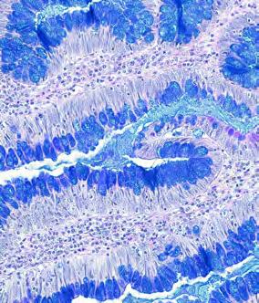
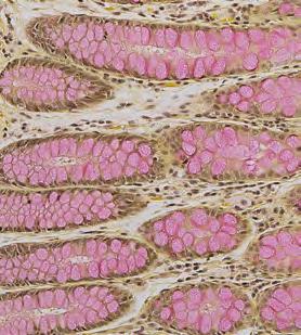
7 Alcian Blue Product information BenchMark Special Stains - Alcian Blue procedure The Alcian Blue Staining Kit uses the differential ph staining properties of alcian blue to demonstrate acid mucopolysaccharides. At ph 2.5, alcian blue stains sulfated mucins, usually epithelial in origin and usually PAS negative, with some staining in connective tissue (subcutaneous tissue in patients with thyroid deficiency, myxedema and in myxomas). It also stains carboxylated sialomucins, found in the mucins of submaxillary glands, small intestine and upper colon, and sulfated and carboxylated acid mucopolysaccharides. Alcian Blue Staining Kit Catalog number: Ordering code: Kit components 1. Alcian Blue contains 1.2% alcian blue in a 3% acetic acid solution 2. Nuclear Fast Red Counterstain contains 1% nuclear fast red and 5% aluminum sulfate Figure 1. Colon stained with Alcian Blue, 200x. 7


8 Alcian Blue Alcian Blue Shortest incubation Default protocol Alternative incubation Longest incubation Alcian Blue: 4-20 min 4 min 8 min 16 min 20 min NFR counterstain: 4-16 min 4 min 4 min 4 min 16 min Figure 2 - Sample images of staining achieved with different protocol time selections. * Key to staining procedures protocol located on page 3 8

9 Congo Red Product information BenchMark Special Stains - Congo Red procedure The Congo Red Staining Kit is a modification of Highman s technique. 1 Congo Red Stain is applied to stain amyloid pink to red with an apple green birefringence under polarized light. A Mayer s hematoxylin solution is used to provide contrasting blue nuclear staining. Congo Red Staining Kit Catalog number: Ordering code: Kit components 1. Congo Red Stain contains 1% Congo red and 70% isopropanol 2. Congo Red Buffer contains 0.5% glycine and 2.0% sodium chloride 3. Congo Red Hematoxylin contains modified Mayer s hematoxylin References 1. Sheehan DC, Hrapchak BB. Theory and Practice of Histotechnology. 2nd edition. St. Louis, MO: C.V. Mosby Company; 1980: Figure 1. Amyloid in lung stained with Congo Red, regular light microscopy, 200x. 9


10 Congo Red Congo Red Regular light microscopy Shortest incubation / Default protocol / Alternative incubation / Longest incubation / Reaction erature: 37-60ºC 37 C 37 C 37 C 60 C Congo Red: min 20 min 24 min 32 min 32 min Hematoxylin: 4-16 min 4 min 12 min 4 min 16 min Figure 2 - Sample images of staining achieved with different protocol time selections. * Key to staining procedures protocol located on page 3 10


11 Congo Red Congo Red Polarized light microscopy Shortest incubation / Default protocol / Intermediate incubation / Longest incubation / Reaction erature: 37-60ºC 37 C 37 C 37 C 60 C Congo Red: min 20 min 24 min 32 min 32 min Hematoxylin: 4-16 min 4 min 12 min 4 min 16 min Figure 3 - Sample images of staining achieved with different protocol time selections. * Key to staining procedures protocol located on page 3 11

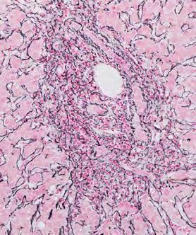
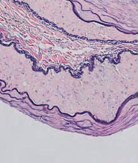
12 Elastic Product information BenchMark Special Stains - Elastic procedure The Elastic Staining Kit is a modification of Hart s method for elastic fibers. A resorcin-fuchsin solution is used to stain elastic fibers dark bluish purple to black. 1 Van Gieson s solution is applied to provide contrasting yellow background tissue while staining the collagen pinkish red. This stain is useful in demonstrating atrophy of elastic fibers in cases of emphysema, as well as the thinning and loss of elastic fibers in arteriosclerosis and other vascular diseases. Elastic Staining Kit Catalog number: Ordering code: Kit components 1. Oxidizer contains less than 1% potassium permanganate 2. Decolorizer contains less than 1% oxalic acid 3. Elastic Tissue Stain contains 66.5% absolute ethanol, 1% hydrochloric acid and 0.8% resorcin fuchsin 4. Elastic Clarifier contains 50% absolute alcohol 5. Van Gieson s solution contains 1.3% picric acid saturated solution References 1. Sheehan DC, Hrapchak BB. Theory and Practice of Histotechnology. 2nd edition. St. Louis, MO: C.V. Mosby Company; 1980: Figure 1. Artery stained with Elastic stain 12


13 Elastic Elastic Shortest incubation times (and default) protocol Longest incubation Hematoxylin: 8-16 min 8 min 16 min Figure 2 - Sample images of staining achieved with different protocol time selections. * Key to staining procedures protocol located on page 3 13

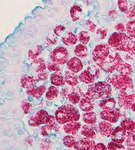
14 Giemsa Product information BenchMark Special Stains - Giemsa procedure The Giemsa Staining Kit is a modification of the original Giemsa stain. 1 Buffered thiazine eosinate solution is used to stain cells differentially with a characteristic blue or pink color. 2 Giemsa Staining Kit Catalog number: Ordering code: Kit components 1. Giemsa Stain contains 0.4% modified Giemsa stain in 70% methanol References 1. C1. Bancroft and Stevens. Theory and Practice of Histological Techniques, 2nd edition. Edinburgh: Churchill-Livingston, Carson F, Hladik C. Histotechnology: A Self Instructional Text, 3rd edition. Hong Kong: American Society for Clinical Pathology Press; Sheehan DC, Hrapchak BB. Theory and Practice of Histotechnology, 2nd edition. St. Louis, MO: C.V. Mosby Company; 1980: Figure 1. Gastric biopsy stained with Giemsa demonstrating H. Pylori organisms, 600x. 14


 in 0.")
 in 0.")
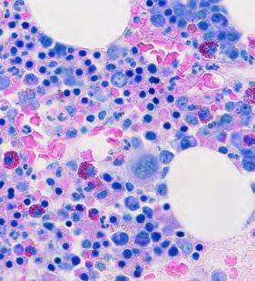
15 Giemsa Product information BenchMark Special Stains Giemsa procedure The Giemsa Staining Kit is a modification of the original Giemsa stain. 2 Buffered thiazine eosinate solution is used to stain cells differentially with a characteristic blue or pink color. 3 Kit components: Giemsa stain contains 0.4% modified Giemsa stain in 70% methanol. Giemsa Staining Kit: Catalog number Ordering code Figure 2. Gastric biopsy with H. Pylori Bone marrow core biopsy Standard post-instrument processing Alternate post-processing Standard post-instrument processing Alternate post-processing 2 x 10 dips (2 x10 seconds) 95% alcohol, followed by four changes of absolute alocohol 1 dip (1 seconds) in 0.1% acetic acid, followed by 2 changes of absolute alcohol 2 x 10 dips (2 x 10 seconds) 95% alcohol, followed by 3-4 changes of absolute alcohol 6 dips (6 seconds) in 0.1% acetic acid, followed by 3-4 changes of absolute alcohol Bone marrow clot (FFPE) Standard post-instrument processing Alternate post-processing 2 x 10 dips (2 x 10 seconds) 95% alcohol, followed by 3-4 changes of absolute alcohol 2 dips (2 seconds) in 0.1% acetic acid, followed by 3-4 changes of absolute alcohol 15

16 GMS II Product information BenchMark Special Stains - GMS II procedure The GMS II Staining Kit is a modification of Gomori s Methenamine Silver procedure. 1 This stain has also been modified by Grocott, and is sometimes called Grocott s Methenamine Silver. 2 GMS II Staining Kit Catalog number: Ordering code: Kit components 1. GMS II Oxidizer contains <6% chromium trioxide 2. GMS II Neutralizer contains <1% sodium bisulfite 3. GMS II Silver A contains <1% silver nitrate 4. GMS II Silver B contains <2% sodium borate and <15% Methenamine 5. GMS II Toner contains <1% gold chloride 6. GMS II Fixer contains <3% sodium thiosulfate 7. GMS II Light Green Counterstain contains <1% light green SF yellowish and <1.5% acetic acid References 1. Bancroft JD, Gamble, M. Theory and Practice of Histological Techniques. 2nd ed. Edinburgh: Churchill- Livingston; Sheehan DC, Hrapchak BB. Theory and Practice of Histotechnology. 2nd ed. St. Louis, MO: C.V. Mosby Company; 1980 Figure 1. Pneumocystus organisms in lung stained with GMS, 200x. 16


17 GMS II GMS II Shortest incubation / Intermediate incubation / Darkest incubation / Reaction erature: 50-60ºC 50 C 52 C 60 C Silver B: 8-16 min 8 min 12 min 16 min Green counterstain: 4-16 min 4 min 4 min 16 min Figure 2 - Sample images of staining achieved with different protocol time selections. * Key to staining procedures protocol located on page 3 17

18 Gram Product information BenchMark Special Stains - Gram procedure The Gram Staining Kit is a modification of the original Gram stain. 1-3 Gram-negative bacteria stain pink to red and grampositive bacteria stain blue to dark purple. Figure 1. Gram stain with Tarttrazine counterstain showing a mixture of Gram-positive and Gram-negative microorganisms, 1000x. (oil) Gram Staining Kit Catalog number: Ordering code: Kit components 1. Gram Crystal Violet contains 1% crystal violet, 0.7% ammonium oxalate monohydrate, and 17.5% reagent alcohol 2. Gram Iodine contains stabilized gram iodine 3. Gram Basic Fuchsin contains 0.15% basic fuchsin 4. Gram Gallego contains 3% formaldehyde and 1.5% acetic acid 5. Gram Tartrazine contains 0.1% tartrazine and 0.25% acetic acid 6. Gram Fast Green contains 0.002% fast green and 0.025% acetic acid References 1. Carson F, Hladik C. Histotechnology: A Self Instructional Text, 3rd edition. Hong Kong: American Society for Clinical Pathology Press; Sheehan DC, Hrapchak BB. Theory and Practice of Histotechnology. 2nd ed. St. Louis, MO: C.V. Mosby Company; Bancroft JD, Gamble, M. Theory and Practice of Histological Techniques. 2nd ed. Edinburgh: Churchill- Livingston;


19 Gram Gram Green Counterstain Shortest incubation Default protocol Longest incubation Optimize Gram-positive: 4 16 min 4 min 8 min 12 min Optimize Gram-negative: 4 12 min 4 min 8 min 16 min Figure 2 - Sample images of staining achieved with different protocol time selections. Appendix, showing gram positive bacteria only. * Key to staining procedures protocol located on page 3 19


20 Gram Gram Yellow Counterstain Shortest incubation Default protocol Longest incubation Optimize Gram-positive: 4 16 min 4 min 8 min 16 min Optimize Gram-negative: 4 12 min 4 min 12 min 12 min Figure 3 - Sample images of staining achieved with different protocol time selections. * Key to staining procedures protocol located on page 3 20

21 Iron Product information BenchMark Special Stains - Iron Staining procedure This iron stain is based on the historic Prussian blue reaction, which has previously been modified by Gomori, Perls and Mallory. 1-2 In the Iron Staining Kit, Iron Reagent A and Iron Reagent B create an acidic ferrocyanide, which reacts with ionic iron in the tissue to produce a bright blue color. Nuclear Fast Red counterstain: is applied to provide a contrasting pink to red background. Iron Staining Kit Catalog number: Ordering code: Kit components 1. Iron Reagent A contains 10% potassium ferrocyanide 2. Iron Reagent B contains less than 2% hydrochloric acid 3. Nuclear Fast Red Counterstain contains less than 1% nuclear fast red and 5% aluminum sulfate References 1. Sheehan DC, Hrapchak BB. Theory and Practice of Histotechnology. 2nd ed. St. Louis, MO: C.V. Mosby Company; Bancroft and Stevens. Theory and Practice of Histological Techniques, 2nd edition. Edinburgh: Churchill-Livingston; 1982 Figure 1. Liver with iron deposits stained with Iron Stain, 200x. 21
 protocol Intermediate incubation")


22 Iron Iron Shortest incubation times (and optimized) protocol Intermediate incubation Longest incubation NFR counterstain: 4-16 min 4 min 12 min 16 min Figure 2- Sample images of staining achieved with different protocol time selections. * Key to staining procedures protocol located on page 3 22


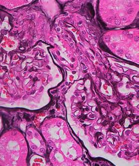
23 Jones H&E & Jones Light Green Product information BenchMark Special Stains - Jones Staining procedure The Jones Staining Kit is a modification of Jones Methenamine Silver procedure. 1 Periodic acid is used to oxidize carbohydrates to aldehyde groups. The combined Jones Silver A and Jones Silver B solutions form a methenamine-silver complex that is easily reduced to metallic silver by the aldehyde groups. Toner reagent contains gold chloride to form a more stable gold complex and remove the yellow tones from the tissue. Fixer, with thiosulfate, stops the reaction and removes any unreduced silver from the section. Two different types of counterstain: (Hematoxylin & Eosin, and Light Green) are available to provide contrasting background to the silver stain. Figure 1. Kidney stained with Jones using Hematoxylin and Eosin counterstain, 200x. H&E Jones Staining Kit Catalog number: Ordering code: Kit components 1. Jones Periodic Acid contains 1% periodic acid 2. Jones Silver A contains 1% silver nitrate 3. Jones Silver B contains <2% sodium borate and less than 14% methenamine 4. Toner contains <1% gold chloride 5. Fixer contains 2% sodium thiosulfate 6. Jones Hematoxylin contains modified Mayer s Hematoxylin (contains sodium iodate and ethylene glycol) 7. Jones Eosin contains <1% Eosin Y in an alcohol solution 8. Jones Periodic Acid contains 1% periodic acid Jones Light Green Staining Kit Catalog number: Ordering code: Kit components 1. Jones Periodic Acid contains 1% periodic acid 2. Jones Silver A contains 1% silver nitrate 3. Jones Silver B contains <2% sodium borate and less than 14% methenamine 4. Toner contains <1% gold chloride 5. Fixer contains 2% sodium thiosulfate 6. Jones Hematoxylin contains modified Mayer s Hematoxylin (contains sodium iodate and ethylene glycol) 7. Jones Light Green contains <1.09% Light Green Y in acetic acid 8. Jones Periodic Acid contains 1% periodic acid References 1. Koski JP. Silver methenamine-borate (SMB): Cost reduction with technical improvement in silver nitrategold chloride impregnations. J Histotechnol. 1981;3:115 Figure 2. Kidney stained with Jones using Light Green counterstain, 200x. 23


24 Jones H&E Jones H&E Shortest incubation / Default protocol / Longest incubation / Reaction erature: 55-60ºC 55 C 60 C 60 C Silver: 8-20 min 8 min 12 min 20 min Hematoxylin: 8-16 min 8 min 8 min 16 min Eosin: 4-12 min 4 min 8 min 12 min Figure 3 - Sample images of staining achieved with different protocol time selections. * Key to staining procedures protocol located on page 3 24


25 Jones Light Green Jones Light Green Shortest incubation / Default protocol / Longest incubation / Reaction erature: 55-60ºC 55 C 60 C 60 C Silver: 8-20 min 8 min 12 min 20 min Hematoxylin: 8-16 min 8 min 12 min 16 min Light Green: 4-16 min 4 min 8 min 16 m Figure 4 - Sample images of staining achieved with different protocol time selections. * Key to staining procedures protocol located on page 3 25
 in formalin fixed, paraffin-embedded tissue. Mucicarmine Staining Kit Catalog number: 860-011 Ordering code: 05279275001 Kit components 1. Mucicarmine stain 2.")
26 Mucicarmine Product information BenchMark Special Stains - Mucicarmine procedure The Mucicarmine Staining Kit is intended for use as a qualitative histologic stain to detect mucopolysaccharides (mucin) in formalin fixed, paraffin-embedded tissue. Mucicarmine Staining Kit Catalog number: Ordering code: Kit components 1. Mucicarmine stain 2. Iron Hematoxylin A contains 95% ethanol and 1% hematoxylin reagent 3. Iron Hematoxylin B contains 1.2% ferric chloride and 1% hydrochloric acid reagent 4. Tartrazine Counterstain contains <1% each of tartrazine and acetic acid reagent Figure 1. Colon adenocarcinoma stained with Mucicarmine stain, 200x. 26


27 Mucicarmine Mucicarmine Shortest incubation Default protocol Alternative incubation Longest incubation Hematoxylin incubation: 8-16 min 8 min 8 min 12 min 16 min Mucicarmine incubation: 8-16 min 8 min 12 min 16 min 16 min Tartrazine incubation: 4-16 min 8 min 4 min 4 min 16 min Figure 12- Sample images of staining achieved with different protocol time selections. * Key to staining procedures protocol located on page 3 27

28 PAS Product information BenchMark Special Stains - PAS Staining procedure The PAS Staining Kit uses Periodic Acid reagent to oxidize glycols to aldehydes. 1 The Schiff s Reagent forms a colorless dialdehyde compound that is transformed to the colored staining of glycol containing cellular components. PAS staining in tissue sections and digestion with a diastase reagent ( / ) is useful as an aid in the diagnosis of glycogen storage. 2 Figure 1. Kidney showing glomerular basement membrane stained with PAS stain, 200x. PAS Staining Kit Catalog number: Ordering code: Kit components 1. Periodic Acid contains less than 1% periodic acid 2. Schiff s Reagent contains 4% sodium bisulfite, 2% dilute hydrochloric acid and 1% pararosaniline chloride. (Three vials provided in the kit) 3. Neutralizer contains less than 1% sodium bisulfite 4. Hematoxylin Counterstain contains modified Mayer s hematoxylin Light Green for PAS Staining Kit Catalog number: Ordering code: Kit components 1. Light Green for PAS contains <1% light green SF yellowish in a 1% acetic acid solution Diastase Kit Catalog number: Ordering code: Kit components 1. Diastase contains 1.2% diastase of malt, with 0.1% sodium azide as a preservative Alcian Blue for PAS Staining Kit Catalog number: Ordering code: Kit components 1. Light Green for PAS contains <1% light green SF yellowish in a 1% acetic acid solution References 1. Carson F, Hladik C. Histotechnology: A Self Instructional Text, 3rd edition. Hong Kong: American Society for Clinical Pathology Press; Thompson SW. Selected Histochemical and Histopathological Methods. Springfield; CC Thomas;


29 PAS PAS Shortest incubation time protocol / Default protocol / Longest incubation / Reaction erature: 37-60ºC 37ºC 45ºC 60ºC Schiffs: min 12 min 20 min 20 min Hematoxylin: 4-12 min 4 min 8 min 8 min 1. Schiff s Reagent is stable for one month after opening. It is recommended that the vial be labeled with the date when opened for the first time. Sealed, unopened Schiff s Reagent is stable until the expiration date printed on the vial label. The kit contains two extra bottles of Schiff s Reagent to allow full use of the kit. As the open vial of Schiff reagent becomes unstable, higher background and less distinct staining of target components may be observed. Figure 2 - Sample images of staining achieved with different protocol time selections. * Key to staining procedures protocol located on page 3 29


30 PAS / Diastase PAS / Diastase Shortest incubation time (and optimized) protocol / Default protocol / Longest incubation / Reaction erature: 37-60ºC 37ºC 50ºC 60ºC Diastase: 4-16 min 4 min 4 min 16 min Schiffs: min 12 min 20 min 20 min Hematoxylin: 4-12 min 4 min 8 min 12 min Figure 3 - Sample images of staining achieved with different protocol time selections. * Key to staining procedures protocol located on page 3 30


31 PAS / Light Green Light Green for PAS Shortest incubation / Default protocol / Alternative incubation / Longest incubation / Reaction erature: 37-60ºC 37 C 45 C 37 C 60 C Schiffs incubation: min 12 min 20 min 20 min 12 min Figure 4 - Sample images of staining achieved with different protocol time selections. * Key to staining procedures protocol located on page 3 31


32 PAS / Alcian Blue Alcian Blue for PAS Shortest incubation Default incubation Alternative incubation Longest incubation Alcian Blue: 8-16 min 8 min 8 min 16 min 16 min Hematoxylin: 4-12 min 0 min 4 min 4 min 12 min Figure 5 - Sample images of staining achieved with different protocol time selections. * Key to staining procedures protocol located on page 3 32

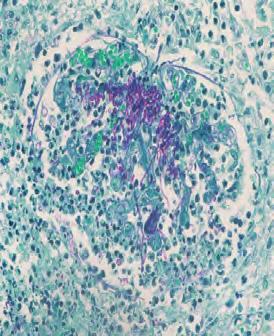
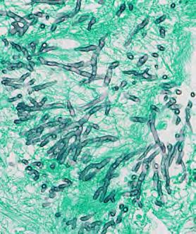
33 Reticulum Product information BenchMark Special Stains - Reticulum II procedure The Reticulum II Staining Kit is a modification of Gordon and Sweets Stain. 1 Oxidizer, with potassium permanganate, oxidizes the tissue to enhance staining of reticular fibers. Decolorizer, with oxalic acid, removes excess potassium permanganate. Sensitizer, with ferric ammonium sulfate, is added to form a metal organic compound. The metal organic compound is replaced by the silver in Reticulum II Silver A. Reducer is applied to develop the deposited silver into visible silver. Toner reagent contains gold chloride for better contrast and clarity. Fixer, with thiosulfate, stops the reaction and removes any unreacted silver from the section. Nuclear Fast Red Counterstain is applied to provide contrasting background. Reticulum II Staining Kit Catalog number: Ordering code: Kit components 1. Oxidizer contains less than 1% potassium permanganate 2. Decolorizer contains less than 1% oxalic acid 3. Sensitizer contains 2% ferric ammonium sulfate 4. Reticulum II Silver A contains less than 1.5% silver carbonate 5. Reticulum II Reducer contains 0.4% formaldehyde 6. Toner contains less than 1% gold chloride 7. Fixer II contains 2% sodium thiosulfate 8. Nuclear Fast Red Counterstain contains less than 1% Nuclear Fast Red and 5.0% aluminum sulfate References 1. Carson F, Hladik C. Histotechnology: A Self Instructional Text, 3rd edition. Hong Kong: American Society for Clinical Pathology Press; 2009 Figure 1. Tonsil stained with Retic stain, 400x. 33


34 Reticulum Reticulum Shortest incubation times (and default) protocol Longest incubation Silver A: 8-16 min 8 min 16 min NFR counterstain: 4-16 min 4 min 4 min Figure 2 - Sample images of staining achieved with different protocol time selections. * Key to staining procedures protocol located on page 3 34

35 Steiner II Product information BenchMark Special Stains - Steiner II procedure The Steiner II Staining Kit is a silver-based kit used in diagnosis of many microorganisms, such as Helicobacter pylori, spirochetes, or Legionella pneumophila. These techniques have been reviewed by Garvey. 1 Figure 1. Gastric Biopsy positive for H. Pylori, 400x. Steiner II Staining Kit Catalog number: Ordering code: Kit components 1. Steiner II Oxidizer contains 0.01% zinc chloride, 10% formalin 2. Steiner II Silver A contains 0.25% silver nitrate 3. Steiner II Fixer contains 1.0% sodium thiosulfate 4. Steiner II Diffuser contains 50% reagent alcohol 5. Steiner II Enhancer contains 5% gum mastic and absolute ethanol 6. Steiner II Clean A contains 95% reagent alcohol 7. Steiner II Reducer contains 0.8% hydroquinone 8. Steiner II Silver B contains 0.20% silver nitrate 9. Steiner II Clean B contains 95% reagent alcohol References 1. Garvey W. Silver Impregnation Techniques to Identify Spirochetes and Other Bacteria. J Histotechnol. 1996;19(3):


36 Steiner II Steiner II Shortest incubation / Intermediate incubation / Longest incubation / Reaction erature: 50-65ºC 50 C 52 C 65 C Silver: 8-16 min 8 min 12 min 16 min Figure 2 - Sample images of staining achieved with different protocol time selections. * Key to staining procedures protocol located on page 3 36

37 Trichrome Blue Product information BenchMark Special Stains - Trichrome Blue procedure The Trichrome Staining Kit is a modification of Masson s trichrome stain. Bouin s solution is applied to tissue sections to intensify the final coloration. Cytoplasm and muscle are stained with Trichrome Red, containing Biebrich Scarlet and acid fuchsin. Nuclei are stained with iron hematoxylin. After application of Trichrome Mordant, the collagen is stained with Trichrome Blue, which contains aniline blue. Trichrome Clarifier, an acetic acid solution, is applied to create a more delicate and transparent shade of color in the tissue section. Figure 1. Kidney stained with Trichrome Blue, 200x. Trichrome Staining Kit Catalog number: Ordering code: Kit components 1. Trichrome Bouin s reagent: A formulation of Bouin s fixative containing formaldehyde, acetic acid and picric acid. Since formalin fixation is not the best fixative when staining with acid dyes, Bouin s fixative is used in a secondary fixation step to correct the ph of the tissue and enable brighter dye staining 2. Trichrome Hematoxylin A and Trichrome Hematoxylin B: Trichrome Hematoxylin A and Trichrome B are two components of the nuclear stain. Trichrome Hematoxylin A is a hematoxylin reagent and Trichrome Hematoxylin B is a ferric chloride reagent. Together they form a complex of iron hematoxylin which stains the nuclei black 3. Trichrome Red: A formulation of the acid dyes Biebrich Scarlet and Acid Fuchsin which stains the red blood cells, muscle and cytoplasmic tissue components 4. Trichrome Mordant: A formulation of the metal salts of phosphotungstic acid and phosphomolybdic acid. It serves as a differentiating agent, removing the excess red from the collagen which is subsequently stained blue with Trichrome Blue 5. Trichrome Blue: A formulation of Aniline Blue used to stain the collagen tissue components 6. Trichrome Clarifier: An acetic acid solution used as a final rinse to remove excess blue and to refine the staining 37
 Optimized protocol for")


38 Trichrome Blue Trichrome Blue Shortest incubation (kidney) Default incubation Darkest incubation (kidney) Optimized protocol for kidney and liver biopsies Bouins: min 32 min 32 min 32 min 32 min Hematoxylin time: 4-24 min 4 min 12 min 24 min 12 min Trichrome Red time: 4-24 min 4 min 12 min 24 min 12 min Mordant: 4-24 min 4 min 12 min 24 min 12 min Blue: 4-24 min 4 min 4 min 24 min 24 min Figure 2 - Sample images of staining achieved with different protocol time selections. * Key to staining procedures protocol located on page 3 38
 Optimized protocol for")


39 Trichrome Blue Trichrome Blue Shortest incubation (kidney) Default incubation Darkest incubation (kidney) Optimized protocol for kidney and liver biopsies Bouins: min 32 min 32 min 32 min 32 min Hematoxylin time: 4-24 min 4 min 12 min 24 min 12 min Trichrome Red time: 4-24 min 4 min 12 min 24 min 12 min Mordant: 4-24 min 4 min 12 min 24 min 12 min Blue: 4-24 min 4 min 4 min 24 min 24 min Figure 3 - Sample images of staining achieved with different protocol time selections. * Key to staining procedures protocol located on page 3 39

40 Trichrome Green Product information BenchMark Special Stains - Trichrome Green procedure Green for Trichrome is a single bottle kit, which is used in conjunction with the Trichrome Staining Kit. The Trichrome Staining Kit is a modification of Masson s Trichrome Stain. Trichrome Bouin s is applied which acts as a mordant to allow penetration of subsequent dyes. Nuclei are stained with Trichrome Hematoxylin A and Trichrome Hematoxylin B (forms a complex of iron hematoxylin). Cytoplasm and muscle is stained with Trichrome Red, containing Biebrich scarlet and acid fuchsin. Trichrome Mordant removes the excess red from the collagen which is stained with Green for Trichrome, which contains fast green. Trichrome Clarifier is an acetic acid rinse used to remove excess green. This kit is optimized for use on the BenchMark Special Stains automated slide stainers. The reagents are applied to tissue on microscope slides and mixed over the entire specimen. The staining reaction is based on the differential effect of acid dye on muscle and collagen. Figure 1. Liver stained with Trichrome Green, 200x. Trichrome Staining Kit Catalog number: Ordering code: Kit components 1. Trichrome Bouin s reagent: A formulation of Bouin s fixative containing formaldehyde, acetic acid and picric acid. Since formalin fixation is not the best fixative to use when staining with acid dyes, Bouin s fixative is used in a secondary fixation step to correct the ph of the tissue and enable brighter dye staining 2. Trichrome Hematoxylin A and Trichrome Hematoxylin B: Trichrome Hematoxylin A and Trichrome B are two components of the nuclear stain. Trichrome Hematoxylin A is a hematoxylin reagent and Trichrome Hematoxylin B is a ferric chloride reagent. Together they form a complex of iron hematoxylin which stains the nuclei black 3. Trichrome Red: A formulation of the acid dyes Biebrich Scarlet and Acid Fuchsin which stains the red blood cells, muscle and cytoplasmic tissue components 4. Trichrome Mordant: A formulation of the metal salts of phosphotungstic acid and phosphomolybdic acid. It serves as a differentiating agent, removing the excess red from the collagen which is subsequently stained green with Fast Green 5. Trichrome Blue: This is not used in Trichrome Green B procedure 6. Trichrome Clarifier: An acetic acid solution used as a final rinse to remove excess green and to refine the staining Green for Trichrome Staining Kit Catalog number: Ordering code: Kit components 1. Trichrome Green reagent: A Fast Green and weak hydrochloric acid solution 40
 Longest incubation (kidney) Optimized protocol")


41 Trichrome Green Trichrome Green Shortest incubation (kidney) Default incubation (kidney) Longest incubation (kidney) Optimized protocol for kidney and liver biopsies Bouins: min 32 min 32 min 32 min 32 min Hematoxylin time: 4-24 min 4 min 4 min 24 min 12 min Trichrome Red time: 4-24 min 4 min 4 min 24 min 12 min Mordant: 4-24 min 4 min 4 min 24 min 12 min Green: 4-32 min 4 min 4 min 24 min 12 min Figure 2 - Sample images of staining achieved with different protocol time selections. * Key to staining procedures protocol located on page 3 41
 Longest incubation (liver) Optimized protocol (for")


42 Trichrome Green Trichrome Green Shortest incubation (liver) Default incubation (liver) Longest incubation (liver) Optimized protocol (for kidney and liver biopsies) (liver) Bouins: min 32 min 32 min 32 min 32 min Hematoxylin time: 4-24 min 4 min 12 min 24 min 12 min Trichrome Red time: 4-24 min 4 min 12 min 24 min 12 min Mordant: 4-24 min 4 min 12 min 24 min 12 min Green: 4-24 min 4 min 4 min 24 min 24 min Figure 3 - Sample images of staining achieved with different protocol time selections. * Key to staining procedures protocol located on page 3 42
43 General technical notes and post-instrument processing Technical notes 1. Known positive tissue controls should be utilized for monitoring the correct performance of processed tissues and test reagents. Control tissue should be fresh autopsy, biopsy or surgical specimen prepared or fixed as soon as possible in a manner identical to test sections. Such tissues should monitor all steps of the analysis, from tissue preparation through staining. Use of a tissue section fixed or processed differently from the test specimen provides control for all reagents and method steps except fixation and tissue processing. The cellular components of other tissue elements may serve as the negative control. 2. You can either bake the slides on the BenchMark Special Stains instrument or use an alternative method off the instrument. Be aware that, with some paraffins, baking at excessively high eratures may adversely affect the staining. Check the erature recommendations for the paraffin used in your lab. 3. Necrotic or autolyzed tissue may exhibit nonspecific staining. 4. At the end of the run there will be residual Liquid Coverslip left on the slide. This will be removed through dehydration with alcohol and xylene. Kit storage information The secondary packaging (box) on each of the special stains kits indicates the appropriate storage conditions for the entire contents of the kit. Individual vials are labeled with specific storage conditions that may include a broader range than indicated on the secondary packaging. Individual reagent vials with a gray collar that are part of a cold storage kit may be stored at room or cold storage (2-30 C). Individual reagent vials that must be stored at 2-8 C have a blue collar. Components that are stored cold must be brought to room erature prior to use to achieve optimal results. Inappropriate storage may shorten the life of the product and cause unsatisfactory results. Refer to the package insert for additional guidance on storage conditions. Do not rinse slides in water and DAWN detergent. 43
44 Roche Diagnostics Deutschland GmbH Sandhofer Strasse 116 DE Mannheim GERMANY Tel: Fax: Ventana Medical Systems, Inc. VENTANA is a trademark of Roche. All other trademarks are the property of their respective owners B
Atlas of Stains. Special Stains on Artisan Link Pro
 Atlas of Stains Special Stains on Artisan Link Pro Intended use Routinely processed samples (paraffin-embedded) may be used. The preferred fixative is neutral buffered formalin. The clinical interpretation
Fixation... Questions 1 Answers 16. Processing... Questions 25 Answers 36. Safety... Questions 67 Answers 73
 Table of Contents Fixation... Questions 1 Answers 16 Processing... Questions 25 Answers 36 Instrumentation... Questions 43 Answers 58 Safety... Questions 67 Answers 73 Laboratory Mathematics & Solution
Preface 1. Fixation and Processing 1
 Contents Preface xi 1. Fixation and Processing 1 Fixation 1 Processing 2 What Should Be Seen in a Well-Fixed, Well-Processed Specimen Stained with Hematoxylin and Eosin 3 Problems Encountered With Fixation
Special Stains. General Reference Guide
 Special Stains General Reference Guide Table of Contents AFB 4 Technical notes and references 6 Gram 32 Technical notes and references 34 Steiner 64 Technical notes and references 66 Alcian Blue 8 Technical
The Oral Histology Series Series 5 Special Stains
 The Oral Histology Series Series 5 Special Stains DAVID E. KLINGMAN, Lt Col, USAF, DC Views and opinions expressed are those of the author(s) and do not reflect official policy or position of the United
3. Staining solutions for histology and cytology Mounting and embedding media... 60
 INDEX 1. Hematoxylin stains... 4 2. Eosin... 6 3. Staining solutions for histology and cytology... 8 4. Reagents I... 48 5. Reagents II... 55 6. Decalcifying solutions... 58 7. Fixatives... 59 8. Formalins...
SURGICAL PATHOLOGY - HISTOLOGY STAINING MANUAL - NERVE TISSUE Page: 1 of 3 BODIAN'S METHOD - NERVE FIBERS PURPOSE: For demonstrating nerve fibers.
 SURGICAL PATHOLOGY - HISTOLOGY Date: STAINING MANUAL - NERVE TISSUE Page: 1 of 3 BODIAN'S METHOD - NERVE FIBERS PURPOSE: For demonstrating nerve fibers. PRINCIPLE: Protargol-S (silver proteinate) is used
Schedule of Accreditation issued by United Kingdom Accreditation Service 2 Pine Trees, Chertsey Lane, Staines-upon-Thames, TW18 3HR, UK
 2 Pine Trees, Chertsey Lane, Staines-upon-Thames, TW18 3HR, UK Cellular Pathology Department University College London Hospital Rockefeller Building University Street London WC1E 6JJ Contact: Gavyn Barrett
Special Staining (I)
") Special Staining (I) Carbohydrates 1- PERIODIC ACID SCHIFF'S (PAS ) Purpose: Glycogen is present in liver, kidney, skeletal and cardiac muscle. The PAS stain is used to demonstrate neutral polysaccharides
Special Staining (I) Carbohydrates 1- PERIODIC ACID SCHIFF'S (PAS ) Purpose: Glycogen is present in liver, kidney, skeletal and cardiac muscle. The PAS stain is used to demonstrate neutral polysaccharides
HISTOPATHOLOGY. Introduction
 HISTOPATHOLOGY Introduction Contacts Services offered Pathology tissue request Laboratory hours Special instructions Histopathology reports List of specimens Introduction The Histopathology section of
PATHOPHYSIOLOGY. DEFINED Involves the study of function that results from disease processes.
 DEFINED Involves the study of function that results from disease processes. What is pathology? Pathology is the branch of medical sciences that treats the essential nature of disease, especially the changes
Carbohydrates and mucins. Dr Phil Bryant, Wales, UK
 Carbohydrates and mucins Dr Phil Bryant, Wales, UK Carbohydrates Provide cells with energy by converting to glucose Excess glucose stored in liver and muscle as glycogen Residual unstored glycogen is turned
BLIZARD INSTITUTE CORE PATHOLOGY ATLAS OF TINCTORIAL STAINS
 BLIZARD INSTITUTE CORE PATHOLOGY ATLAS OF TINCTORIAL STAINS Contents Introduction... 3 Background to Tinctorial Stains... 3 Haematoxylin and Eosin Stain (H&E)... 3 Connective Tissue Stains... 4 Nucleic
2014 CURRENT ISSUES IN PATHOLOGY
 2014 CURRENT ISSUES IN PATHOLOGY SPECIAL STAINS IN LIVER BIOPSY PATHOLOGY Sanjay Kakar, MD University of California, San Francisco Trichrome stain : (1) Assess degree of fibrosis. H&E stain is not reliable
Microscopy PART - 6. Microscopy. Products for bacteriology. Products for cytology. Products for hematology. Products for histology
 PART - 6 Products for bacteriology Products for cytology Products for hematology Products for histology Universal reagents for For more details contact : Merck Specialities Private Limited Tel : +91 22
ABOUT US WORLDWIDE DISTRIBUTION ICON KEY STAIN LINE PRODUCT LIST
 ABOUT US WORLDWIDE DISTRIBUTION ICON KEY STAIN LINE PRODUCT LIST BQCKit Company is one of the three differentiated business lines of Bioquochem S.L. We are a Spanish biotechnology company specialized
European Pharmacopoeia solutions
 Reagena manufactures numerous Ph.Eur compliant solutions for use in quality control, production and identification. These products include buffers, stabilizers, identification reagents, controls, calibrators,
-26- MATERIALS AND METHODS
 -26- MATERIALS AND METHODS The pollutant : Sevin (1-naphthyl N-methyl carbamate) was used at a concentrations of 0.5 and 1.0 mg/l. At these concentrations, the insecticide was completely soluble in water.
What in the world is Histotechnology? Karen Stiffler, MA, HTL Program Director for Histotechnology
 What in the world is Histotechnology? Karen Stiffler, MA, HTL Program Director for Histotechnology The Basics of Histology Histology: the study of body tissues "histo" is from the Greek "histos" meaning
Special Stains in Dermatopathology
 Application Special Stains in Dermatopathology Jameel Ahmad Brown, MD Bruce R. Smoller, MD Fellow: Division of Dermatopathology University of Arkansas for Medical Sciences 4301 W. Markham St. Little Rock,
Mucin Histochemistry Study of the Prostate in Normal and Malignant Lesions
 ISSN 2231-4261 ORIGINAL ARTICLE Mucin Histochemistry Study of the Prostate in Normal and Malignant Lesions 1* 1 1 1 Manoj P.Ambali, Megha A. Doshi, Pratibha P. Patil, Shweta H. Chavan 1 Department of Anatomy,
CATALOG Parview Road Middleton, WI
 CATALOG 2505 Parview Road Middleton, WI 53562-2579 800-383-7799 www.newcomersupply.com February 2018 ABOUT US Mission Keeping it real: Our job is to help you get your job done! Everything we do is designed
Periodic Acid-Schiff-Light Green Stain to Detect Glomerular Protein Deposits by Routine Light Microscopy
 Periodic Acid-Schiff-Light Green Stain to Detect Glomerular Protein Deposits by Routine Light Microscopy CHARLES N. GAMBLE, M.D. Department of Pathology, Sutter Memorial Hospital, Sacramento, California
FD Rapid MultiStain Kit
 Quality & Excellence since 1996 FD Rapid MultiStain Kit An effective histological staining system with multiple functions designed for easy use in all types of neuroscience laboratories User Manual PK
HRP cytochemistry. Division of Radiooncology, Deutsches Krebsforschungszentrum, Heidelberg, Germany
 HRP cytochemistry WOLF D. KUHLMANN, M.D. Division of Radiooncology, Deutsches Krebsforschungszentrum, 69120 Heidelberg, Germany A range of substrates is available for the cytochemical staining of peroxidase
Pepsin Solution ready-to-use
 SIE HABEN DIE VISION, WIR HABEN DIE SUBSTANZ. Pepsin Solution Single component Pepsin Solution: only one component refrigerator stable Pepsin is a commonly used digestive enzyme for immunohistochemical
Schedule of Accreditation issued by United Kingdom Accreditation Service 2 Pine Trees, Chertsey Lane, Staines-upon-Thames, TW18 3HR, UK
 2 Pine Trees, Chertsey Lane, Staines-upon-Thames, TW18 3HR, UK Calderdale and Huddersfield NHS Foundation Trust Calderdale Royal Hospital Salterhebble Halifax HX3 0PW United Kingdom Contact: Dr Richard
Yara Saddam. Amr Alkhatib. Ihsan
 1 Yara Saddam Amr Alkhatib Ihsan NOTE: Yellow highlighting=correction/addition to the previous version of the sheet. Histology (micro anatomy) :- the study of tissues and how they are arranged into organs.
School Of Medicine Biomedical Sciences
 School Of Medicine Biomedical Sciences COSHH Risk Assessment Form Before any experimental procedure involving the use of chemicals or reagents is undertaken, potential hazards must be identified and an
A Histochemical Study of Epithelial Mucin
 A Histochemical Study of Epithelial Mucin in the Chick Chorioallantois JAMES L. CONKLIN Department of Anatomy, The University of Michigan, Ann Arbor, Michigun ABSTRACT Histochemical methods demonstrate
TRACP & ALP double-stain Kit
 Table of Content I. Description... 2 II. Introduction... 2 III. Principles... 2 IV. Kit components... 3 V. Storage... 3 VI. Preparation of reagents... 3 VII. Methods... 4-7 Cell fixation... 4 Activity
White paper Comparative analysis of H&E stain quality between the VENTANA HE 600 system and a traditional linear stainer
 White paper Comparative analysis of H&E stain quality between the VENTANA HE 600 and a traditional linear stainer 1 11/5/2015 8:55:39 AM Introduction Most tissue-based diagnostic decisions in anatomic
Tabla de resistencia de agentes químicos
 Tabla de resistencia de agentes químicos www.placka.com.ar / Teléfono [0341] 2080706 Maipú 670 / S2114AKC Coronel Domínguez Dpto. Rosario - Pcia. Santa Fe - República Argentina A Producto Químico Acetic
BioSciences. Peripheral Blood Smear Preparation. Blood Smear Preparation Materials
 PolyFacts Vol. 5 No. 1 BioSciences Use the Wright Stain High Quality StainRITE Ready-to-Use Stains for Hematology Peripheral blood smear (peripheral blood film) is a glass microscope slide coated with
3. PRELIMINARY PHYTOCHEMICAL SCREENING
 93 3. PRELIMINARY PHYTOCHEMICAL SCREENING 3.1 INTRODUCTION All the drugs- Ayurvedic, Unani and Herbal extracts were subjected to preliminary phytochemical screening to test the presence of alkaloids, carbohydrates
Ch 3: Observing Microorganisms Through a Microscope
 Ch 3: Observing Microorganisms Through a Microscope SLOs Review the metric units of measurement Define total magnification and resolution Explain how electron and light microscopy differ Differentiate
 Index A ABC method. See avidin-biotin enzyme complex method. absolute alcohol and celloidin embedding, 40 for Luxol fast blue staining, 213 rapid processing with, 38 absolute alcohol hematoxylin for Russell
FIXATION OF TISSUES MODULE 5.1 INTRODUCTION OBJECTIVES 5.2 AIMS OF FIXATION 5.3 PRINCIPLE OF FIXATION. Notes
 MODULE Fixation of Tissues 5 FIXATION OF TISSUES 5.1 INTRODUCTION It is a process by which the cells or tissues are fixed in chemical and partly physical state so that they can withstand subsequent treatment
Evaluation of a Cold Staining Method for Detecting Acid Fast Bacilli in the Sputum
 International Journal of Current Microbiology and Applied Sciences ISSN: 2319-7706 Volume 5 Number 6 (2016) pp. 125-129 Journal homepage: http://www.ijcmas.com Original Research Article http://dx.doi.org/10.20546/ijcmas.2016.506.015
An Evaluation of Xylene-free Processing of Tissues From the Central Nervous System Using the PelorisTM Dual Retort Rapid Tissue Processor
 An Evaluation of Xylene-free Processing of Tissues From the Central Nervous System Using the PelorisTM Dual Retort Rapid Tissue Processor Geoffrey Rolls Leica Microsystems, iosystems Division, Melbourne,
Progress in the Development of Microscopical Techniques for Diagnostic Pathology
 Journal of Histotechnology ISSN: 0147-8885 (Print) 2046-0236 (Online) Journal homepage: https://www.tandfonline.com/loi/yhis20 Progress in the Development of Microscopical Techniques for Diagnostic Pathology
BRIEFING. Nonharmonized attributes: Identification, Heavy metals, Characters, Labeling, Bacterial endotoxins, Sterility, Storage.
 BRIEFING Citric Acid, Anhydrous, page 872 of PF 28(3) [May June 2002]. The European Pharmacopoeia is the coordinating pharmacopeia for the international harmonization of the compendial standards for the
ON THE PRESENCE OF A CILIATED COLUMNAR EPITHELIAL CELL TYPE WITHIN THE BOVINE CERVICAL MUCOSA 1
 ON THE PRESENCE OF A CILIATED COLUMNAR EPITHELIAL CELL TYPE WITHIN THE BOVINE CERVICAL MUCOSA 1 R. I. Wordinger, 2 J. B. Ramsey, I. F. Dickey and I. R. Hill, Jr. Clemson University, Clemson, South Carolina
Instruction Manual Updated 8/27/2013 Ver. 1.1
 Water Analysis Kit Part No. 144-95 Instruction Manual Updated 8/27/2013 Ver. 1.1 OFI Testing Equipment, Inc. 11302 Steeplecrest Dr. Houston, Texas 77065 U.S.A. Tele: 832.320.7300 Fax: 713.880.9886 www.ofite.com
DROP TEST SODIUM NITRITE (1 drop = 40 ppm)
") 1 x 5011 Instruction 1 x 9198R Sample Tube, Graduated (25 ml) w/ cap & red dot, plastic 1 x R-0819-C Ferroin Indicator, 2 oz, DB 2 x R-0820-C CAN Solution, 2 oz, DB SODIUM NITRITE (1 drop = 40 ppm) Instr.
1 x 5011 Instruction 1 x 9198R Sample Tube, Graduated (25 ml) w/ cap & red dot, plastic 1 x R-0819-C Ferroin Indicator, 2 oz, DB 2 x R-0820-C CAN Solution, 2 oz, DB SODIUM NITRITE (1 drop = 40 ppm) Instr.
(Writing model for laboratory note book)
") Paper: Lab 50 Syllabus *************************************************************************** Experiment: Organic Qualitative analysis 1) Detection of elements (Nitrogen, Sulphur and halogens). 2)
Paper: Lab 50 Syllabus *************************************************************************** Experiment: Organic Qualitative analysis 1) Detection of elements (Nitrogen, Sulphur and halogens). 2)
BRIEFING Assay + + +
 BRIEFING Sodium Starch Glycolate, NF 22 page 2933 and page 3202 of PF 22(6) [Nov. Dec. 1996]. The United States Pharmacopeia is the coordinating pharmacopeia for the international harmonization of the
SUPPLEMENTARY MATERIAL. Sample preparation for light microscopy
 SUPPLEMENTARY MATERIAL Sample preparation for light microscopy To characterize the granulocytes and melanomacrophage centers, cross sections were prepared for light microscopy, as described in Material
Change to read: BRIEFING
 BRIEFING Dibasic Calcium Phosphate Dihydrate, USP 29 page 359. The Japanese Pharmacopoeia is the coordinating pharmacopeia for the international harmonization of the compendial standards for the Dibasic
» Monohydrate Citric Acid contains one molecule of water of hydration. It contains not less than 99.5 percent and not more than 100.
 BRIEFING Citric Acid, Monohydrate. The European Pharmacopoeia is the coordinating pharmacopeia for the international harmonization of the compendial standards for the Citric Acid, Monohydrate monograph,
(A) PCR primers (arrows) designed to distinguish wild type (P1+P2), targeted (P1+P2) and excised (P1+P3)14-
 PCR primers (arrows) designed to distinguish wild type (P1+P2), targeted (P1+P2) and excised (P1+P3)14-") 1 Supplemental Figure Legends Figure S1. Mammary tumors of ErbB2 KI mice with 14-3-3σ ablation have elevated ErbB2 transcript levels and cell proliferation (A) PCR primers (arrows) designed to distinguish
1 Supplemental Figure Legends Figure S1. Mammary tumors of ErbB2 KI mice with 14-3-3σ ablation have elevated ErbB2 transcript levels and cell proliferation (A) PCR primers (arrows) designed to distinguish
Instruction Number: 5681
 Instruction Number: 5681 Component Description > COMPONENT SHEET > K 1690 COMBINATION BOILER/COOLING SYSTEM Alkalinity P/T (HCl) 1 x 5229 Instruction *1 x 9198 Sample Tube, Graduated, 25 ml, plastic w/cap
Techniques for Viewing Pollen Tubes in Angiosperm Flowers
 The University of Akron IdeaExchange@UAkron Honors Research Projects The Dr. Gary B. and Pamela S. Williams Honors College Spring 2016 Techniques for Viewing Pollen Tubes in Angiosperm Flowers Cameron
APPENDIX 1 ETHICAL CLEARANCE
 APPENDIX 1 ETHICAL CLEARANCE 75 APPENDIX 2 76 PROCEDURE FOR PREPARING OF LIVER HISTOLOGY SLIDES Overview: Histology involves the use of a set of techniques to examine the morphology, architecture and composition
Schedule of Accreditation issued by United Kingdom Accreditation Service 2 Pine Trees, Chertsey Lane, Staines-upon-Thames, TW18 3HR, UK
 Schedule of Accreditation 2 Pine Trees, Chertsey Lane, Staines-upon-Thames, TW18 3HR, UK Department of Eye Pathology 1 st Floor, Cayton Street Building UCL Institute of Ophthalmology 11-43 Bath Street
Non-hematogenous endogenous pigments
 Non-hematogenous endogenous pigments 0 This group contains the following : 1. Melanins. 2. Lipofuscins. 3. Chromaffin. 4. Pseudomelanosis. 5. Dubin-Johnson pigments. 6. Ceroid-type lipofuscins. 7. Hamazaki-Weisenberg
H.Pylori IgG
 DIAGNOSTIC AUTOMATION, INC. 21250 Califa Street, Suite 102 and116, Woodland Hills, CA 91367 Tel: (818) 591-3030 Fax: (818) 591-8383 onestep@rapidtest.com technicalsupport@rapidtest.com www.rapidtest.com
MULTIPLE CHOICE. Choose the one alternative that best completes the statement or answers the question.
 Exam Name MULTIPLE CHOICE. Choose the one alternative that best completes the statement or answers the question. 1) A nanometer would be a suitable unit of measurement for which of the following? 1) A)
H.Pylori IgG Cat # 1503Z
 DIAGNOSTIC AUTOMATION, INC. 23961 Craftsman Road, Suite D/E/F, Calabasas, CA 91302 Tel: (818) 591-3030 Fax: (818) 591-8383 onestep@rapidtest.com technicalsupport@rapidtest.com www.rapidtest.com See external
Annexure III SOLUTIONS AND REAGENTS
 Annexure III SOLUTIONS AND REAGENTS A. STOCK SOLUTIONS FOR DNA ISOLATION 0.5M Ethylene-diamine tetra acetic acid (EDTA) (ph=8.0) 1M Tris-Cl (ph=8.0) 5M NaCl solution Red cell lysis buffer (10X) White cell
H.pylori IgA Cat #
 DIAGNOSTIC AUTOMATION, INC. 23961 Craftsman Road, Suite D/E/F, Calabasas, CA 91302 Tel: (818) 591-3030 Fax: (818) 591-8383 onestep@rapidtest.com technicalsupport@rapidtest.com www.rapidtest.com See external
DROP TEST P/M & P/T ALKALINITY (1 drop = 10 ppm)
") 1 x 5067G Instruction 1 x 9198G Sample Tube, Graduated (25 ml) w/ cap & green dot, plastic 1 x R-0637-C Methyl Orange Indicator, 2 oz, DB 1 x R-0638G-A Phenolphthalein Indicator,.75 oz w/ green cap, DB
1 x 5067G Instruction 1 x 9198G Sample Tube, Graduated (25 ml) w/ cap & green dot, plastic 1 x R-0637-C Methyl Orange Indicator, 2 oz, DB 1 x R-0638G-A Phenolphthalein Indicator,.75 oz w/ green cap, DB
Schedule of Accreditation issued by United Kingdom Accreditation Service 2 Pine Trees, Chertsey Lane, Staines-upon-Thames, TW18 3HR, UK
 2 Pine Trees, Chertsey Lane, Staines-upon-Thames, TW18 3HR, UK Laboratory locations: Department of Histopathology Royal United Hospitals NHS Foundation Trust Combe Park Bath BA1 3NG Contact: Lesley Shipway
Special stains in liver pathology
 Current Issues in Surgical Pathology 2014 Special stains in liver pathology Which, why, how Really? Sanjay Kakar, MD University of California, San Francisco Outline Which stains Why the stain is done How
Appendix 1. A. Procedure for preparing histopathology slides. The liver removed and stored immediately in buffered formalin 10 % for
 Appendix 1 A. Procedure for preparing histopathology slides. The liver removed and stored immediately in buffered formalin 10 % for histopathological examination. The tissue fixed for at least 48 hours
ab Oil Red O (Lipid Stain)
") Version 2 Last updated 19 December 2018 ab150678 Oil Red O (Lipid Stain) For the histological visualization of fat cells and neutral fat. View kit datasheet: www.abcam.com/ab150678 (use www.abcam.cn/ab150678
Version 2 Last updated 19 December 2018 ab150678 Oil Red O (Lipid Stain) For the histological visualization of fat cells and neutral fat. View kit datasheet: www.abcam.com/ab150678 (use www.abcam.cn/ab150678
Cable Ties-Material Selection Ordering Guide
 Introduction Thomas & Betts offers TY-RAP cable ties and accessories in a wide variety of materials, each suited for specific environments. The purpose of this document, therefore, is to assist you in
Corning BioCoat Matrigel Invasion Chamber
 Corning BioCoat Matrigel Invasion Chamber Catalog No. 354480, 354481 Guidelines for Use Discovery Labware, Inc., Two Oak Park, Bedford, MA 01730, Tel: 1.978.442.2200 (U.S.) CLSTechServ@Corning.com www.corning.com/lifesciences
EXERCISE 3 Carbon Compounds
 LEARNING OBJECTIVES EXERCISE 3 Carbon Compounds Perform diagnostic tests to detect the presence of reducing sugars (Benedict s), starch (Lugol s), protein (Biuret), lipid (SudanIV) and sodium chloride
Clinical Chemistry (CHE 221)
") Clinical Chemistry (CHE 221) Experiment # 14 Blood Alcohol Determination by Gas Chromatography and by Reaction with Alcohol Dehydrogenase Name Date Performed Date Submitted Partners Name(s) Partners Name(s)
Clinical Chemistry (CHE 221) Experiment # 14 Blood Alcohol Determination by Gas Chromatography and by Reaction with Alcohol Dehydrogenase Name Date Performed Date Submitted Partners Name(s) Partners Name(s)
See external label 2 C-8 C Σ=96 tests Cat # 1505Z. MICROWELL ELISA H.Pylori IgA Cat # 1505Z
 DIAGNOSTIC AUTOMATION, INC. 23961 Craftsman Road, Suite E/F, Calabasas, CA 91302 Tel: (818) 591-3030 Fax: (818) 591-8383 onestep@rapidtest.com technicalsupport@rapidtest.com www.rapidtest.com See external
Mucoprotein-containing histiocytes (muciphages)
") J. clin. Path. (1966), 19, 368 Mucoprotein-containing histiocytes (muciphages) in the rectum J. G. AZZOPARDI AND D. J. EVANS' From the Department of Morbid Anatomy, Postgraduate Medical School, London
J. clin. Path. (1966), 19, 368 Mucoprotein-containing histiocytes (muciphages) in the rectum J. G. AZZOPARDI AND D. J. EVANS' From the Department of Morbid Anatomy, Postgraduate Medical School, London
H. pylori IgM. Cat # H. pylori IgM ELISA. ELISA: Enzyme Linked Immunosorbent Assay. ELISA - Indirect; Antigen Coated Plate
 DIAGNOSTIC AUTOMATION, INC. 23961 Craftsman Road, Suite D/E/F, Calabasas, CA 91302 Tel: (818) 591-3030 Fax: (818) 591-8383 onestep@rapidtest.com technicalsupport@rapidtest.com www.rapidtest.com H. pylori
Examination of Chemicals in Trap Cases. (Phenolphthalein)
") Introduction Examination of Chemicals in Trap Cases (Phenolphthalein) Although a number of different techniques using different chemicals such as fluorescent dyes, starch powder, phenolphthalein powders
Introduction Examination of Chemicals in Trap Cases (Phenolphthalein) Although a number of different techniques using different chemicals such as fluorescent dyes, starch powder, phenolphthalein powders
Comparative Histochemical Study of Mucin in Colonic Goblet Cells of Albino Rat, Goat & Dog
 IOSR Journal of Dental and Medical Sciences (IOSR-JDMS) e-issn: 2279-0853, p-issn: 2279-0861.Volume 15, Issue 4 Ver. XIII (Apr. 2016), PP 48-53 www.iosrjournals.org Comparative Histochemical Study of Mucin
Study of Melanin Bleaching After Immunohistochemistry of Melanin-containing Tissues. Hongwu Shen, MD and Wenqiao Wu, MD
 TECHNICAL ARTICLE Study of Melanin Bleaching After Immunohistochemistry of Melanin-containing Tissues Hongwu Shen, MD and Wenqiao Wu, MD Abstract: Melanin may interfere with immunohistochemical staining.
Multi-Site Performance Summary of. AEROSPRAY TB SLIDE STAINER/CYTOCENTRIFUGE (Model 7722)
") Multi-Site Performance Summary of AEROSPRAY TB SLIDE STAINER/CYTOCENTRIFUGE (Model 7722) Abstract The new Aerospray TB Slide Stainer/Cytocentrifuge - Model 7722 (ELITechGroup Inc., www.elitechgroup.com)
Multi-Site Performance Summary of AEROSPRAY TB SLIDE STAINER/CYTOCENTRIFUGE (Model 7722) Abstract The new Aerospray TB Slide Stainer/Cytocentrifuge - Model 7722 (ELITechGroup Inc., www.elitechgroup.com)
Studies of differential staining with acid dyes in the human adenohypophysis
 467 Studies of differential staining with acid dyes in the human adenohypophysis By A. W. F. FISHER and D. BULMER (From the Anatomy Department, University of Manchester) Summary With Mallory techniques
Staining Technology and Bright- Field Microscope Use
 Staining Technology and Bright- Field Microscope Use 2 Abstract We will introduce bright-field microscope use, practice Gram staining with foodborne pathogens, and practice endospore staining with Bacillus
Human Saliva as a Convenient Source of Ribonuclease. By S. BRADBURY
 Human Saliva as a Convenient Source of Ribonuclease 323 By S. BRADBURY (From the Cytological Laboratory, Department of Zoology, University Museum, Oxford) SUMMARY Saliva, heated to 80 C for 10 minutes
H.Pylori IgM Cat # 1504Z
 DIAGNOSTIC AUTOMATION, INC. 23961 Craftsman Road, Suite D/E/F, Calabasas, CA 91302 Tel: (818) 591-3030 Fax: (818) 591-8383 onestep@rapidtest.com technicalsupport@rapidtest.com www.rapidtest.com See external
SOLUTION TEST STRIPS
 SOLUTION TEST STRIPS Patented AHPHLD8 REF066 (17/055) Virox_InsertBook_PreventionHLD8_06F.indd 1 2017-04-21 11:14 Virox_InsertBook_PreventionHLD8_06F.indd 2 2017-04-21 11:14 INTENDED USE: The Prevention
ab Luxol Fast Blue Stain (Myelin Stain)
") Version 3 Last updated 25 June 2018 ab150675 Luxol Fast Blue Stain (Myelin Stain) For the histological identification of basic neuronal structures in brain or spinal cord sections. This product is for
Version 3 Last updated 25 June 2018 ab150675 Luxol Fast Blue Stain (Myelin Stain) For the histological identification of basic neuronal structures in brain or spinal cord sections. This product is for
Assessment of the distribution of AMG positive material in the brain of largemouth bass, Micropterus salmoides
 University of Tennessee, Knoxville Trace: Tennessee Research and Creative Exchange University of Tennessee Honors Thesis Projects University of Tennessee Honors Program 5-2009 Assessment of the distribution
Contributions to Anatomic Pathology, over the years
 Contributions to Anatomic Pathology, over the years Anatomic Pathology, part 1 G.B. Morgagni Xavier Bichat Rudolf Wirchow Anatomic Pathology, part 1 Anatomic pathology materials: morphological samples
Instructions for Performing In-Office Lab Tests
 Instructions for Performing In-Office Lab Tests SALIVARY PH- STATIC MEASUREMENT Testing must be done at least 30 minutes from any food or beverage. 1. Simply place a ph testing strip in the patient s mouth
It s not just water! What is Urinalysis?
 It s not just water! An introduction to Urinalysis What is Urinalysis? Urinalysis or the analysis of urine is one of the oldest laboratory procedures in the practice of medicine. It is a good test for
Hydroponics TEST KIT MODEL AM-41 CODE 5406
 Hydroponics TEST KIT MODEL AM-41 CODE 5406 TABLE OF CONTENTS Page Contents List...4 Dilution Procedure...6 Test Procedures: ph... 6 Nitrate Nitrogen...7 Phosphorus... 7 Potassium... 7 Ammonia Nitrogen...8
Pathology stains and solvents. A complete range of stains and solvents for use in Histology, Microbiology, Haematology and Cytology
 Pathology stains and solvents A complete range of stains and solvents for use in Histology, Microbiology, Haematology and Cytology Australian and New Zealand pathologists rely on Thermo Fisher Scientific
APPENDICES. Appendix A Morphometric analysis guideline of frog and its spinal cord. Snout-to-vent length
 APPENDICES Appendix A Morphometric analysis guideline of frog and its spinal cord Snout-to-vent length Dorsal view of Fejervarya limnocharis Total length C T L S Ventral view of frog spinal cord (C: length
Thyroid Stimulating Hormone (S-TSH) Thyroid Stimulating
 Thyroid Stimulating") ab108659 Thyroid Stimulating Hormone (S-TSH) Human ELISA Kit Instructions for Use For the quantitative measurement of Human Thyroid Stimulating Hormone (S-TSH) concentrations in serum. This product is
ab108659 Thyroid Stimulating Hormone (S-TSH) Human ELISA Kit Instructions for Use For the quantitative measurement of Human Thyroid Stimulating Hormone (S-TSH) concentrations in serum. This product is
HISTOLOGY SPECIMEN COLLECTION MANUAL
 Lee Memorial Health System Lee County, FL CLINICAL LABORATORY HISTOLOGY SPECIMEN COLLECTION MANUAL Abdominal Fluid Minimum Volume No fixative in an appropriate sized container. 1 ml The container must
HBeAg and HBeAg Ab ELISA Kit
 HBeAg and HBeAg Ab ELISA Kit Catalog Number KA0290 96 assays Version: 17 Intended for research use only www.abnova.com Table of Contents Introduction... 3 Intended Use... 3 Principle of the Assay... 3
LIST OF ORGANS FOR HISTOPATHOLOGICAL ANALYSIS:!! Neural!!!!!!Respiratory:! Brain : Cerebrum,!!! Lungs and trachea! Olfactory, Cerebellum!!!!Other:!
 LIST OF ORGANS FOR HISTOPATHOLOGICAL ANALYSIS:!! Neural!!!!!!Respiratory:! Brain : Cerebrum,!!! Lungs and trachea! Olfactory, Cerebellum!!!!Other:! Spinal cord and peripheral nerves! Eyes, Inner ear, nasal
Molecular Probes Introducing 14 new probes
 Molecular Probes Introducing 14 new probes Gene and Chromosome Probes Dual Colour ISH INFORM HER2 Dual ISH DNA Probe Cocktail Assay Product Part Number INFORM HER2 Dual ISH DNA Probe Cocktail 800-4422
Cytological evaluation of effusion fluid with cell block technique and cytology smears among Sudanese patients
 EUROPEAN ACADEMIC RESEARCH Vol. IV, Issue 3/ June 2016 ISSN 2286-4822 www.euacademic.org Impact Factor: 3.4546 (UIF) DRJI Value: 5.9 (B+) Cytological evaluation of effusion fluid with cell block technique
Zurn Chemical Resistance Information Flo-Thru Drainage Systems
 Chart below shows chemical resistance of polyester and vinylester to select chemicals. For a more complete list or for other chemicals, contact your Zurn Sales Representative. Zurn Chemical Resistance
Ordering Physician. Collected REVISED REPORT. Performed. IgG IF, Renal MCR. Lambda IF, Renal MCR. C1q IF, Renal. MCR Albumin IF, Renal MCR
 RenalPath Level IV Wet Ts IgA I Renal IgM I Renal Kappa I Renal Renal Bx Electron Microscopy IgG I Renal Lambda I Renal C1q I Renal C3 I Renal Albumin I Renal ibrinogen I Renal Mayo Clinic Dept. of Lab
IHC Polymer. BioGenex Website. Presented for: Presented by: Date: BioGenex Tech Support 2016
 IHC Polymer Presented for: Presented by: Date: BioGenex Website BioGenex Tech Support 2016 Immunohistochemistry (IHC) Quick Guide# Super SensitiveTM Polymer-HRP kits. Baking An-gen Retrieval DeWax Overnight,
Compara've Medicine Animal Histology Services. Glossary of Terms
 Compara've Medicine Animal Histology Services Glossary of Terms About this guide This guide is intended to provide clarifica'on on standard histologic terminology. Included are examples of different sample