The Oral Histology Series Series 5 Special Stains
|
|
|
- Sydney Craig
- 6 years ago
- Views:
Transcription


1 The Oral Histology Series Series 5 Special Stains DAVID E. KLINGMAN, Lt Col, USAF, DC Views and opinions expressed are those of the author(s) and do not reflect official policy or position of the United States Air Force, Department of Defense, or US Government.

2 Reticulin stain: Highlights reiculin fibers (which are argyrophilic) in parenchymal tissues in organs such as liver and spleen; demonstrates growth patterns of neoplasms

3 Reticulin stain: Highlights reiculin fibers (which are argyrophilic) in parenchymal tissues in organs such as liver and spleen; demonstrates growth patterns of neoplasms

4 Elastic/Verhoeff s van Gieson stain: Demonstrates atrophy of elastic tissue in cases of emphysema and thinning/loss of 4 elastic fibers in arteriosclerosis, temporal arteritis and other vascular diseases
")
5 Masson s trichrome stain: Differentiates collagen fibers from smooth muscle; routine stain for liver and kidney biopsy. Increases seen in collagen diseases (e.g. cirrhosis of liver)
")
6 Masson s trichrome stain: Differentiates collagen fibers from smooth muscle; routine stain for liver and kidney biopsy. Increases seen in collagen diseases (e.g. cirrhosis of liver)
: Demonstrates")


7 Periodic acid-schiff stain (with or without diastase): Demonstrates glycogen and polysaccharides; glycogen will be digested with diastase (diastase labile) versus mucins which will not digest (diastase resistant)
: Demonstrates glycogen and")
 versus mucins which will")
8 Periodic acid-schiff stain (with or without diastase): Demonstrates glycogen and polysaccharides; glycogen will be digested with diastase (diastase labile) versus mucins which will not digest (diastase resistant)

9 Best s carmine stain: Red stain which highlights glycogen 9

10 Mucicarmine stain: Highlights bacteria (red); highlights mucin (useful in identifying mucus cells, 10 for example, in mucoepidermoid carcinoma)

11 Alcian green stain: Highlights cartilage 11


12 Grocott s methenamine silver stain: Highlights the cell wall of microorganisms (useful for identifying fungal organisms)


13 Acid fast stain: Highlights certain bacteria, particularly Mycobacterial species

14 Acid fast stain: Highlights certain bacteria, particularly Mycobacterial species

15 Acid fast stain: Highlights certain bacteria, particularly Mycobacterial species

16 Acid fast stain: Highlights certain bacteria, particularly Mycobacterial species

17 Auramine rhodamine stain: A fluorescence stain, used to highlight acid fast bacilli such as Mycobacterium 17

18 Auramine rhodamine stain: A fluorescence stain, used to highlight acid fast bacilli such as Mycobacterium 18

19 Gram stain: Differentiates bacteria into gram-positive and gram-negative species 19

20 Gram stain: Differentiates bacteria into gram-positive and gram-negative species 20

21 Giemsa stain: Used in peripheral blood smears 21

22 Giemsa stain: Used in peripheral blood smears 22

23 Giemsa stain: Used in peripheral blood smears

24 Giemsa stain: Used in peripheral blood smears

25 Giemsa stain: Used in peripheral blood smears

26 Iron stain (Prussian blue): Highlights ferric ions in tissues, such as bone marrow samples

27 Iron stain (Prussian blue): Highlights ferric ions in tissues, such as bone marrow samples

28 Grimelius (Pascual s modified) stain: Identifies modified argyrophil cells found in larger numbers in same general locations 28 as argentaffin cells; differentiates carcinoid tumors and neuroendocrine tumors such as Merkel cell carcinoma

29 Fontana Masson stain: Identifies argentaffin cells and melanin pigment 29

30 Warthin-Starry stain: Stains spirochetes

31 Warthin-Starry stain: Stains spirochetes

32 Warthin-Starry stain: Stains spirochetes

33 Warthin-Starry stain: Stains spirochetes

34 Phosphotungstic acid-haematoxylin (PTAH) / Malloy s stain: Aids in diagnosing/identifying muscle cross-striations, fibrin, and mitochondria-rich cells 34

35 Phosphotungstic acid-haematoxylin (PTAH) / Malloy s stain: Aids in diagnosing/identifying muscle cross-striations, fibrin, and mitochondria-rich cells 35

36 Phosphotungstic acid-haematoxylin (PTAH) / Malloy s stain: Aids in diagnosing/identifying muscle cross-striations, fibrin, and mitochondria-rich cells 36

37 Phosphotungstic acid-haematoxylin (PTAH) / Malloy s stain: Aids in diagnosing/identifying muscle cross-striations, fibrin, and mitochondria-rich cells 37

38 Palmgren stain: Identifies nerve axons 38

39 Bodian stain: Demonstrates nerve fibers 39

40 Luxol fast blue/cresyl violet stain: Demonstrates myelin sheath 40

41 Bielschowsky stain: Demonstrates neurofibrillary tangles, nerve fibers, and senile plaques; useful 41 in diagnosis of Alzheimer s Disease

42 Congo red stain: Useful as a cytoplasm and erythrocyte stain; amyloid will demonstrate apple green birefringence when viewed under polarized light

43 Congo red stain: Useful as a cytoplasm and erythrocyte stain; amyloid will demonstrate apple green birefringence when viewed under polarized light

44 Crystal violet stain: Demonstrates amyloid

45 Thioflavin T stain: Fluorescent stain which demonstrates amyloid; useful in Alzheimer s diagnosis 45

46 Sudan IV stain: Stains lipids, triglycerides, and lipoproteins 46
")
47 Oil Red O stain: Demonstrates fat/lipids in fresh (frozen) tissue 47

48 Jones methenamine silver stain: Demonstrates/highlights basement membrane in renal glomerules 48
; identifies mineralization within tissues")
49 Von Kossa stain: Stains Ca++ (appears deep-blue to purple); identifies mineralization within tissues 49

50 Leder stain: Detects the enzyme chloracetate esterase found in granulocytes, including mast cells; 50 positive staining is seen in CLL vs. CML
51 other stains 51

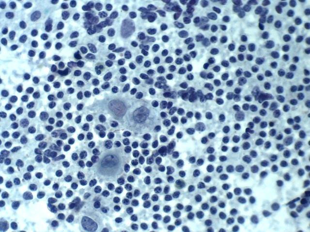
52 52 DiffQuik
53 53 Papanicoloau
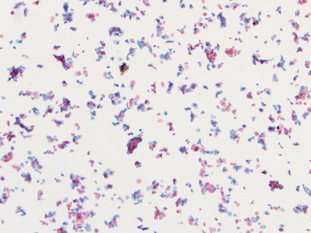
54 54 Papanicoloau
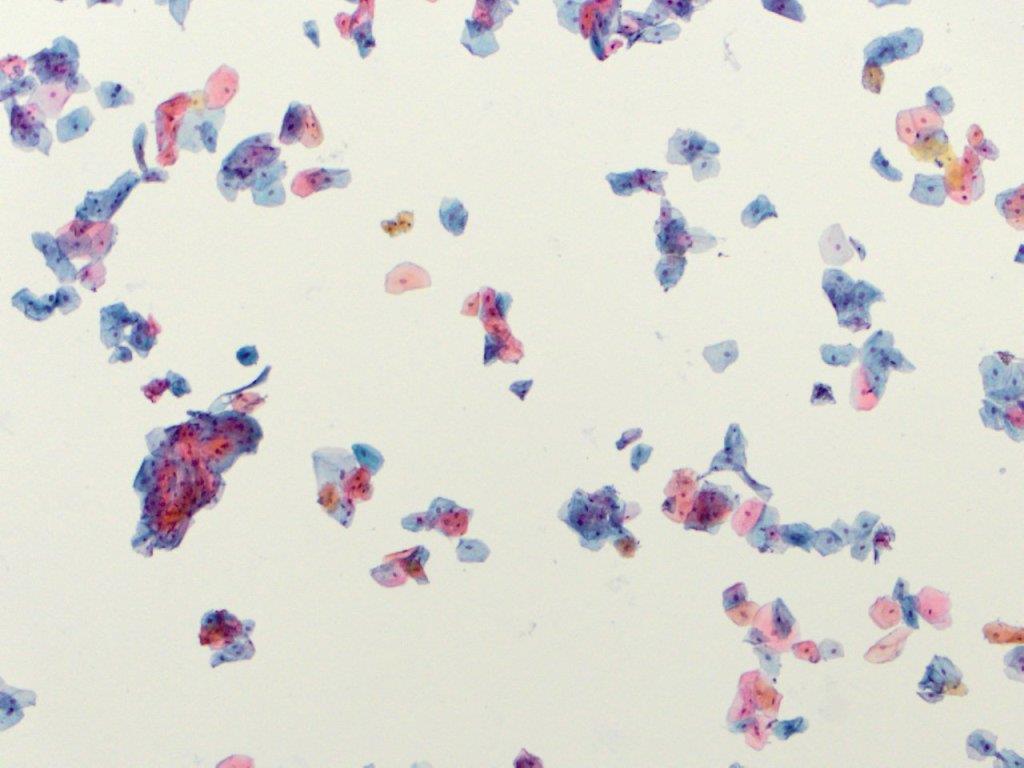
55 55 Papanicoloau
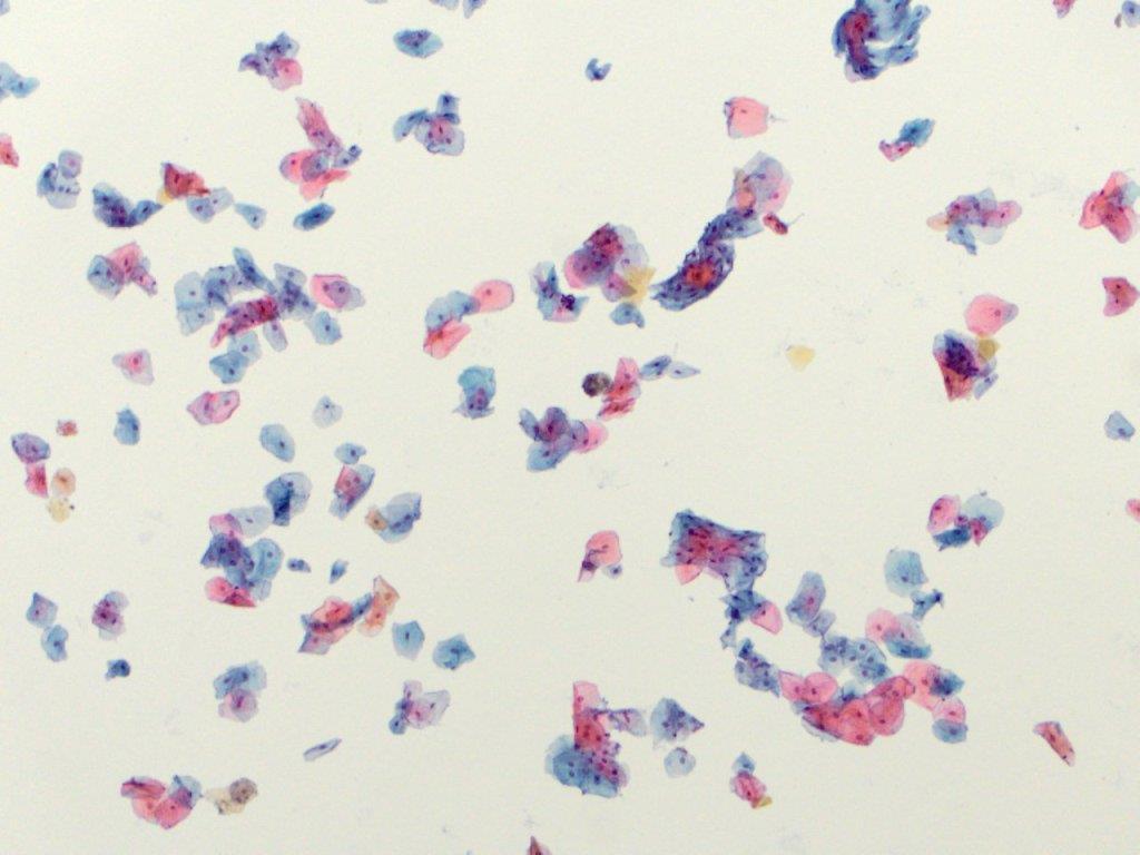
56 56 Papanicoloau
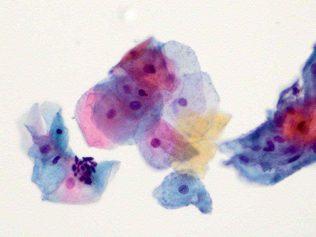
57 57 Papanicoloau
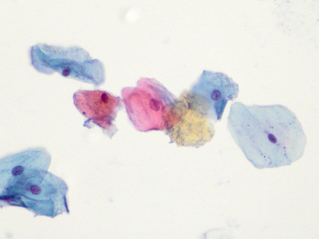
58 58 Papanicoloau

59 59 toluidine blue

60 60 hematoxylin and eosin
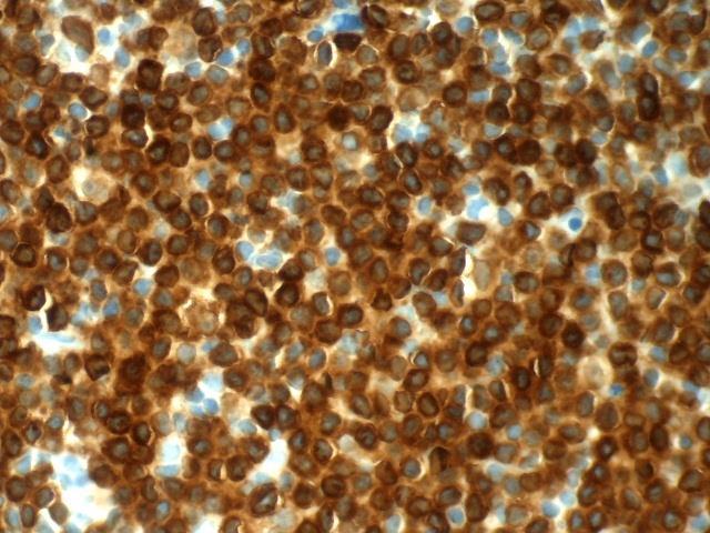
61 61 immunohistochemistry
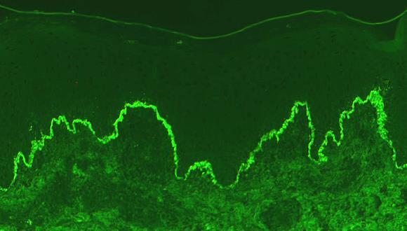
62 62 immunofluorescence
Fixation... Questions 1 Answers 16. Processing... Questions 25 Answers 36. Safety... Questions 67 Answers 73
 Table of Contents Fixation... Questions 1 Answers 16 Processing... Questions 25 Answers 36 Instrumentation... Questions 43 Answers 58 Safety... Questions 67 Answers 73 Laboratory Mathematics & Solution
Atlas of Stains. Special Stains on Artisan Link Pro
 Atlas of Stains Special Stains on Artisan Link Pro Intended use Routinely processed samples (paraffin-embedded) may be used. The preferred fixative is neutral buffered formalin. The clinical interpretation
BLIZARD INSTITUTE CORE PATHOLOGY ATLAS OF TINCTORIAL STAINS
 BLIZARD INSTITUTE CORE PATHOLOGY ATLAS OF TINCTORIAL STAINS Contents Introduction... 3 Background to Tinctorial Stains... 3 Haematoxylin and Eosin Stain (H&E)... 3 Connective Tissue Stains... 4 Nucleic
Schedule of Accreditation issued by United Kingdom Accreditation Service 2 Pine Trees, Chertsey Lane, Staines-upon-Thames, TW18 3HR, UK
 2 Pine Trees, Chertsey Lane, Staines-upon-Thames, TW18 3HR, UK Cellular Pathology Department University College London Hospital Rockefeller Building University Street London WC1E 6JJ Contact: Gavyn Barrett
UNDERSTANDING SPECIAL STAINS
 Vet Times The website for the veterinary profession https://www.vettimes.co.uk UNDERSTANDING SPECIAL STAINS Author : MELANIE DOBROMYLSKYJ Categories : Vets Date : April 14, 2014 MELANIE DOBROMYLSKYJ explains
Preface 1. Fixation and Processing 1
 Contents Preface xi 1. Fixation and Processing 1 Fixation 1 Processing 2 What Should Be Seen in a Well-Fixed, Well-Processed Specimen Stained with Hematoxylin and Eosin 3 Problems Encountered With Fixation
Schedule of Accreditation issued by United Kingdom Accreditation Service 2 Pine Trees, Chertsey Lane, Staines-upon-Thames, TW18 3HR, UK
 2 Pine Trees, Chertsey Lane, Staines-upon-Thames, TW18 3HR, UK 05/09/2018 Cellular Pathology Level 2 Esher Wing Galsworthy Road Kingston upon Thames KT2 7QB Contact: Dr Sussan Gharaie Tel: +44 (0)208 934
Schedule of Accreditation issued by United Kingdom Accreditation Service 2 Pine Trees, Chertsey Lane, Staines-upon-Thames, TW18 3HR, UK
 2 Pine Trees, Chertsey Lane, Staines-upon-Thames, TW18 3HR, UK Paediatric Histopathology Western Bank Sheffield S10 2TH United Kingdom Contact: Tel: +44 (0) 1142 717240 Fax: +44 (0) 1142 706121 E-Mail:
Special Stains in Dermatopathology
 Application Special Stains in Dermatopathology Jameel Ahmad Brown, MD Bruce R. Smoller, MD Fellow: Division of Dermatopathology University of Arkansas for Medical Sciences 4301 W. Markham St. Little Rock,
Schedule of Accreditation issued by United Kingdom Accreditation Service 2 Pine Trees, Chertsey Lane, Staines-upon-Thames, TW18 3HR, UK
 Schedule of Accreditation 2 Pine Trees, Chertsey Lane, Staines-upon-Thames, TW18 3HR, UK Department of Eye Pathology 1 st Floor, Cayton Street Building UCL Institute of Ophthalmology 11-43 Bath Street
Schedule of Accreditation issued by United Kingdom Accreditation Service 2 Pine Trees, Chertsey Lane, Staines-upon-Thames, TW18 3HR, UK
 2 Pine Trees, Chertsey Lane, Staines-upon-Thames, TW18 3HR, UK Laboratory locations: Department of Histopathology Royal United Hospitals NHS Foundation Trust Combe Park Bath BA1 3NG Contact: Lesley Shipway
Schedule of Accreditation issued by United Kingdom Accreditation Service 2 Pine Trees, Chertsey Lane, Staines-upon-Thames, TW18 3HR, UK
 2 Pine Trees, Chertsey Lane, Staines-upon-Thames, TW18 3HR, UK Blizard Institute Core Pathology Pathology and Pharmacy Building Second Floor 80 Newark Street London E1 2ES Contact: Pauline Levey Tel: +44
BenchMark Special Stains. Product guide
 BenchMark Special Stains Product guide Quick reference table Product name Ordering code Catalog number Tissue thickness Tests per kit Total vials in kit package *Not all vials in the kit are used at one
 Index A ABC method. See avidin-biotin enzyme complex method. absolute alcohol and celloidin embedding, 40 for Luxol fast blue staining, 213 rapid processing with, 38 absolute alcohol hematoxylin for Russell
Special Staining (I)
") Special Staining (I) Carbohydrates 1- PERIODIC ACID SCHIFF'S (PAS ) Purpose: Glycogen is present in liver, kidney, skeletal and cardiac muscle. The PAS stain is used to demonstrate neutral polysaccharides
Special Staining (I) Carbohydrates 1- PERIODIC ACID SCHIFF'S (PAS ) Purpose: Glycogen is present in liver, kidney, skeletal and cardiac muscle. The PAS stain is used to demonstrate neutral polysaccharides
Schedule of Accreditation issued by United Kingdom Accreditation Service 2 Pine Trees, Chertsey Lane, Staines-upon-Thames, TW18 3HR, UK
 United Kingdom Accreditation Service 2 Pine Trees, Chertsey Lane, Staines-upon-Thames, TW18 3HR, UK North Tyneside General Hospital Rake Lane North Shields Tyne & Wear NE29 8NH Contact: Ian Taylor Tel:
Carbohydrates and mucins. Dr Phil Bryant, Wales, UK
 Carbohydrates and mucins Dr Phil Bryant, Wales, UK Carbohydrates Provide cells with energy by converting to glucose Excess glucose stored in liver and muscle as glycogen Residual unstored glycogen is turned
3. Staining solutions for histology and cytology Mounting and embedding media... 60
 INDEX 1. Hematoxylin stains... 4 2. Eosin... 6 3. Staining solutions for histology and cytology... 8 4. Reagents I... 48 5. Reagents II... 55 6. Decalcifying solutions... 58 7. Fixatives... 59 8. Formalins...
2014 CURRENT ISSUES IN PATHOLOGY
 2014 CURRENT ISSUES IN PATHOLOGY SPECIAL STAINS IN LIVER BIOPSY PATHOLOGY Sanjay Kakar, MD University of California, San Francisco Trichrome stain : (1) Assess degree of fibrosis. H&E stain is not reliable
HISTOPATHOLOGY. Introduction
 HISTOPATHOLOGY Introduction Contacts Services offered Pathology tissue request Laboratory hours Special instructions Histopathology reports List of specimens Introduction The Histopathology section of
PRICE LIST August 2017-July 2018
 PRICE LIST August 2017-July 2018 Necropsy... 1 Cytology... 2 Biopsy... 2 Histology special stains... 3 Small animal profiles... 4 Equine profiles... 5 Large animal profiles... 5 Standard biochemistry s...
PRICE LIST August 2018-July 2019
 PRICE LIST August 2018-July 2019 Necropsy... 1 Cytology... 2 Biopsy... 2 Histology special stains... 3 Small animal profiles... 4 Equine profiles... 5 Large animal profiles... 5 Standard biochemistry s...
Schedule of Accreditation issued by United Kingdom Accreditation Service 2 Pine Trees, Chertsey Lane, Staines-upon-Thames, TW18 3HR, UK
 2 Pine Trees, Chertsey Lane, Staines-upon-Thames, TW18 3HR, UK Lister Hospital Contact: Heather Taylor Coreys Mill Lane Tel: +44 (0) 01438 285086 Stevenage E-Mail: Heathertaylor3@nhs.net SG1 4AB Website:
CHAPTER. V SUMMARY AND CONCLUSIONS. during postnatal period and to provide elasticity during prenatal and early
 CHAPTER. V SUMMARY AND CONCLUSIONS The present study was conducted on 40 samples, each of thymus and Spleen of goat from prenatal to four months and above age. The small pieces from each thymus and spleen
Schedule of Accreditation issued by United Kingdom Accreditation Service 2 Pine Trees, Chertsey Lane, Staines-upon-Thames, TW18 3HR, UK
 2 Pine Trees, Chertsey Lane, Staines-upon-Thames, TW18 3HR, UK Calderdale and Huddersfield NHS Foundation Trust Calderdale Royal Hospital Salterhebble Halifax HX3 0PW United Kingdom Contact: Dr Richard
Progress in the Development of Microscopical Techniques for Diagnostic Pathology
 Journal of Histotechnology ISSN: 0147-8885 (Print) 2046-0236 (Online) Journal homepage: https://www.tandfonline.com/loi/yhis20 Progress in the Development of Microscopical Techniques for Diagnostic Pathology
Schedule of Accreditation issued by United Kingdom Accreditation Service 2 Pine Trees, Chertsey Lane, Staines-upon-Thames, TW18 3HR, UK
 2 Pine Trees, Chertsey Lane, Staines-upon-Thames, TW18 3HR, UK Department of Cellular Pathology Contact: Karen Wignall Royal Preston Hospital Tel: +44 (0) 1772 523108 Sharoe Green Lane E-Mail: karen.wignall@lthtr.nhs.uk
Microscopy PART - 6. Microscopy. Products for bacteriology. Products for cytology. Products for hematology. Products for histology
 PART - 6 Products for bacteriology Products for cytology Products for hematology Products for histology Universal reagents for For more details contact : Merck Specialities Private Limited Tel : +91 22
Saito, Yutaka; Tsuchiyama, Hideo. Citation Acta medica Nagasakiensia. 1988, 33
 NAOSITE: Nagasaki University's Ac Title Author(s) Histochemical Nature of Eosinophili Adrenal Medulla Kawai, Kioko; Senba, Masachika; Shi Yoshida, Kuniko; Nakatani, Akira; K Saito, Yutaka; Tsuchiyama,
Schedule of Accreditation issued by United Kingdom Accreditation Service 2 Pine Trees, Chertsey Lane, Staines-upon-Thames, TW18 3HR, UK
 2 Pine Trees, Chertsey Lane, Staines-upon-Thames, TW18 3HR, UK Cellular Pathology Laboratory Contact: Breege Nicholson Queen Elizabeth Hospital Tel: +44 (0) 208 333 3000 ext. 8478 Stadium Road E-Mail:
This is Learning Component 6 in Learning Module 1. We will show examples of features ( things ) including mineral deposits, urates, pigments, dust,
 including mineral deposits, urates, pigments, dust,") This is Learning Component 6 in Learning Module 1. We will show examples of features ( things ) including mineral deposits, urates, pigments, dust, plant material, and amyloid. 1 Calcium salts are the
This is Learning Component 6 in Learning Module 1. We will show examples of features ( things ) including mineral deposits, urates, pigments, dust, plant material, and amyloid. 1 Calcium salts are the
Compara've Medicine Animal Histology Services. Glossary of Terms
 Compara've Medicine Animal Histology Services Glossary of Terms About this guide This guide is intended to provide clarifica'on on standard histologic terminology. Included are examples of different sample
HISTOLOGY SPECIMEN COLLECTION MANUAL
 Lee Memorial Health System Lee County, FL CLINICAL LABORATORY HISTOLOGY SPECIMEN COLLECTION MANUAL Abdominal Fluid Minimum Volume No fixative in an appropriate sized container. 1 ml The container must
Special Stains. General Reference Guide
 Special Stains General Reference Guide Table of Contents AFB 4 Technical notes and references 6 Gram 32 Technical notes and references 34 Steiner 64 Technical notes and references 66 Alcian Blue 8 Technical
Fig. 59 Malignant phaeochromocytoma, hepatic metastasis.
 Fig. 59 Malignant phaeochromocytoma, hepatic metastasis. X 120 Hyperte nsion Fig. 60 Malignant sympathetic paraganglioma, lymph node metastasis Primary in bladder. x 1 20 Hypertension Fig. 61 Malignant
Bone Marrow Pathology. Part 1. R.S. Riley, M.D., Ph.D.
 Bone Marrow Pathology Part 1 R.S. Riley, M.D., Ph.D. Bone Marrow Pathology Bone marrow basics Red cell diseases White cell diseases Other diseases Bone Marrow Pathology Bone marrow basics Hematopoiesis
Schedule of Accreditation issued by United Kingdom Accreditation Service 2 Pine Trees, Chertsey Lane, Staines-upon-Thames, TW18 3HR, UK
 2 Pine Trees, Chertsey Lane, Staines-upon-Thames, TW18 3HR, UK Issue No: 002 Issue date: 05 March 2018 Berkshire and Surrey Pathology Services Department of Histopathology Wexham Park Hospital Slough Berkshire
Schedule of Accreditation issued by United Kingdom Accreditation Service 2 Pine Trees, Chertsey Lane, Staines-upon-Thames, TW18 3HR, UK
 2 Pine Trees, Chertsey Lane, Staines-upon-Thames, TW18 3HR, UK Department of Cellular Pathology Dorset County Hospital Williams Avenue Dorchester DT1 2JY Contact: Sharon Wood Tel: +44 (0)1305 254326 E-Mail:
Schedule of Accreditation issued by United Kingdom Accreditation Service 2 Pine Trees, Chertsey Lane, Staines-upon-Thames, TW18 3HR, UK
 2 Pine Trees, Chertsey Lane, Staines-upon-Thames, TW18 3HR, UK 142-144 New Cavendish Street W1W 6YF Contact: Mr Tel: +44 (0) 20 7299 4490 Fax: +44 (0)20 7299 4491 E-Mail: Neil.Barrett@unilabs.com Website:
Schedule of Accreditation issued by United Kingdom Accreditation Service 2 Pine Trees, Chertsey Lane, Staines-upon-Thames, TW18 3HR, UK
 Schedule of Accreditation 2 Pine Trees, Chertsey Lane, Staines-upon-Thames, TW18 3HR, UK Department of Cellular Pathology Pathology Centre Queen Alexandra Hospital Portsmouth PO6 3LY Contact: Julie Williams
ABOUT US WORLDWIDE DISTRIBUTION ICON KEY STAIN LINE PRODUCT LIST
 ABOUT US WORLDWIDE DISTRIBUTION ICON KEY STAIN LINE PRODUCT LIST BQCKit Company is one of the three differentiated business lines of Bioquochem S.L. We are a Spanish biotechnology company specialized
TEST MENU BY SPECIALTY
 1 TEST MENU BY SPECIALTY Breast Pathology Surgical excisions, needle core biopsies, and plastic surgery accepted from all sites Assessment of margins Axillary lymph node dissections Sentinel lymph nodes
Schedule of Accreditation issued by United Kingdom Accreditation Service High Street, Feltham, Middlesex, TW13 4UN, UK
 21-47 High Street, Feltham, Middlesex, TW13 4UN, UK Cellular Pathology Pathology Department Northampton General Hospital Cliftonville Northampton Northamptonshire NN1 5BD Contact: Mary Pendleton Tel: +44
Schedule of Accreditation issued by United Kingdom Accreditation Service 2 Pine Trees, Chertsey Lane, Staines-upon-Thames, TW18 3HR, UK
 2 Pine Trees, Chertsey Lane, Staines-upon-Thames, TW18 3HR, UK Department of Cellular Pathology Forth Valley Royal Hospital Stirling Road Larbert FK5 4WR Contact: Suzanne Ferra Tel: 01324 566643 Tel: +44
CATALOG Parview Road Middleton, WI
 CATALOG 2505 Parview Road Middleton, WI 53562-2579 800-383-7799 www.newcomersupply.com February 2018 ABOUT US Mission Keeping it real: Our job is to help you get your job done! Everything we do is designed
Schedule of Accreditation - draft issued by United Kingdom Accreditation Service 2 Pine Trees, Chertsey Lane, Staines-upon-Thames, TW18 3HR, UK
 - draft 2 Pine Trees, Chertsey Lane, Staines-upon-Thames, TW18 3HR, UK Royal Bournemouth & Christchurch Hospitals NHS Foundation Cellular Pathology Department Royal Bournemouth Hospital Castle Lane East
Schedule of Accreditation issued by United Kingdom Accreditation Service 2 Pine Trees, Chertsey Lane, Staines-upon-Thames, TW18 3HR, UK
 2 Pine Trees, Chertsey Lane, Staines-upon-Thames, TW18 3HR, UK The Princess Alexandra Hospital NHS Trust The Michael Letcher Department of Cellular Pathology The Princess Alexandra Hospital NHS Trust Hamstel
Schedule of Accreditation issued by United Kingdom Accreditation Service 2 Pine Trees, Chertsey Lane, Staines-upon-Thames, TW18 3HR, UK
 2 Pine Trees, Chertsey Lane, Staines-upon-Thames, TW18 3HR, UK Department of Neuropathology Contact: Catherine Rowe Pathology Science Building (Phase II) Tel: +44 (0)1179 505 050 Southmead Hospital E-Mail:catherine.rowe@nbt.nhs.uk
Mucoprotein-containing histiocytes (muciphages)
") J. clin. Path. (1966), 19, 368 Mucoprotein-containing histiocytes (muciphages) in the rectum J. G. AZZOPARDI AND D. J. EVANS' From the Department of Morbid Anatomy, Postgraduate Medical School, London
J. clin. Path. (1966), 19, 368 Mucoprotein-containing histiocytes (muciphages) in the rectum J. G. AZZOPARDI AND D. J. EVANS' From the Department of Morbid Anatomy, Postgraduate Medical School, London
THIOFLAVIN-T FOR AMYLOID DETECTION
 THE AMERICAN JOURNAL OP CLINICAL PATHOLOGY Copyright 67 by The Williams & Wilkins Co. Vol. 7, No. Printed in U.S.A. THIOFLAVIN-T FOR AMYLOID DETECTION S. M. SAEED, M.D., AND GERALD FINE, M.D. Department
Neuropathology Inflammation, Infection, Demyelination in the CNS
 Neuropathology Inflammation, Infection, Demyelination in the CNS PathoBasic 2016-09-20 Jürgen Hench Inflammation in the CNS inflammation generally as a reaction against pathogen, substance, necrotic, or
Schedule of Accreditation issued by United Kingdom Accreditation Service 2 Pine Trees, Chertsey Lane, Staines-upon-Thames, TW18 3HR, UK
 2 Pine Trees, Chertsey Lane, Staines-upon-Thames, TW18 3HR, UK Department of Clinical Neuropathology King s College Hospital NHS Foundation Trust Denmark Hill London SE5 9RS Contact: Lawrence Doey Tel:
Schedule of Accreditation issued by United Kingdom Accreditation Service 2 Pine Trees, Chertsey Lane, Staines-upon-Thames, TW18 3HR, UK
 2 Pine Trees, Chertsey Lane, Staines-upon-Thames, TW18 3HR, UK Cellular Pathology Bedford Hospital NHS Trust Kempston Road Bedford MK42 9DJ Contact: Neil Cully Tel: +44 (0) 2907182500 Fax: +44 (0) 2071888313
PATHOPHYSIOLOGY. DEFINED Involves the study of function that results from disease processes.
 DEFINED Involves the study of function that results from disease processes. What is pathology? Pathology is the branch of medical sciences that treats the essential nature of disease, especially the changes
Special stains in liver pathology
 Current Issues in Surgical Pathology 2014 Special stains in liver pathology Which, why, how Really? Sanjay Kakar, MD University of California, San Francisco Outline Which stains Why the stain is done How
THE NAME OF THE STAINS:
 THE NAME OF THE STAINS: HISTOLOGY HISTORY PART 2 Jean Mitchell, BS, HT (ASCP) Newcomer Supply Middleton, Wisconsin jmitchell@newcomersupply.com Jane Parr, BS, HT (ASCP) National Jewish Health & University
PRACTICAL HISTOPATHOLOGY IN MOUSE MODELS OF HUMAN DISEASE: GUIDES TO PHENOTYPING THE GENETICALLY ALTERED MOUSE
 PRACTICAL HISTOPATHOLOGY IN MOUSE MODELS OF HUMAN DISEASE: GUIDES TO PHENOTYPING THE GENETICALLY ALTERED MOUSE http://mousepheno.ucsd.edu/ https://www.ncbi.nlm.nih.gov/pmc/articles/pmc3693904/ 1. Approval
Microscopic Examination of Urine
 Download http://www.vetlab.com/kova.htm Definition of urine sediment: all solid materials suspended in the urine - a semiquantative evaluation of the urine sediment Significance of formed elements in the
Non-hematogenous endogenous pigments
 Non-hematogenous endogenous pigments 0 This group contains the following : 1. Melanins. 2. Lipofuscins. 3. Chromaffin. 4. Pseudomelanosis. 5. Dubin-Johnson pigments. 6. Ceroid-type lipofuscins. 7. Hamazaki-Weisenberg
The Differentiation of Yeast and Yeast-Like Forms in Human Tissues. Introduction. Histochemical Stains Used to Detect Fungi. Histopathologic Diagnoses
 The Differentiation of Yeast and Yeast-Like Forms in Human Tissues Gary W. Procop, MD Chair, Clinical Pathology Staff, Anatomic Pathology Director, Molecular Microbiology, Mycology, and Parasitology Cleveland
Amyloid neuropathy: (A) scattered Congo Red positive material in endoneurium displays apple green birefringence under polarized light.
 scattered Congo Red positive material in endoneurium displays apple green birefringence under polarized light.") Cơ vân Cơ vân Cơ vân (cắt dọc) Amyloid neuropathy: (A) scattered Congo Red positive material in endoneurium displays apple green birefringence under polarized light. (A, Congo Red). Amyloid neuropathy:
Cơ vân Cơ vân Cơ vân (cắt dọc) Amyloid neuropathy: (A) scattered Congo Red positive material in endoneurium displays apple green birefringence under polarized light. (A, Congo Red). Amyloid neuropathy:
Nerve. Jacqueline Brooks, HTL, SCT, MB (ASCP) Q IHC
 Q IHC") Nerve Jacqueline Brooks, HTL, SCT, MB (ASCP) Q IHC 1 Housekeeping 2 IHCs (Neural & Neuroendocrine Calcitonin Chromogranin A Gastrin Glial Fibrillary Acidic Protein (GFAP) Glucagon Insulin Myelin Basic
Nerve Jacqueline Brooks, HTL, SCT, MB (ASCP) Q IHC 1 Housekeeping 2 IHCs (Neural & Neuroendocrine Calcitonin Chromogranin A Gastrin Glial Fibrillary Acidic Protein (GFAP) Glucagon Insulin Myelin Basic
Practical Histology. Lab 3: Connective tissue
 Practical Histology Lab 3: Connective tissue Connective tissues Connective tissue provides structural support for the body by binding cells and tissues together to form organs. It also provides metabolic
Schedule of Accreditation issued by United Kingdom Accreditation Service 2 Pine Trees, Chertsey Lane, Staines-upon-Thames, TW18 3HR, UK
 Schedule of ccreditation United Kingdom ccreditation Service 2 Pine Trees, Chertsey Lane, Staines-upon-Thames, TW18 3HR, UK Issue No: 003 Issue date: 27 pril 2018 ccredited to Leighton Hospital Middlewich
Autopsy findings in 51 year-old man with mantle cell lymphoma
 Autopsy findings in 51 year-old man with mantle cell lymphoma Bobbi S. Pritt, MD, MSc Professor of Laboratory Medicine and Pathology Mayo Clinic Disclosure of Relevant Financial Relationships USCAP requires
NACC Vascular Consortium. NACC Vascular Consortium. NACC Vascular Consortium
 NACC Vascular Consortium NACC Vascular Consortium Participating centers: Oregon Health and Science University ADC Rush University ADC Mount Sinai School of Medicine ADC Boston University ADC In consultation
Schedule of Accreditation issued by United Kingdom Accreditation Service 2 Pine Trees, Chertsey Lane, Staines-upon-Thames, TW18 3HR, UK
 2 Pine Trees, Chertsey Lane, Staines-upon-Thames, TW18 3HR, UK Christie Hospital Manchester M20 4BX Contact: Neil Wrathall Tel: +44 (0) 161 918 7264 Fax: +44 (0) 161 446 8549 E-Mail: neil.wrathall@christie.nhs.uk
Hemosiderin. Livia Vida 2018
 Hemosiderin Livia Vida 2018 Questions Histochemical caracteristics of the different pigments. Exogenous pigments. Hemoglobinogenic pigments. Causes and forms of jaundice. Hemoglobinogenic pigments. Pathological
Individual cells Extracellular matrix
 Connective Tissue Connective Tissue Elements Individual cells Extracellular matrix»fibers» Collagen» Elastic» Reticular»Ground Substance» PG (proteoglycans)» GAG (glycosaminoglycan)» GP (glycoprotein)
Schedule of Accreditation issued by United Kingdom Accreditation Service 2 Pine Trees, Chertsey Lane, Staines-upon-Thames, TW18 3HR, UK
 2 Pine Trees, Chertsey Lane, Staines-upon-Thames, TW18 3HR, UK Great Ormond Street Hospital for Children NHS Foundation Trust Department of Histopathology Floor 3 Camelia Botnar Laboratories, Great Ormond
Schedule of Accreditation issued by United Kingdom Accreditation Service 2 Pine Trees, Chertsey Lane, Staines-upon-Thames, TW18 3HR, UK
 2 Pine Trees, Chertsey Lane, Staines-upon-Thames, TW18 3HR, UK Department of Cellular Pathology Pathology Centre C37 New Cross Hospital Wolverhampton Road Wolverhampton WV10 0QP United Kingdom Contact:
Schedule of Accreditation issued by United Kingdom Accreditation Service 2 Pine Trees, Chertsey Lane, Staines-upon-Thames, TW18 3HR, UK
 2 Pine Trees, Chertsey Lane, Staines-upon-Thames, TW18 3HR, UK operating as North East Essex and Suffolk Pathology Service (NEESPS) Cellular Pathology Contact: Lynn Partridge Ipswich Hospital Tel: +44
Campylobacter like organisms on the gastric mucosa:
 J Clin Pathol 1984;37:1002-1006 Campylobacter like organisms on the gastric mucosa: culture, histological, and serological studies DM JONES,* AM LESSELLS,t JOAN ELDRIDGE* From the *Public Health Laboratory
Serotonin- and Somatostatin-Positive Goblet Cell Carcinoid of the Duodenum
 2012 66 4 351 356 Serotonin- and Somatostatin-Positive Goblet Cell Carcinoid of the Duodenum a b* c c c a a b d a c b d 352 Ohara et al. received remedies at another hospital. Hematemesis then recurred
Histo lab 7. Special connective tissue is derived from the mesoderm (mesenchyme).
.") Histo lab 7 Special connective tissue is derived from the mesoderm (mesenchyme). If we have high density of fibers, we call it dense connective tissue. (Fibers are more than the ground substance). If we
Histo lab 7 Special connective tissue is derived from the mesoderm (mesenchyme). If we have high density of fibers, we call it dense connective tissue. (Fibers are more than the ground substance). If we
Overview of Disease Mechanisms in the Eye. Christopher M Reilly, DVM, MAS, DACVP Basic Science Course June 7 8, 2016 NC State University
 Overview of Disease Mechanisms in the Eye Christopher M Reilly, DVM, MAS, DACVP Basic Science Course June 7 8, 2016 NC State University * Outline Tips on pathology submissions General processes and definitions
Tissues 10/21/2016. Epithelial Tissue
 Tissues This is a generalized cell diagram. It shows the anatomy of a cell, but most cells do not actually look like this. Cells can have a wide variety of shapes and sizes, depending on their function.
Flow Cytometry. What is flow cytometry?
 Flow Cytometry Flow Cytometry What is flow cytometry? Flow cytometry is a popular laser-based technology to analyze the characteristics of cells or particles. It is predominantly used to measure fluorescence
HISTOLOGY Lecture TWO DR. ASHRAF SAID
 HISTOLOGY Lecture TWO DR. ASHRAF SAID Start Of this lecture TISSUES TISSUE: A DEFINITION A group of connected and interdependent cells that cooperate to perform a specific function CONNECTIVE TISSUE The
Human Anatomy and Physiology- Problem Drill 04: Tissues of the Body
 Human Anatomy and Physiology- Problem Drill 04: Tissues of the Body Question No. 1 of 10 A biopsy sample is obtained from a lesion on the right cheek of a male patient. A technician in the histology lab
Atrophy. Dystrophy. II. practical training 2 rd year Dentistry. Lucie Tučková
 Atrophy. Dystrophy. II. practical training 2 rd year Dentistry Lucie Tučková Atrophy Decrease in size of the cell or organ Reduction in cell size and/or cell number, or both Atrophic cells may have diminished
They cells can not function death.
 Jenna Hellack Jan 2001 Tissues What do you think happens when the cells use up their food and oxygen before there is time to replenish it? They cells can not function death. Blood Cell Cancer cell Plant
Basic Tissue Types and Functions
 Tissues Histology Basic Tissue Types and Functions 1) Epithelial tissue covering 2) Connective tissue support 3) Muscle tissue movement 4) Nervous tissue control Epithelial Tissue 1) Covers a body surface
Case Report Intracytoplasmic Crystalline Inclusions in the Hepatocytes of an Antelope
 SAGE-Hindawi Access to Research Veterinary Medicine International Volume 2010, Article ID 373698, 4 pages doi:10.4061/2010/373698 Case Report Intracytoplasmic Crystalline Inclusions in the Hepatocytes
Histological Spectrum of Pure Neuritic Leprosy:
 Department of Histopathology Postgraduate Institute of Medical Education & Research Chandigarh,India Histological Spectrum of Pure Neuritic Leprosy: Experience at Tertiary Care Centre Dr Uma Nahar INTRODUCTION
Tissues. Tissues - Overview. Bio211 Laboratory 2. Epithelial and Connective Tissues
 Bio211 Laboratory 2 Epithelial and Connective Tissues 1 Tissues Tissues to be examined under the microscope Epithelial Tissue (p. 79 Lab Manual) [TODAY] Connective Tissue (p. 93 Lab Manual) [TODAY] Muscle/Nervous
Argentaffin and non-argentaffin carcinoid tumours
 J. clin. Path. (1968), 21, 60 Argentaffin and non-argentaffin carcinoid tumours of the appendix F. E. DISCHE From Dulwich Hospital Pathology Department (King's College Hospital), London SYNOPSIS Tumours
BioSciences. Peripheral Blood Smear Preparation. Blood Smear Preparation Materials
 PolyFacts Vol. 5 No. 1 BioSciences Use the Wright Stain High Quality StainRITE Ready-to-Use Stains for Hematology Peripheral blood smear (peripheral blood film) is a glass microscope slide coated with
Mucin Histochemistry Study of the Prostate in Normal and Malignant Lesions
 ISSN 2231-4261 ORIGINAL ARTICLE Mucin Histochemistry Study of the Prostate in Normal and Malignant Lesions 1* 1 1 1 Manoj P.Ambali, Megha A. Doshi, Pratibha P. Patil, Shweta H. Chavan 1 Department of Anatomy,
Schedule of Accreditation issued by United Kingdom Accreditation Service 2 Pine Trees, Chertsey Lane, Staines-upon-Thames, TW18 3HR, UK
 National Hospital for Neurology & Neurosurgery UCLH Queen Square London WC1N 3BG United Kingdom Contact: Linda Herbert Tel: +44 (0) 2034567890 E-Mail: linda.herbert@uclh.nhs.uk Website: www.uclh.nhs.uk
Histology Review Can you identify the Cell Structures? Can you identify the Stain? Can you identify the Cell type?
 Histology Review Can you identify the Cell Structures? Can you identify the Stain? Can you identify the Cell type? 2.01 Border of Epithelia (fluorescence) M A: lamina basalis B: epithelium C: other tissues
Extracellular degeneration
 Extracellular degeneration By Dr. Hemn Hassan Othman PhD, Pathology Fall 2016 1/17/2017 1 Extracellular Degenerations I / Hyaline Degeneration (Hyalinization): The ward hyaline is derived from the Latin
TISSUE PATHWAYS FOR INFLAMMATORY AND NON-NEOPLASTIC DERMATOSES AND NON-NEOPLASTIC LESIONS
 The Royal College of Pathologists Pathology: the science behind the cure TISSUE PATHWAYS FOR INFLAMMATORY AND NON-NEOPLASTIC DERMATOSES AND NON-NEOPLASTIC LESIONS September 2008 Unique document number
Chapter 1: Cells and Tissues
 Chapter 1: Cells and Tissues Cells and Tissues Carry out all chemical activities needed to sustain life Cells are the building blocks of all living things Tissues are groups of cells that are similar in
Body Tissues. Cells are specialized for particular functions Tissues - groups of cells with similar structure. and function Four primary tissue types:
 Chapter 3 Tissues Body Tissues Cells are specialized for particular functions Tissues - groups of cells with similar structure and function Four primary tissue types: Epithelium Connective tissue Nervous
vinous Varieties of So-called Mammary Carcinoid Tumors Solid and
 and vinous Varieties of Socalled Mammary Carcinoid Tumors EDWIN R. FISHER, M.D., ALKA S. PALEKAR, MD., AND NSABP COLLABORATORS Fisher, Edwin R., Palekar, Alka S., and NSABP collaborators: and mucinous
Ordering Physician. Collected REVISED REPORT. Performed. IgG IF, Renal MCR. Lambda IF, Renal MCR. C1q IF, Renal. MCR Albumin IF, Renal MCR
 RenalPath Level IV Wet Ts IgA I Renal IgM I Renal Kappa I Renal Renal Bx Electron Microscopy IgG I Renal Lambda I Renal C1q I Renal C3 I Renal Albumin I Renal ibrinogen I Renal Mayo Clinic Dept. of Lab
[General Pathology] Introduction to Pathology
![[General Pathology] Introduction to Pathology](/thumbs/73/69585662.jpg "[General Pathology] Introduction to Pathology") Introduction to Pathology Pathology: Literally translated, pathology is the study (logos) of disease (pathos, suffering). It involves the investigation of the causes of disease and the associated changes
Introduction to Pathology Pathology: Literally translated, pathology is the study (logos) of disease (pathos, suffering). It involves the investigation of the causes of disease and the associated changes
Presented by: Dr. Giuseppe Molinaro Dr. Davide De Biase
 Presented by: Dr. Giuseppe Molinaro Dr. Davide De Biase Dog Spayed Female LABRADOR RETRIEVER 3 Years old VACCINATIONS ANTIPARASITIC COMMERCIAL DIET VOMITING FOR A MONTH DULLNESS WEIGHT LOSS INAPPETANCE
What is histology? HISTOLOGY
 Introduction to Histology What is histology? HISTOLOGY histo = tissue ogy = study So HISTOLOGY = the study of tissues! What is a TISSUE? Tissues are groups of cells with specialized structural and functional