Clinical Differential Diagnosis 4/16/2018 DERMATOPATHOLOGY OF THE GENITALIA AND BREAST NO CONFLICTS TO DISCLOSE
|
|
|
- Phillip Rich
- 5 years ago
- Views:
Transcription
1 DERMATOPATHOLOGY OF THE GENITALIA AND BREAST JOHN S. METCALF, MD Professor of Pathology and Dermatology MUSC NO CONFLICTS TO DISCLOSE Clinical Differential Diagnosis Plaques: Erythematous Inflammatory Non-infectious» Eczematous dermatitis (irritant contact dermatitis, allergic contact dermatitis, neurodermatitis )» Psoriasis» Lichen Planus and fixed drug Infectious» Bacterial» Dermatophytes» Candidiasis Neoplastic Squamous Cell Carcinoma in situ (Bowen s Disease) Paget s Disease (mammary and extramammary) Inflammatory carcinoma Cutaneous T-Cell Lymphoma (Mycosis Fungoides) 1
, superficial")


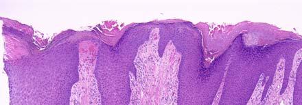
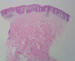
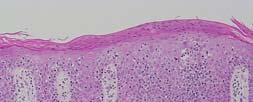
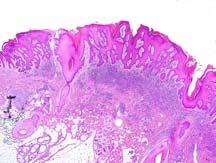
2 Spongiotic Dermatitis Acute: Cornified layers show normal architecture; epidermis of normal thickness, but with intercellular edema (spongiosis), superficial perivascular infiltrate (sometimes with eosinophils) Subacute: Usually areas of parakeratosis with serum, some epidermal hyperplasia, less pronounced spongiosis Chronic: Compact orthokeratosis sometimes with areas of parakeratosis, irregular psoriasiform epidermal hyperplasia, sometimes residual spongiosis Acute Spongiotic Dermatitis 2

3 Spongiotic dermatitis with spongiotic vesiculation 3


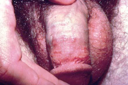
4 Eczematous dermatitis (contact dermatitis) 4


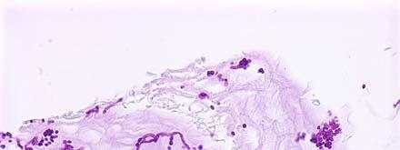
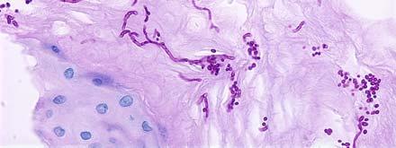
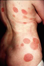
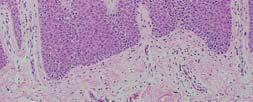
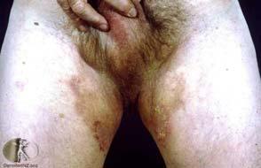
5 Psoriasis Erythrasma Corynebacterium minutissimum Superfical fungal infection and candidiasis Superfical inflammatory infiltrate, sometimes with neutrophils Cornified layers contain spores and/hyphae or pseudohyphae Diastase Periodic Acid- Schiff stain may be helpful but sometimes not necessary 5

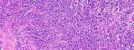
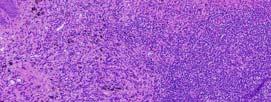
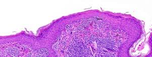
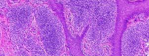
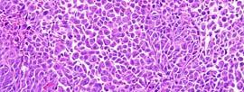
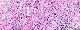
6 CTCL- MF 6


 Fixed Drug")
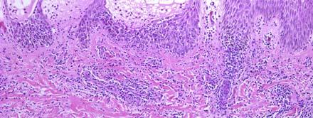
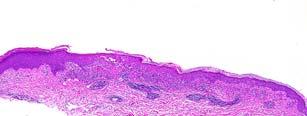
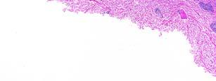
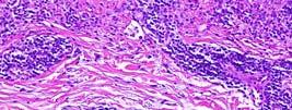
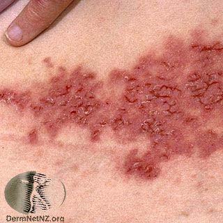
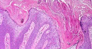
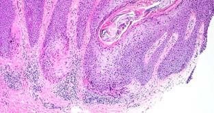
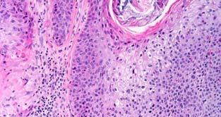
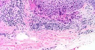
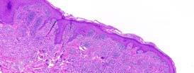
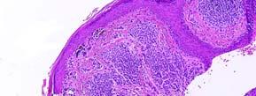
7 Lichenoid Lichen Planus Lichen Sclerosis Plasma cell balanitis Lichen planus-like keratosis (breast) Fixed Drug Erythema Multiforme 7


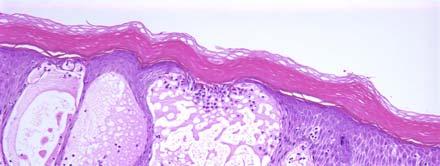
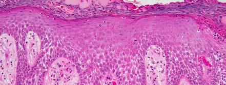
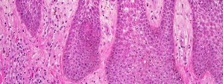
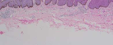
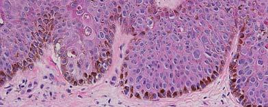
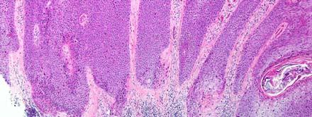
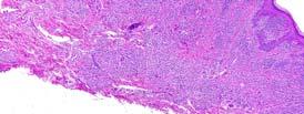
8 Lichen Planus and Lichen Sclerosis DDX Early stage disease: Both show lichenoid interface inflammation and early on may be impossible to differentiate; however: Loss of basal keratinocytes with squamatization of basal layer, necrotic keratinocytes, and saw-toothing usually not seen in LS Homoginization and sclerosis of papillary dermis often can be identified in early stage LS, but usually not in LP Lichen Planus Lichen Planus 8


9 Lichen Planus Lichen Planus Lichen planus-like keratosis 9

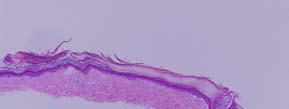

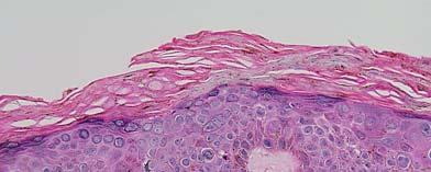
10 Lichen sclerosis Lichen sclerosis Lichen sclerosis 10
 Fixed Drug")

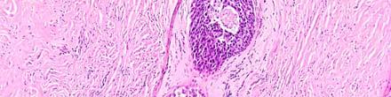
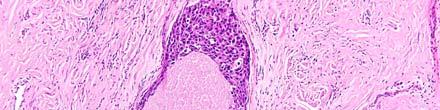
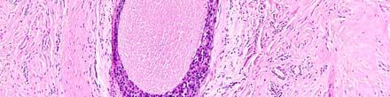
11 Plasma cell balanitis (Zoon s) Plasma cell balanitis Fixed Drug Reaction and Erythema Multiforme 11


12 Erythema multiforme Fixed drug Fixed drug 12


13 Fixed drug VESICULOBULLOUS DISORDERS Pemphigoid Bullous Cicatricial Pemphigus Vulgaris Vegitans Hailey-Hailey disease Darier s disease Acantholytic dermatosis of the vulvocrural area Bullous pemphigoid 13

14 Pemphigus vulgaris Hailey-Hailey (benign familial pemphigus) Hailey-Hailey 14


 and")
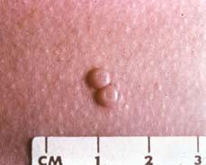
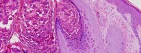
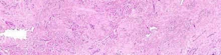
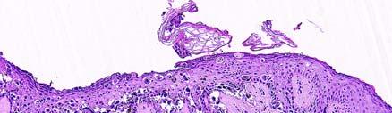
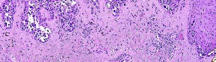
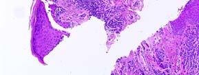
15 Darier s (acantholytic dyskeratosis) Clinical Differential Diagnosis Papules and papulovesicles Inflammatory Non-infectious Flat-topped: lichen planus Fixed drug Infectious Condyloma lata Molluscum contagiosum Herpes Simplex (I and II) and Varicella Zoster Condyloma Lata 15


16 16
17 Clinical Differential Diagnosis Ulcers: Superficial: Infectious: Syphilis Herpes Granuloma inguinale Non-infections Lipschutz ulcer Excoriation or trauma Deep Pyoderma gangrenosum Necrotizing fasciitis 17


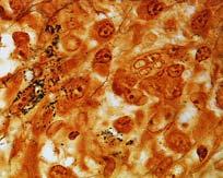

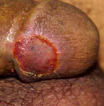
18 Steiner Primary syphilis T. Pal Granuloma inguinale 18
 Often")
19 Pyoderma Gangrenosum vs. Necrotizing Fasciitis and Fournier s Gangrene Pyoderma Gangrenosum Erythematous pustule or nodule becoming an ulcer with undermined edges Sometimes initiated by trauma (or surgery) Often an associated systemic illness such as Ulcerative Colitis, Diabetes Often begins as sterile follicular inflammation with abscess formation Pyoderma gangrenosum 19


20 Pyoderma gangrenosum Necrotizing Fasciitis of the Genitalia (Fournier s Gangrene) Usually associated with diabetes High mortality rate Often polymicrobial as opposed to extragenital NF which is usually caused by Group A strep Deep vessel thrombosis and necrosis of deep soft tissues Positive tissue and blood cultures usually Fournier s gangrene 20

21 Necrotizing fasciitis Necrotizing fasciitis Necrotizing fasciitis 21
 Clinical Differential Diagnosis Nodules and tumors Ectopic")
22 DDX NF: Often deep vessel thrombosis with necrosis and edema of septa and fascia, positive blood and tissue cultures Often minimal epidermal change PG: Often subepidermal edema, undermining inflammation at edge of ulcer Usually no deep vessel thrombosis or deep necrosis Cultures are negative (unless secondarily infected) Clinical Differential Diagnosis Nodules and tumors Ectopic glandular tissue Adnexal tumors Benign Malignant Invasive Carcinomas Mesenchymal tumors Metastatic tumors Hidradenoma papilliferum (MLG adenoma) 22

23 Accessory nipplevulva Adenosis-like glandular proliferation of vulva 23


24 ER P63 Invasive adenocarcinoma with lobular features- vulva 24


25 Adenoid cystic carcinoma (Bartholin s Duct) 25
 Neoplastic Benign Benign keratoses")
 Condylomata Borderline")
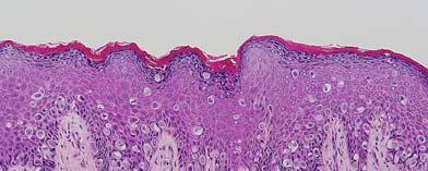
26 Clinical Differential Diagnosis Papules (con t) Neoplastic Benign Benign keratoses (seborrheic keratosis or seborrheic keratosislike) Condylomata Borderline Bowenoid papulosis 26
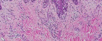
27 Seborrheic Keratosis-like Lesions Lesions having the architecture and histologic features characteristic of seborrheic keratosis, but occurring of skin in the genital region, are frequently associated with Human Papilloma Virus infection. 27

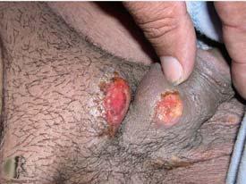
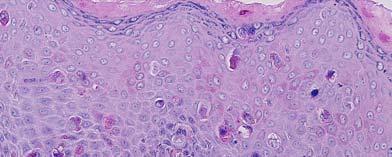
28 BOWENOID PAPULOSIS Predilection for sexually active young adults In males, commonly involves glans and foreskin; in females, vulvar lesions which are often pigmented and bilateral Uncommonly gives rise to invasive SCC Usually associated with HPV 16 Differentiation from Bowen s Disease histologically may be impossible 28
29 Bowen s Disease Cutaneous squamous cell carcinoma in situ Predilection for sun-damaged skin (solar irradiation) In genital region, associated chiefly with HPV types 16,18, 31, 33, and others Expanding asymptomatic erythematous plaque 29
 Bowen s Disease which is associated")


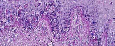
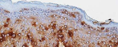
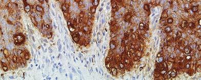
30 Bowen s disease Bowen s disease Carcinoma of the Vulva Two pathways: Classic VIN (uvin) Bowen s Disease which is associated with HPV infections (especially types 16 and 18) p16 immunostain usually diffusely positive; high proliferative index Differentiated (simplex) VIN Often accompanied by chronic inflammatory dermatoses Lichen sclerosis Lichen simplex chronicus p-53 immunostain of limited help 30
31 uvin uvin uvin 31

32 uvin uvin 32


33 uvin uvin uvin- Bowen s disease with Pagetoid histology 33

34 uvin with invasion 34


35 Lichen sclerosis with adjacent SCC Differentiated VIN associated with Lichen Sclerosis 35
36 Verrucous Carcinoma of the Genitalia Vulvar verrucous carcinomas are usually not associated with HPV and are typically associated with chronic inflammatory dermatoses. However, HPV (6, 11, 16, 18) have implicated in some verrucous carcinomas of the glans penis and the genitocrural areas- proposed name warty (condylomatous) squamous cell carcinoma Pure verrucous carcinoma is generally slow growing, locally destructive, but seldom metastasizes 36
37 Verrucous Carcinoma of the Genitalia: Strict Criteria Well differentiated verrucous/papillary carcinoma Minimal nuclear atypia Blunt epithelial-stromal interface Absence of co-existing conventional keratinizing SCC 37


38 Expanding Erythematous or Pigmented Plaques with or without associated tumors Epithelial: Paget s Disease Mammary Extramammary Bowen s Disease 38

 H. Jacobaeus concluded that Paget s D.")

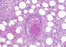
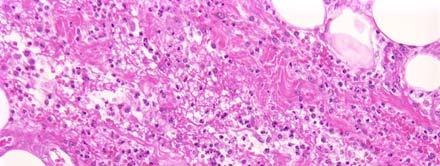
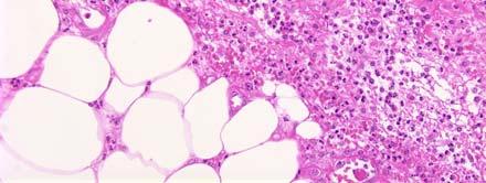
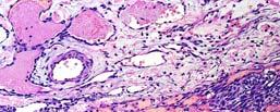
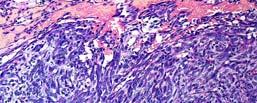
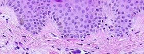
39 Paget s Disease (mammary and extramammary) (1874) Sir James Paget described an eruption of the nipple/areola complex with characteristics of ordinary chronic eczema or psoriasis which was followed by the development of breast cancer. (1904) H. Jacobaeus concluded that Paget s D. was a carcinoma from its conception, originating from the breast parenchyma and extending along the duct system to the skin. In fewer than 5% of cases is no underlying breast carcinoma found. 39


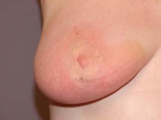
40 Paget s disease of breast Duct carcinoma in situ with overlying Paget s disease 40


41 Pigmented Paget s disease- Breast 41


42 SOX-10 CK5/6 CEA Extramammary Paget s Disease Tends to occur in sites with a high density of apocrine glands. Erythematous, enlarging plaque. Female predominance with vulva being most common site. 42

 If no underlying carcinoma is found: In situ adnexal")

43 Extramammary Paget s Disease: Histogenesis: Not thought to be uniform: 25% - underlying adnexal adenocarcinoma 10-15% - internal malignancy (rectum, cervix, prostate, bladder, etc.) If no underlying carcinoma is found: In situ adnexal carcinoma Origin from pluripotential cells in epidermis Extramammary Paget s disease Extramammary Paget s disease 43
44 Extramammary Paget s disease ZOON S BALANITIS AND VULVITIS AB-D-PAS CEA 44
45 Extramammary Paget s disease CK7 cck7 45
; aka labia")
46 Clinical Differential Diagnosis Pigmented lesions Benign Melanotic macules Melanocytic nevi (special site features) Malignant Pigmented Bowen s disease Pigmented Paget s disease Melanoma Genital melanotic macule (melanosis); aka labia lentigo 46
47 Characteristics of Special Site Nevi Usually occur in young women A mostly nested junctional component: Nests variably sized, but may be large, and oriented vertically or horizontally Cells within nests may be dyshesive Sometimes a crowded junctional component with ill-defined nests and single unit melanocytes Pagetoid spread present in a minority of cases, but is focal and centrally located Histologic Patterns of Special Site Nevi 1. Nested: large nests perpendicular or parallel to DEJ 2. Dyshesive: junctional nests approaching confluence, with cellular dyshesion 3. Crowded: poorly defined nests with interspersed single cells, obscuring DEJ Additional Features Concerning features: Epithelioid cells with prominent nucleoli Occasional pagetoid scatter, centrally Dermal fibrosis Reassuring features: Symmetry Sharp dermarcation Maturation of dermal component Low dermal proliferation 47


48 Vulvar nevus- 37 y.o. Special site nevus of breast 48


49 Special site nevus, breast, with congenital features (38 y.o) 49

50 Sp. Site nevus (congenital features) 32y.o. man 50


51 Sp. Site nevusinguinal crease- 6 y.o. 6 y.o. girl (inguinal crease) 51
52 ssuprapubic nevus 52
53 Vulvar Melanoma 53
54 Vulvar melanoma- 88 y.o. woman 54


55 Melanoma of Breast- 46 y.o. woman 55
56 Molecular Diagnosis 1) Multiple chromosomal gains or losses can be detected by FISH or CGH and can be of assistance in differentiating melanoma from melanocytic nevi. 2) Current FISH probes are fairly successful in differentiating histologically benign nevi from melanomas. 3) However up to this point, in histologically ambiguous melanocytic tumors, FISH test results do not generally show unequivocal features of melanoma or nevi, but rather show inconsistencies (borderline melanocytic tumors). 56
 Atypical")
 C-MYC")


57 Endothelial Tumors of the Skin of the Breast (post-irradiation) Atypical vascular proliferation Mitoses usually lacking (Ki67 may be helpful) C-MYC negative Post-irradiation angiosarcoma Mitoses usually present C-MYC usually positive Lymphangiosarcoma arising in the setting of chronic lymphedema (Stewart-Treves) Post-irrad AVL (Ki67) 57


 58")
58 Post irradiation angiosarcoma Post-irradiation angiosarcoma Post-irrad angiosarc (Ki67) 58

59 C-MYC Stewart-Treves 59
60 Inflammatory Carcinoma of the Breast 60
61 61


62 Case presentation 66 y.o. woman with a biopsy of right breast skin. History of lung cancer with metastasis. 62

")
63 Pitfalls in Diagnosis of Breast Carcinoma (related to skin) Experience will teach you things you wish you didn t have to learn - Harv Read Case Presentation 56 y.o. woman with a core needle biopsy of the left breast (12:00, 1 cm from nipple) Clinical Information: Breast mass- fibroadenoma vs debris-filled cyst vs cancer 63

 resected from")
64 Triple negative The rest of the story: Previously: pt3b N2c melanoma (with satellites) resected from right face 64


65 MART-1- breast SOX10- lung Case Presentation: 60 y.o. woman with a history of poorly differentiated invasive duct carcinoma of the breast (with negative nodes) five years ago, presented with a well defined right axillary mass. Core needle biopsy performed. GATA 3 and CK7 positive 65
66 Lumpectomy performed with axillary dissection (negative) 66
67 67
68 Nodule arising adjacent to mastectomy scar Nodular hidradenoma 68
:81:347-355. Weedon s Skin Pathology (4 th Ed). Editor: James W. Patterson.")

 Two distinct pathways to development of squamous cell carcinoma of the vulva.")

69 Selected References Smith GL, Bunker CB, and Dinneen MD. Fournier s Gangrene (Review). British Journal of Urology (1998):81: Weedon s Skin Pathology (4 th Ed). Editor: James W. Patterson. Churchill Livingstone Reyes M C and Cooper K. An update on vulvar intraepithelial neoplasia: terminology and a practical approach to diagnosis. J Clin Pathol 2014 Apr.;67(4): Ueda Y, et. Al. (Review article) Two distinct pathways to development of squamous cell carcinoma of the vulva. Journal of skin cancer. Vol 2011 Mason AR, et al. Nevi of Special Sites. Clinics in laboratory Medicine, 2011, 31: Muhlbauer A, et.al. The correlation of the standard 5 probe FISH assay with melanocytic tumors of uncertain malignant potential. Annals of Diagnostic Pathology. June pp Hoda SA, et al. Rosen s Breast Pathology, 4 th Ed. Wolters Kluver2014. Chapter 39 (Sarcoma), pp
Benign and malignant epithelial lesions: Seborrheic keratosis: A common benign pigmented epidermal tumor occur in middle-aged or older persons more
 Benign and malignant epithelial lesions: Seborrheic keratosis: A common benign pigmented epidermal tumor occur in middle-aged or older persons more common on the trunk; but extremities, head and neck are
Pathology of the skin. Dr Fónyad László, 1sz. Patológiai és Kísérleti Rákkutató Intézet, SE
 Pathology of the skin Dr Fónyad László, 1sz. Patológiai és Kísérleti Rákkutató Intézet, SE The skin Biggest organ Kb. 1.8 nm Kb. 10 kg Most frequent site for tumor development (BCC) Pathology of the skin
Basal cell carcinoma 5/28/2011
 Goal of this Presentation A practical approach to the diagnosis of cutaneous carcinomas and their mimics Thaddeus Mully, MD University of California San Francisco To review common non-melanoma skin cancers
Histopathology: skin pathology
 Histopathology: skin pathology These presentations are to help you identify, and to test yourself on identifying, basic histopathological features. They do not contain the additional factual information
Dermatopathology: The tumor is composed of keratinocytes which show atypia, increase mitoses and abnormal mitoses.
 Squamous cell carcinoma (SCC): A common malignant tumor of keratinocytes arising in the epidermis, usually from a precancerous condition: 1- UV induced actinic keratosis, usually of low grade malignancy.
Pathology of the skin. 2nd Department of Pathology, Semmelweis University
 Pathology of the skin 2nd Department of Pathology, Semmelweis University Histology of the skin Epidermis: Stratum corneum Stratum granulosum Stratum spinosum Stratum basale Dermis: papillary and reticular
04/09/2018. Squamous Cell Neoplasia and Precursor Lesions. Agenda. Squamous Dysplasia. Squamo-proliferative lesions. Architectural features
 Squamous Cell Neoplasia and Precursor Lesions Jennifer L. Hunt, MD, MEd Aubrey J. Hough Jr, MD, Endowed Professor of Pathology Chair of Pathology and Laboratory Medicine University of Arkansas for Medical
Simulators of melanoma
 Simulators of melanoma Philip E. LeBoit, M.D. Depts. of Pathology and Dermatology University of California, San Francisco Simulators of melanoma Simulators of melanoma in situ Melanocytic Non-melanocytic
VULVAR CARCINOMA. Page 1 of 5
 VULVAR CARCINOMA EXAMPLE OF A VULVAR CARCINOMA USING PROPOSED TEMPLATE Case: Invasive squamous cell carcinoma arising in D-VIN Tumor in left labia major Left partial vaginectomy and sentinel lymph node
Actinic keratosis (AK): Dr Sarma s simple guide
: Dr Sarma s simple guide") Actinic keratosis (AK): Dr Sarma s simple guide Actinic keratosis is a very common lesion that you will see in your day-to-day practice. First, let me explain the name Actinic keratosis. It means keratosis
Actinic keratosis (AK): Dr Sarma s simple guide Actinic keratosis is a very common lesion that you will see in your day-to-day practice. First, let me explain the name Actinic keratosis. It means keratosis
Genital Lesions in Dermatopathology
 Genital Lesions in Dermatopathology Janis M. Taube, MD Director of Dermatopathology Associate Professor of Dermatology and Pathology Johns Hopkins University SOM Overview Vulvovaginal pathology Inflammatory
Chapter 6 Squamous Cell Carcinoma: Variants and Challenges
 Chapter 6 Squamous Cell Carcinoma: Variants and Challenges Michael B. Morgan EPIDEMIOLOGY: Second most common skin cancer, rare in the dark-skinned races. ETIOLOGY: Ultraviolet light, HPV infection. PATHOGENESIS:
Squamous Cell Neoplasia and Precursor Lesions
 Squamous Cell Neoplasia and Precursor Lesions Jennifer L. Hunt, MD, MEd Aubrey J. Hough Jr, MD, Endowed Professor of Pathology Chair of Pathology and Laboratory Medicine University of Arkansas for Medical
Diseases of the vulva
 Diseases of the vulva 1. Bartholin Cyst - Infection of the Bartholin gland produces an acute inflammation within the gland (adenitis) and may result in an abscess. Bartholin duct cysts - Are relatively
BAP-oma & BEYOND MICHAEL A NOWAK, MD
 BAP-oma & BEYOND MICHAEL A NOWAK, MD CONFLICTS No conflicts with the content of this lecture BAP-oma Wiesner 2011: Families with multiple tan dome-shaped papules of head, neck, trunk, and extremities.
Genital Lesions in Dermatopathology
 Genital Lesions in Dermatopathology Janis M. Taube, MD Director of Dermatopathology Associate Professor of Dermatology and Pathology Johns Hopkins University SOM Overview Vulvovaginal lesions Non-Neoplastic
Inflammatory Dermatoses of the Vulva for the General/Gyn Pathologist with emphasis in the lichenoid pattern
 Inflammatory Dermatoses of the Vulva for the General/Gyn Pathologist with emphasis in the lichenoid pattern By Konstantinos Linos MD, FCAP, FASDP Bone, Soft Tissue and Dermatopathology Assistant Professor
Appendageal skin tumors
 Appendageal skin tumors Ibrahim Khalifeh, M.D. Associate Professor Department of Pathology American University of Beirut Medical Center Beirut, Lebanon Appendageal tumors Neoplasms whose differentiation
Nomenclature (terminology) refers to the naming of
 refers to the naming of") 2011 ISSVD Terminology and Classification of Vulvar Dermatological Disorders: An Approach to Clinical Diagnosis Peter J. Lynch, MD, Micheline Moyal-Barracco, MD, James Scurry, MD, and Colleen Stockdale,
2011 ISSVD Terminology and Classification of Vulvar Dermatological Disorders: An Approach to Clinical Diagnosis Peter J. Lynch, MD, Micheline Moyal-Barracco, MD, James Scurry, MD, and Colleen Stockdale,
11/1/18. Age and Vulvar Pathology ANATOMY. Prepuce Clitoris Vestibule Hart line. Labia. minora. Labia. majora. Fourchette.
 Age and Vulvar Pathology Maria Angelica Selim, MD Professor of Pathology and Dermatology Director Dermatopathology Unit Duke University Medical Center Labia minora Labia majora Bartholin s duct Prepuce
Mucinoses Diverse group of disorders which have in common deposition of basophilic, finely granular and stringy material in the connective tissues of
 Cutaneous Mucinoses Nathan C. Walk, M.D. Mucinoses Diverse group of disorders which have in common deposition of basophilic, finely granular and stringy material in the connective tissues of the dermis.
Dysplasia, Mimics and Other Controversies
 Dysplasia, Mimics and Other Controversies Mary S. Richardson, MD Dept. of Pathology Medical University of South Carolina Charleston, SC Notice of Faculty Disclosure In accordance with ACGME guidelines,
Clinically Microscopically Pathogenesis: autoimmune not lifetime
 Vulvar Diseases: Can be divided to non-neoplastic and neoplastic diseases. The neoplastic diseases are much less common. Of those, squamous cell carcinoma is the most common. most common in postmenopausal
MECHANISMS OF HUMAN DISEASE: LABORATORY SESSION PATHOLOGY OF THE SKIN LAB. Friday, February 12, :30 am 11:00 am
 MECHANISMS OF HUMAN DISEASE: LABORATORY SESSION PATHOLOGY OF THE SKIN LAB Friday, February 12, 2012 9:30 am 11:00 am FACULTY COPY GOALS: Describe the basic clinical and morphologic features of various
المركب النموذج--- سبيتز وحمة = Type Spitz's Nevus, Compound SPITZ NEVUS 1 / 7
 SPITZ NEVUS 1 / 7 Epidemiology An annual incidence rate of 1.4 cases of Spitz nevus per 100,000 individuals has been estimated in Australia, compared with 25.4 per 100,000 individuals for cutaneous melanoma
Important Decisions in Dermatopathology: The Clinico- Pathologic Correlation. Dermatopathology Specialists Needed. Changing Trends
 Important Decisions in Dermatopathology: The Clinico- Pathologic Correlation Uma Sundram, MD, PhD Departments of Pathology and Dermatology Stanford University May 29, 2008 Dermatopathology Specialists
Gross appearance of nodular hyperplasia in material obtained from suprapubic prostatectomy. Note the multinodular appearance and the admixture of
 Tiền liệt tuyến Tiền liệt tuyến Gross appearance of nodular hyperplasia in material obtained from suprapubic prostatectomy. Note the multinodular appearance and the admixture of solid and microcystic areas.
When Immunostains Can Get You in Trouble: Gynecologic Pathology p16: Panacea or Pandora s Box?
 When Immunostains Can Get You in Trouble: Gynecologic Pathology p16: Panacea or Pandora s Box? Teri A. Longacre, MD Stanford Medicine Stanford California pi6 in Gynecologic Pathology: Panacea or Pandora
Acantholytic Anaplastic Extramammary Paget s Disease: A Case Report and Review of the Literature
 Ann Dermatol Vol. 23, Suppl. 2, 2011 http://dx.doi.org/10.5021/ad.2011.23.s2.s226 CASE REPORT Acantholytic Anaplastic Extramammary Paget s Disease: A Case Report and Review of the Literature Yu-Jin Oh,
Desmoplastic Melanoma R/O BCC. Clinical Information. 74 y.o. man with lesion on left side of neck r/o BCC
 R/O BCC Sabine Kohler, M.D. Professor of Pathology and Dermatology Dermatopathology Service Stanford University School of Medicine Clinical Information 74 y.o. man with lesion on left side of neck r/o
Diseases of the breast (1 of 2)
") Diseases of the breast (1 of 2) Introduction A histology introduction Normal ducts and lobules of the breast are lined by two layers of cells a layer of luminal cells overlying a second layer of myoepithelial
Diseases of the breast (1 of 2) Introduction A histology introduction Normal ducts and lobules of the breast are lined by two layers of cells a layer of luminal cells overlying a second layer of myoepithelial
Mody. AIS vs. Invasive Adenocarcinoma of the Cervix
 Common Problems in Gynecologic Pathology Michael T. Deavers, M.D. Houston Methodist Hospital, Houston, Texas Common Problems in Gynecologic Pathology Adenocarcinoma in-situ (AIS) of the Cervix vs. Invasive
MECHANISMS OF HUMAN DISEASE: LABORATORY SESSION PATHOLOGY OF THE SKIN LAB. Friday, February 13, :30 am 11:00 am
 MECHANISMS OF HUMAN DISEASE: LABORATORY SESSION PATHOLOGY OF THE SKIN LAB Friday, February 13, 2009 9:30 am 11:00 am FACULTY COPY GOALS: Describe the basic clinical and morphologic features of various
Self assesment Case 21
 17-18 MAY 2018 London Dermatopathology Symposium 2018 Self assesment Case 21 MARC HASPESLAGH CASE 21 1802-50585 48 year old lady with eczematous lesions at ear helix and red patch on nose bridge since
Index. Angiosarcoma diagnosis, 47 lymphedema-related vs. non-lymphedemarelated, 48
 A Acneiform rash biopsy, 134 cetuximab, EGFR, 132 133 diagnosis, 131 patient history, 131 134 treatment, 134 135 Acne vulgaris, 109 AGA. See Androgenetic alopecia Alopecia areata, 148 American Joint Committee
PRINCESS MARGARET CANCER CENTRE CLINICAL PRACTICE GUIDELINES GYNECOLOGIC CANCER VULVAR
 PRINCESS MARGARET CANCER CENTRE CLINICAL PRACTICE GUIDELINES GYNECOLOGIC CANCER VULVAR Last Revision Date July 2015 1 Site Group: Gynecologic Cancer Vulvar Author: Dr. Stephane Laframboise 1. INTRODUCTION
Whitney A. High, MD, JD, MEng
 ADS Dermatopathology Meeting 2014 Selected Adnexal Tumors Whitney A. High, MD, JD, MEng Associate Professor, Dermatology & Pathology Director of Dermatopathology (Dermatology) University of Colorado School
Lichenoid Tissue Reaction in Malignant Melanoma A Potential Diagnostic Pitfall
 natomic Pathology / LICHENOID TISSUE RECTION IN MLIGNNT MELNOM Lichenoid Tissue Reaction in Malignant Melanoma Potential Diagnostic Pitfall CPT Scott R. Dalton, MC, US, 1,3 Capt Matt. aptista, USF, MC,
NEOPLASMS OF THE SURFACE EPITHELIUM (KERATINOCYTES)
") NEOPLASMS OF THE SURFACE EPITHELIUM (KERATINOCYTES) Papillary Lesions Precancerous Lesions Keratinocyte Proliferations Carcinomas Melanotic Lesions Melanomas Normal Mucosa Keratin layer Spinous layer Basal
NEOPLASMS OF THE SURFACE EPITHELIUM (KERATINOCYTES) Papillary Lesions Precancerous Lesions Keratinocyte Proliferations Carcinomas Melanotic Lesions Melanomas Normal Mucosa Keratin layer Spinous layer Basal
MULLERIAN PAPILLOMA ENTITY RECOGNITION FAILURE 04/04/2016 OUT OF SIGHT, OUT OF MIND: LESSER KNOWN LESIONS OF THE VULVOVAGINAL TRACT
 OUT OF SIGHT, OUT OF MIND: LESSER KNOWN LESIONS OF THE VULVOVAGINAL TRACT 23 rd ANNUAL SEMINAR IN PATHOLOGY 30 APRIL 2016 W. Dwayne Lawrence MD MSc (Path.) Chief of Pathology and Laboratory Medicine Women
Mousa. Israa Ayed. Abdullah AlZibdeh. 0 P a g e
 1 Mousa Israa Ayed Abdullah AlZibdeh 0 P a g e Breast pathology The basic histological units of the breast are called lobules, which are composed of glandular epithelial cells (luminal cells) resting on
Table of Contents: Part 1 Medical Dermatology. Chapter 1 Acneiform Disorders. Acne. Acne Vulgaris. Pomade Acne. Steroid Acne
 Table of Contents: Part 1 Medical Dermatology Chapter 1 Acneiform Disorders Acne Acne Vulgaris Pomade Acne Steroid Acne Infantile Acne Pediatric Perspectives Neonatal Acne (Acne Neonatorum) Pediatric Perspectives
BREAST PATHOLOGY MCQS
 BREAST PATHOLOGY MCQS 1) :The most important factor in breast enlargement during pregnancy is A. stromal edema B. secretion of chorionic gonadotropin C. glandular hyperplasia D. proliferation of stroma
Dermatopathology. Dr. Rafael Botella Estrada. Hospital La Fe de Valencia
 Dermatopathology Dr. Rafael Botella Estrada. Hospital La Fe de Valencia Melanoma and mimics Dr. Martin Mihm Malignant lesions result from the accumulation of mutations Class I lesions (benign) Class II
IT S FUNDAMENTAL MY DEAR WATSON! A SHERLOCKIAN APPROACH TO DERMATOLOGY
 IT S FUNDAMENTAL MY DEAR WATSON! A SHERLOCKIAN APPROACH TO DERMATOLOGY Skin, Bones, and other Private Parts Symposium Dermatology Lectures by Debra Shelby, PhD, DNP, FNP-BC, FADNP, FAANP Debra Shelby,
Malignant tumors of melanocytes: Part 1. Deba P Sarma, MD., Omaha
 Malignant tumors of melanocytes: Part 1 Deba P Sarma, MD., Omaha The melanocytic tumor is one of the most difficult and confusing areas in Dematopathology. It is true that most (95%) of such lesions are
Disorders of the vulva
 Vulval lesions Disorders of the vulva Terminology standardised by the International Society for the Study of Vulvovaginal Disease(ISSVD) Classification 1.Nonneoplastic epithelial disorders of vulva Lichen
Benign versus Cancerous Lesions How to tell the difference FMF 2014 Christie Freeman MD, CCFP, DipPDerm, MSc
 1 Benign versus Cancerous Lesions How to tell the difference FMF 2014 Christie Freeman MD, CCFP, DipPDerm, MSc Benign lesions Seborrheic Keratoses: Warty, stuck-on Genetics and birthdays Can start in late
Vulva Inflammatory Disorders Lichen Planus Fixed Drug Eruption Erythema Multiforme Plasmacytosis Mucosae (Zoon) Lichen Sclerosus Allergic Contact
 Lichen Sclerosus Allergic Contact") Vulva Inflammatory Disorders Lichen Planus Fixed Drug Eruption Erythema Multiforme Plasmacytosis Mucosae (Zoon) Lichen Sclerosus Allergic Contact Dermatitis Psoriasis Lichen Simplex Chronicus Foreign Body
Vulva Inflammatory Disorders Lichen Planus Fixed Drug Eruption Erythema Multiforme Plasmacytosis Mucosae (Zoon) Lichen Sclerosus Allergic Contact Dermatitis Psoriasis Lichen Simplex Chronicus Foreign Body
BREAST PATHOLOGY. Fibrocystic Changes
 BREAST PATHOLOGY Lesions of the breast are very common, and they present as palpable, sometimes painful, nodules or masses. Most of these lesions are benign. Breast cancer is the 2 nd most common cause
Clinicopathologic Self- Assessment S003 AAD 2017
 Clinicopathologic Self- Assessment S003 AAD 2017 Clay J. Cockerell, M.D. Director, Cockerell Dermatopathology Director, Division of Dermatopathology UT Southwestern Medical Center July 2017 No relevant
Atlas of Eyelid and Conjunctival Tumors
 Atlas of Eyelid and Conjunctival Tumors Jerry A. Shields, M.D. Director, Ocular Oncology Service Wills Eye Hospital Professor of Ophthalmology Thomas Jefferson University Philadelphia, Pennsylvania Carol
Uropathology January Jon Oxley
 Uropathology January 2012 Jon Oxley Background to seminar These slides were available to view via the web from scanned slides The junior pathologists answered questions on them via the web The answers
Diploma examination. Dermatopathology: First paper. Tuesday 21 March Candidates must answer FOUR questions ONLY. Time allowed: Three hours
 Dermatopathology: First paper Tuesday 21 March 2017 1. Discuss the role of fluorescent in-situ hybridization (FISH) and emerging molecular techniques in the diagnosis of cutaneous melanocytic lesions,
5/21/2018. Prostate Adenocarcinoma vs. Urothelial Carcinoma. Common Differential Diagnoses in Urological Pathology. Jonathan I.
 Common Differential Diagnoses in Urological Pathology Jonathan I. Epstein Prostate Adenocarcinoma vs. Urothelial Carcinoma 1 2 NKX3.1 NKX3.1 3 4 5 6 Proposed ISUP Recommendations Option to use PSA as a
LYMPHATIC DRAINAGE AXILLARY (MOSTLY) INTERNAL MAMMARY SUPRACLAVICULAR
 INTERNAL MAMMARY SUPRACLAVICULAR") BREAST LYMPHATIC DRAINAGE AXILLARY (MOSTLY) INTERNAL MAMMARY SUPRACLAVICULAR HISTOLOGY LOBE: (10 in whole breast) LOBULE: (many per lobe) ACINUS/I, aka ALVEOLUS/I: (many per lobule) DUCT(S): INTRA- or
BREAST LYMPHATIC DRAINAGE AXILLARY (MOSTLY) INTERNAL MAMMARY SUPRACLAVICULAR HISTOLOGY LOBE: (10 in whole breast) LOBULE: (many per lobe) ACINUS/I, aka ALVEOLUS/I: (many per lobule) DUCT(S): INTRA- or
Disorders of Cell Growth & Neoplasia. Histopathology Lab
 Disorders of Cell Growth & Neoplasia Histopathology Lab Paul Hanna April 2010 Case #84 Clinical History: 5 yr-old, West Highland White terrier. skin mass from axillary region. has been present for the
Gross Appearance & Histology of Skin Cancer. Kyle Mannion M.D. January 21, 2005
 Gross Appearance & Histology of Skin Cancer Kyle Mannion M.D. January 21, 2005 Actinic Keratosis 5-20% will develop squamous/basal cell ca Almost solely from solar damage Usually develop during 4 th decade
Vulvar Disease Clinical Cases
 Vulvar Disease Clinical Cases Hope K. Haefner, MD Professor University of Michigan Health System February 27, 2015 Cartagena Conflicts of Interest Hope Haefner, MD is on the Advisory Board of Merck Co.,
!! 2 to 3% of All New Visceral Cancers.!! Peak Incidence is 6th Decade!! M:F = 2:1
 !! Kathleen M. O Toole, M.D.!! 2 to 3% of All New Visceral Cancers!! Peak Incidence is 6th Decade!! M:F = 2:1!! Grossly is a Bright Yellow, Necrotic Mass with a Pseudocapsule 1 !!Conventional RCC! Clear
ISPUB.COM. Seborrheic Keratosis: A Pictorial Review of the Histopathologic Variations. D Sarma, S Repertinger
 ISPUB.COM The Internet Journal of Dermatology Volume 7 Number 2 Seborrheic Keratosis: A Pictorial Review of the Histopathologic Variations D Sarma, S Repertinger Citation D Sarma, S Repertinger.. The Internet
Proceedings of the Southern European Veterinary Conference - SEVC -
 Close this window to return to IVIS www.ivis.org Proceedings of the Southern European Veterinary Conference - SEVC - Sep. 30-Oct. 3, 2010, Barcelona, Spain Next SEVC Conference: Sep. 30-Oct. 2, 2011 -
Breast pathology. 2nd Department of Pathology Semmelweis University
 Breast pathology 2nd Department of Pathology Semmelweis University Breast pathology - Summary - Benign lesions - Acute mastitis - Plasma cell mastitis / duct ectasia - Fat necrosis - Fibrocystic change/
CASE 4 21/07/2017. Ectopic Prostatic Tissue in Cervix. Female 31. LLETZ for borderline nuclear abnormalities
 Female 31 CASE 4 LLETZ for borderline nuclear abnormalities PSA Ectopic Prostatic Tissue in Cervix AJSP 2006;30;209-215 usually incidental microscopic finding usually in ectocervical stroma? developmental
2 to 3% of All New Visceral Cancers Peak Incidence is 6th Decade M:F = 2:1 Grossly is a Bright Yellow, Necrotic Mass with a Pseudocapsule
 GENITOURINARY PATHOLOGY Kathleen M. O Toole, M.D. Renal Cell Carcinoma 2 to 3% of All New Visceral Cancers Peak Incidence is 6th Decade M:F = 2:1 Grossly is a Bright Yellow Necrotic Mass Grossly is a Bright
Penile cancer teams in UK. Common variants. Penile cancer teams. Basaloid squamous carcinoma. The Pathology of Penile Tumours
 The Pathology of Penile Tumours Dr Jonathan H Shanks The Christie NHS Foundation Trust, Manchester, UK Penile cancer teams in UK 12 centres for penile cancer work (10 in England and Wales, 2 in Scotland)
Contents. 3 Diagnostic Tests and Studies Introduction Examination... 27
 Contents 1 Normal Anatomy... 1 1.1 Introduction... 1 1.2 Surface Landmarks... 1 1.3 Oral Mucosa... 3 1.4 Tongue... 5 1.5 Floor of Mouth... 6 1.6 Palate... 6 1.7 Dentition... 7 1.8 Temporomandibular Joint...
CLINICAL SIGNIFICANCE OF BENIGN EPITHELIAL CHANGES
 Papillomas. Papillomas are composed of multiple branching fibrovascular cores, each having a connective tissue axis lined by luminal and myoepithelial cells ( Fig. 23-11 ). Growth occurs within a dilated
Periocular Malignancies
 Periocular Malignancies Andrew Gurwood, O.D., F.A.A.O., Dipl. Marc Myers, O.D., F.A.A.O. Drs. Myers and Gurwood have no financial interests to disclose. Course Description Discussion of the most common
NEVI: A PROBLEM OF MISDIAGNOSIS*
 NEVI: A PROBLEM OF MISDIAGNOSIS* MARTIN SWERDLOW, M.D. Department of Pathology J Michael Reese Hospital, Chicago, Illinois The recurring discrepancy between the clinical and pathologic diagnosis of nevus
Diploma Examination. Dermatopathology: First paper. Tuesday 20 March Candidates must answer FOUR questions. Time allowed: 3 hours
 Dermatopathology: First paper Tuesday 20 March 2018 Candidates must answer FOUR questions Time allowed: 3 hours 1. Give an account of the genetic aberrations encountered in Spitzoid neoplasms and how these
Rash Decisions Approach to the patient with a skin condition
 National Conference for Nurse Practitioners April 25, 2014 Rash Decisions Approach to the patient with a skin condition Margaret A. Bobonich, DNP, FNP C, DCNP, FAANP Assistant Professor, Case Western Reserve
4Ps LUMPS AND BUMPS B.L.&T. BUMPS, LUMPS, AND TATTOOS. Most Common BUMP in the oral cavity Fibroma INTERDENTAL PAPILLAE LESIONS
 B.L.&T. BUMPS, LUMPS, AND TATTOOS LUMPS AND BUMPS DIFFERENTIAL DIAGNOSIS FOR LUMPS AND BUMPS Traumatic Fibroma Papilloma Epulis Fissuratum Inflammatory Papillary Hyperplasia Lesions of Attached Gingiva
Breast Pathology. Breast Development
 Breast Pathology Lecturer: Hanina Hibshoosh, M.D. Reading: Kumar, Cotran, Robbins, Basic Pathology, 6th Edition, pages 623-635 Breast Development 5th week - thickening of the epidermis - milk line 5th
Premalignant skin tumours
 Chapter 14: Premalignant skin tumours page: 434 Premalignant skin tumours page: 435 Solar keratoses (senile keratoses) Raised red and well-defined plaques with a rough surface covered in scales of varying
Clinicopathologic Self-Assessment
 Clinicopathologic Self-Assessment Handout Symposium (S003), July 27 th 2017 Maija Kiuru MD PhD Assistant Professor, Departments of Dermatology & Pathology University of California Davis CASE 1: History
David B. Troxel, MD. Common Medicolegal Situations: Misdiagnosis of Melanoma
 Common Medicolegal Situations: Misdiagnosis of Melanoma David B. Troxel, MD Medical Director, The Doctors Company, Napa, California Clinical Professor Emeritus, University of California at Berkeley Past
Dermoscopy: Recognizing Top Five Common In- Office Diagnoses
 Dermoscopy: Recognizing Top Five Common In- Office Diagnoses Vu A. Ngo, DO Department of Family Medicine and Dermatology Choctaw Nation Health Services Authority Learning Objectives Introduction to dermoscopy
Cutaneous Adnexal Tumors
 Cutaneous Adnexal Tumors Lesions with Predominant Follicular Differentiation Special Emphasis on Basal Cell Carcinoma 2014-04-01 Prof. Dr. med. Katharina Glatz Pathologie Cutaneous Adnexal Tumors Hair
PATHOLOGY OF THE SKIN 2. Tumours of the skin
 PATHOLOGY OF THE SKIN 2. Tumours of the skin Máirín E. McMenamin MB MRCPI FRCPath Dip (Dermatopathol) RCPath St. James s Hospital and University of Dublin, Trinity College Tumour (Neoplasia) Benign or
Papillary Lesions of the breast
 Papillary Lesions of the breast Emad Rakha Professor of Breast Pathology The University of Nottingham Papillary lesions of the breast are a heterogeneous group of disease, which are characterised by neoplastic
Benign Mimics of Malignancy in Breast Pathology
 Arthur Purdy Stout Society of Surgical Pathologists Companion Meeting Benign Mimics of Malignancy in Breast Pathology Stuart J. Schnitt, M.D. Beth Israel Deaconess Medical Center and Harvard Medical School,
Melanocytic Lesions: Use of Immunohistochemistry and Special Studies Napa Valley 2018
 Melanocytic Lesions: Use of Immunohistochemistry and Special Studies Napa Valley 2018 Victor G. Prieto, MD, PhD Professor Depts. of Pathology and Dermatology University of Texas - MD Anderson Cancer Center
Mousa. Lina Al-Lawama. Enas Ajarma. 0 P a g e
 3 Mousa Lina Al-Lawama Enas Ajarma 0 P a g e رب اشرح لي صدري ويسر لي أمري واحلل عقدة من لساني يفقهوا قولي -Last time we were talking about vulvar pathology (non neoplastic epithelial disorder :lichen chronicus
Pathology. Skin Tumor. Bayan N. Mohammad 15/10/2015. Mohammad al-orjani. Page 0 of 23
 #7 35 Pathology Skin Tumor Bayan N. Mohammad 15/10/2015 Mohammad al-orjani Page 0 of 23 بسم هللا الرحمن الرحيم GREETINGS This lecture is about skin tumors, all the slides are included and every slide will
UNIVERSITY OF MEDICINE AND PHARMACY OF CRAIOVA FACULTY OF MEDICINE DOCTORAL THESIS SUMMARY
 UNIVERSITY OF MEDICINE AND PHARMACY OF CRAIOVA FACULTY OF MEDICINE DOCTORAL THESIS SUMMARY CLINICAL, HISTOPATHOLOGICAL AND IMMUNOHISTOCHEMICAL STUDY OF THE EPITHELIAL PRECANCEROUS LESIONS PRECURSORS OF
Common Benign Lesions and Skin Cancers. 22nd May 2015 Dr Mark Foley
 Common Benign Lesions and Skin Cancers 22nd May 2015 Dr Mark Foley Thank you for downloading this file. This intended to supplement the presentation given at the NZ Wound Care Conference, it is not intended
Vulvar Carcinoma. Definition: Cases should be classified as carsinoma of the vulva when the primary site growth is in the vulva Malignant melanoma sho
 Carcinoma Vulva & Vagina Subdivisi Onkologi Ginekologi Bagian Obgin FK USU Vulvar Carcinoma. Definition: Cases should be classified as carsinoma of the vulva when the primary site growth is in the vulva
Diagnoses of Cases 1. Lentigo, other melanosis and the acquired nevus 2. Variations on the acquired nevus 3. Dermal melanocytosis
 Diagnoses of Cases 1. Lentigo, other melanosis and the acquired nevus 1 1A. Lentigo simplex 4 1B. Psoralens and ultraviolet A (PUVA) lentigo 6 1C. Solar lentigo 8 1D. Café au lait macule 10 1E. Ink-spot
NEOPLASIA. 3. Which of the following tumour is benign a. Chondrosarcoma b. Osteochondroma c. Chondroblastoma d. Ewing s tumour e.
 NEOPLASIA 1. malignant neoplasms a. are independent of hormonal influence b. are always composed of homogenous cell lines c. arise from differentiated cells by a process of anaplasia d. display abnormal
Case Report Scrotal Apocrine Adenocarcinoma with Pagetoid Phenomenon and Inguinal Lymph Node Metastases
 Volume 2016, Article ID 8353745, 4 pages http://dx.doi.org/10.1155/2016/8353745 Case Report Scrotal Apocrine Adenocarcinoma with Pagetoid Phenomenon and Inguinal Lymph Node Metastases Aristeidis Hristos
Management of pediatric melanocytic lesions
 Open Journal of Clinical & Medical Case Reports Management of pediatric melanocytic lesions Volume 3 (2017) Issue 8 ISSN 2379-1039 Jin Kim, BS; Emmanuel Gabriel MD, PhD; Weiguo Liu MD, PhD; Lin Lin MD,
Epithelial tumors. Dr. F.F. Khuzin, PhD Dr. M.O. Mavlikeev
 Epithelial tumors Dr. F.F. Khuzin, PhD Dr. M.O. Mavlikeev Epithelial tumors Tumors from the epithelium are the most frequent among tumors. There are 2 group features of these tumors: The presence in most
1/14/2018. Objectives
 2018 Pathology CME Cutaneous Hematopathology Maui, HI Jan 18 th 26 th Pseudolymphomas Alejandro A. Gru, M.D. Assistant Professor of Pathology & Dermatology Dermatopathology Division and Fellowship Director
Maligna Melanoma and Atypical Fibroxanthoma: An Unusual Collision Tumour G Türkcü 1, A Keleş 1, U Alabalık 1, D Uçmak 2, H Büyükbayram 1 ABSTRACT
 Maligna Melanoma and Atypical Fibroxanthoma: An Unusual Collision Tumour G Türkcü 1, A Keleş 1, U Alabalık 1, D Uçmak 2, H Büyükbayram 1 ABSTRACT Two different neoplasia in the same biopsy material called
Lesion Imaging Characteristics Mass, Favoring Benign Circumscribed Margins Intramammary Lymph Node
 Lesion Imaging Characteristics Mass, Favoring Benign Circumscribed Margins Intramammary Lymph Node Oil Cyst Mass, Intermediate Concern Microlobulated Margins Obscured Margins Mass, Favoring Malignant Indistinct
Slide seminar. Asist. Prof. Jože Pižem, MD, PhD Institute of Pathology Medical Faculty, University of Ljubljana
 Slide seminar Asist. Prof. Jože Pižem, MD, PhD Institute of Pathology Medical Faculty, University of Ljubljana Case 5 A 57-year-old man with a dermal/subcutaneous lesion on the scalp, which was interpreted
Cluster designation 5 staining of normal and non-lymphoid neoplastic skin*
 J Cutan Pathol 2005: 32: 50 54 Copyright # Blackwell Munksgaard 2005 Blackwell Munksgaard. Printed in Denmark Journal of Cutaneous Pathology Cluster designation 5 staining of normal and non-lymphoid neoplastic
Synonyms. Nephrogenic metaplasia Mesonephric adenoma
 Nephrogenic Adenoma Synonyms Nephrogenic metaplasia Mesonephric adenoma Definition Benign epithelial lesion of urinary tract with tubular, glandular, papillary growth pattern Most frequently in the urinary