Digestive system. Dr. Sami Zaqout. IUG
|
|
|
- Darrell Jordan
- 6 years ago
- Views:
Transcription

1 Digestive system
2 Digestive system Digestive tract Associated glands Oral cavity Salivary glands Esophagus Liver Stomach Pancreas Small and large intestines Rectum and anus

3 General Structure of the Digestive Tract Mucosa Submucosa Muscularis Serosa
 Chagas' disease (Trypanosoma")
4 Abnormal innervation of the bowel Hirschsprung disease (congenital megacolon) Chagas' disease (Trypanosoma cruzi)
5 The Oral Cavity Stratified squamous epithelium Keratinized Nonkeratinized Gingiva Soft palate Hard palate Lips Cheeks Floor of the mouth
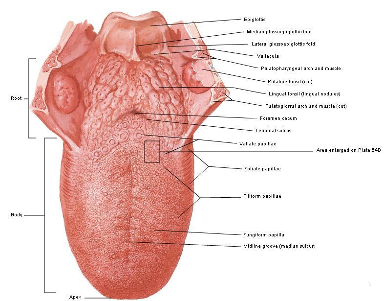
6 Tongue

7 Papillae Papillae Filiform Papillae Fungiform Papillae Foliate Papillae Circumvallate Papillae
 glands drain their contents into the deep groove that encircles the periphery of each papilla.")
8 Circumvallate Papillae They are distributed in the V region in the posterior portion of the tongue. Numerous serous (von Ebner's) glands drain their contents into the deep groove that encircles the periphery of each papilla. The glands also secrete a lipase that probably prevents the formation of a hydrophobic layer over the taste buds that would hinder their function.
 Ion channels (salty and")
9 Taste bud Tastants dissolved in saliva contact the taste cells through the pore interacting with: Taste receptors: (sweet and bitter tastes) Ion channels (salty and sour tastes)
10 Pharynx A transitional space between the oral cavity and the respiratory and digestive systems. Lined by stratified nonkeratinized squamous epithelium in the region continuous with the esophagus Lined by ciliated pseudostratified columnar epithelium containing goblet cells in the regions close to the nasal cavity. Contains the tonsils. The mucosa of also has many small mucous salivary glands in its lamina propria. The constrictor and longitudinal muscles of the pharynx are located outside this layer.

11 Teeth & Associated Structures
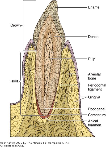
12 Teeth
 The organic matrix of dentin is secreted by")
13 Dentin Calcified tissue Type I collagen fibrils Glycosaminoglycans Phosphoproteins Phospholipids Calcium salts (hydroxyapatite) The organic matrix of dentin is secreted by odontoblasts
.")
14 Odontoblast Polarized protein-secreting cells with secretion granules in the apical cytoplasm and a basal nucleus. Odontoblasts have slender, branched apical extensions the odontoblast processes (Tomes' fibers). The matrix produced by odontoblasts is initially unmineralized and is called predentin

15 Odontoblast Odontoblast processes gradually become longer as the dentin becomes thicker. Running in small canals called dentinal tubules that are extensively branched near the junction between dentin and enamel.
16 Dentin Vs. Bone Dentin is a calcified tissue that is harder than bone because of its higher content of calcium salts (70% of dry weight). Unlike bone, dentin persists as a mineralized tissue long after destruction of the odontoblasts.
17 Enamel Enamel is the hardest component of the human body. 96% mineral hydroxyapatite crystals strontium, magnesium, lead, fluoride 1% organic material amelogenins and enamelins 3% water Enamel consists of elongated rods or columns enamel rods (prisms) that are bound together by interrod enamel.

18 Ameloblasts Enamel matrix is secreted by cells called ameloblasts. These tall columnar cells possess numerous mitochondria in the region below the nucleus. Rough endoplasmic reticulum and a welldeveloped Golgi complex are found above the nucleus. Each ameloblast has an apical extension, known as a Tomes' process, containing numerous secretory granules that contain the proteins that make up the enamel matrix.

19 Pulp Odontoblasts Fibroblasts Thin collagen fibrils Ground substance that contains glycosaminoglycans Pulp is a highly innervated and vascularized tissue.
20 Periodontium Cementum Periodontal ligament Alveolar bone Gingiva
21 Cementum Cementum covers the dentin of the root and is similar in composition to bone. Haversian systems and blood vessels are absent. It is thicker in the apical region of the root, where there are cementocytes cells with the appearance of osteocytes. Like osteocytes they are encased in lacunae Unlike those cells cementocytes do not communicate through canaliculi, and their nourishment comes from the periodontal ligament. Like bone tissue, cementum is labile and reacts to the stresses to which it is subjected by resorbing old tissue or producing new tissue.

22 Periodontal Ligament Special type of connective tissue whose fibers penetrate the cementum of the tooth and bind it to the bony walls of its socket while permitting limited movement of the tooth. It has high rate of collagen renewal.

23 Alveolar Bone It is an immature type of bone (primary bone). Many of the collagen fibers of the periodontal ligament are arranged in bundles that penetrate this bone and the cementum (Sharpey's fibers)
24 Gingiva Mucous membrane firmly bound to the periosteum. Composed of stratified squamous epithelium and lamina propria containing numerous connective tissue papillae. Junctional epithelium bound to the tooth enamel by means of a cuticle that resembles a thick basal lamina and forms the epithelial attachment of Gottlieb. The epithelial cells are attached to this cuticle by hemidesmosomes. Between the enamel and the epithelium is the gingival sulcus.
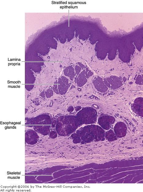
25 Esophagus

26 Junction of the esophagus with the stomach
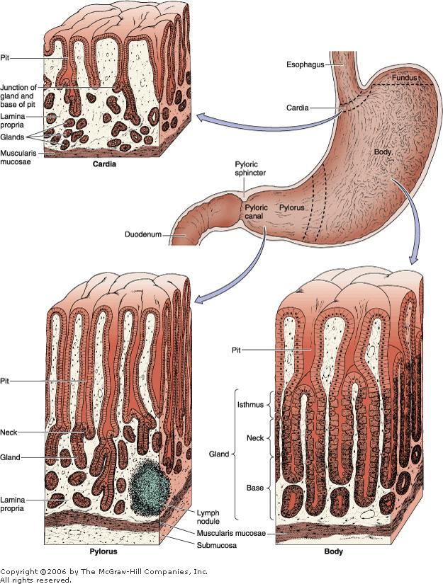
27 Stomach

28

29 Surface-lining cells Bicarbonate


30 Mucous Neck Cells Mucus secretion

31 Oxyntic (Parietal) Cells Hydrochloric acid Potassium chloride Gastric intrinsic factor

32 Atrophic gastritis Both parietal and chief cells are much less numerous, and the gastric juice has little or no acid or pepsin activity. Pernicious anemia
")
33 Oxyntic (Parietal) Cells

34 Chief (Zymogenic) Cells Pepsinogen Lipase


35 Enteroendocrine Cells Somatostatin Serotonin Gastrin

36 Pylorus Has deep gastric pits into which the branched, tubular pyloric glands open. Pyloric glands have longer pits and shorter coiled secretory portions. These glands secrete mucus as well as appreciable amounts of the enzyme lysozyme. Gastrin (G) cells which release gastrin are enteroendocrine cells intercalated among the mucous cells of pyloric glands. Other enteroendocrine cells D cells secrete somatostatin, which inhibits the release of some other hormones, including gastrin.

37 Junction of the stomach with the duodenum

38 Small Intestine

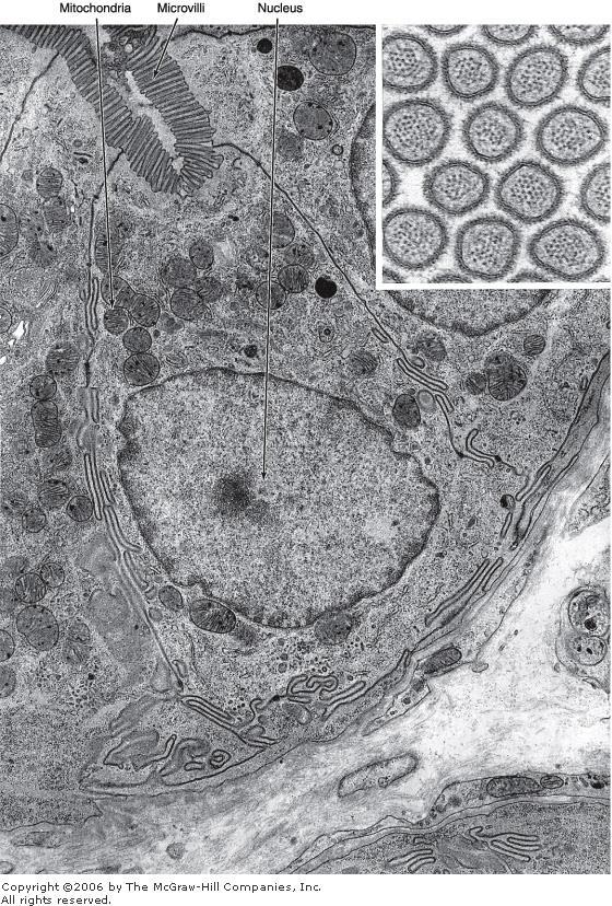
39 Absorptive cells or Enterocytes Disaccharidases peptidases

40 Structure of a microvillus

41 Goblet cells Interspersed between the absorptive cells. They are less abundant in the duodenum and increase in number as they approach the ileum. Produce acid glycoproteins of the mucin type

42 Paneth's cells Lysozyme
")
43 Immunological protection of the intestine M (microfold) cells

44 Endocrine Cells of the Intestine closed type open type
45 Principal Enteroendocrine Cells in the Gastrointestinal Tract

46 Lamina Propria Through Serosa


47 Duodenal or Brunner's Glands Alkaline


48 Peyer's Patches

49 Large Intestine


50 Absorptive and mucous goblet cells


51 Large Intestine Muscularis

52 Rectoanal junction

53 Cell Renewal in the Gastrointestinal Tract
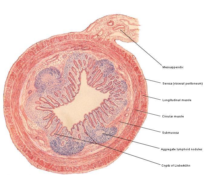

54 Appendix
 Some proteins such as the carcinoembryonic antigen produced exclusively by malign")
55 Cancer of the Digestive Tract Approximately 90 95% of malignant tumors of the digestive system are derived from intestinal or gastric epithelial cells. Malignant tumors of the large bowel are derived almost exclusively from its glandular epithelium (adenocarcinomas) Some proteins such as the carcinoembryonic antigen produced exclusively by malign cells are very important for the diagnosis of cancer.
56 Organs Associated with Digestive Tract Salivary glands Pancreas Liver Gallbladder Wet and lubricate Produce digestive enzymes Produces bile Stores the bile Initiate the digestion Secrete hormones Metabolism and inactivation Secrete germicidal protective substances Synthesis of blood proteins Buffering function

57 Salivary Glands

58 Serous cells Usually pyramidal in shape. Broad base resting on the basal lamina. Narrow apical surface with short, irregular microvilli facing the lumen. They exhibit characteristics of polarized protein-secreting cells. Usually form a spherical mass of cells called acinus.

59 Mucous cells Usually cuboidal to columnar in shape. Their nuclei are oval and pressed toward the bases of the cells. Secret glycoproteins mucins Mucous cells are most often organized as tubules. The mucous cells form tubules, but their ends are capped by serous cells, which constitute the serous demilunes.

60 Myoepithelial cells Found between the basal lamina and the basal plasma membrane of the cells forming secretory end pieces and intercalated ducts. Sometimes called basket cells

61 Duct system Intralobular ducts Intercalated ducts Striated ducts Interlobular ducts Pseudostratified or stratified cuboidal epithelium Stratified columnar epithelium The main duct Nonkeratinized stratified squamous epithelium
62 Salivary Glands Small salivary glands Large salivary glands Parotid Submandibular Sublingual

63 Parotid Gland Branched acinar gland. Its secretory portion is composed exclusively of serous cells. Containing secretory granules that are rich in proteins and have a high amylase activity. Intercalated and striated ducts are easily observed.

64 Submandibular (Submaxillary) Gland Branched tubuloacinar gland formed of serous and mucous cells. The serous cells are the main component. Serous cells are responsible for the weak amylolytic activity. The cells that form the demilunes in the submandibular gland secrete the enzyme lysozyme. Striated ducts are easily observed but intercalated ducts are very short.

65 Sublingual Gland Branched tubuloacinar gland formed of serous and mucous cells. Mucous cells predominate in this gland. Intralobular ducts are not as well developed as in other major salivary glands.
66 Minor Salivary Glands Nonencapsulated glands distributed throughout the oral mucosa and submucosa. Saliva is produced by small groups of secretory units and is conducted to the oral cavity by short ducts, with little modification of its content. Minor salivary glands are usually mucous. The small serous glands present in the posterior region of the tongue (von Ebner's glands) are the only exception. Lymphocyte agregates are commonly observed within minor salivary glands, associated with IgA secretion.
67 Pancreas The pancreas is a mixed exocrine-endocrine gland that produces digestive enzymes and hormones. Enzymes are stored and released by cells of the exocrine portion, arranged in acini. The hormones are synthesized in clusters of endocrine epithelial cells known as islets of Langerhans

68 Pancreas Absence of striated ducts Presence of the islets of Langerhans in the pancreas. The initial portions of intercalated ducts penetrate the lumens of the acini. Intercalated ducts are tributaries of larger intralobular ducts that form larger interlobular ducts.

69 Pancreas The exocrine pancreatic acinus is composed of several serous cells surrounding a lumen. These cells are highly polarized, with a spherical nucleus, and are typical proteinsecreting cells.

70 Pancreas secretions The exocrine pancreas secretes ml of isosmotic alkaline fluid per day containing: Water Ions Proteases Amylase Lipases Phospholipase A2 Nucleases The majority of the enzymes are stored as proenzymes in the secretory granules of acinar cells.
71 Pancreas Secretions Regulation Gastric acid in the intestinal lumen is a strong stimulus for secretin release. Secretin promoting the secretion of an abundant alkaline fluid rich in electrolytes and poor in enzyme activity. The release of cholecystokinin is triggered by the presence of long-chain fatty acids, gastric acid, and certain essential amino acids in the intestinal lumen. Cholecystokinin promotes secretion of a less abundant but enzyme-rich fluid acting mainly in the extrusion of zymogen granules.
72 Liver The liver is the second-largest organ of the body and the largest gland. The liver is the organ in which nutrients absorbed in the digestive tract are processed and stored for use by other parts of the body. Most of its blood (70-80%) comes from the portal vein, arising from the stomach, intestines, and spleen; the smaller percentage (20-30%) is supplied by the hepatic artery. Bile is an exocrine secretion of the liver that is important for toxic substances elimination and lipid digestion. The liver also has the very important function of producing plasma proteins, such as albumin, other carrier proteins, coagulation factors, and growth factors.
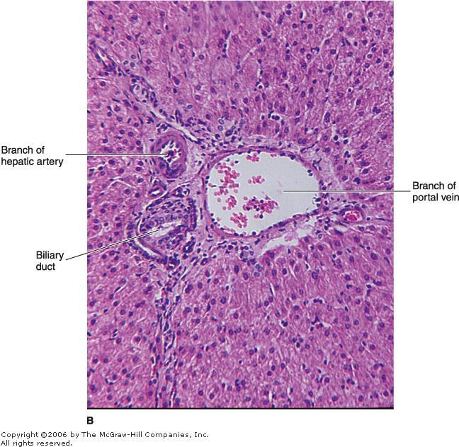
73 Stroma The liver is covered by a thin connective tissue capsule (Glisson's capsule) that becomes thicker at the hilum, where the portal vein and the hepatic artery enter the organ and where the right and left hepatic ducts and lymphatics exit. These vessels and ducts are surrounded by connective tissue all the way to their termination (or origin) in the portal spaces between the liver lobules. At this point, a delicate reticular fiber network that supports the hepatocytes and sinusoidal endothelial cells of the liver lobules is formed.

74 The Liver Lobule

75 The Liver Lobule Hepatocyte Portal spaces Central vein Liver sinusoids Space of Disse Kupffer cells Fat-storing cells Portal Vein System Arterial System

76 The Liver Lobule

77 The Heterogeneity of Hepatocytes
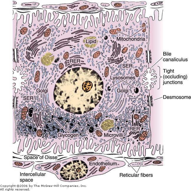
78 The Hepatocyte


79 The Hepatocyte Protein synthesis and Carbohydrate storage Secretion of bile acids

80 The Hepatocyte Secretion of bilirubin


81 Bile Duct System
82 Liver Regeneration Despite its slow rate of cell renewal, the liver has an extraordinary capacity for regeneration. The loss of hepatic tissue by surgical removal or from the action of toxic substances triggers a mechanism by which hepatocytes begin to divide, continuing until the original mass of tissue is restored.

83 Liver Cirrhosis The regenerated liver tissue is usually well organized, exhibiting the typical lobular arrangement and replacing the functions of the destroyed tissue. However, when there is continuous or repeated damage to hepatocytes over a long period of time, the multiplication of liver cells is followed by a pronounced increase in the amount of connective tissue.


84 Biliary Tract

85 Gallbladder The wall of the gallbladder consists of: a mucosa composed of simple columnar epithelium and lamina propria a layer of smooth muscle a perimuscular connective tissue layer a serous membrane

86 Gallbladder The mucosa has abundant folds that are particularly evident when the gallbladder is empty. The epithelial cells are rich in mitochondria. All these cells are capable of secreting small amounts of mucus.
87 Tumors of the Digestive Glands Most malignant tumors of the liver derive from hepatic parenchyma or epithelial cells of the bile duct. It may be associated with a variety of acquired disorders, such as chronic viral hepatitis (B or C) and cirrhosis. In the exocrine pancreas, most tumors arise from ductal epithelial cells.
88 Thank you
Organs Associated with the Digestive Tract. Dr. Emad I H Shaqoura M.D, M.Sc. Anatomy Faculty of Medicine, IUG March, 2016
 Organs Associated with the Digestive Tract Dr. Emad I H Shaqoura M.D, M.Sc. Anatomy Faculty of Medicine, IUG March, 2016 2 Salivary Glands Salivary Glands Major 90% of saliva Minor 10% of saliva Parotid
HISTOLOGY VIRTUAL LABORATORY GASTROINTESTINAL SYSTEM
 HISTOLOGY VIRTUAL LABORATORY GASTROINTESTINAL SYSTEM LIP (Slides GI 1, 2) Identify the outer portion lined by stratified squamous (keratinized) epithelium. Note the hair follicles and sebaceous glands
Objectives. Describe the cells of the GI tract and their function. Differentiate between different parts of the GI tract
 GI Histology 1 Objectives Describe the cells of the GI tract and their function Describe the histological features of each part of the GI tract. Differentiate between different parts of the GI tract Appreciate
The Digestive System. Chapter 25
 The Digestive System Chapter 25 Introduction Structure of the digestive system A tube that extends from mouth to anus Accessory organs are attached Functions include Ingestion Movement Digestion Absorption
Connective tissue The Digestive System
 Connective tissue The Digestive System Part 1 Structure of digestive system Functions Basic Structure of the Alimentary Canal Wall Tube is made up of four layers: 1. Mucosa 2. Submucosa 3. Muscularis externa
DIGESTIVE SYSTEM II ACCESSORY DIGESTIVE ORGANS
 DIGESTIVE SYSTEM II ACCESSORY DIGESTIVE ORGANS Dr. Larry Johnson Texas A& M University Objectives Distinguish between the parotid and submandibular salivary glands. Understand and identify the structural
Connective tissue The Digestive System
 Connective tissue The Digestive System Part 1 Structure of digestive system Functions Basic Structure of the Alimentary Canal Wall Tube is made up of four layers: 1. Mucosa 2. Submucosa 3. Muscularis externa
Slide 154: Pancreas, H&E
 Slide 154: Pancreas, H&E the pancreas, located adjacent to the duodenum, is a mixed exocrine and endocrine gland; it is usually readily identifiable by the presence of the interspersed endocrine pancreatic
The Digestive System. Chapter 16. Introduction. Overview of Digestive System. Histological Organization. Movement and Mixing of Digestive Materials
 The Digestive System Chapter 16 Introduction Structure of the digestive system A tube that extends from mouth to anus Accessory organs are attached Functions include Ingestion Movement Digestion Absorption
Tongue In the buccal cavity of the digestive system
 Tongue In the buccal cavity of the digestive system same layers as those of tubular organs Mucosa, submucosa, and muscularis muscularis = the muscularis externa no muscularis mucosa 1 Tongue ling = tongue
The Digestive System and Body Metabolism Premedical Biology
 The Digestive System and Body Metabolism Premedical Biology Copyright 2003 Pearson Education, Inc. publishing as Benjamin Cummings The Digestive System and Body Digestion Metabolism Breakdown of ingested
General Structure of Digestive Tract
 Dr. Nabil Khouri General Structure of Digestive Tract Common Characteristics: Hollow tube composed of a lumen whose diameter varies. Surrounded by a wall made up of 4 principal layers: Mucosa Epithelial
Digestive System. Presented by: Dr M. Arianmanesh PhD in Reproductive and Developmental Biology Dept. of Anatomical Sciences
 Digestive System Presented by: Dr M. Arianmanesh PhD in Reproductive and Developmental Biology Dept. of Anatomical Sciences Today we will discuss: Histological layers of alimentary canal Oral cavity Lip
Oral cavity Lab exercises
 Oral cavity Lab exercises Slide #190 (GT-1-32). Oral cavity, goat. large conical buccal papillae stratified squamous epithelium keratinized or non-keratinized no muscularis mucosae connective tissue represents
Digestive system L 4. Lecturer Dr. Firdous M. Jaafar Department of Anatomy/Histology section
 Digestive system L 4 Lecturer Dr. Firdous M. Jaafar Department of Anatomy/Histology section objectives 1-Describe the structure of liver. 2-Define liver lobule, and identify its zones. 3-Define portal
Exercise. Digestive System. Digestive system function. 1. Define the following terms: a. Chemical digestionb. Mechanical digestionc.
 Exercise 7 The Digestive System NAME: DATE: INSTRUCTOR: SECTION: Digestive system function 1. Define the following terms: a. Chemical digestionb. Mechanical digestionc. Ingestiond. Digestione. Absorptionf.
Chapter 12 The Digestive Glands
 Chapter 12 The Digestive Glands Lyu Zhengmei Department of Histology and Embryology, Anhui Medical University Components of digestive glands large salivary glands, pancreas, liver, gallbladder. These organs
Two main groups Alimentary canal continuous coiled hollow tube Accessory digestive organs
 Digestion Breakdown of ingested food Absorption of nutrients into the blood Metabolism Production of cellular energy (ATP) Constructive and degradative cellular activities Two main groups Alimentary canal
Digestive System 7/15/2015. Outline Digestive System. Digestive System
 Digestive System Biology 105 Lecture 18 Chapter 15 Outline Digestive System I. Functions II. Layers of the GI tract III. Major parts: mouth, pharynx, esophagus, stomach, small intestine, large intestine,
GI Histology Lab 1. Prepared by: Zeina Kalaji
 GI Histology Lab 1 Prepared by: Zeina Kalaji Lip ORAL MUCOSA -Arrow shows labial salivary glands in the submucosa. VERMILLION transitional zone. SKIN Stratified Squamous epithelium, keratinized -Arrow
Tissue: The Living Fabric: Part A
 PowerPoint Lecture Slides prepared by Janice Meeking, Mount Royal College C H A P T E R 4 Tissue: The Living Fabric: Part A Tissues Groups of cells similar in structure and function Types of tissues Epithelial
Paneth Cells. Road Map to the Finish. No Review this Friday. Today 11/29 Finish digestion/accessory organs. Wednesday 12/1 Immune System I
 Road Map to the Finish No Review this Friday Today 11/29 Finish digestion/accessory organs Wednesday 12/1 Immune System I Paneth Cells - base of intestinal glands -! large -! intense acidophilic granules
Epithelium. Four primary tissue types:
 Epithelium Four primary tissue types: Epithelial (covering) Connective (support) Nervous (control) Muscular (movement) Smooth muscle Cardiac muscle Skeletal muscle 1 Epithelial Tissue Features Epithelial
DIGESTIVE. CHAPTER 17 Lecture: Part 1 Part 2 BIO 212: ANATOMY & PHYSIOLOGY II
 BIO 212: ANATOMY & PHYSIOLOGY II 1 CHAPTER 17 Lecture: DIGESTIVE Part 1 Part 2 Dr. Lawrence G. Altman www.lawrencegaltman.com Some illustrations are courtesy of McGraw-Hill. Processes of DIGESTION Mechanical
Chapter 14: The Digestive System
 Chapter 14: The Digestive System Digestive system consists of Muscular tube (digestive tract) alimentary canal Accessory organs teeth, tongue, glandular organs 6 essential activities 1. 2. 3. 4. 5. 6.
Digestive system L 2. Lecturer Dr. Firdous M. Jaafar Department of Anatomy/Histology section
 Digestive system L 2 Lecturer Dr. Firdous M. Jaafar Department of Anatomy/Histology section objectives 1-Describe the general structure of digestive tract: a-mucosa. b-submucosa. c-muscularis externa d-adventitia
Dr. Abeer.c.Yousif. Histology -2 nd stage. What is histology?
 What is histology? Histology is the science of microscopic anatomy of cells and tissues, in Greek language Histo= tissue and logos = study and it's tightly bounded to molecular biology, physiology, immunology
Chapter 9. The digestive system. Glossary. Louise McErlean
 Chapter 9 The digestive system Louise McErlean Glossary Absorption Process whereby the products of digestion move into the blood or lymph fluid. Acini glands Produce pancreatic juice. Amylase Carbohydrate
The Digestive System. What is the advantage of a one-way gut? If you swallow something, is it really inside you?
 The Digestive System What is the advantage of a one-way gut?! If you swallow something, is it really inside you? Functions and Processes of the Digestive System: Move nutrients, water, electrolytes from
Epithelial Tissue. Functions include: 1. Protection 4. Absorption 2. Secretion 5. Filtration 3. Sensory reception
 Tissues There are 4 primary tissue types in the human body: 1. Epithelial (covering/lining) 2. Connective (support) 3. Muscle (movement) 4. Nervous (control) Epithelium Epithelial Tissue Covers the surface
DIGESTIVE SYSTEM ALIMENTARY CANAL / GI TRACT & ACCESSORY ORGANS. Mar 16 10:34 PM
 DIGESTIVE SYSTEM ALIMENTARY CANAL / GI TRACT & ACCESSORY ORGANS Mar 16 10:34 PM 1 I. Digestive System Functions > Ingestion the taking in of food > Propulsion movement caused by force > Digestion breakdown
Glandular Epithelium. Dr. Hersh Abdul Ham-Karim BVM&S, PG Dip, MSc and PhD
 Glandular Epithelium Dr. Hersh Abdul Ham-Karim BVM&S, PG Dip, MSc and PhD Glandular Epithelium Groups of surface cells differentiate, proliferate, and penetrate underlying connective tissue. Their main
Includes mouth, pharynx, esophagus, stomach, small intestine, large intestine, rectum, anus. Salivary glands, liver, gallbladder, pancreas
 Chapter 14 The Digestive System and Nutrition Digestive System Brings Nutrients Into the Body The digestive system includes Gastrointestinal (GI) tract (hollow tube) Lumen: space within this tube Includes
(b) Stomach s function 1. Dilution of food materials 2. Acidification of food (absorption of dietary Fe in small intestine) 3. Partial chemical digest
 Stomach s function 1. Dilution of food materials 2. Acidification of food (absorption of dietary Fe in small intestine) 3. Partial chemical digest") (1) General features a) Stomach is widened portion of gut-tube: between tubular and spherical; Note arranged of smooth muscle tissue in muscularis externa. 1 (b) Stomach s function 1. Dilution of food
(1) General features a) Stomach is widened portion of gut-tube: between tubular and spherical; Note arranged of smooth muscle tissue in muscularis externa. 1 (b) Stomach s function 1. Dilution of food
MICROSTRUCTURES LIPS TOOTH TONGUE OESOPHAGUS STOMACH, CARDIAC, PYLORIC FUNDIC GLANDS
 MICROSTRUCTURES LIPS TOOTH TONGUE OESOPHAGUS STOMACH, CARDIAC, PYLORIC FUNDIC GLANDS HUMAN ANATOMY: MICROSTRUCTURES CLASSIFICATION: LOCATION AND BOUNDARIES, FORM, FUNCTION, MICROSCOPIC STRUCTURE: A hollow
I. The Alimentary Canal (GI track)
") A. About 9 meters long B. Passes through the ventral cavity. C.Movements of the Tube 1. Mixing movements- smooth muscles contract rhythmically. 2. Propelling movements- a wavelike motion called peristalsis.
A. About 9 meters long B. Passes through the ventral cavity. C.Movements of the Tube 1. Mixing movements- smooth muscles contract rhythmically. 2. Propelling movements- a wavelike motion called peristalsis.
Lab activity manual Histology of the digestive system
 Lab activity manual Histology of the digestive system Jeanne Adiwinata Pawitan Prerequisite: Histology of the 4 basic tissues In this module we learn about the histology of the digestive system, from the
Alimentary Canal (I)
") Alimentary Canal (I) Esophagus and Stomach (Objectives) By the end of this lecture, the student should be able to discuss the microscopic structure in correlation with the function of the following organs:
Alimentary Canal (I) Esophagus and Stomach (Objectives) By the end of this lecture, the student should be able to discuss the microscopic structure in correlation with the function of the following organs:
Large Intestine. The large intestine consists of a mucosal membrane with no folds except in its distal (rectal) portion
 portion") GI Histology 3 Large Intestine The large intestine consists of a mucosal membrane with no folds except in its distal (rectal) portion No villi are present in this portion of the intestine The intestinal
GI Histology 3 Large Intestine The large intestine consists of a mucosal membrane with no folds except in its distal (rectal) portion No villi are present in this portion of the intestine The intestinal
HISTOLOGY. GIT Block 432 Histology Team. Lecture 1: Alimentary Canal (1) (Esophagus & Stomach) Done by: Ethar Alqarni Reviewed by: Ibrahim Alfuraih
 (Esophagus & Stomach) Done by: Ethar Alqarni Reviewed by: Ibrahim Alfuraih") HISTOLOGY Lecture 1: Alimentary Canal (1) (Esophagus & Stomach) Done by: Ethar Alqarni Reviewed by: Ibrahim Alfuraih Color Guide: Black: Slides. Red: Important. Green: Doctor s notes. Blue: Explanation.
HISTOLOGY Lecture 1: Alimentary Canal (1) (Esophagus & Stomach) Done by: Ethar Alqarni Reviewed by: Ibrahim Alfuraih Color Guide: Black: Slides. Red: Important. Green: Doctor s notes. Blue: Explanation.
Tissues. tissue = many cells w/ same structure and function. cell shape aids its function tissue shape aids its function
 Tissues tissue = many cells w/ same structure and function cell shape aids its function tissue shape aids its function Histology = study of tissues 4 types of tissues Epithelial coverings contact openings
Bio 104 Digestive System
 13 Lecture Outline: Digestive System Hole s HAP [Chapters 17 & 18] General Characteristics of the Alimentary Canal A. Functions 1. Ingestion 2. Mechanical digestion 3. Chemical digestion 4. Propulsion
Soft palate elevates, closing off the nasopharynx. Hard palate Tongue Bolus Epiglottis. Glottis Larynx moves up and forward.
 The Cephalic Phase Chemical and mechanical digestion begins in the mouth Saliva is an exocrine secretion Salivary secretion is under autonomic control Softens and lubricates food Chemical digestion: salivary
Dr Nadine Gravett School of Anatomical Sciences Room 2B10B
 Dr Nadine Gravett School of Anatomical Sciences Room 2B10B Nadine.Gravett@wits.ac.za Oral cavity Mechanical breakdown Formation of bolus Oesophagus Conduit from mouth to stomach Stomach Digestion Temporary
Epithelia will be discussed according to the following scheme: Type Number of layers Shape Line drawing. Squamous Cuboidal Columnar
 Epithelia Epithelia will be discussed according to the following scheme: Type Number of layers Shape Line drawing Simple Squamous Cuboidal Columnar Covering and Lining epithelium Pseudostratified Stratified
Tissue: The Living Fabric
 PowerPoint Lecture Slide Presentation by Vince Austin Human Anatomy & Physiology FIFTH EDITION Elaine N. Marieb Chapter 4 Tissue: The Living Fabric Part A Tissues Groups of cells similar in structure and
Small intestine. Small intestine
 General features Tubular organ longest part; 5-6 m most of chemical digestion absorption of nutrients reabsorption of H2O occurs. Two structural features; maximize the lumenal surface area villi microvilli
BIO 116 Anatomy & Physiology II Practice Assignment 3 - The Lymphatic, Immune and Digestive Systems This is not a required assignment
 BIO 116 Anatomy & Physiology II Practice Assignment 3 - The Lymphatic, Immune and Digestive Systems This is not a required assignment 1. Which are components of the lymphatic system? a: Thyroid gland b:
The doctor mentioned a few things about the esophagus from the previous lecture:
 السالم عليكم [HISOLOGY 2] April 27, 2014 The doctor mentioned a few things about the esophagus from the previous lecture: Esophagus - It is about 25 cm in length (from the incisor it is 45 cm) Histological
Gastrointestinal Anatomy and Physiology. Bio 219 Napa Valley College Dr. Adam Ross
 Gastrointestinal Anatomy and Physiology Bio 219 Napa Valley College Dr. Adam Ross Functions of digestive system Digestion Breakdown of food (chemically) using enzymes, acid, and water Absorption Nutrients,
Esophagus. Transport is achieved by peristaltic contractions and relaxation of the esophageal sphincters (upper and lower)
") GI Histology 2 Esophagus is a muscular tube whose function is to transport foodstuffs from the mouth to the stomach and to prevent the retrograde flow of gastric contents Transport is achieved by peristaltic
GI Histology 2 Esophagus is a muscular tube whose function is to transport foodstuffs from the mouth to the stomach and to prevent the retrograde flow of gastric contents Transport is achieved by peristaltic
1. Approximately 21 ft. long: duodenum (one ft.), jejunum (eight ft.), and ileum (twelve ft.)
, jejunum (eight ft.), and ileum (twelve ft.)") IV. Small Intestines A. General features and functions 1. Approximately 21 ft. long: duodenum (one ft.), jejunum (eight ft.), and ileum (twelve ft.) 2. Functions: move forward chyme, continue digestion,
IV. Small Intestines A. General features and functions 1. Approximately 21 ft. long: duodenum (one ft.), jejunum (eight ft.), and ileum (twelve ft.) 2. Functions: move forward chyme, continue digestion,
Energy, Chemical Reactions and Enzymes
 Phosphorylation Hydrolysis Energy, Chemical Reactions and Enzymes Chapter 2 (selections) What is Energy? Energy is the capacity to do work Potential Energy Kinetic Energy Chemical Bond Energy Like a rechargeable
Histology Notes -Part 1: Epithelial Tissues
 Introduction Group of cells w/ similar structure & function = TISSUE Four Basic Tissue Types 1. Epithelial-covers 2. Connective-supports 3. Muscular*-produces movement (will discuss in the muscular system
Laboratory exercises for abdominal organs
 Laboratory exercises for abdominal organs Slide #77 (C007- H- 107A). Pancreas, dog. pancreatic islets CENTROACINAR CELLS ARE THE BEGINNING CELLS OF THE INTERCALATED DUCTS THAT DRAIN THE SECRETORY ACINI
Nutrition. Autotrophs. plants, some protists & bacteria producers
 Nutrition Autotrophs plants, some protists & bacteria producers Nutrition Heterotrophs animals, fungi, some protists & bacteria consumers Animal Nutrition Most obtain food by ingestion take in their food
Histology = the study of tissues. Tissue = a complex of cells that have a common function
 { EPITHELIAL TISSUE Histology = the study of tissues Tissue = a complex of cells that have a common function The Four Primary Tissue Types: Epithelium (epithelial tissue) covers body surfaces, lines body
Histology Lab. looking at microscopic pictures of tissues, for more information use Junqueira book and you can use BlueHistolgy website
 Done By: Aseel Twaijer & Laith Sorour Histology Lab *These notes help in differentiating tissues and you must read them while looking at microscopic pictures of tissues, for more information use Junqueira
Tissues. Definition. A group of similar cells and their intercellular substances specialized to perform a specific function.
 Chapter 4 - Tissues Tissues Definition A group of similar cells and their intercellular substances specialized to perform a specific function. Tissues Epithelial covers exposed surfaces, lines internal
The Digestive System Laboratory
 The Digestive System Laboratory 1 The Digestive Tract The alimentary canal is a continuous tube stretching from the mouth to the anus. Liver Gallbladder Small intestine Anus Parotid, sublingual, and submaxillary
Digestive Anatomy Lab
 Digestive Anatomy Lab In-Lab Exercises I have included the word list in this document. Any descrepencies between this document and the wordlist, you should default to this document. There is a lot of repetition
Chapter 1: Cells and Tissues
 Chapter 1: Cells and Tissues Cells and Tissues Carry out all chemical activities needed to sustain life Cells are the building blocks of all living things Tissues are groups of cells that are similar in
Organs Histology D. Sahar AL-Sharqi. Digestive System
 Digestive System The digestive system consists of the digestive tract oral cavity, esophagus, stomach, small and large intestines, and anus and its associated glands salivary glands, liver, and pancreas.
Tissues. tissue = many cells w/ same structure and function. cell shape aids function tissue shape aids function. Histology = study of tissues
 Tissues tissue = many cells w/ same structure and function cell shape aids function tissue shape aids function Histology = study of tissues 4 types of tissues Epithelial coverings contact openings Connective
Dana Alrafaiah. Dareen Abu Shalbak. Mohammad Almuhtaseb. 1 P a g e
 2 Dana Alrafaiah Dareen Abu Shalbak Mohammad Almuhtaseb 1 P a g e Esophagus: A muscular tube that is 25 cm long, but if measured from the incisors it would be 45cm long. Extends from C6 of cervical vertebra,
Glandular Epithelium. Dr. Heba Kalbouneh Associate Professor of Anatomy and Histology
 Glandular Epithelium Dr. Heba Kalbouneh Associate Professor of Anatomy and Histology Glands Glandular epithelia are tissues formed by cells specialized to produce secretion. Secretion: if substances produced
DIGESTIVE. CHAPTER 17 Lecture: Part 1 Part 2 BIO 212: ANATOMY & PHYSIOLOGY II
 BIO 212: ANATOMY & PHYSIOLOGY II CHAPTER 17 Lecture: DIGESTIVE Part 1 Part 2 Dr. Lawrence G. Altman www.lawrencegaltman.com Some illustrations are courtesy of McGraw-Hill. SMALL INTESTINE DUODENUM > JEJUNUM
c. Heparin sulfate proteoglycans d. Type IV collagen e. Type II collagen 3. The following statements concerning regular bronchioles are correct: a.
 This is just some examples of questions they could ask on the exam. For the multiple parts questions you would probably have 8-10 parts but they should tell you the exact pattern in one of the lectures.
General functions of digestive system. Ch. 15 The Digestive System. General histology of the wall of the digestive tract. Overview of digestive organs
 Overall idea: obtain nutrients from food (for energy and raw materials for synthesis), and defecate the leftover waste 2 types of organs involved: 1. Parts of the digestive tract (= a long muscular tube
Chapter 26 The Digestive System
 Chapter 26 The Digestive System Digestive System Gastroenterology is the study of the stomach and intestine. Digestion Catabolism Absorption Anabolism The actions of the digestive system are controlled
Chapter 4 - Epithelial Tissues
 Chapter 4 - Epithelial Tissues Tissues Definition A group of closely associated cells that work together to perform a specific function Types Epithelial - covering Connective - support Muscle - movement
The Digestive system
 The Digestive system The GI tract (gastrointestinal tract) Mouth Pharynx Esophagus Stomach Small intestine Large intestine Anus The accessory digestive organs Supply secretions contributing to the breakdown
Body Tissues. Cells are specialized for particular functions Tissues - groups of cells with similar structure. and function Four primary tissue types:
 Chapter 3 Tissues Body Tissues Cells are specialized for particular functions Tissues - groups of cells with similar structure and function Four primary tissue types: Epithelium Connective tissue Nervous
BIOL& 253 Lab Manual for Practical #2 Page 1 Rausch. For all slides, know a function for structures marked with a single asterisk (*).
.") BIOL& 253 Lab Manual for Practical #2 Page 1 Rausch Lab equipment: slides, models SLIDES For all slides, know a function for structures marked with a single asterisk (*). DIGESTIVE SYSTEM Layers of the
BIOL& 253 Lab Manual for Practical #2 Page 1 Rausch Lab equipment: slides, models SLIDES For all slides, know a function for structures marked with a single asterisk (*). DIGESTIVE SYSTEM Layers of the
Digestive Lecture Test Questions Set 4
 Digestive Lecture Test Questions Set 4 1. Which of the following is not associated directly with the small intestine: a. villi b. circular folds c. microvilli d. haustrae e. secretin 2. The largest (longest)
Cell and Tissue Types. Epithelial, Connective, Muscle, Nerve
 Cell and Tissue Types Epithelial, Connective, Muscle, Nerve Objectives Explain the major stages of the cell cycle and cellular division (mitosis). Describe specific events occurring in each of the phases
A deep groove encircles the body of the circumvallate papilla. Serous (von Ebner s) glands (serous) drain into the base of this groove.
 glands (serous) drain into the base of this groove.") By Dr. Raja Ali A deep groove encircles the body of the circumvallate papilla. Serous (von Ebner s) glands (serous) drain into the base of this groove. The flow of fluid from these glands serves to wash
By Dr. Raja Ali A deep groove encircles the body of the circumvallate papilla. Serous (von Ebner s) glands (serous) drain into the base of this groove. The flow of fluid from these glands serves to wash
DIGESTIVE SYSTEM CLASS NOTES. tube along with several
 DIGESTIVE SYSTEM CLASS NOTES Digestion Breakdown of food and the of nutrients in the bloodstream. Metabolism Production of for and cellular activities. The digestive system is composed of the canal which
Basic Tissue Types and Functions
 Tissues Histology Basic Tissue Types and Functions 1) Epithelial tissue covering 2) Connective tissue support 3) Muscle tissue movement 4) Nervous tissue control Epithelial Tissue 1) Covers a body surface
Epithelial Lecture Test Questions
 Epithelial Lecture Test Questions 1. Which of the following free surfaces lack(s) epithelia: a. lung alveoli (air sacs) b. hard palate c. joint cavities d. abdominal cavity e. salivary gland ducts 2. Which
Gastric Contrac,le Ac,vity. Regula,on of Gastric Emptying
 Gastric Contrac,le Ac,vity Figure 23.18 Regula,on of Gastric Emptying Gastric emptying is regulated by: Neural enterogastric reflex Hormonal (enterogastrone) mechanisms In the presence of gastric gastrin
ACTIVITY 11: RESPIRATORY AND DIGESTIVE SYSTEMS RESPIRATORY SYSTEM
 ACTIVITY 11: RESPIRATORY AND DIGESTIVE SYSTEMS OBJECTIVES: 1) How to get ready: Read Chapters 25 and 26, McKinley et al., Human Anatomy, 4e. All text references are for this textbook. 2) Identify structures
5 Dr. Heba Kalbouneh
 5 Dr. Heba Kalbouneh Glandular epithelium Gland: Is a collection of epithelial cells the secrets a certain product, like: proteins, lipids and carbohydrates. Secretion : A certain material that is produced
The Digestive System and Body Metabolism
 PowerPoint Lecture Slide Presentation by Patty Bostwick-Taylor, Florence-Darlington Technical College The Digestive System and Body Metabolism 14PART C Accessory Digestive Organs Teeth Salivary glands
RESPIRATORY SYSTEM. described: pp. 744,746 fig. 25.1, described: p. 746 fig described: p. 776 fig. 26.3
 ACTIVITY 11: RESPIRATORY AND DIGESTIVE SYSTEMS OBJECTIVES: 1) How to get ready: Read Chapters 25 and 26, McKinley et al., Human Anatomy, 5e. All text references are for this textbook. 2) Identify structures
Lab activity manual - Histology of the digestive system. Lab activity 1: esophagus stomach - small intestines
 Lab activity manual - Histology of the digestive system Jeanne Adiwinata Pawitan Prerequisite: Histology of the 4 basic tissues In this module we learn about the histology of the digestive system, from
The Digestive System
 The Digestive System Identify the Structure and Function. Mesentery of the Large Intestine The mesentery functions to connect the visceral organs to the abdominal wall. Identify the Structure. Nasal Cavity
Digestion and Absorption. Food:
 Digestion and Absorption Food: Food is a basic requirement of all living beings. Food provides energy for different activities in the body. Food also provides organic materials for growth and repair. Carbohydrates,
Chapter 15 Digestion and Nutrition
 Chapter 15 Digestion and Nutrition Digestive System: Digestion refers to the mechanical and chemical breakdown of foods so that nutrients can be absorbed by cells. The digestive system consists of the
2. SECRETIONS OF THE DIGESTIVE TRACT
 2. SECRETIONS OF THE DIGESTIVE TRACT SECRETORY GLANDS AND CELLS The alimentary tract produces a large variety and quantity of substances which contribute to digesting the food, and protecting and regulating
MICROSTRUCTURES SMALL INTESTIN LARGE INTESTIN PANCREAS LIVER GALLBLADDER SALIVARY GLANDS ADRENALS THYROID AND PARATHYROID GLANDS
 MICROSTRUCTURES SMALL INTESTIN LARGE INTESTIN PANCREAS LIVER GALLBLADDER SALIVARY GLANDS ADRENALS THYROID AND PARATHYROID GLANDS HUMAN ANATOMY: MICROSTRUCTURES CLASSIFICATION: LOCATION AND BOUNDARIES,
Tissues. groups of cells similar in structure and function 4 types. epithelium connective muscle nervous
 Tissues groups of cells similar in structure and function 4 types epithelium connective muscle nervous Epithelial Tissue lining covering glandular Functions protection absorption filtration secretion Epithelium
2. SECRETIONS OF THE DIGESTIVE TRACT
 2. SECRETIONS OF THE DIGESTIVE TRACT SECRETORY GLANDS AND CELLS The alimentary tract produces a large variety and quantity of substances which contribute to digesting the food, and protecting and regulating
PRACTICAL ROADMAP. GLANDS AFFECTING LIFESTYLE WJ van der Spuy & T Tshabalala
 PRACTICAL ROADMAP GLANDS AFFECTING LIFESTYLE WJ van der Spuy & T Tshabalala GLANDS AFFECTING LIFESTYLE Submandibular gland (salivary gland) Liver Pancreas Hypophysis (pituitary gland) Thyroid Suprarenal
Chapter 23: The Digestive System
 Chapter 23: The Digestive System I. OVERVIEW Consists of alimentary canal and accessory organs. GIT (gastrointestinal tract) and teeth, tongue, gallbladder, salivary glands, liver and pancreas. (A) Digestive
the serous membranes lining the peritoneal cavity continuously produce what?
 Basic A & P II Dr. L. Bacha Chapter Outline (Martini & Nath 2010) - two groups of organs form the digestive system (see Fig. 22-1): 1. digestive tract what is it also called? list the organs that make
Glandular Epithelium. Dr. Heba Kalbouneh Assistant Professor of Anatomy and Histology
 Glandular Epithelium Dr. Heba Kalbouneh Assistant Professor of Anatomy and Histology Glands Gla dular epithelia are tissues for ed y ells spe ialized to produ e se retio. Secretion: if substances produced
- Digestion occurs during periods of low activity - Produces more energy than it uses. - Mucosa
 Introduction Digestive System Chapter 29 Provides processes to break down molecules into a state easily used by cells - A disassembly line: Starts at the mouth and ends at the anus Digestive functions
Body Tissues Pearson Education, Inc.
 Body Tissues Tissues Groups of cells with similar structure and function Four primary types: Epithelial tissue (epithelium).1 Connective tissue.2 Muscle tissue.3 Nervous tissue.4 Epithelial Tissues Locations:
BIO 139 ANATOMY AND PHYSIOLOGY II THE DIGESTIVE SYSTEM
 BIO 139 ANATOMY AND PHYSIOLOGY II THE DIGESTIVE SYSTEM MARY CATHERINE FLATH, Ph.D. DIGESTIVE SYSTEM ORGANS MOUTH PHARYNX ESOPHAGUS STOMACH SMALL INTESTINE LARGE INTESTINE SALIVARY GLANDS PANCREAS LIVER