Connective tissue The Digestive System
|
|
|
- Letitia Baker
- 6 years ago
- Views:
Transcription

1 Connective tissue The Digestive System Part 1

2 Structure of digestive system
3 Functions
 or Adventitia")
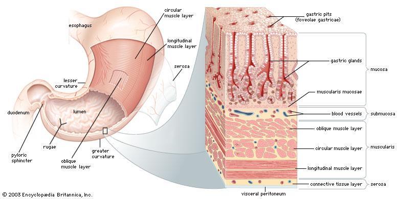
4 Basic Structure of the Alimentary Canal Wall Tube is made up of four layers: 1. Mucosa 2. Submucosa 3. Muscularis externa 4. Serosa (Peritoneum) or Adventitia

 Lamina propria loose connective tissue Muscularis mucosae")
5 Mucosa The innermost wall of the alimentary tube. Consists of: Epithelium - usually simple columnar epithelium with goblet cells; may be stratified squamous if protection is needed (e.g. esophagus) Lamina propria loose connective tissue Muscularis mucosae takes part in the formation of folds


6 Submucosa Made up of loose connective tissue. Contains submucosal (Meissner s) nervous plexus and blood vessels, sometimes glands.
 nervous plexus in between Responsible for peristalsis (controlled by the nerve")
7 Muscularis externa Usually two layers of smooth muscle: inner circular layer outer longitudinal layer. Myenteric (Auerbach s) nervous plexus in between Responsible for peristalsis (controlled by the nerve plexus)
.")
8 Outer membrane A serous membrane/peritoneum consisting of the mesothelium (simple squamous epithelium), and a small amount of underlying loose connective tissue. Or adventitia consisting only of connective tissue is found where the wall of the tube is directly attached or fixed to adjoining structures (i.e., body wall and certain organs).

9 Enteric nervous system
10 The Alimentary Canal

11 Pharynx Common respiratory and digestive pathway (both air and swallowed food and drinks pass through). Stratified squamous non-keratinized epithelium Lamina propria contains many elastic fibers No muscularis mucosae No submucosa Striated muscle in the muscularis externa

12 Esophagus Fixed muscular tube that delivers food and liquid from the pharynx to the stomach.

13 Esophagus Epithelium - stratified squamous Mucosal and submucosal glands of the esophagus secrete mucus to lubricate and protect the luminal wall. Esophageal glands proper lie in the submucosa. These glands are scattered along the length of the esophagus but are somewhat more concentrated in the upper half. Esophageal cardiac glands are named for their similarity to the cardiac glands of the stomach and are found in the lamina propria of the mucosa. Muscularis externa in the upper third consists of striated muscle, mixture of striated and smooth muscle in the middle third and only smooth muscle in the lower third. Those glands near the stomach tend to protect the esophagus from regurgitated acid gastric contents. Under certain conditions, however, they are not fully effective, and excessive reflux results in pyrosis, a condition more commonly known as heartburn.

14 Stomach Esophagus Junction


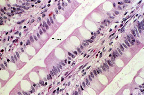
15 Stomach An expandable, muscular bag, that keeps swallowed food. Food can stay in the stomach for 2 hours or more. Food in the stomach is broken down chemically, by gastric juice, and mechanically, by contraction of the three layers of smooth muscle in the muscularis externa. The broken up food at the end of this process is called chyme.
16 Stomach The mucosa is relatively thick, lined by simple columnar epithelium, contains numerous tubular glands. In the empty contracted stomach, the mucosa makes longitudinal folds rugae. Gastric pits - funnel-shaped invaginations of the epithelium continuous at their base with the tubular glands. The muscularis mucosae is also thick. Glands are absent in the submucosa. The muscularis externa is made up of 3 layers: inner oblique, middle circular, external - longitudinal.
17 Stomach

18 Epithelial cells of the Stomach Surface mucous cells line the inner surface of the stomach and the gastric pits. The mucous protects against abrasion from rougher components of the chyme. Additionally, its high bicarbonate and potassium concentration protects the epithelium from the acidic content of the gastric juice. mucus + bicarbonates = physiologic gastric mucosa barrier Surface mucous cells are renewed approximately every 3 to 5 days.

 contains pyloric")
19 The region around the cardia contains the cardiac glands. The region, which includes the fundus and corpus, contains the gastric glands proper (also called fundic glands). The distal region of the stomach (pylorus) contains pyloric glands. Gastric glands

20 Cells of gastric glands Mucous neck cells secrete mucous, predominant in cardiac and pyloric glands. Stem cells in the base of the pit and in the neck of the gland, divide rapidly and replace dead cells. Parietal cells upper half of the gland, secrete hydrochloric acid (HCl) and intrinsic factor Chief cells most numerous in the lower part of the gland, secrete pepsinogen, absent in pyloric and cardiac glands. Enteroendocrine cells lower part of the gland, secrete hormones to regulate digestion
 Because HCl is bacteriostatic, most of the bacteria entering the stomach with the ingested food are")
 Intrinsic")
21 Parietal cells Hydrochloric acid (HCl), which gives the gastric juice a low ph (1.0 to 2.0) Because HCl is bacteriostatic, most of the bacteria entering the stomach with the ingested food are destroyed. However, some bacteria can adapt to the low ph of the gastric contents (Helicobacter pylori) Intrinsic factor, a glycoprotein that binds to vitamin B12. Lack of intrinsic factor leads to pernicious anemia and vitamin B-12 deficiency

22 Chief cells Pepsinogen a precursor enzyme which is converted into an active enzyme Pepsin, under the influence of HCl at a ph lower than 5.

23 Enteroendocrine cells Secrete multiple hormonal products which influence several digestive system organs: Gastrin Histamine Cholecystokinin Somatostatin. Pyloric Gastric Gland stained for Gastrin Cells
24

25 Small Intestine Consist of three parts: duodenum, jejunum, ileum. Main functions are: digestion, absorption of nutrients and production of gastrointestinal hormones.
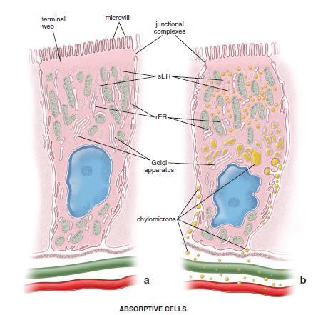
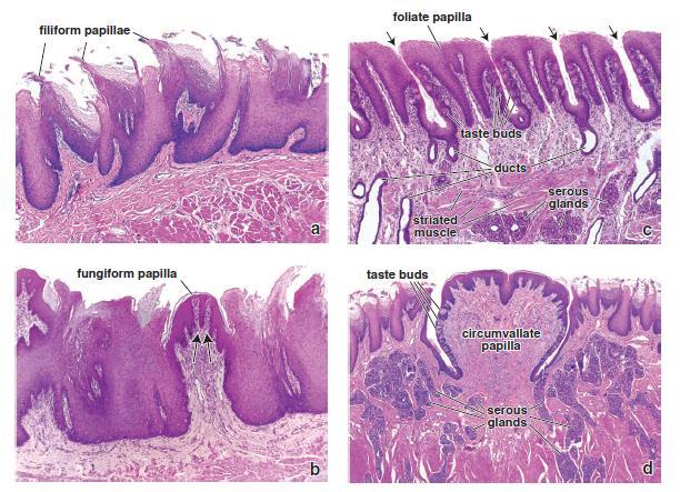
26 Small Intestine The mucosa is highly folded, providing a huge surface area for absorption: large circular folds called plicae circulares smaller finger like mucosal projections, called villi, cover circular folds the lining columnar epithelial cells have fine projections on their apical surfaces called microvilli. Between the villi there are crypts, called crypts of Lieberkuhn, which are short glands. The lamina propria has a rich vascular and lymphatic network, which absorbs the digestive products, and there is a muscularis mucosae layer immediately at the base of the crypts. There are multiple lymphoid aggregations called Peyer's patches The muscularis externa layer contains two layers of smooth muscle for continuous peristaltic activity.


27 Plicae circulares villi, and microvilli increase the absorptive surface area of the small intestine.

28 The Small Intestine


29 The intestinal glands, or crypts of Lieberkühn, are simple tubular structures that extend from the muscularis mucosae through the thickness of the lamina propria, where they open onto the luminal surface of the intestine at the base of the villi
, modified")
30 At least five types of cells are found in intestinal mucosal epithelium: Enterocytes, whose primary function is absorption Goblet cells, unicellular mucinsecreting glands Paneth cells, whose primary function is to maintain mucosal innate immunity by secreting antimicrobial substances Enteroendocrine cells, which produce various paracrine and endocrine hormones M cells (microfold cells), modified enterocytes that cover enlarged lymphatic nodules in the lamina propria
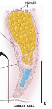
31 Enterocytes Goblet cells represent unicellular glands

32 Paneth Cells at the base of the Crypts Paneth cells play a role in regulation of normal bacterial flora of the small intestine. They contain: Lysozyme that digests the cell walls of certain groups of bacteria. Defensins that function as mediators of CD8 T- lymphocytes.

33 M cells (microfold cells), modified enterocytes that cover enlarged lymphatic nodules Ileum Peyer s Patches in the Submucosa

34 Comparative histology of Small Intestine

35 Large Intestine The principal functions of the large intestine are reabsorption of electrolytes and water and elimination of undigested food and waste. The mucosa of the large intestine has a smooth surface; neither plicae circulares nor villi are present. It contains numerous straight tubular intestinal glands (crypts of Lieberkühn) that extend through the full thickness of the mucosa The mucosal epithelium of the large intestine generally contains the same cell types as the small intestine. But more goblet cells. And no Paneth cells, which are normally absent in humans.

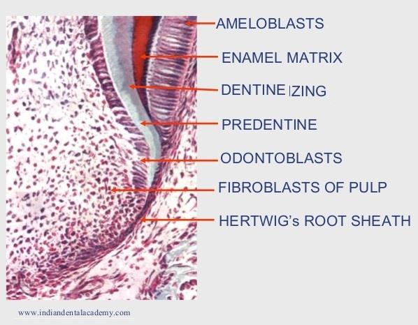
36 Colon Mucus Membrane
37 Accessory digestive organs Oral Cavity Salivary glands Liver and gallbladder Pancreas


38 The Tongue
39

40 Filiform papillae are the smallest and most numerous in humans. They are conical, elongated projections of connective tissue that are covered with highly keratinized stratified squamous epithelium. This epithelium does not contain taste buds. The papillae serve only a mechanical role. Fungiform papillae, as the name implies, are mushroomshaped projections located on the dorsal surface of the tongue. They project above the filiform papillae. They tend to be more numerous near the tip of the tongue. Taste buds are present in the stratified squamous epithelium on the dorsal surface of these papillae.

41 Foliate papillae consist of parallel low ridges separated by deep mucosal clefts, which are aligned at right angles to the long axis of the tongue. They occur on the lateral edge of the tongue. In aged individuals, the foliate papillae may not be recognized; in younger individuals, they are easily found on the posterior lateral surface of the tongue. Taste buds are present Circumvallate papillae are the large, dome-shaped structures that reside in the mucosa just anterior to the sulcus terminalis The human tongue has 8 to 12 of these papillae. Each papilla is surrounded by invagination lined with stratified squamous epithelium that contains numerous taste buds.

42 Taste buds are present on fungiform, foliate, and circumvallate papillae. Taste buds appear as oval, pale-staining bodies. A small opening onto the epithelial surface at the apex of the taste bud is called the taste pore. Three principal cell types are found in taste buds: Neuroepithelial (sensory) cells are the most numerous cells in the taste bud. Supporting cells are less numerous. Basal cells are small cells located in the basal portion of the taste bud.
43 Structure of Teeth Crown - exposed surface of tooth Neck - boundary between root and crown Enamel - outer surface Dentin bone-like, but noncellular Pulp cavity - hollow with blood vessels and nerves Root canal - canal length of root Gingival sulcus - where gum and tooth meet Periodontal Ligament keeps teeth in side the alveoli
44 Teeth consist of three layers of specialized tissues Enamel, a hard, thin, translucent layer of acellular mineralized tissue that covers the crown of the tooth. Dentin, the most abundant dental tissue; it lies deep to the enamel in the crown and cementum in the root. Its unique tubular structure and biochemical composition support the more rigid enamel and cementum overlying the surface of the tooth. Cementum, is a thin layer of bonelike material that is secreted by cementocytes, cells that closely resemble osteocytes.

45 Enamel Enamel is the hardest substance in the body; it consists of 96-98% calcium hydroxyapatite. It is secreted by cells called ameloblasts. These cells are absent after birth (no regeneration) Enamel is composed of enamel rods that span the entire thickness of the enamel layer.

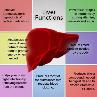
46 Dentin is a calcified material that forms most of the tooth substance. Dentin contains less hydroxyapatite than enamel, about 70%, but more than is found in bone and cementum. Dentin is secreted by odontoblasts that form an epithelial layer over the inner surface of the dentin Dentin


47 Teeth development Oral ectoderm ameloblasts Mesenchyme odontoblasts, cementoblasts, pulp


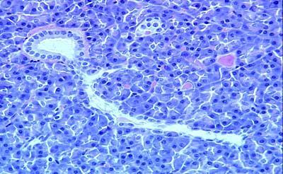
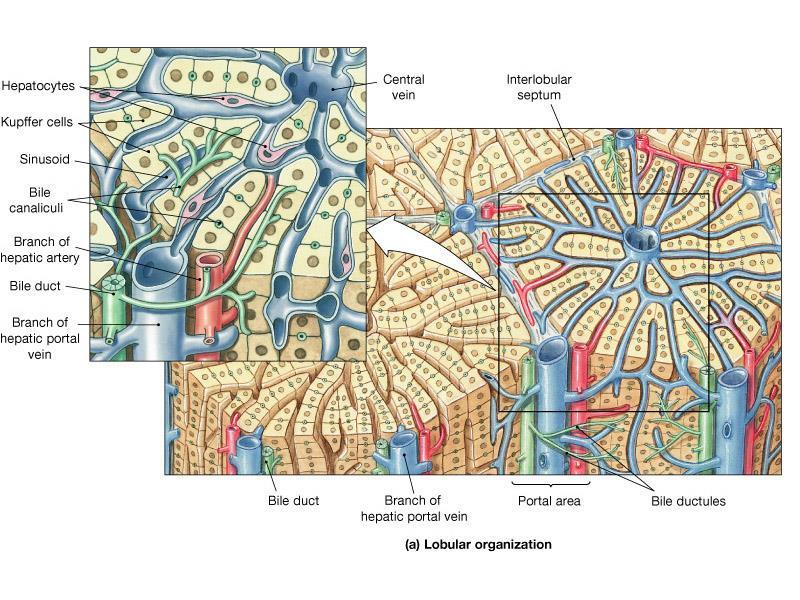
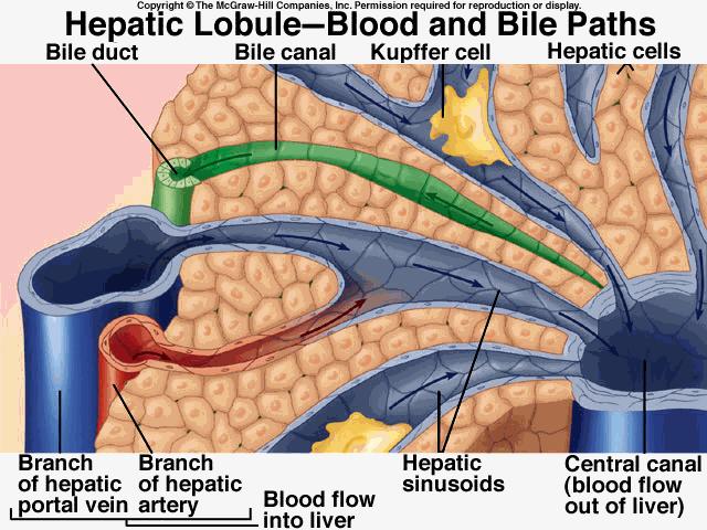
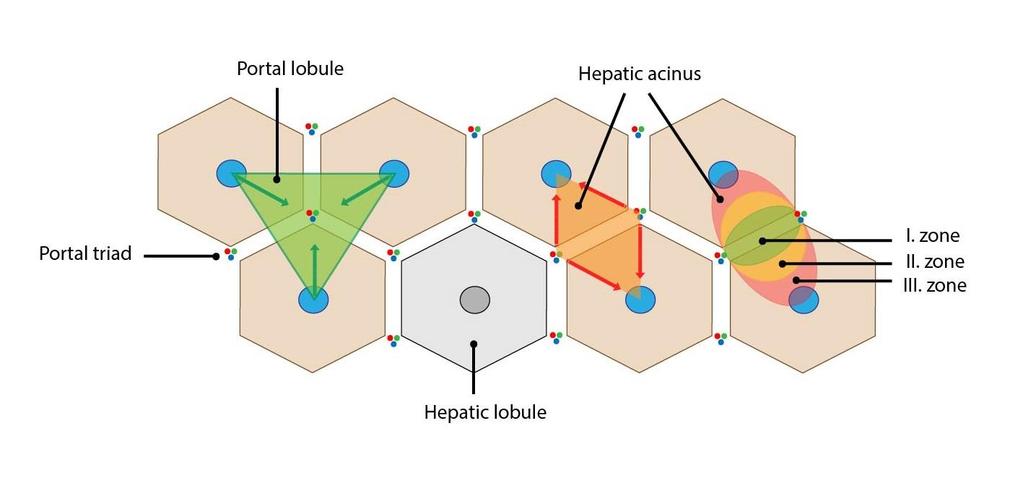
48 Bud stage Cells of oral ectoderm proliferate and form an invagination into the underlining mesenchyme epithelial tooth bud.
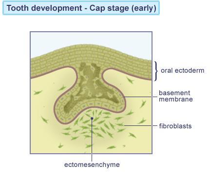
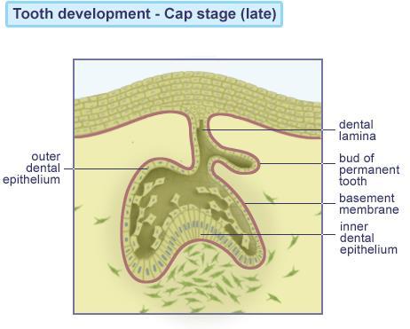

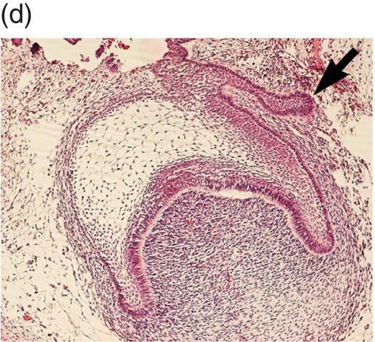
49 Cap stage

 Stratum intermidiate")

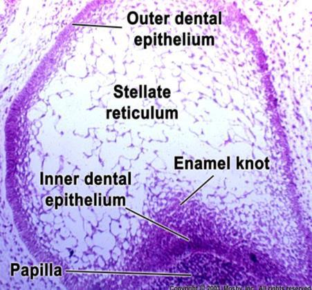
50 Bell stage Enamel organ Outer enamel epithelium (permanent teeth bud) Inner enamel epithelium (ameloblasts) Stratum intermidiate Mesenchyme - odontoblasts Cervical loop junction of inner and outer enamel epithelium

51 Ectoderm-mesenchyme interaction Ameloblasts stimulate odontoblasts to synthesize predentin Calcification of predentin Enamel production by ameloblasts
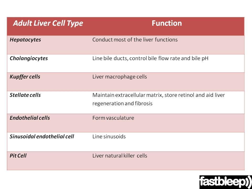
, fibroblasts (periodontum), osteoblasts")
52 Root development Formation of Hertwig s root sheath (inner and outer enamel epithelium of the cervical loop) Stimulation of predentin synthesis Predentin calcification Root sheath degeneration Dentin stimulates differentiation of surrounding mesenchymal cells into cementoblasts (cementum), fibroblasts (periodontum), osteoblasts (bone)
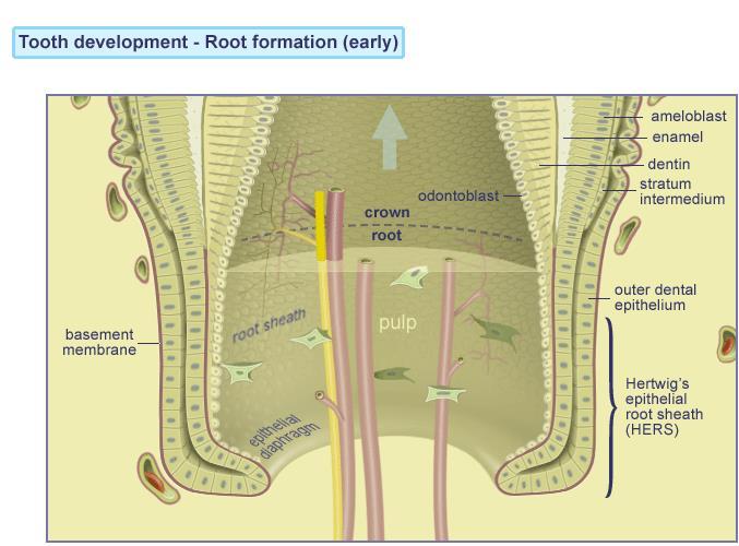
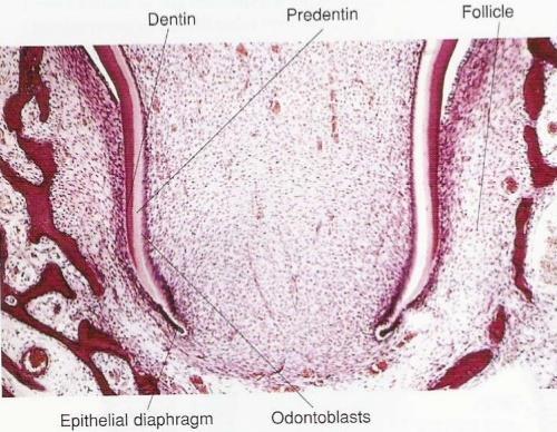
53 Root development

54

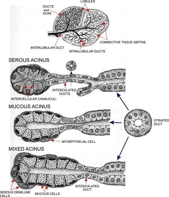
55 Salivary glands Major glands parotid, submandibular, sublingual. Minor glands throughout the mucosa of the oral cavity Saliva is a mixture of mucus and serous fluids, each produced to various extents in various glands. Also contains salivary amylase, (starts to break down starch) lysozyme (antibacterial) and IgA antibodies.

56 The parotid glands are completely serous. Serous cells are protein-secreting cells. The submandibular glands are mixed glands that are mostly serous in humans The small sublingual glands are mixed glands that are mostly mucous secreting in humans. Mucous cells are mucin-secreting cells.
57

58 Salivary Ducts The lumen of the salivary acinus is continuous with that of a duct system that may have as many as three sequential segments: Intercalated duct, which leads from the acinus Striated duct, so-called because of the presence of striations, the infoldings of the basal plasma membrane of the columnar cells that form the duct Excretory ducts, which are the larger ducts that empty into the oral cavity

59 Salivary Ducts

60 Liver Gall Bladder Stomach Duodenum Pancreas


61 Liver is the biggest gland
62

63 Histology: Liver Lobule and Portal Tract
 -Bile Duct (B) -Lymphatic vessels (L) S =")
64 Liver Portal Tract -Portal vein (PV) -Hepatic artery (A) -Bile Duct (B) -Lymphatic vessels (L) S = sinusoids
65
66
67

68 Epithelial and sinusoidal cells Bile duct cell
69

70 The pancreas performs both exocrine and endocrine functions. The exocrine secretory glands drain pancreatic juice into the pancreatic ducts and, from there, ultimately into the duodenum. The secretion is essential for the digestion and absorption of proteins, fats and carbohydrates.

71 Exocrine part Acinar cells Several types of digestive enzymes e.g., trypsin, lipase, amylase


72 Features: Islets of Langerhan s Centroacinar cells No striated ducts Centroacinar cells - In the lumen of each acinus, at the origin of intercalated duct - Secrete bicorbonate-rich fluid

73 Pancreas Secretory acini & Islet of Langerhans Lobule

74 Pancreas Secretory acini w/ zymogen granules
 C centroacinar")
75 Pancreas Intercalated Duct (D) C centroacinar cell

76 Pancreas - Intercalating Duct (arrow)

77 Pancreas Interlobular Duct

78 Pancreas Intralobular Duct
79 The endocrine pancreas is responsible for the production and secretion of glucagon and insulin, which take place in specialized cells of the islets of Langerhans.

80 Thank you for attention
Connective tissue The Digestive System
 Connective tissue The Digestive System Part 1 Structure of digestive system Functions Basic Structure of the Alimentary Canal Wall Tube is made up of four layers: 1. Mucosa 2. Submucosa 3. Muscularis externa
HISTOLOGY VIRTUAL LABORATORY GASTROINTESTINAL SYSTEM
 HISTOLOGY VIRTUAL LABORATORY GASTROINTESTINAL SYSTEM LIP (Slides GI 1, 2) Identify the outer portion lined by stratified squamous (keratinized) epithelium. Note the hair follicles and sebaceous glands
The Digestive System. Chapter 25
 The Digestive System Chapter 25 Introduction Structure of the digestive system A tube that extends from mouth to anus Accessory organs are attached Functions include Ingestion Movement Digestion Absorption
Alimentary Canal (I)
") Alimentary Canal (I) Esophagus and Stomach (Objectives) By the end of this lecture, the student should be able to discuss the microscopic structure in correlation with the function of the following organs:
Alimentary Canal (I) Esophagus and Stomach (Objectives) By the end of this lecture, the student should be able to discuss the microscopic structure in correlation with the function of the following organs:
HISTOLOGY. GIT Block 432 Histology Team. Lecture 1: Alimentary Canal (1) (Esophagus & Stomach) Done by: Ethar Alqarni Reviewed by: Ibrahim Alfuraih
 (Esophagus & Stomach) Done by: Ethar Alqarni Reviewed by: Ibrahim Alfuraih") HISTOLOGY Lecture 1: Alimentary Canal (1) (Esophagus & Stomach) Done by: Ethar Alqarni Reviewed by: Ibrahim Alfuraih Color Guide: Black: Slides. Red: Important. Green: Doctor s notes. Blue: Explanation.
HISTOLOGY Lecture 1: Alimentary Canal (1) (Esophagus & Stomach) Done by: Ethar Alqarni Reviewed by: Ibrahim Alfuraih Color Guide: Black: Slides. Red: Important. Green: Doctor s notes. Blue: Explanation.
Digestive system L 2. Lecturer Dr. Firdous M. Jaafar Department of Anatomy/Histology section
 Digestive system L 2 Lecturer Dr. Firdous M. Jaafar Department of Anatomy/Histology section objectives 1-Describe the general structure of digestive tract: a-mucosa. b-submucosa. c-muscularis externa d-adventitia
(b) Stomach s function 1. Dilution of food materials 2. Acidification of food (absorption of dietary Fe in small intestine) 3. Partial chemical digest
 Stomach s function 1. Dilution of food materials 2. Acidification of food (absorption of dietary Fe in small intestine) 3. Partial chemical digest") (1) General features a) Stomach is widened portion of gut-tube: between tubular and spherical; Note arranged of smooth muscle tissue in muscularis externa. 1 (b) Stomach s function 1. Dilution of food
(1) General features a) Stomach is widened portion of gut-tube: between tubular and spherical; Note arranged of smooth muscle tissue in muscularis externa. 1 (b) Stomach s function 1. Dilution of food
The Digestive System and Body Metabolism Premedical Biology
 The Digestive System and Body Metabolism Premedical Biology Copyright 2003 Pearson Education, Inc. publishing as Benjamin Cummings The Digestive System and Body Digestion Metabolism Breakdown of ingested
The doctor mentioned a few things about the esophagus from the previous lecture:
 السالم عليكم [HISOLOGY 2] April 27, 2014 The doctor mentioned a few things about the esophagus from the previous lecture: Esophagus - It is about 25 cm in length (from the incisor it is 45 cm) Histological
General Structure of Digestive Tract
 Dr. Nabil Khouri General Structure of Digestive Tract Common Characteristics: Hollow tube composed of a lumen whose diameter varies. Surrounded by a wall made up of 4 principal layers: Mucosa Epithelial
Includes mouth, pharynx, esophagus, stomach, small intestine, large intestine, rectum, anus. Salivary glands, liver, gallbladder, pancreas
 Chapter 14 The Digestive System and Nutrition Digestive System Brings Nutrients Into the Body The digestive system includes Gastrointestinal (GI) tract (hollow tube) Lumen: space within this tube Includes
The Digestive System. Chapter 16. Introduction. Overview of Digestive System. Histological Organization. Movement and Mixing of Digestive Materials
 The Digestive System Chapter 16 Introduction Structure of the digestive system A tube that extends from mouth to anus Accessory organs are attached Functions include Ingestion Movement Digestion Absorption
Dana Alrafaiah. Dareen Abu Shalbak. Mohammad Almuhtaseb. 1 P a g e
 2 Dana Alrafaiah Dareen Abu Shalbak Mohammad Almuhtaseb 1 P a g e Esophagus: A muscular tube that is 25 cm long, but if measured from the incisors it would be 45cm long. Extends from C6 of cervical vertebra,
Chapter 9. The digestive system. Glossary. Louise McErlean
 Chapter 9 The digestive system Louise McErlean Glossary Absorption Process whereby the products of digestion move into the blood or lymph fluid. Acini glands Produce pancreatic juice. Amylase Carbohydrate
Chapter 14: The Digestive System
 Chapter 14: The Digestive System Digestive system consists of Muscular tube (digestive tract) alimentary canal Accessory organs teeth, tongue, glandular organs 6 essential activities 1. 2. 3. 4. 5. 6.
DIGESTIVE. CHAPTER 17 Lecture: Part 1 Part 2 BIO 212: ANATOMY & PHYSIOLOGY II
 BIO 212: ANATOMY & PHYSIOLOGY II 1 CHAPTER 17 Lecture: DIGESTIVE Part 1 Part 2 Dr. Lawrence G. Altman www.lawrencegaltman.com Some illustrations are courtesy of McGraw-Hill. Processes of DIGESTION Mechanical
Digestive System. Presented by: Dr M. Arianmanesh PhD in Reproductive and Developmental Biology Dept. of Anatomical Sciences
 Digestive System Presented by: Dr M. Arianmanesh PhD in Reproductive and Developmental Biology Dept. of Anatomical Sciences Today we will discuss: Histological layers of alimentary canal Oral cavity Lip
Two main groups Alimentary canal continuous coiled hollow tube Accessory digestive organs
 Digestion Breakdown of ingested food Absorption of nutrients into the blood Metabolism Production of cellular energy (ATP) Constructive and degradative cellular activities Two main groups Alimentary canal
consists of: Muscular, hollow tube (= digestive tract ) + Various accessory organs
 + Various accessory organs") DIGESTIVE SYSTEM consists of: Muscular, hollow tube (= digestive tract ) + Various accessory organs FUNCTION Individual parts function in: ingestion mechanical digestion chemical and enzymatic digestion
DIGESTIVE SYSTEM consists of: Muscular, hollow tube (= digestive tract ) + Various accessory organs FUNCTION Individual parts function in: ingestion mechanical digestion chemical and enzymatic digestion
Small intestine. Small intestine
 General features Tubular organ longest part; 5-6 m most of chemical digestion absorption of nutrients reabsorption of H2O occurs. Two structural features; maximize the lumenal surface area villi microvilli
Chapter 26 The Digestive System
 Chapter 26 The Digestive System Digestive System Gastroenterology is the study of the stomach and intestine. Digestion Catabolism Absorption Anabolism The actions of the digestive system are controlled
Gastrointestinal Anatomy and Physiology. Bio 219 Napa Valley College Dr. Adam Ross
 Gastrointestinal Anatomy and Physiology Bio 219 Napa Valley College Dr. Adam Ross Functions of digestive system Digestion Breakdown of food (chemically) using enzymes, acid, and water Absorption Nutrients,
DIGESTIVE SYSTEM ALIMENTARY CANAL / GI TRACT & ACCESSORY ORGANS. Mar 16 10:34 PM
 DIGESTIVE SYSTEM ALIMENTARY CANAL / GI TRACT & ACCESSORY ORGANS Mar 16 10:34 PM 1 I. Digestive System Functions > Ingestion the taking in of food > Propulsion movement caused by force > Digestion breakdown
Organs Histology D. Sahar AL-Sharqi. Digestive System
 Digestive System The digestive system consists of the digestive tract oral cavity, esophagus, stomach, small and large intestines, and anus and its associated glands salivary glands, liver, and pancreas.
Digestive System 7/15/2015. Outline Digestive System. Digestive System
 Digestive System Biology 105 Lecture 18 Chapter 15 Outline Digestive System I. Functions II. Layers of the GI tract III. Major parts: mouth, pharynx, esophagus, stomach, small intestine, large intestine,
Objectives. Describe the cells of the GI tract and their function. Differentiate between different parts of the GI tract
 GI Histology 1 Objectives Describe the cells of the GI tract and their function Describe the histological features of each part of the GI tract. Differentiate between different parts of the GI tract Appreciate
Slide 154: Pancreas, H&E
 Slide 154: Pancreas, H&E the pancreas, located adjacent to the duodenum, is a mixed exocrine and endocrine gland; it is usually readily identifiable by the presence of the interspersed endocrine pancreatic
Exercise. Digestive System. Digestive system function. 1. Define the following terms: a. Chemical digestionb. Mechanical digestionc.
 Exercise 7 The Digestive System NAME: DATE: INSTRUCTOR: SECTION: Digestive system function 1. Define the following terms: a. Chemical digestionb. Mechanical digestionc. Ingestiond. Digestione. Absorptionf.
Bio 104 Digestive System
 13 Lecture Outline: Digestive System Hole s HAP [Chapters 17 & 18] General Characteristics of the Alimentary Canal A. Functions 1. Ingestion 2. Mechanical digestion 3. Chemical digestion 4. Propulsion
Anatomy & Histology of The Small intestine
 Anatomy & Histology of The Small intestine Prof. Abdulameer Al-Nuaimi E-mail: a.al-nuaimi@sheffield.ac.uk E. mail: abdulameerh@yahoo.com Jejunum Ileum Histology: Duodenum, jejunum, and ileum
Digestive system. Dr. Sami Zaqout. IUG
 Digestive system Digestive system Digestive tract Associated glands Oral cavity Salivary glands Esophagus Liver Stomach Pancreas Small and large intestines Rectum and anus General Structure of the Digestive
Dr Nadine Gravett School of Anatomical Sciences Room 2B10B
 Dr Nadine Gravett School of Anatomical Sciences Room 2B10B Nadine.Gravett@wits.ac.za Oral cavity Mechanical breakdown Formation of bolus Oesophagus Conduit from mouth to stomach Stomach Digestion Temporary
The Digestive System Laboratory
 The Digestive System Laboratory 1 The Digestive Tract The alimentary canal is a continuous tube stretching from the mouth to the anus. Liver Gallbladder Small intestine Anus Parotid, sublingual, and submaxillary
Small Intestine, Large Intestine and anal cannel
 Small Intestine, Large Intestine and anal cannel 32409 Small intestine Large intestine Small intestine General Structure of the Digestive Tract rat 32409 Epithelium with goblet cells and absorptive cells
DIGESTIVE SYSTEM II ACCESSORY DIGESTIVE ORGANS
 DIGESTIVE SYSTEM II ACCESSORY DIGESTIVE ORGANS Dr. Larry Johnson Texas A& M University Objectives Distinguish between the parotid and submandibular salivary glands. Understand and identify the structural
Lab activity manual Histology of the digestive system
 Lab activity manual Histology of the digestive system Jeanne Adiwinata Pawitan Prerequisite: Histology of the 4 basic tissues In this module we learn about the histology of the digestive system, from the
General functions of digestive system. Ch. 15 The Digestive System. General histology of the wall of the digestive tract. Overview of digestive organs
 Overall idea: obtain nutrients from food (for energy and raw materials for synthesis), and defecate the leftover waste 2 types of organs involved: 1. Parts of the digestive tract (= a long muscular tube
Lab activity manual - Histology of the digestive system. Lab activity 1: esophagus stomach - small intestines
 Lab activity manual - Histology of the digestive system Jeanne Adiwinata Pawitan Prerequisite: Histology of the 4 basic tissues In this module we learn about the histology of the digestive system, from
(A) Diarrhea. (B) Stomach cramps. (C) Dehydration due to excess fluid loss. (D) A, B, and C are correct. (E) Only answer B is correct.
 Diarrhea. (B) Stomach cramps. (C) Dehydration due to excess fluid loss. (D) A, B, and C are correct. (E) Only answer B is correct.") Human Anatomy - Problem Drill 21: The Digestive System Question No. 1 of 10 1. A 26-year-old male is treated in the emergency department for severe gastrointestinal disturbance. Which of the following
Human Anatomy - Problem Drill 21: The Digestive System Question No. 1 of 10 1. A 26-year-old male is treated in the emergency department for severe gastrointestinal disturbance. Which of the following
DIGESTIVE TRACT ESOPHAGUS
 DIGESTIVE TRACT From the lower esophagus to the lower rectum four fundamental layers comprise the wall of the digestive tube: mucosa, submucosa, muscularis propria (externa), and adventitia or serosa (see
Digestive Anatomy Lab
 Digestive Anatomy Lab In-Lab Exercises I have included the word list in this document. Any descrepencies between this document and the wordlist, you should default to this document. There is a lot of repetition
Digestive System. In one end and out the other.
 Digestive System In one end and out the other. Overview Every cell in the body needs nourishment, yet most cells cannot leave their position in the body and travel to a food source, so the food must be
Esophagus. Transport is achieved by peristaltic contractions and relaxation of the esophageal sphincters (upper and lower)
") GI Histology 2 Esophagus is a muscular tube whose function is to transport foodstuffs from the mouth to the stomach and to prevent the retrograde flow of gastric contents Transport is achieved by peristaltic
GI Histology 2 Esophagus is a muscular tube whose function is to transport foodstuffs from the mouth to the stomach and to prevent the retrograde flow of gastric contents Transport is achieved by peristaltic
I. The Alimentary Canal (GI track)
") A. About 9 meters long B. Passes through the ventral cavity. C.Movements of the Tube 1. Mixing movements- smooth muscles contract rhythmically. 2. Propelling movements- a wavelike motion called peristalsis.
A. About 9 meters long B. Passes through the ventral cavity. C.Movements of the Tube 1. Mixing movements- smooth muscles contract rhythmically. 2. Propelling movements- a wavelike motion called peristalsis.
The Digestive System
 The Digestive System Identify the Structure and Function. Mesentery of the Large Intestine The mesentery functions to connect the visceral organs to the abdominal wall. Identify the Structure. Nasal Cavity
الله الر ح م ن الر ح يم مسب
 بسم رلا هللارلا هللا This is the second histology lecture in the GI system. In this lecture, we will discuss the histology of the esophagus, stomach, and small intestine so prepare yourself.this sheet
Principles of Anatomy and Physiology
 Principles of Anatomy and Physiology 14 th Edition CHAPTER 24 The Digestive System Introduction The purpose of this chapter is to Identify the anatomical components of the digestive system as well as their
Oral cavity Lab exercises
 Oral cavity Lab exercises Slide #190 (GT-1-32). Oral cavity, goat. large conical buccal papillae stratified squamous epithelium keratinized or non-keratinized no muscularis mucosae connective tissue represents
The Digestive System. What is the advantage of a one-way gut? If you swallow something, is it really inside you?
 The Digestive System What is the advantage of a one-way gut?! If you swallow something, is it really inside you? Functions and Processes of the Digestive System: Move nutrients, water, electrolytes from
Digestive System Module 4: The Stomach *
 OpenStax-CNX module: m49286 1 Digestive System Module 4: The * Donna Browne Based on The by OpenStax This work is produced by OpenStax-CNX and licensed under the Creative Commons Attribution License 4.0
NURSE-UP DIGESTIVE SYSTEM AKA G.I. SYSTEM
 NURSE-UP DIGESTIVE SYSTEM AKA G.I. SYSTEM The digestive system is used for breaking down food into nutrients which then pass into the circulatory system and are taken to where they are needed in the body.
MICROSTRUCTURES LIPS TOOTH TONGUE OESOPHAGUS STOMACH, CARDIAC, PYLORIC FUNDIC GLANDS
 MICROSTRUCTURES LIPS TOOTH TONGUE OESOPHAGUS STOMACH, CARDIAC, PYLORIC FUNDIC GLANDS HUMAN ANATOMY: MICROSTRUCTURES CLASSIFICATION: LOCATION AND BOUNDARIES, FORM, FUNCTION, MICROSCOPIC STRUCTURE: A hollow
DIGESTIVE SYSTEM CLASS NOTES. tube along with several
 DIGESTIVE SYSTEM CLASS NOTES Digestion Breakdown of food and the of nutrients in the bloodstream. Metabolism Production of for and cellular activities. The digestive system is composed of the canal which
GASTROINTESTINAL PHYSIOLOGY PHYSIOLOGY DEPARTMENT KAMPALA INTERNATIONAL UNIVERSITY DAR ES SALAAM TANZANIA
 GASTROINTESTINAL PHYSIOLOGY PHYSIOLOGY DEPARTMENT KAMPALA INTERNATIONAL UNIVERSITY DAR ES SALAAM TANZANIA Anatomy of the GI Tract The GI tract is essentially a hollow tube connecting the mouth to the anus.
GI Histology Lab 1. Prepared by: Zeina Kalaji
 GI Histology Lab 1 Prepared by: Zeina Kalaji Lip ORAL MUCOSA -Arrow shows labial salivary glands in the submucosa. VERMILLION transitional zone. SKIN Stratified Squamous epithelium, keratinized -Arrow
Nutrition. Autotrophs. plants, some protists & bacteria producers
 Nutrition Autotrophs plants, some protists & bacteria producers Nutrition Heterotrophs animals, fungi, some protists & bacteria consumers Animal Nutrition Most obtain food by ingestion take in their food
3/16/2016. Food--mixture of carbohydrates, proteins, and lipids
 Food--mixture of carbohydrates, proteins, and lipids Food being broken down into small molecules Takes place in the alimentary canal Complete digestive system 4 layers of tissue (in book) Lumen 1) MECHANICAL/PHYSICAL--
Week 12 - Outline. Outline. Digestive System I Major Organs. Overview of Digestive System
 Outline Week 12 - Digestive System I Major Organs Copyright The McGraw-Hill Companies, Inc. Permission required for reproduction or display. Digestive Tract Function GI Tract Structure Regulation of the
MCAT Biology Problem Drill 20: The Digestive System
 MCAT Biology Problem Drill 20: The Digestive System Question No. 1 of 10 Question 1. During the oral phase of swallowing,. Question #01 A. Initially, the food bolus is moved to the back of the tongue and
1. Approximately 21 ft. long: duodenum (one ft.), jejunum (eight ft.), and ileum (twelve ft.)
, jejunum (eight ft.), and ileum (twelve ft.)") IV. Small Intestines A. General features and functions 1. Approximately 21 ft. long: duodenum (one ft.), jejunum (eight ft.), and ileum (twelve ft.) 2. Functions: move forward chyme, continue digestion,
IV. Small Intestines A. General features and functions 1. Approximately 21 ft. long: duodenum (one ft.), jejunum (eight ft.), and ileum (twelve ft.) 2. Functions: move forward chyme, continue digestion,
BIO 116 Anatomy & Physiology II Practice Assignment 3 - The Lymphatic, Immune and Digestive Systems This is not a required assignment
 BIO 116 Anatomy & Physiology II Practice Assignment 3 - The Lymphatic, Immune and Digestive Systems This is not a required assignment 1. Which are components of the lymphatic system? a: Thyroid gland b:
Energy, Chemical Reactions and Enzymes
 Phosphorylation Hydrolysis Energy, Chemical Reactions and Enzymes Chapter 2 (selections) What is Energy? Energy is the capacity to do work Potential Energy Kinetic Energy Chemical Bond Energy Like a rechargeable
Gastrointestinal System!
 Gastrointestinal System! Assoc. Prof. Prasit Suwannalert, Ph.D. (Email: prasit.suw@mahidol.ac.th)! Objectives: After learning, student should be able to describe and discuss in topics of! 1. Anatomical
DIGESTIVE. CHAPTER 17 Lecture: Part 1 Part 2 BIO 212: ANATOMY & PHYSIOLOGY II
 BIO 212: ANATOMY & PHYSIOLOGY II CHAPTER 17 Lecture: DIGESTIVE Part 1 Part 2 Dr. Lawrence G. Altman www.lawrencegaltman.com Some illustrations are courtesy of McGraw-Hill. SMALL INTESTINE DUODENUM > JEJUNUM
The Digestive system
 The Digestive system The GI tract (gastrointestinal tract) Mouth Pharynx Esophagus Stomach Small intestine Large intestine Anus The accessory digestive organs Supply secretions contributing to the breakdown
Tongue In the buccal cavity of the digestive system
 Tongue In the buccal cavity of the digestive system same layers as those of tubular organs Mucosa, submucosa, and muscularis muscularis = the muscularis externa no muscularis mucosa 1 Tongue ling = tongue
An overview of the digestive system. mouth pharynx esophagus stomach small intestine large intestine rectum anus
 An overview of the digestive system mouth pharynx esophagus stomach small intestine large intestine rectum anus Why GIT? What are the main steps in the digestive process? Ingestion intake of food via the
Digestive System Lecture Notes Read Ch 14; review questions start on page 477 S/A # 4, 5, 7, 8, 10, 11, 12, 13, 16, 17, 19, 20, 21, 22, 26, 35
 The PRINCIPLE structure of the digestive system is the Alimentary Canal (Gastrointestinal Tract) = hollow tube much like a hallway in a home. I. Functions of the Digestive System a. Ingestion: Putting
Digestion and Absorption. Food:
 Digestion and Absorption Food: Food is a basic requirement of all living beings. Food provides energy for different activities in the body. Food also provides organic materials for growth and repair. Carbohydrates,
The Digestive System and Body Metabolism
 14 PART B The Digestive System and Body Metabolism PowerPoint Lecture Slide Presentation by Jerry L. Cook, Sam Houston University ESSENTIALS OF HUMAN ANATOMY & PHYSIOLOGY EIGHTH EDITION ELAINE N. MARIEB
- Digestion occurs during periods of low activity - Produces more energy than it uses. - Mucosa
 Introduction Digestive System Chapter 29 Provides processes to break down molecules into a state easily used by cells - A disassembly line: Starts at the mouth and ends at the anus Digestive functions
Chapter 16. Lecture and Animation Outline
 Chapter 16 Lecture and Animation Outline To run the animations you must be in Slideshow View. Use the buttons on the animation to play, pause, and turn audio/text on or off. Please Note: Once you have
the serous membranes lining the peritoneal cavity continuously produce what?
 Basic A & P II Dr. L. Bacha Chapter Outline (Martini & Nath 2010) - two groups of organs form the digestive system (see Fig. 22-1): 1. digestive tract what is it also called? list the organs that make
Topic 6: Human Physiology
 Topic 6: Human Physiology 6.1 Digestion and Absorption D.1 Human Nutrition D.2 Digestion Essential Understandings: The structure of the digestive system allows it to move, digest, and absorb food. A balanced
Histology Lab. looking at microscopic pictures of tissues, for more information use Junqueira book and you can use BlueHistolgy website
 Done By: Aseel Twaijer & Laith Sorour Histology Lab *These notes help in differentiating tissues and you must read them while looking at microscopic pictures of tissues, for more information use Junqueira
Soft palate elevates, closing off the nasopharynx. Hard palate Tongue Bolus Epiglottis. Glottis Larynx moves up and forward.
 The Cephalic Phase Chemical and mechanical digestion begins in the mouth Saliva is an exocrine secretion Salivary secretion is under autonomic control Softens and lubricates food Chemical digestion: salivary
Dorsum of the tongue. Oral Part exhibit lingual papillae of the 4 types. Oral Part of Tongue divided into Left & right halves by shallow median groove
 Histology of TONGUE Figure 22.13 Dorsum of the tongue Oral Part of Tongue divided into Left & right halves by shallow median groove Oral Part exhibit lingual papillae of the 4 types a. filiform papillae,
Glandular Epithelium. Dr. Hersh Abdul Ham-Karim BVM&S, PG Dip, MSc and PhD
 Glandular Epithelium Dr. Hersh Abdul Ham-Karim BVM&S, PG Dip, MSc and PhD Glandular Epithelium Groups of surface cells differentiate, proliferate, and penetrate underlying connective tissue. Their main
Practical Histology o
 Practical Histology o 1.. Contents: Histology of the : Stomach Esophagus Small intestine Large intestine Liver Gallbladder Exocrine pancreas Spleen GNT Block Things you need to know before the exam : o
Digestive System II - Lower tract Revised
 ANAT D502 Basic Histology Digestive System II - Lower tract Revised 10.12.12 Outline: I. Small intestine II. Enterocyte digestion II. Hepatic portal system IV. Large intestine V. Enteric nervous system
The Digestive System. Basic process of digestion. Mouth and Teeth 10/30/2016
 The Digestive System Basic process of digestion 1. Ingestion: animal eats food. 2. Digestion: animal body breaks food down. Mechanical digestion: chewing (mastication). Chemical digestion: enzymes and
The Human Body: Digestive System
 Directions: Fill in the blanks. 1. Digestive System Is a series of hollow organs joined in a long, twisting tube from the mouth to the anus Carries out digestion which is the process of breaking down food
BELLWORK DEFINE: PERISTALSIS CHYME RUGAE Remember the structures of the digestive system 1
 BELLWORK DEFINE: PERISTALSIS CHYME RUGAE 2.07 Remember the structures of the digestive system 1 STANDARD 8) Outline basic concepts of normal structure and function of all body systems, and explain how
University of Buea. Faculty of Health Sciences. Programme in Medicine
 Faculty of Health Sciences University of Buea Wednesday, 28 th January 2009 Time: 8 00-10 00 Programme in Medicine MED 303 (Gastrointestinal Physiology) EXAMS (2008-2009) Identify the letter of the choice
- Digestion occurs during periods of low activity - Produces more energy than it uses. 3 Copyright 2016 by Elsevier Inc. All rights reserved.
 Introduction Digestive System Chapter 29 Provides processes to break down molecules into a state easily used by cells - A disassembly line: Starts at the mouth and ends at the anus Digestive functions
Chapter 23: The Digestive System
 Chapter 23: The Digestive System I. OVERVIEW Consists of alimentary canal and accessory organs. GIT (gastrointestinal tract) and teeth, tongue, gallbladder, salivary glands, liver and pancreas. (A) Digestive
The Digestive System and Body Metabolism
 PowerPoint Lecture Slide Presentation by Patty Bostwick-Taylor, Florence-Darlington Technical College The Digestive System and Body Metabolism 14PART C Accessory Digestive Organs Teeth Salivary glands
Bio 322 Human Anatomy Objectives for the laboratory exercise Digestive System
 Bio 322 Human Anatomy Objectives for the laboratory exercise Digestive System Required reading before beginning this lab: Saladin, KS: Human Anatomy 5 th ed (2017) Chapter 24 For this lab you will use
The Digestive System. Prepares food for use by all body cells.
 The Digestive System Prepares food for use by all body cells. Digestion The chemical breakdown of complex biological molecules into their component parts. Lipids to fatty acids Proteins to individual amino
A deep groove encircles the body of the circumvallate papilla. Serous (von Ebner s) glands (serous) drain into the base of this groove.
 glands (serous) drain into the base of this groove.") By Dr. Raja Ali A deep groove encircles the body of the circumvallate papilla. Serous (von Ebner s) glands (serous) drain into the base of this groove. The flow of fluid from these glands serves to wash
By Dr. Raja Ali A deep groove encircles the body of the circumvallate papilla. Serous (von Ebner s) glands (serous) drain into the base of this groove. The flow of fluid from these glands serves to wash
Anatomy and Physiology: The Unity of Form and Function
 Chapter 25 Anatomy and Physiology: The Unity of Form and Function Second Edition Kenneth S. Saladin (c) The McGraw-Hill Companies, Inc. Structures and Location A. Location from mouth through thoracic
The Digestive System
 The Digestive System s Big Book of Handouts Digestion and nutrition Specific Learning outcomes B11-2-01: Identify major structures and functions of the human digestive system from a diagram, model, or
Respiratory & Digestive Organs of the Head and Neck, Human;
 Name Date Lab Exercise 5: Lab Exercise 6: Lab Exercise 7: Lab Exercise 8: Respiratory & Digestive Organs of the Head and Neck, Human; Histology of the Respiratory System Digestive System Models, Human
/30/17 Ch 8: Muscular System 1. Table of Contents # Date Title Page # 03/13/17 Ch 10: Somatic and Special Senses 53
 Table of Contents # Date Title Page # 1. 01/30/17 Ch 8: Muscular System 1 2. 3. 4. 5. 6. 7. 02/14/17 Ch 9: Nervous System 12 03/13/17 Ch 10: Somatic and Special Senses 53 03/27/17 Ch 11: Endocrine System
ANATOMY & PHYSIOLOGY ONLINE COURSE - SESSION 13 THE DIGESTIVE SYSTEM
 ANATOMY & PHYSIOLOGY ONLINE COURSE - SESSION 13 THE DIGESTIVE SYSTEM The digestive system also known as the alimentary canal or gastrointestinal tract consists of a series of hollow organs joined in a
Section 1.1: What is the function of digestion?
 Section 1.1: What is the function of digestion? When you have completed this section, you should be able to: Describe the overall function of the GI tract. Describe the processes involved in digestion.
Digestive Lecture Test Questions Set 4
 Digestive Lecture Test Questions Set 4 1. Which of the following is not associated directly with the small intestine: a. villi b. circular folds c. microvilli d. haustrae e. secretin 2. The largest (longest)
The stomach is formed of three parts: -
 The stomach is formed of three parts: - (a) CARDIAC STOMACH: - It receives the oesophagus through Cardiac aperture guarded by a cardiac sphincter which prevents regurgitation of food. (b) FUNDIC PART:
Human Biology. Digestive System
 Human Biology Digestive System Digestion - Defined Prepares food for use by all body cells The physical and/or chemical breakdown of food Did you know: the average person eats more than 500kg of food per
Lesson Overview The Digestive System
 30.3 THINK ABOUT IT The only system in the body that food actually enters is the digestive system. So how does food get to the rest of the body after the process of digestion? Functions of the Digestive