Op#c Nerve Head & Re#nal Imaging
|
|
|
- Beverly Gallagher
- 5 years ago
- Views:
Transcription
1 Op#c Nerve Head & Re#nal Imaging Dr Cesar Carrillo October, 2014 Vien#ane/NOC **Disclaimer** The images contained in this presenta5on are not my own, they can be found on the web
2 OUTLINE Indica5ons Confocal laser scanning tomography (Optos) Confocal laser scanning tomography with polarimetry (GDx) Confocal tomography (HRT) Op5cal coherence tomography (OCT) 2
! hydroxychloroquine (Plaquenil)! vigabatrin (Sabril) toxicity!")
3 Optic Nerve Head and Retinal Imaging - Indications Medically necessary for documen5ng the appearance of the op5c nerve head and re5na Pa5ents with :! glaucoma, glaucoma suspects! posterior vitreous detachment screening for:! chloroquine (Aralen)! hydroxychloroquine (Plaquenil)! vigabatrin (Sabril) toxicity! other diseases macular hole, diabe5c re5nopathy, and pseudotumor cerebri
4 Optic Nerve Head and Retinal Imaging Op5c nerve imaging for glaucoma more frequently than once per year is considered not medically necessary Methods: o Confocal laser scanning tomography (Optos) o Confocal laser scanning tomography with polarimetry (GDx nerve fiber layer tes5ng or analysis) o Confocal tomography (HRT) o Op5cal coherence tomography (OCT)
 Non-")
5 Confocal Laser Scanning Tomography (Optos) Ultra- Widefield (UWF) imaging Documen5ng Monitoring ocular pathology in the periphery > 80% view (200⁰) Non- contact

6 Confocal Laser Scanning Tomography with Polarimetry (GDx) GDx nerve fiber layer tes5ng or analysis Confocal laser scanning tomography with polarimetry


7 Confocal Tomography (HRT) Observa5on &documenta5on of the ONH Laser take 3- dimensional photo Area of the op5c disc and rim, volume of the cup

8 Optical Coherence Tomography (OCT)
9 Op5cal Coherence Tomography OCT Diagnos5c imaging technique that examines living 5ssue non- invasively. It is based on a complex analysis of the reflec5on of low coherence radia5on from the 5ssue under examina5on Real 5me cross sec5onal analysis
10 Op5cal Coherence Tomography OCT Qualita5ve and quan5ta5ve analysis of the re5na Qualita5ve analysis includes descrip5on by loca5on, a descrip5on of form and structure, iden5fica5on of anomalous structures, and observa5on of the reflec5ve quali5es of the re5na

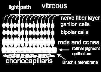
11 OCT Quan5ta5ve analysis involves measurements of the re5na, specifically re5nal thickness and volume, and nerve fiber layer thickness. This is possible because the OCT so_ware is able to iden5fy and "trace" two key layers of the re5na, the NFL and RPE
 in a single \"scan pass o Each A-")
12 OCT How does it work? o 128 to 768 axial samples (A- scans) in a single "scan pass o Each A- scan has 1024 data points and is 2mm long (deep)
13 OCT Resolu5on: When all of the A- scans are combined into one image, the image has a resolving power of about 10 microns ver5cally and 20 microns horizontally Compare that to the resolu5on of a good ophthalmic ultrasound at 100 microns

14 OCT Protocols: OCT has several built- in protocols for scanning the re5na and the op5c nerve head A protocol is simply a pre- determined procedure or method
15 OCT Scan Protocol Types: Line Circle Radial Lines
16 OCT The "line" scan simply scans in a single, straight line. The length of the line can be changed as well as the scan angle

17 OCT The "circle" scans in a circle instead of a line
18 OCT The "radial lines" scans 6 consecu5ve line scans in a star pagern
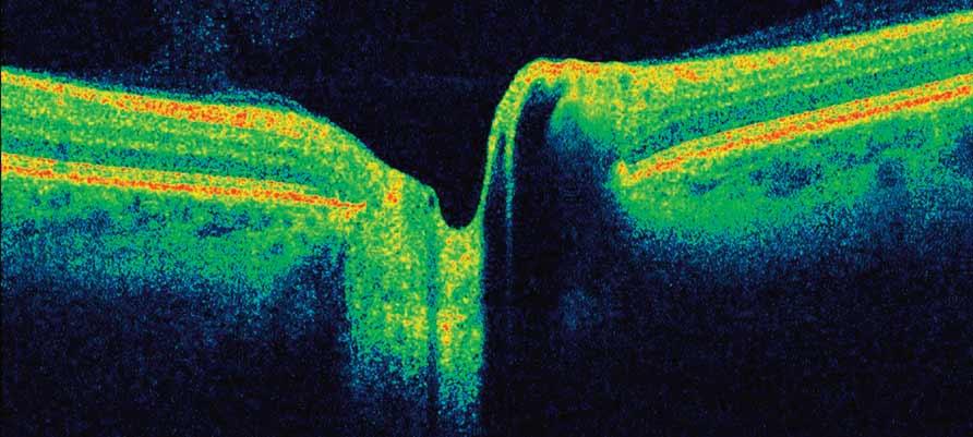
19 Re5nal Anatomy Compared to OCT The vitreous is the black space on the top of the image We can iden5fy the fovea by the normal depression The nerve fiber layer (NFL) and the re5nal pigment epithelium (RPE) are easily iden5fiable layers as they are more highly reflec5ve than the other layers of the re5na
20 Re5nal Anatomy Compared to OCT This higher reflec5vity is represented by the "hoger" colors (red, yellow, orange, white) in the false color representa5on of the OCT The middle layers of the re5na, between the NFL and RPE, are much less easily iden5fiable in the scan


21 Re5nal Anatomy Compared to OCT

22 Re5nal Anatomy Compared to OCT For purposes of analysis, the OCT image of the re5na can be subdivided ver5cally into four regions the pre- re5na the epi- re5na the intra- re5na the sub- re5na
23 The Pre- Re5nal Profile A normal pre- re5nal profile is black space Normal vitreous space is translucent The small, faint, bluish dots in the pre- re5nal space is "noise" This is an electronic aberra5on created by increasing the sensi5vity of the instrument to beger visualize low reflec5ve structures
24 The pre- re5nal profile Anomalous structures: pre- re5nal membrane epi- re5nal membrane vitreo- re5nal strands vitreo- re5nal trac5on pre- re5nal neovascular membrane pre- papillary neovascular membrane

25 The Over- All Re5nal Profile

26 A Pre- Re5nal Membrane With Trac5on on the Fovea
27 A Pigment Epithelial Detachment is Causing the Convexity

28 Aside from the re5nal detachment, no5ce the underlying concave curvature of the re5na, sugges5ng the long eye of a significant myopic
29 The foveal profile The normal foveal profile is a slight depression in the surface of the re5na
30 Deforma5ons in the Foveal Profile macular pucker macular pseudo- hole macular lamellar hole macular cyst macular hole, stage 1 (no depression, cyst present) macular hole, stage 2 (par5al rupture of re5na, increased thickness) macular hole, stage 3 (hole extends to RPE, increased thickness, some fluid) macular hole, stage 4 (complete hole, edema at margins, complete PVD)

31 Macular Cyst

32 Macular Hole - Stage 2

33 Macular Hole- Stage 3

34 Macular Hole- Stage 4 Operculum Suspended by the Hyaloid Membrane
35 The Macular Profile The macular profile can, and o_en does, include the fovea as it's center
36 Deforma5ons in the Macular Profile Serous re5nal detachment (RD) Serous re5nal pigment epithelial detachment (PED) Hemorrhagic pigment epithelial detachment

37 Serous Re5nal Pigment Epithelial Detachment (PED)
38 Intra- Re5nal Anomalies in the Macular Profile Choroidal neovascular membrane Diffuse intra- re5nal edema Cystoid macular edema Drusen Hard exudates Scar 5ssue Atrophic degenera5on Sub- re5nal fibrosis RPE tear

39 Choroidal Neovascular Membrane

40 Cystoid Macular Edema Caused by Diabe5c Maculopathy

41 Sub- Re5nal Fibrosis
42 OCT and Fluorescein Angiography in Re5nal Diagnosis FAs provide excellent characteriza5on of re5nal blood flow over 5me, as well as size and extent informa5on on the x and y axis (north- south, east- west) The OCT gives us informa5on in the z (depth) axis, telling us what layers of the re5na are affected


43 OCT and Fluorescein Angiography in Re5nal Diagnosis


44 OCT and Fluorescein Angiography in Re5nal Diagnosis
45 OCT Scans for Qualita5ve Analysis of the Re5na The Fast Macular Thickness Scan (FMTS, FMTM, or FMT scan) The Line Scan The Cross Hair Scan (3mm and 6mm)
46 The Fast Macular Thickness Scan The Fast Macular Thickness Scan consists of 6 radial line scans in a spoke pagern. It is a low resolu5on scan that was designed for quan5ta5ve analysis (thickness and volume)
47 The Fast Macular Thickness Scan The FMT scan is placed over the area of interest, which is usually the macula. When scanning the macula, the pa5ent simply looks at the fixa5on target. The center of the FMT scan lines up with the fixa5on target by default A scan is saved and then reviewed with any of the re5na analysis tools
48 The Line Scan The line scan is par5cularly useful because of it's flexibility. The length of the line can be changed, the angle of the line can be changed, and the line can be dragged with the mouse to any posi5on or angle on the video screen

49 The Cross Hair Scan Cross Hair Scan performs a high resolu5on horizontal line scan and then automa5cally flips to a ver5cal line scan without having to exit the protocol This is a common technique used in B- scan ultrasonography
50 Serial FMT Scans Over Time One of the most useful func5ons of the OCT is the ability to take volume measurement over 5me For example, a FMT scan before treatment for AMD, and FMT scans at various intervals a_er treatment Successful treatment should be followed by a decrease in re5nal thickness and volume
51 Op5c Nerve Head Scans When evalua#ng the glaucoma suspect or the glaucoma pa#ent, two parameters that the ophthalmologist is interested in are the characteris#cs of the op#c nerve cup and the thickness of the nerve fiber layer surrounding the op#c nerve head The Fast Op#c Disc scan The Fast RNFL Thickness scan
52 The Fast Op5c Disc Scan The op5c cup profile can be evaluated by capturing a "Fast Op5c Disc" scan The pa5ent fixes on the target, which is automa5cally placed at the edge of the scan window so that the op5c nerve is viewed toward the center of the video window The operator then moves the scan so that the star pagern is centered on the op5c nerve head

53 The Fast Op5c Disc Scan

54 The Op5c Nerve Scan can be Analyzed with the Op5c Nerve Head Analysis" Protocol
55 The Fast RNFL Thickness Scan Nerve fiber layer thickness can be evaluated with the "Fast RNFL Thickness" scan This is a circular scan that requires the operator to place the circle so that the center of the circle is centered on the op5c nerve head

56 The analysis so_ware places lines on the top and bogom of the nerve fiber layer and the distance between the two lines is interpreted to be the thickness of the nerve fiber layer
57 Care must be take to make sure that the image is captured with the circle centered on the op5c nerve The placement of the circle can make a big difference in the analysis of the nerve fiber layer thickness

, but the")
58 These two scans (OD) are of a normal eye. The scan in the first analysis is well centered and the RNFL thickness falls within the normal range. The scan in the second analysis is of the same eye (OD), but the scan is not well centered. The analysis is abnormal (black arrows)
59 OCT A good quality OCT scan has good reflec5vity from edge to edge The "hoger" colors (orange, red, white, yellow) are maximized Generally, the re5na should be in the lower por5on of the scan window so that the vitreous can be images as well
60 OCT
61 Scanning Tips Communicate with the doctor regarding the size and loca5on of the pathology of interest Refer to other images of the pathology, e.g. color photos and FA Review past OCT exams and repeat scan types used before Dilate the eye well?? The pa5ent must keep the forehead against the bar and the chin in the chinrest, with teeth together. Use the marker on the headrest to align the pa5ent ver5cally. The outer canthus should be even with the line
62 Scanning Tips Use the two bugons near the joys5ck for freezing and saving scans. This saves you from having to juggle the joys5ck and the mouse Minimize pa5ent fa5gue by keeping scan 5me to a minimum Never scan an eye for more than 10 minutes Keep the cornea lubricated. Use ar5ficial tears and have the pa5ent blink when you are not saving a scan pass Move the instrument on the x and y axis (using the joys5ck) to work around opaci5es
63 Scanning Tips Alignment and focus are used to maximize the quality of the scan Alignment begins with centering and zooming in on the "football" shaped reflex in the video image The ini5al lens- to- subject distance is achieved when the re5nal image fills the video screen This is similar to the image you see when doing re5na photography. At this point, your agen5on should shi_ to the scan window
64
Optical Coherence Tomography in Diabetic Retinopathy. Mrs Samantha Mann Consultant Ophthalmologist Clinical Lead of SEL-DESP
 Optical Coherence Tomography in Diabetic Retinopathy Mrs Samantha Mann Consultant Ophthalmologist Clinical Lead of SEL-DESP Content OCT imaging Retinal layers OCT features in Diabetes Some NON DR features
Re(nal and OCT Grand Rounds
 Op#cal Coherence Tomography Op(cal: Light- based Re(nal and OCT Grand Rounds Steven Ferrucci, OD, FAAO Chief, Optometry Sepulveda VA Professor, SCCO/MBKU Coherence: property of light waves in which the
Mark Dunbar: Disclosure
 Important Things to Understand About OCT Mark T. Dunbar, O.D., F.A.A.O. Bascom Palmer Eye Institute University of Miami, School of Medicine Mark Dunbar: Disclosure Optometry Advisory Board for: Allergan
OPTIC DISC PIT Pathogenesis and Management OPTIC DISC PIT
 OPTIC DISC PIT Pathogenesis and Management Abdel-Latif Siam Ain Shams University Cairo Egypt OPTIC DISC PIT Congenital pit is an atypical coloboma usually located on the temporal edge of the disc, associated
Do You See What I See!!! Shane R. Kannarr, OD
 Do You See What I See!!! Shane R. Kannarr, OD skannarr@kannarreyecare.com Define Specialty Testing Additional Test to: Prove/Disprove Diagnosis To monitor progression of a condition To document a condition
Moving forward with a different perspective
 Moving forward with a different perspective The Leader In Vision Diagnostics Offers A New Perspective Marco has served the eyecare community by offering exceptional lane products and automated high tech
OCT Image Analysis System for Grading and Diagnosis of Retinal Diseases and its Integration in i-hospital
 Progress Report for1 st Quarter, May-July 2017 OCT Image Analysis System for Grading and Diagnosis of Retinal Diseases and its Integration in i-hospital Milestone 1: Designing Annotation tool extraction
Advances in OCT Murray Fingeret, OD
 Disclosures Advances in OCT Murray Fingeret, OD Consultant Alcon, Allergan, Bausch & Lomb, Carl Zeiss Meditec, Diopsys, Heidelberg Engineering, Reichert, Topcon Currently Approved OCT Devices OCT Devices
8/6/17. Disclosures Aerie Pharmaceuticals Alcon BioTissue Diopsys Optovue Shire
 Nathan Lighthizer, O.D., F.A.A.O. Associate Professor Assistant Dean for Clinical Care Director of Continuing Education Chief of Specialty Care Clinics Oklahoma College of Optometry Tahlequah, OK lighthiz@nsuok.edu
The College of Optometrists - Learning outcomes for the Professional Certificate in Medical Retina
 Learning outcomes for the Professional Certificate in Medical Retina, incorporating diabetic retinopathy screening and age related macular degeneration The professional certificate is a prerequisite to
Re)nal and OCT Grand Rounds. What's new in OCT? Principles of AngioVue OCTA. Vascular Imaging No Referral Needed 3/9/18. Spectral Domain: Many Op3ons
nal and OCT Grand Rounds. What's new in OCT? Principles of AngioVue OCTA. Vascular Imaging No Referral Needed 3/9/18. Spectral Domain: Many Op3ons") Spectral Domain: Many Op3ons Re)nal and OCT Grand Rounds Steven Ferrucci, OD, FAAO Chief, Optometry Sepulveda VA Professor, SCCO/MBKU Ease of use Customer support Integra)on of other technology FAF Color
Spectral Domain: Many Op3ons Re)nal and OCT Grand Rounds Steven Ferrucci, OD, FAAO Chief, Optometry Sepulveda VA Professor, SCCO/MBKU Ease of use Customer support Integra)on of other technology FAF Color
What Is O.C.T. and Why Should I Give A Rip? OCT & Me How Optical Coherence Tomography Changed the Life of a Small Town Optometrist 5/19/2014
 OCT & Me How Optical Coherence Tomography Changed the Life of a Small Town Optometrist Email: myoder@wcoil.com Mark A. Yoder, O.D. 107 N. Main Street PO Box 123 Bluffton, OH 45817 @yoderod 115.02 Histoplasma
Structural examina.on: Imaging
 ManaMa: Glaucoma Structural examina.on: Imaging Luís Abegão Pinto, MD, PhD Department of Ophthalmology CHLC Lisbon Faculty of Medicine, Lisbon University 1 11-10- 2013 Structural changes Qualitative changes
OCT Interpretation in Retinal Disease
 OCT Interpretation in Retinal Disease Jay M. Haynie, OD, FAAO Financial Disclosure I have received honoraria or am on the advisory board for the following companies: Carl Zeiss Meditec Advanced Ocular
OCT Angiography in Primary Eye Care
 OCT Angiography in Primary Eye Care An Image Interpretation Primer Julie Rodman, OD, MS, FAAO and Nadia Waheed, MD, MPH Table of Contents Diabetic Retinopathy 3-6 Choroidal Neovascularization 7-9 Central
Cirrus TM HD-OCT. Details defi ne your decisions
 Cirrus TM HD-OCT Details defi ne your decisions 2 With high-defi nition OCT Carl Zeiss Meditec takes you beyond standard spectral domain Built on 10 years experience at the vanguard of innovation, Carl
Clinically Significant Macular Edema (CSME)
") Clinically Significant Macular Edema (CSME) 1 Clinically Significant Macular Edema (CSME) Sadrina T. Shaw OMT I Student July 26, 2014 Advisor: Dr. Uwaydat Clinically Significant Macular Edema (CSME) 2
Clinically Significant Macular Edema (CSME) 1 Clinically Significant Macular Edema (CSME) Sadrina T. Shaw OMT I Student July 26, 2014 Advisor: Dr. Uwaydat Clinically Significant Macular Edema (CSME) 2
The Quick Guide to OCT Mastery 50 Real Cases with Expert Analysis
 OPTICAL COHERENCE TOMOGRAPHY The Quick Guide to OCT Mastery 50 Real Cases with Expert Analysis VOL 1 Sanjay Sharma, MD, FRCS, MSc (Epid), MBA Ophthalmologist, Epidemiologist Queen s University, Canada
PRIMUS 200 from ZEISS The essential OCT
 PRIMUS 200 from ZEISS The essential OCT Seeing beyond the surface. ZEISS PRIMUS 200 // INNOVATION MADE BY ZEISS Clear Visualization. Advanced Technology. Reliability. Essential elements of your first OCT.
The Human Eye. Cornea Iris. Pupil. Lens. Retina
 The Retina Thin layer of light-sensitive tissue at the back of the eye (the film of the camera). Light rays are focused on the retina then transmitted to the brain. The macula is the very small area in
Fundus Autofluorescence. Jonathan A. Micieli, MD Valérie Biousse, MD
 Fundus Autofluorescence Jonathan A. Micieli, MD Valérie Biousse, MD The retinal pigment epithelium (RPE) has many important functions including phagocytosis of the photoreceptor outer segments Cone Rod
Optical Coherence Tomograpic Features in Idiopathic Retinitis, Vasculitis, Aneurysms and Neuroretinitis (IRVAN)
") Columbia International Publishing Journal of Ophthalmic Research (2014) Research Article Optical Coherence Tomograpic Features in Idiopathic Retinitis, Vasculitis, Aneurysms and Neuroretinitis (IRVAN)
Columbia International Publishing Journal of Ophthalmic Research (2014) Research Article Optical Coherence Tomograpic Features in Idiopathic Retinitis, Vasculitis, Aneurysms and Neuroretinitis (IRVAN)
PRIMUS 200 from ZEISS The essential OCT
 EN 00_00I The contents of the brochure may differ from the current status of approval of the product in your country. Please contact your regional representative for more information. Subject to change
Course # Getting to Know Your OCT
 Course # 140 Getting to Know Your OCT Course Title: Lecturer: Getting to Know Your OCT Brad Sutton, OD, FAAO IU School of Optometry Financial Disclosures No financial disclosures Optical Coherence Tomography-OCT
Re)nal and OCT Grand Rounds
nal and OCT Grand Rounds") Disclosures Re)nal and OCT Grand Rounds Steven Ferrucci, OD, FAAO Chief, Optometry Sepulveda VA Professor, SCCO/MBKU Speakers bureau and/or Advisory Board for: Alcon Allergan AutoGenomics B&L Heidelberg
Disclosures Re)nal and OCT Grand Rounds Steven Ferrucci, OD, FAAO Chief, Optometry Sepulveda VA Professor, SCCO/MBKU Speakers bureau and/or Advisory Board for: Alcon Allergan AutoGenomics B&L Heidelberg
Introducing ANGIOVUE ESSENTIAL. Built on the Avanti Widefield OCT Platform. OCT Angiography for Primary Eye Care
 Introducing ANGIOVUE ESSENTIAL Built on the Avanti Widefield OCT Platform OCT Angiography for Primary Eye Care Transform Your View of the Retina OCT Angiography (OCTA) is a quick non-invasive test that
Cirrus TM HD-OCT. Details define your decisions
 Cirrus TM HD-OCT Details define your decisions 2 With high-definition OCT Carl Zeiss Meditec takes you beyond standard spectral domain Built on 10 years experience at the vanguard of innovation, Carl Zeiss
DOME SHAPED MACULOPATHY. Ιωάννης Ν. Βαγγελόπουλος Χειρ. Οφθαλμίατρος - Βόλος
 DOME SHAPED MACULOPATHY Ιωάννης Ν. Βαγγελόπουλος Χειρ. Οφθαλμίατρος - Βόλος DOME SHAPED MACULOPATHY-DEFINITIONS The entity Dome Shaped Macula ( DSM ) was first described by Gaucher and associates in 2008
Amber Priority. Image Library
 Amber Priority Image Library Amber flag Diabetic Maculopathy (M1) Pre-proliferative Diabetic Retinopathy (R2) Old, treated and now inactive DR (R1/M0/P1or R0/M0/P1) Where only partial or incomplete images
Dr/ Marwa Abdellah EOS /16/2018. Dr/ Marwa Abdellah EOS When do you ask Fluorescein angiography for optic disc diseases???
 When do you ask Fluorescein angiography for optic disc diseases??? 1 NORMAL OPTIC DISC The normal optic disc on fluorescein angiography is fluorescent due to filling of vessels arising from the posterior
evaluation of vitreoretinal adhesions in exudative AMD using optical coherence tomography
 evaluation of vitreoretinal adhesions in exudative AMD using optical coherence tomography Dr. Mahmoud Alaa Abouhusssein, FRCO Lecturer of ophthalmology, Alexandria university Dr. Amir Ramadan Gomaa, MD
Optical Coherence Tomography (OCT) in Uveitis Piergiorgio Neri, BMedSc, MD, PhD Head Ocular Immunology Unit
 in Uveitis Piergiorgio Neri, BMedSc, MD, PhD Head Ocular Immunology Unit") The Eye Clinic Polytechnic University of Marche Head: Prof Alfonso Giovannini November, 1991 Optical Coherence Tomography (OCT) in Uveitis Piergiorgio Neri, BMedSc, MD, PhD Head Ocular Immunology Unit
The Eye Clinic Polytechnic University of Marche Head: Prof Alfonso Giovannini November, 1991 Optical Coherence Tomography (OCT) in Uveitis Piergiorgio Neri, BMedSc, MD, PhD Head Ocular Immunology Unit
Re(nal and OCT Grand Rounds
 Disclosure Statement Re(nal and OCT Grand Rounds Steven Ferrucci, OD, FAAO Chief, Optometry Sepulveda VA Professor, SCCO/MBKU Speakers bureau/advisory Board Allergan Alcon AutoGenomics B&L Centervue Heidelberg
Learn Connect Succeed. JCAHPO Regional Meetings 2016
 Learn Connect Succeed JCAHPO Regional Meetings 2016 pearls and pitfalls of ophthalmic imaging JCHAPO 2016 Conference Vikas Chopra, M.D. Medical Director, UCLA Doheny Eye Centers Pasadena Principal Investigator,
Visualize. Analyze. Personalize. OCT + OCTA
 Visualize. Analyze. Personalize. OCT + OCTA A New Approach to Protecting Vision AngioVue OCT Angiography brings valuable new information to clinical practice. Non-invasive visualization of retinal vasculature.
The World s fastest OCT. As simple as pressing. the start button
 The World s fastest OCT As simple as pressing the start button lution continues Optopol engineering team, designers of the first commercially available Spectral Domain OCT in the world, are proud to present
Overview. Macular OCT Artifact Study
 Imaging Artifacts Sarah Moyer, CRA, OCT-C Director, Ophthalmic Imaging Kittner Eye Center University of North Carolina Chapel Hill, NC Disclose financial interest now Overview Sarah s Thoughts on Artifacts
Beyond the C/D Ratio: Evaluating a Glaucomatous Optic Nerve. Marcus Gonzales, OD, FAAO Cedar Springs Eye Clinic COPE ID#: GL
 Beyond the C/D Ratio: Evaluating a Glaucomatous Optic Nerve Marcus Gonzales, OD, FAAO Cedar Springs Eye Clinic COPE ID#: 27809-GL Points to Remember Glaucoma affects the ONH in characteristic patterns
OCT Angiography The Next Frontier
 Choroid Retina avascular 5/13/2017 OCT Angiography The Next Frontier Pierce Kenworthy OD, FAAO June 9, 2017 OCT Angiography (OCTA) 2016 Non-invasive, motion contrast imaging Represents erythrocyte movement
 measure of your overall performance. An isolated glucose test is helpful to let you know what your sugar level is at one moment, but it doesn t tell you whether or not your diabetes is under adequate control
ATLAS OF OCT. Retinal Anatomy in Health & Pathology by Neal A. Adams, MD. Provided to you by:
 ATLAS OF OCT Retinal Anatomy in Health & Pathology by Neal A. Adams, MD Provided to you by: Atlas of OCT The OCT Atlas is written by Neal A. Adams, MD, and produced by Heidelberg Engineering, Inc. to help
Patient AB. Born in 1961 PED
 Clinical Atlas Patient AB Born in 1961 PED Autofluorescence Dilated 45 EasyScan Zero-dilation IR 45 Fundus Dilated 45 In the fundus photos (Canon CX1) the PED is not able to be seen. However, the extent
History/principles of the OCT What does the normal retinal OCT look like Vitreal disorders Retinal/RPE disorders Choroidal disorders
 Nathan Lighthizer, O.D., F.A.A.O. Assistant Professor Assistant Dean for Clinical Care Director of Continuing Education Chief of Specialty Care Clinics Chief of Electrodiagnostics Clinic Oklahoma College
A COMPLETE GUIDE TO THE CLARUS 500 ULTRA-WIDEFIELD RETINAL CAMERA
 A COMPLETE GUIDE TO THE CLARUS 500 ULTRA-WIDEFIELD RETINAL CAMERA BY DANIEL EPSHTIEN, OD Originally published on newgradoptometry.com Ultra-widefield retinal imaging has had a contentious path to acceptance
Visualize. Analyze. Personalize. OCT + OCTA. with
 Visualize. Analyze. Personalize. OCT + OCTA with Avanti Widefield OCT with AngioVue OCTA Imaging Comprehensive Structural and Functional Imaging in a Single Imaging Platform Comprehensive OCT Imaging The
FUNDUS EXAMINATION. Dr Cesar Carrillo
 FUNDUS EXAMINATION Dr Cesar Carrillo October, 2014 Vien%ane/NOC **Disclaimer** The images contained in this presenta4on are not my own, they can be found on the web Normal Fundus Funduscopy Techniques/
Re)nal and OCT Grand Rounds
nal and OCT Grand Rounds") Op#cal Coherence Tomography Op)cal: Light- based Re)nal and OCT Grand Rounds Steven Ferrucci, OD, FAAO Chief, Optometry Sepulveda VA Professor, SCCO/MBKU Coherence: property of light waves in which the
Op#cal Coherence Tomography Op)cal: Light- based Re)nal and OCT Grand Rounds Steven Ferrucci, OD, FAAO Chief, Optometry Sepulveda VA Professor, SCCO/MBKU Coherence: property of light waves in which the
Optical Coherence Tomography: Pearls for the Anterior Segment Surgeon Basic Science Michael Stewart, M.D.
 Optical Coherence Tomography: Pearls for the Anterior Segment Surgeon Basic Science Michael Stewart, M.D. Disclosure OCT Optical Coherence Tomography No relevant financial relationships I will refer to
PART 1: GENERAL RETINAL ANATOMY
 PART 1: GENERAL RETINAL ANATOMY General Anatomy At Ora Serrata At Optic Nerve Head Fundoscopic View Of Normal Retina What Is So Special About Diabetic Retinopathy? The WHO definition of blindness is
A Patient s Guide to Diabetic Retinopathy
 Diabetic Retinopathy A Patient s Guide to Diabetic Retinopathy 840 Walnut Street, Philadelphia PA 19107 www.willseye.org Diabetic Retinopathy 1. Definition Diabetic retinopathy is a complication of diabetes
Widefield Retinal Imaging with Auto Fluorescence Technology in the Optometric Practice
 Widefield Retinal Imaging with Auto Fluorescence Technology in the Optometric Practice This course will define ultra-widefield retinal imaging and autofluorescence for the attendee. Will show how it is
ZEISS AngioPlex OCT Angiography. Clinical Case Reports
 Clinical Case Reports Proliferative Diabetic Retinopathy (PDR) Case Report 969 PROLIFERATIVE DIABETIC RETINOPATHY 1 1-year-old diabetic female presents for follow-up of proliferative diabetic retinopathy
Vitreomacular interface disorders. Ghanbari MD 1393:10:25
 Vitreomacular interface disorders Ghanbari MD 1393:10:25 Human vitreous after dissection of the sclera, choroid, and retina. Lamellar structure of the posterior vitreous cortex (PVC) in the monkey. V =
RETINAL CONDITIONS RETINAL CONDITIONS
 GENERAL INFORMATION RETINAL CONDITIONS RETINAL CONDITIONS WHAT ARE RETINAL CONDITIONS? Retinal conditions affect the light-sensitive tissue at the back of eye known as the retina. They include diseases
Posterior Segment Update
 Posterior Segment Update Featured Speaker: Dr. Kyle Cheatham, FAAO, DIP ABO DISCLOSURE STATEMENT We have no direct financial or proprietary interest in any companies, products or services mentioned in
Incorporating OCT Angiography Into Patient Care
 Incorporating OCT Angiography Into Patient Care Beth A. Steele, OD, FAAO OCT A: Introduction Isolates microvascular circulation from OCT image data Axial resolution = 5 microns (i.e. fine capillaries visible)
The Measure of Confidence
 Heidelberg_936357.qxd:Layout 1 5/9/08 12:01 PM 12:02 Page 1 (Cyan (Magenta (Yellow (Black (UV Five Powerful Solutions to Fit Your Practice PowerCheck Glaucoma FastCheck+ GPS Software and Retina Edema Index
Audit of Macular Hole Surgery, Visual Outcome Prediction on OCT Appearance of Macular Hole
 International Journal of Ophthalmology & Visual Science 2017; 2(4): 93-97 http://www.sciencepublishinggroup.com/j/ijovs doi: 10.11648/j.ijovs.20170204.13 Audit of Macular Hole Surgery, Visual Outcome Prediction
Is OCT-A Needed As An Investigative Tool During The Management Of Diabetic Macular Edema
 Is OCT-A Needed As An Investigative Tool During The Management Of Diabetic Macular Edema Ayman M Khattab MD, FRCS Professor of Ophthalmology Cairo University Diabetic Macular Edema (DME) Diabetic macular
Contents. Chapter 1 Common Pitfalls in the Use of Optical Coherence Tomography for Macular Diseases Lihteh Wu and Teodoro Evans
 Contents Chapter 1 Common Pitfalls in the Use of Optical Coherence Tomography for Macular Diseases Lihteh Wu and Teodoro Evans 1.1 Introduction... 1 1.2 Limitations of Time-Domain OCT... 2 1.2.1 Acquisition
SOCT Copernicus REVO. * - Currently import and overlay are avaibale in manual mode only
 SOCT Copernicus REVO Easy Operation (Full auto & Auto mode) Auto alignment (Z-position, C-gate, Focus, Tomogram) Voice guide (support patient through examination) Powerful analysis tools Enhanced tomograms
4/19/2018 FUNDUS AUTOFLUORESCENCE. Fluorescence Imaging. Fundus Autofluorescence (FAF) Fluorescence. Fluorescence
 Fluorescence. Fluorescence") I have no financial or proprietary interest in the subject matter of this presentation. FUNDUS AUTOFLUORESCENCE Timothy J. Bennett, CRA, OCT-C, FOPS Penn State Eye Center Hershey, PA Fluorescence Imaging
I have no financial or proprietary interest in the subject matter of this presentation. FUNDUS AUTOFLUORESCENCE Timothy J. Bennett, CRA, OCT-C, FOPS Penn State Eye Center Hershey, PA Fluorescence Imaging
Case report 12/10/2014. Delphine Lam ; Dr Mayer Srour Service d ophtalmologie Professeur E.Souied Université Paris Est
 Case report 12/10/2014 Delphine Lam ; Dr Mayer Srour Service d ophtalmologie Professeur E.Souied Medical history Man, 75 years old Complaint: Vision loss in left eye in June 2014 Past ophthalmologic history:
When optical coherence tomography (OCT)
") Macular Imaging: SD-OCT in nterior Segment Surgical Practice Many pathologic processes of the macula can be visualized or quantified only with this modality. y Steven G. Safran, MD When optical coherence
Macular Imaging: SD-OCT in nterior Segment Surgical Practice Many pathologic processes of the macula can be visualized or quantified only with this modality. y Steven G. Safran, MD When optical coherence
Diabetes & Your Eyes
 Diabetes & Your Eyes Diabetes is a disease that occurs when the pancreas does not secrete enough insulin or the body is unable to process it properly. Insulin is the hormone that regulates the level of
OCT in the Diagnosis and Follow-up of Glaucoma
 OCT in the Diagnosis and Follow-up of Glaucoma Karim A Raafat MD. Professor Of Ophthalmology Cairo University Hmmmm! Do I have Glaucoma or not?! 1 Visual Function 100% - N Gl Structure : - 5000 axon /
Maculogix Honorarium Speaker Science Based Health Honorarium Speaker OptoVue Honorarium Speaker B&L Honorarium Advisor Allergan. Genentech.
 Disclosures Commercial Interest Nature of Relevant Financial Relationship Fundus evalua1on from Photography to Op1cal Coherence Angiography Leo Semes, OD, FAAO Heart of America 2018 Maculogix Honorarium
OCT Interpretation. Financial Disclosure. Jay M. Haynie, OD, FAAO. OCT Image Layers 7/21/2014
 OCT Interpretation Jay M. Haynie, OD, FAAO Financial Disclosure I have received honoraria or am on the advisory board for the following companies: Olympia Tacoma Renton Kennewick - Washington Carl Zeiss
Tips and Tactics for Retinal Imaging
 Tips and Tactics for Retinal Imaging Retinal Imaging Fundus camera Scanning Laser Ophthalmoscope (cslo) SD-Optical Coherence Tomography Timothy J. Bennett, CRA, OCT-C, FOPS Penn State Eye Center Hershey,
Glaucoma: Diagnostic Modalities
 Glaucoma: Diagnostic Modalities - Dr. Barun Kumar Nayak, Dr. Sarika Ramugade Glaucoma is a leading cause of blindness in the world, especially in older people. Early detection and treatment by ophthalmologist
IN NICU OCT UTILIZES A CONCEPT KNOWN AS INTERFEROMETRY APPLICATIONS FOR OCT THE PRIMARY USE IN THE EYE - RETINA
 2016 25 YEARS OF OPTICAL COHERENCE TOMOGRAPHY OPTICAL COHERENCE TOMOGRAPHY IN NICU Marcin Stopa, MD, PhD, FEBO Department of Ophthalmology, Chair of Ophthalmology and Optometry. Poznan University of Medical
Medical Policy An independent licensee of the Blue Cross Blue Shield Association
 Scanning Computerized Ophthalmic Diagnostic Imaging Devices Page 1 of 36 Medical Policy An independent licensee of the Blue Cross Blue Shield Association Title: Scanning Computerized Ophthalmic Diagnostic
tracking progression we can better manage our patients. Like any tool, any instrument you ve got to
 EIYESS ALBEINUTI, MD 1 As we know OCT has become very instrumental in taking care of glaucoma patients whether we have the ability to objectively image the RNFL and therefore pickup earlier signs of damage
Goals. Glaucoma PARA PEARL TO DO. Vision Loss with Glaucoma
 Glaucoma Janet R. Fett, OD Drs. Kincaid, Fett and Tharp So Sioux City, NE eyewear21@hotmail.com Goals Understand Glaucoma Disease process Understand how your data (objective and subjective) assists in
Local Coverage Determination (LCD): Scanning Computerized Ophthalmic Diagnostic Imaging (SCODI) (L34431)
: Scanning Computerized Ophthalmic Diagnostic Imaging (SCODI) (L34431)") Local Coverage Determination (LCD): Scanning Computerized Ophthalmic Diagnostic Imaging (SCODI) (L34431) Links in PDF documents are not guaranteed to work. To follow a web link, please use the MCD Website.
Local Coverage Determination (LCD): Scanning Computerized Ophthalmic Diagnostic Imaging (SCODI) (L34431) Links in PDF documents are not guaranteed to work. To follow a web link, please use the MCD Website.
Retinal Tear and Detachment
 Retinal Tear and Detachment Introduction The retina is the layer of tissue in the back of the eye that is responsible for vision. It is attached to the choroid tissue, which supplies the retina with blood.
af Diagnostic Atlas A Retinal Reference Guide Building The Retina Company
 af Diagnostic Atlas A Retinal Reference Guide Building The Retina Company af Diagnostic Atlas A Retinal Reference Guide Optos core devices produce ultra-widefield (UWF ), high resolution digital images
OPHTHALMOLOGICAL DISORDERS
 Telephone No.: 24622495 Telegraphic Address: Aeronautical: VIDDYAYX Commercial: AIRCIVIL NEW DELHI E Mail: dri@dgca.nic.in Fax:01124629211 GOVERNMENT OF INDIA AERONAUTICAL INFORMATION SERVICE DIRECTOR
Corporate Medical Policy
 Corporate Medical Policy Glaucoma, Evaluation by Ophthalmologic Techniques File Name: Origination: Last CAP Review: Next CAP Review: Last Review: glaucoma_evaluation_by_ophthalmologic_techniques 3/2001
R&M Solutions
 Mohamed Hosny El-Bradey, MD., Assistant Professor of Ophthalmology, Tanta University. Wael El Haig, MD., Professor of Ophthalmology. Zagazeeg University. 1 Myopic CNV is considered the most common vision
1/31/2018. Course Objectives. Diagnostic Testing. Optic Nerve Damage ANATOMY AND PHYSIOLOGY OF A GLAUCOMA WORK-UP/TONOMETRY TECHNICIAN: -SDP
 ANATOMY AND PHYSIOLOGY OF A GLAUCOMA WORK-UP/TONOMETRY KNOW THE DISEASE PROCESS TECHNICIAN: EXPLAIN PROCESS OF EXAMINATION Presenters: Dana McMahan, COA Nicole Smith, COA Engage with patient s, help alleviate
THE EYE: RETINA AND GLOBE
 Neuroanatomy Suzanne Stensaas February 24, 2011, 10:00-12:00 p.m. Reading: Waxman Ch. 15. Your histology and gross anatomy books should be useful. Reading: Histology of the Eye from any histology book
1. AGENCY USE ONLY (Leave blank) 2. REPORT DATE 3. REPORT TYPE AND DATES COVERED FINAL 01 May 93 TO 30 Apr 95. mwwin i ii ^.. IIM«-»- "«'» ' ' i» 1
 2. REPORT DATE 3. REPORT TYPE AND DATES COVERED FINAL 01 May 93 TO 30 Apr 95. mwwin i ii ^.. IIM«-»- «'» ' ' i» 1") REPORT DOCUMENTATION PAGI AFOSR-TR- OiOl Public reoortma burden for this colleaion of information is estimated to average i nour per respor aathenna and maintaining the data needed, and completing and
REPORT DOCUMENTATION PAGI AFOSR-TR- OiOl Public reoortma burden for this colleaion of information is estimated to average i nour per respor aathenna and maintaining the data needed, and completing and
SOUTH-EAST EUROPEAN JOURNAL of OPHTHALMOLOGY 2015; 1 (1) 34 40
 34 40") Review article SOUTH-EAST EUROPEAN JOURNAL of OPHTHALMOLOGY 2015; 1 (1) 34 40 Retinal nerve fiber layer versus peripapillary capillary density assessment A powerful tool for detecting optic nerve head
Review article SOUTH-EAST EUROPEAN JOURNAL of OPHTHALMOLOGY 2015; 1 (1) 34 40 Retinal nerve fiber layer versus peripapillary capillary density assessment A powerful tool for detecting optic nerve head
HHS Public Access Author manuscript Ophthalmic Surg Lasers Imaging Retina. Author manuscript; available in PMC 2016 January 14.
 High-Speed Ultrahigh-Resolution OCT of Bruch s Membrane in Membranoproliferative Glomerulonephritis Type 2 Mehreen Adhi, MD, Sarah P. Read, MD, PhD, Jonathan J. Liu, PhD, James G. Fujimoto, PhD, and Jay
Yasser R. Serag, MD Tamer Wasfi, MD El- Saied El-Dessoukey, MD Magdi S. Moussa, MD Anselm Kampik, MD
 Microperimetric Evaluation of Brilliant Blue G- assisted Internal Limiting Membrane Peeling By Yasser R. Serag, MD Tamer Wasfi, MD El- Saied El-Dessoukey, MD Magdi S. Moussa, MD Anselm Kampik, MD The internal
Ganglion cell analysis by optical coherence tomography (OCT) Jonathan A. Micieli, MD Valérie Biousse, MD
 Jonathan A. Micieli, MD Valérie Biousse, MD") Ganglion cell analysis by optical coherence tomography (OCT) Jonathan A. Micieli, MD Valérie Biousse, MD Figure 1. Normal OCT of the macula (cross section through the line indicated on the fundus photo)
Ganglion cell analysis by optical coherence tomography (OCT) Jonathan A. Micieli, MD Valérie Biousse, MD Figure 1. Normal OCT of the macula (cross section through the line indicated on the fundus photo)
The MP-1 Microperimeter Clinical Applications in Retinal Pathologies
 The MP-1 Microperimeter Clinical Applications in Retinal Pathologies Nelson R. Sabates, MD Director, Retina/Vitreous Service Vice-Chairman Department of Ophthalmology University of Missouri Kansas City
Authors. Introduction. Introduction. Materials and Methods. Objective 10/27/2015
 Idiopathic Polypoidal Choroidal Vasculopathy (IPCV) in Thai Population Presenting with Choroidal Neovascularization (CNV) A multicenter study Authors Yonrawee Piyacomn 1, Chavakij Bhoomibunchoo 1, Yosanan
Ultrasound B-Scan for Posterior Segment Evaluation
 Retina Ultrasound B-Scan for Posterior Segment Evaluation Shalini Singh MS Shalini Singh MS, Manisha Agarwal MS, Aditya Bansal DNB Dr. Shroff s Charity Eye Hospital, New Delhi B reproducible investigation
Ocular imaging in acquired retinopathy with multiple myeloma
 Ocular imaging in acquired retinopathy with multiple myeloma ABDELRAHMAN GABER SALMAN MD- FRCS (GLASG)- MRCS (ED) ASSOCIATE PROFESSOR AIN SHAMS UNIVERSITY EVRS 2015 Immunogammopathies Immunogammopathies
Prior Authorization Review Panel MCO Policy Submission
 Prior Authorization Review Panel MCO Policy Submission A separate copy of this form must accompany each policy submitted for review. Policies submitted without this form will not be considered for review.
Splitting of Differentials: a New Type of Foveal Retinoschisis
 Splitting of Differentials: a New Type of Foveal Retinoschisis Tobin Ansel, OD, MS Belinda Weinberg, OD Nancy Shenouda-Awad, OD, FAAO Abstract West Haven VA Medical Center Stellate Nonhereditary Idiopathic
Disclosures. Definitions. Goals. Imaging and glaucoma 3/22/2016
 Pinakin Davey OD, PhD, FAAO Professor and Director of Research Disclosures Principal investigator for ivue OCT trial Principal investigator Topcon FDA trials for Maestro and OCT 2000 Consultant for Topcon
DRI OCT Triton Series A Multimodal Swept Source OCT
 DRI OCT Triton Series A Multimodal Swept Source OCT Color Red-Free FA FAF Posterior Anterior See what others can t see. A Multimodal Swept Source OCT DEEP RANGE IMAGING Swept Source OCT imaging massively
Spontaneous Large Serous Retinal Pigment Epithelial Tear
 This is an Open Access article licensed under the terms of the Creative Commons Attribution-NonCommercial-NoDerivs 3.0 License (www.karger.com/oa-license), applicable to the online version of the article
VITREOMACULAR UPDATE FOR THE PRIMARY CARE OD
 VITREOMACULAR UPDATE FOR THE PRIMARY CARE OD VITREOMACULAR UPDATE FOR THE PRIMARY CARE OD 1 2 DISCLOSURE STATEMENT I have received lecture honoraria from TearScience. I have no direct financial or proprietary
Title: OCT Analysis Workshop: Interpretation of OCT printouts
 Title: OCT Analysis Workshop: Interpretation of OCT printouts Authors: David Yang, OD, FAAO Staff Optometrist, VA Palo Alto Health Care System Associate Clinical Professor, UC Berkeley School of Optometry
Abstract title: Vision loss from myelinated retinal nerve fiber layer with maculopathy. Authors: Man Kin (Eric) Chow, OD Lori Vollmer, OD, FAAO
 Chow, OD Lori Vollmer, OD, FAAO") Abstract title: Vision loss from myelinated retinal nerve fiber layer with maculopathy. Authors: Man Kin (Eric) Chow, OD Lori Vollmer, OD, FAAO Joseph Sowka, OD, FAAO General Topic: Ocular Disease Primary
Abstract title: Vision loss from myelinated retinal nerve fiber layer with maculopathy. Authors: Man Kin (Eric) Chow, OD Lori Vollmer, OD, FAAO Joseph Sowka, OD, FAAO General Topic: Ocular Disease Primary
SEGMENTATION OF MACULAR LAYERS IN OCT DATA OF TOPOLOGICALLY DISRUPTED MACULA
 SEGMENTATION OF MACULAR LAYERS IN OCT DATA OF TOPOLOGICALLY DISRUPTED MACULA Athira S C 1, Reena M Roy 2 1 P G Scholar, L.B.S Institute of Technology for women Poojappura Trivandrum, 2 Assistant Professor,