Supplementary Materials for
|
|
|
- Tracy Pitts
- 5 years ago
- Views:
Transcription

1 advances.sciencemag.org/cgi/content/full/1/10/e /dc1 Supplementary Materials for Structural-functional connectivity deficits of neocortical circuits in the Fmr1 /y mouse model of autism Matthias G. Haberl, Valerio Zerbi, Andor Veltien, Melanie Ginger, Arend Heerschap, Andreas Frick This PDF file includes: Published 20 November 2015, Sci. Adv. 1, e (2015) DOI: /sciadv Fig. S1. High-field 11.7-T DT-MRI measurements of the white and gray matter of adult Fmr1 /y and wild-type littermate mice. Fig. S2. Tracing of the input to V1. Fig. S3. Schematic representation of the experimental strategy for the 3D anatomical registration of projection neurons into V1. Fig. S. Functional connectivity matrix of wild-type and Fmr1 /y mice. Legends for movies S1 and S2 Other Supplementary Material for this manuscript includes the following: (available at advances.sciencemag.org/cgi/content/full/1/10/e /dc1) Movie S1 (.mp format). Slice view and 3D view, illustrating the 3D mouse brain model with the combined positions of all retrogradely (input; green) and anterogradely (local; red) labeled neurons in wild-type mice. Movie S2 (.mp format). Slice view and 3D view, illustrating the 3D mouse brain model with the combined positions of all retrogradely (input; green) and anterogradely (local; red) labeled neurons in Fmr1 /y mice.
2 Supplementary Materials A Eva WT Fmr1 -/y FMI Genu/Body EC Splenium/FMJ Eva Au CG HC M1 RS S1 S2 SN Thal Vi B Fmr1 KO AD AD FMI Genu/Body EC Splenium/FMJ Au CG HC M1 RS S1 S2 SN Thal Vi Fig. S1. High-field 11.7T DT-MRI measurements of the white and gray matter of adult Fmr1 -/y and wild-type littermate mice. Tensor images were collectively acquired in several horizontal planes from +2.0 to -.0 mm from the bregma, with an interplane distance of 0.5 mm (WT: n = 12; Fmr1 -/y : n = 7). Neither the first eigenvalue (A; Eva1, which represents the diffusion along the primary diffusion direction) nor the average diffusivity (B; AD, which represents the average of the diffusion along the three main directions and captures the size of the tensor) were significantly different for the corpus callosum, any cortical areas or the thalamus. Values were measured on individual planes and grouped into Splenium/Forceps major of the corpus callosum (FMJ), external capsule IC), Genu/Body of the corpus callosum, Forceps minor of the corpus callosum (FMI) for the corpus callosum, and per brain area for the cortex and thalamus. Data are presented as the mean ± SEM. Statistical significance was determined using multiple t-tests corrected for multiple comparisons using the Holm-Sidak method with alpha = Key: auditory cortex (Au), cingulate cortex (CG), hippocampus (HC), primary motor cortex (M1), retrosplenial cortex (RS), primary somatosensory cortex (S1), secondary somatosensory cortex (S2), substantia nigra (SN), visual cortex (Vi).


3 A/P: mm 2 Bregma A/P: mm A/P: mm A/P: mm V2ML 0 DLG A/P: mm A/P: mm A/P: mm A/P: mm LO A/P: mm A/P: mm A M2 Cg1 RSA LGP M2 IC LGP S1 BF S1 BF MCPO RSA Au1-8 V1 L1 2 L2/3 6 AuD Au1 100µm Au


4 B VO AI MCPO LGP IC A/P: mm A/P: mm A/P: mm A/P: mm LGP V2MM DLG 250µm Bregma A/P: mm A/P: mm V2ML - -6 V1-8 L1 0 2 L2/3 6 AuD A/P: mm 100µm SRad Contralateral Visual Cortex V1 Cortex Hippocampus RSA L1 L2 L6 2 A/P: mm A/P: mm Thalamus L6
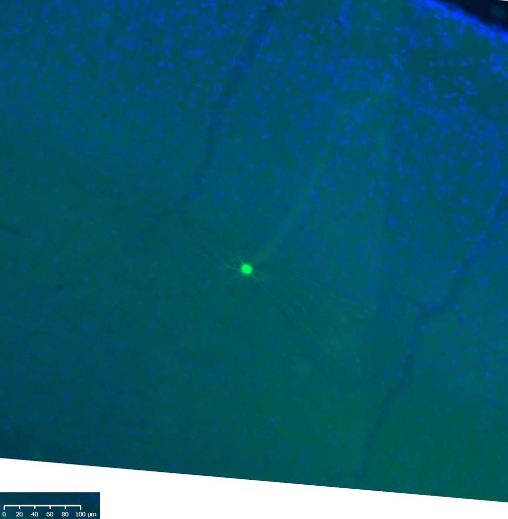
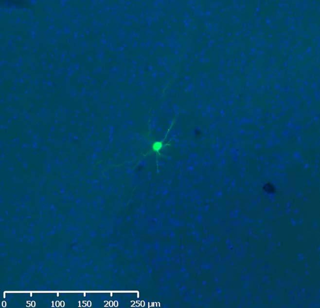
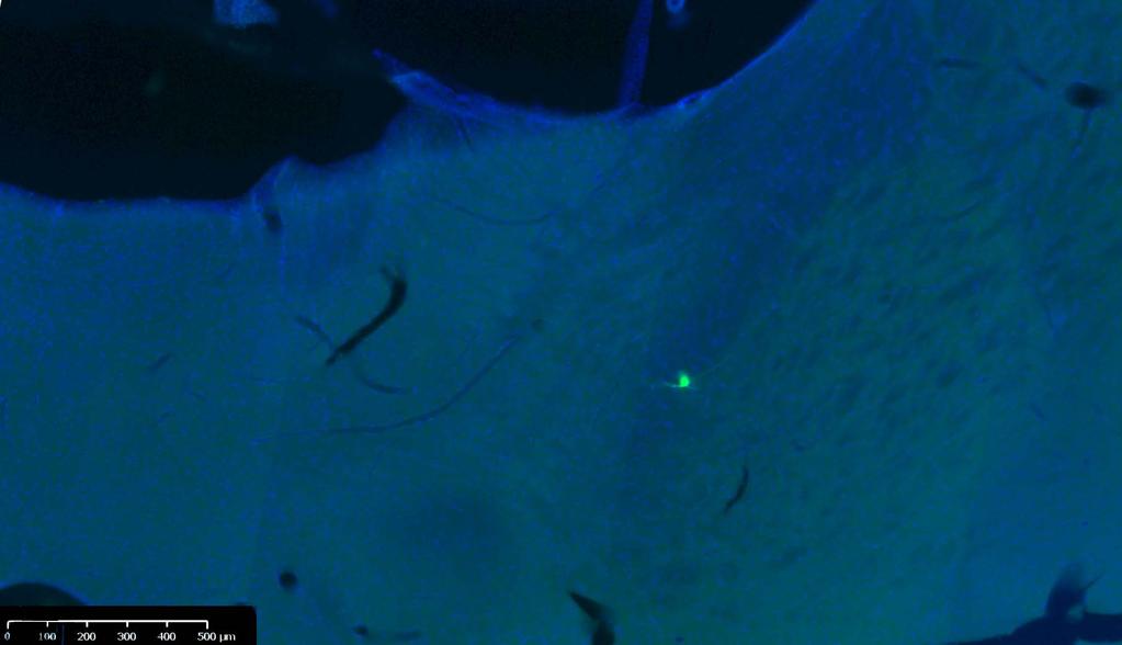
5 Fig. S2 Tracing of the input to V1. Representative coronal sections of various brain areas with projecting cells sending input to V1, from one WT (A) and one Fmr1 -/y (B) mouse. Green cells: retrograde rabies virus RV G-eGFP (RV G) infected cells expressing egfp; Red cells: anterograde rabies virus RV G-mCherry(VSV G RtmC ) infected cells expressing mcherry; Blue cellular nuclei: DAPI staining. Key: L1-6: layers 1-6 of the neocortex; Au1: primary auditory cortex; AuD: secondary auditory cortex dorsal area; Cg1: cingulate cortex area 1; DLG: dorsal lateral geniculate nucleus of the thalamus; LGP: lateral globus pallidus; LO: lateral orbital cortex; M2: secondary motor cortex; MCPO: magnocellular preoptic nucleus; RSA: retrosplenial agranular cortex; S1 BF: primary somatosensory cortex barrel field; SRad: stratum radiatum of the hippocampus; : secondary visual cortex lateral area; V2ML: secondary visual cortex mediolateral area; V2MM: secondary visual cortex mediomedial area; VO: ventral orbital cortex. Scale bars: 1 mm (main images) and 25µm (insets), unless otherwise noted.


6 0 A Tracer injections Retrograde tracing Sectioning B Slide scanning slices, 50μm thickness: >7.5mm tissue DLG V1 AuD Au1 C Overlay and Transfer to brain model Brain model D Connectivity calculations Distance Center of injection site #x y z comment Distance from injection site in m Mo M2 Ctx 363, Mo M2 Ctx 3291, Mo M2 Ctx 3635, Mo M2 Ctx 3595, Mo M2 Ctx 336, Mo M2 Ctx 3251, Mo M2 Ctx 333,11 Area Weight
7 Fig. S3. Schematic representation of the experimental strategy for the 3D anatomical registration of projection neurons into V1. (A) Stereotaxic injection permits introduction of the viral tracers necessary for retrograde labeling (green). The inclusion of an anterograde tracer (red) marks the site of injection. Following sufficient time for viral expression, the animal is sacrificed, the brain fixed and then subsequently sectioned to permit microscopy. (B) A slide scanning approach permits the rapid acquisition of sections from the entire fore-brain. (C) Labeled neurons are detected manually from individual sections overlaid with a brain atlas and assigned a numerical coordinate, used for the generation of a 3D model (D). The 3D model is then used to calculate connectivity features (distance of projection neurons (retrogradely labeled, green) from injection site calculated from centre of mass of red-labeled neurons).
8 A LGP L LGP R PF L PF R VPN L VPN R DH L DH R VH L VH R Au L Au R Mo L Mo R S1 L S1 R V1 L V1 R RS Pi L Pi R Am L Am R PtA L PtA R CPu L CPu R DLG L DLG R WT DH L DH R VH L VH R Au L Au R Mo L Mo R S1 L S1 R V1 L V1 R RS Pi L Pi R Am L Am R Fmr1 -/y PtA L PtA R CPu L CPu R DLG L DLG R LGP L LGP R PF L PF R VPN L VPN R 20 z-score 10 0 B Difference Fmr1 - WT Statistical differences PtA L PtA R CPu L CPu R DLG L DLG R LGP L LGP R PF L PF R VPN L VPN R -/y DH L DH R VH L VH R Au L Au R Mo L Mo R S1 L S1 R V1 L V1 R RS Pi L Pi R Am L Am R DH L DH R VH L VH R Au L Au R Mo L Mo R S1 L S1 R V1 L V1 R RS Pi L Pi R Am L Am R PtA L PtA R CPu L CPu R DLG L DLG R LGP L LGP R PF L PF R VPN L VPN R Fmr1 -/y > WT Fmr1 -/y > WT p < 0.05 p < 0.01 p < Fig S. Functional connectivity matrix of wild-type and Fmr1 -/y mice. (A) fmri measurements under light anaesthesia were performed in Fmr1 -/y (n=7) and WT (n=10) Mice. (B) The intracortical functional connectivity is reduced in Fmr1 -/y mice, in particular between the sensory cortical areas
9 (Au, Mo, S1, and V1). None of the analyzed areas showed a significant increase in functional connectivity. Key: Dorsal hippocampus (DH), ventral hippocampus (VH), auditory cortex (Au), motor cortex (Mo), primary somatosensory cortex (S1), primary visual cortex (V1), retrosplenial cortex (RS), Piriform Cortex (Pi), Amygdala (Amy), Pretectal Area (PtA), Caudate Putamen (CPu), lateral geniculate nucleus (DLG), globus pallidus (LGP), parafascicular nucleus (PF), ventral posterolateral nucleus and ventral posteromedial nucleus (VPN). Right (R) and left hemisphere (L). Supplementary Movie S1. Slice view and 3D view, illustrating the 3D mouse brain model with the combined positions of all retrogradely- (input; green) and anterogradely (local; red) labeled neurons in wild-type mice. Supplementary Movie S2. Slice view and 3D view, illustrating the 3D mouse brain model with the combined positions of all retrogradely- (input; green) and anterogradely (local; red) labeled neurons in Fmr1 -/y mice.
Nature Neuroscience doi: /nn Supplementary Figure 1. Characterization of viral injections.
 Supplementary Figure 1 Characterization of viral injections. (a) Dorsal view of a mouse brain (dashed white outline) after receiving a large, unilateral thalamic injection (~100 nl); demonstrating that
Nature Neuroscience: doi: /nn Supplementary Figure 1. Distribution of starter cells for RV-mediated retrograde tracing.
 Supplementary Figure 1 Distribution of starter cells for RV-mediated retrograde tracing. Parcellation of cortical areas is based on Allen Mouse Brain Atlas and drawn to scale. Thick white curves, outlines
Nature Neuroscience: doi: /nn Supplementary Figure 1. Large-scale calcium imaging in vivo.
 Supplementary Figure 1 Large-scale calcium imaging in vivo. (a) Schematic illustration of the in vivo camera imaging set-up for large-scale calcium imaging. (b) High-magnification two-photon image from
10/3/2016. T1 Anatomical structures are clearly identified, white matter (which has a high fat content) appears bright.
 appears bright.") H2O -2 atoms of Hydrogen, 1 of Oxygen Hydrogen just has one single proton and orbited by one single electron Proton has a magnetic moment similar to the earths magnetic pole Also similar to earth in that
H2O -2 atoms of Hydrogen, 1 of Oxygen Hydrogen just has one single proton and orbited by one single electron Proton has a magnetic moment similar to the earths magnetic pole Also similar to earth in that
For more information about how to cite these materials visit
 Author(s): Peter Hitchcock, PH.D., 2009 License: Unless otherwise noted, this material is made available under the terms of the Creative Commons Attribution Non-commercial Share Alike 3.0 License: http://creativecommons.org/licenses/by-nc-sa/3.0/
Cerebral Cortex 1. Sarah Heilbronner
 Cerebral Cortex 1 Sarah Heilbronner heilb028@umn.edu Want to meet? Coffee hour 10-11am Tuesday 11/27 Surdyk s Overview and organization of the cerebral cortex What is the cerebral cortex? Where is each
Chapter 3. Structure and Function of the Nervous System. Copyright (c) Allyn and Bacon 2004
 Allyn and Bacon 2004") Chapter 3 Structure and Function of the Nervous System 1 Basic Features of the Nervous System Neuraxis: An imaginary line drawn through the center of the length of the central nervous system, from the
Chapter 3 Structure and Function of the Nervous System 1 Basic Features of the Nervous System Neuraxis: An imaginary line drawn through the center of the length of the central nervous system, from the
Nature Neuroscience: doi: /nn.4642
 Supplementary Figure 1 Recording sites and example waveform clustering, as well as electrophysiological recordings of auditory CS and shock processing following overtraining. (a) Recording sites in LC
Leah Militello, class of 2018
 Leah Militello, class of 2018 Objectives 1. Describe the general organization of cerebral hemispheres. 2. Describe the locations and features of the different functional areas of cortex. 3. Understand
Prof. Saeed Abuel Makarem & Dr.Sanaa Alshaarawy
 Prof. Saeed Abuel Makarem & Dr.Sanaa Alshaarawy 1 Objectives By the end of the lecture, you should be able to: Describe the anatomy and main functions of the thalamus. Name and identify different nuclei
Outline of the next three lectures
 Outline of the next three lectures Lecture 35 Anatomy of the human cerebral cortex gross and microscopic cell types connections Vascular supply of the cerebral cortex Disorders involving the cerebral cortex
Nature Neuroscience: doi: /nn Supplementary Figure 1
 Supplementary Figure 1 Atlas representations of the midcingulate (MCC) region targeted in this study compared against the anterior cingulate (ACC) region commonly reported. Coronal sections are shown on
Supplementary Materials
 Supplementary Materials Fig. S1. Weights of full-dose treatment groups comparing 1 st, 2 nd, and 3 rd generation gene replacement therapy. Mice were treated at p1 with 4x10 11 GC of the three different
Psyc 311A, fall 2008 Conference week 3 TA: Jürgen Germann
 Psyc 311A, fall 2008 Conference week 3 TA: Jürgen Germann e-mail: jurgen.germann@mcgill.ca Overview: 1. Meninges 2. Cerebral cortex-cytoarchitecture 3. Diencephalon (thalamus/hypothalamus) (this replaces
Nature Neuroscience: doi: /nn Supplementary Figure 1
 Supplementary Figure 1 Distribution of GlyT2::eGFP fibers in the mouse thalamus at three different coronal levels. Note the innervation centered in the rostral (CL, PC) and caudal (PF) nuclear groups of
Introduction to the Central Nervous System: Internal Structure
 Introduction to the Central Nervous System: Internal Structure Objective To understand, in general terms, the internal organization of the brain and spinal cord. To understand the 3-dimensional organization
For more information about how to cite these materials visit
 Author(s): Peter Hitchcock, PH.D., 2009 License: Unless otherwise noted, this material is made available under the terms of the Creative Commons Attribution Non-commercial Share Alike 3.0 License: http://creativecommons.org/licenses/by-nc-sa3.0/
Basal Ganglia. Introduction. Basal Ganglia at a Glance. Role of the BG
 Basal Ganglia Shepherd (2004) Chapter 9 Charles J. Wilson Instructor: Yoonsuck Choe; CPSC 644 Cortical Networks Introduction A set of nuclei in the forebrain and midbrain area in mammals, birds, and reptiles.
Zhu et al, page 1. Supplementary Figures
 Zhu et al, page 1 Supplementary Figures Supplementary Figure 1: Visual behavior and avoidance behavioral response in EPM trials. (a) Measures of visual behavior that performed the light avoidance behavior
Biological Bases of Behavior. 3: Structure of the Nervous System
 Biological Bases of Behavior 3: Structure of the Nervous System Neuroanatomy Terms The neuraxis is an imaginary line drawn through the spinal cord up to the front of the brain Anatomical directions are
CISC 3250 Systems Neuroscience
 CISC 3250 Systems Neuroscience Levels of organization Central Nervous System 1m 10 11 neurons Neural systems and neuroanatomy Systems 10cm Networks 1mm Neurons 100μm 10 8 neurons Professor Daniel Leeds
Brain anatomy and artificial intelligence. L. Andrew Coward Australian National University, Canberra, ACT 0200, Australia
 Brain anatomy and artificial intelligence L. Andrew Coward Australian National University, Canberra, ACT 0200, Australia The Fourth Conference on Artificial General Intelligence August 2011 Architectures
Regional and Lobe Parcellation Rhesus Monkey Brain Atlas. Manual Tracing for Parcellation Template
 Regional and Lobe Parcellation Rhesus Monkey Brain Atlas Manual Tracing for Parcellation Template Overview of Tracing Guidelines A) Traces are performed in a systematic order they, allowing the more easily
Telencephalon (Cerebral Hemisphere)
") Telencephalon (Cerebral Hemisphere) OUTLINE The Cortex - Lobes, Sulci & Gyri - Functional Subdivisions - Limbic Lobe & Limbic System The Subcortex - Basal Ganglia - White Matter (Internal Capsule) - Relations
Telencephalon (Cerebral Hemisphere) OUTLINE The Cortex - Lobes, Sulci & Gyri - Functional Subdivisions - Limbic Lobe & Limbic System The Subcortex - Basal Ganglia - White Matter (Internal Capsule) - Relations
Vision II. Steven McLoon Department of Neuroscience University of Minnesota
 Vision II Steven McLoon Department of Neuroscience University of Minnesota 1 Ganglion Cells The axons of the retinal ganglion cells form the optic nerve and carry visual information into the brain. 2 Optic
PSY 302: CHAPTER 3 NOTES THE BRAIN (PART II) - 9/5/17. By: Joseline
 - 9/5/17. By: Joseline") PSY 302: CHAPTER 3 NOTES THE BRAIN (PART II) - 9/5/17 By: Joseline Left 3 MAJOR FISSURES : 2HEMISPHERES Right Lateral Ventricle Central Fissure Third Ventricle Sulcus Lateral Fissure Gyros Fissure- Fissures
PSY 302: CHAPTER 3 NOTES THE BRAIN (PART II) - 9/5/17 By: Joseline Left 3 MAJOR FISSURES : 2HEMISPHERES Right Lateral Ventricle Central Fissure Third Ventricle Sulcus Lateral Fissure Gyros Fissure- Fissures
The human brain. of cognition need to make sense gives the structure of the brain (duh). ! What is the basic physiology of this organ?
. ! What is the basic physiology of this organ?") The human brain The human brain! What is the basic physiology of this organ?! Understanding the parts of this organ provides a hypothesis space for its function perhaps different parts perform different
The human brain The human brain! What is the basic physiology of this organ?! Understanding the parts of this organ provides a hypothesis space for its function perhaps different parts perform different
The Neuroscience of Addiction: A mini-review
 The Neuroscience of Addiction: A mini-review Jim Morrill, MD, PhD MGH Charlestown HealthCare Center Massachusetts General Hospital Disclosures Neither I nor my spouse/partner has a relevant financial relationship
Supplementary Figure 1
 Supplementary Figure 1 Localization of virus injections. (a) Schematic showing the approximate center of AAV-DIO-ChR2-YFP injection sites in the NAc of Dyn-cre mice (n=8 mice, 16 injections; caudate/putamen,
Pediatric MS MRI Study Methodology
 General Pediatric MS MRI Study Methodology SCAN PREPARATION axial T2-weighted scans and/or axial FLAIR scans were obtained for all subjects when available, both T2 and FLAIR scans were scored. In order
Nature Neuroscience: doi: /nn Supplementary Figure 1. Visualization of AT1a-positive cells using AT1a lacz/+ mouse.
 Supplementary Figure 1 Visualization of AT1a-positive cells using AT1a lacz/+ mouse. (a f) Immunohistochemical detection of β-gal in the mouse brain. Coronal sections at the respective anteroposterior
Anatomy of the basal ganglia. Dana Cohen Gonda Brain Research Center, room 410
 Anatomy of the basal ganglia Dana Cohen Gonda Brain Research Center, room 410 danacoh@gmail.com The basal ganglia The nuclei form a small minority of the brain s neuronal population. Little is known about
BASAL GANGLIA. Dr JAMILA EL MEDANY
 BASAL GANGLIA Dr JAMILA EL MEDANY OBJECTIVES At the end of the lecture, the student should be able to: Define basal ganglia and enumerate its components. Enumerate parts of Corpus Striatum and their important
9.14 Class 32 Review. Limbic system
 9.14 Class 32 Review Limbic system 1 Lateral view Medial view Brainstem, sagittal section Sensory- Perceptual Motor Behavior Major functional modules of the CNS Motivation Courtesy of MIT Press. Used with
Supplemental Information. A Labeled-Line Neural Circuit. for Pheromone-Mediated Sexual Behaviors in Mice
 Neuron, Volume Supplemental Information A Labeled-Line Neural Circuit for Pheromone-Mediated Sexual Behaviors in Mice Kentaro K. Ishii, Takuya Osakada, Hiromi Mori, Nobuhiko Miyasaka, Yoshihiro Yoshihara,
Biological Bases of Behavior : Quiz 3
 Biological Bases of Behavior : Quiz 3 1. One of the oldest methods used in neuroscience to study brain function is a. stereotaxic surgery. b. autoradiography. c. experimental ablation. d. immunocytochemistry.
ZBED6 expression pattern during embryogenesis and in the central nervous system
 ZBED6expressionpatternduringembryogenesisandin thecentralnervoussystem AxelEricsson 2010 DepartmentofNeuroscience DevelopmentalgeneticsUppsalaUniversity Supervisors:KlasKullanderandMartinLarhammar 1 P
Visual system invades the endbrain: pathways to striatum and cortex (continued) Why this happened in evolution
 Why this happened in evolution") Visual system invades the endbrain: pathways to striatum and cortex (continued) Why this happened in evolution What were the adaptive advantages? Visual information reaching the striatum directly: Advantages
Visual system invades the endbrain: pathways to striatum and cortex (continued) Why this happened in evolution What were the adaptive advantages? Visual information reaching the striatum directly: Advantages
Transcranial Pulsed Ultrasound Stimulates Intact Brain Circuits
 Neuron, Volume 66 Supplemental Information Transcranial Pulsed Ultrasound Stimulates Intact Brain Circuits Yusuf Tufail, Alexei Matyushov, Nathan Baldwin, Monica L. Tauchmann, Joseph Georges, Anna Yoshihiro,
Systems Neuroscience Dan Kiper. Today: Wolfger von der Behrens
 Systems Neuroscience Dan Kiper Today: Wolfger von der Behrens wolfger@ini.ethz.ch 18.9.2018 Neurons Pyramidal neuron by Santiago Ramón y Cajal (1852-1934, Nobel prize with Camillo Golgi in 1906) Neurons
I: To describe the pyramidal and extrapyramidal tracts. II: To discuss the functions of the descending tracts.
 Descending Tracts I: To describe the pyramidal and extrapyramidal tracts. II: To discuss the functions of the descending tracts. III: To define the upper and the lower motor neurons. 1. The corticonuclear
Nature Neuroscience: doi: /nn Supplementary Figure 1. Diverse anorexigenic signals induce c-fos expression in CEl PKC-δ + neurons
 Supplementary Figure 1 Diverse anorexigenic signals induce c-fos expression in CEl PKC-δ + neurons a-c. Quantification of CEl c-fos expression in mice intraperitoneal injected with anorexigenic drugs (a),
Chapter 2: Studies of Human Learning and Memory. From Mechanisms of Memory, second edition By J. David Sweatt, Ph.D.
 Chapter 2: Studies of Human Learning and Memory From Mechanisms of Memory, second edition By J. David Sweatt, Ph.D. Medium Spiny Neuron A Current Conception of the major memory systems in the brain Figure
Wenqin Hu, Cuiping Tian, Tun Li, Mingpo Yang, Han Hou & Yousheng Shu
 Distinct contributions of Na v 1.6 and Na v 1.2 in action potential initiation and backpropagation Wenqin Hu, Cuiping Tian, Tun Li, Mingpo Yang, Han Hou & Yousheng Shu Supplementary figure and legend Supplementary
Nsci 2100: Human Neuroanatomy 2017 Examination 3
 Name KEY Lab Section Nsci 2100: Human Neuroanatomy 2017 Examination 3 On this page, write your name and lab section. On your bubble answer sheet, enter your name (last name, space, first name), internet
SUPPLEMENTARY INFORMATION
 SUPPLEMENTARY INFORMATION doi:10.1038/nature12107 Supplementary Figure 1. CLARITY preserves GFP and TdTomato signals. (a) 3D rendering of a 1mm-thick Thy1-EGFP M line mouse brain block processed by CLARITY
Lack of GPR88 enhances medium spiny neuron activity and alters. motor- and cue- dependent behaviors
 Lack of GPR88 enhances medium spiny neuron activity and alters motor- and cue- dependent behaviors Albert Quintana, Elisenda Sanz, Wengang Wang, Granville P. Storey, Ali D. Güler Matthew J. Wanat, Bryan
P. Hitchcock, Ph.D. Department of Cell and Developmental Biology Kellogg Eye Center. Wednesday, 16 March 2009, 1:00p.m. 2:00p.m.
 Normal CNS, Special Senses, Head and Neck TOPIC: CEREBRAL HEMISPHERES FACULTY: LECTURE: READING: P. Hitchcock, Ph.D. Department of Cell and Developmental Biology Kellogg Eye Center Wednesday, 16 March
Hallucinations and conscious access to visual inputs in Parkinson s disease
 Supplemental informations Hallucinations and conscious access to visual inputs in Parkinson s disease Stéphanie Lefebvre, PhD^1,2, Guillaume Baille, MD^4, Renaud Jardri MD, PhD 1,2 Lucie Plomhause, PhD
Thalamic nuclei. Each thalamus has several well defined borders: Introduction. Thalamus
 Thalamic nuclei Introduction For the successful completion of any task, some sort of recognition, identification and organisation is needed. Imagine what would happen if employees in a team would just
Supplementary Online Material Supplementary Table S1 to S5 Supplementary Figure S1 to S4
 Supplementary Online Material Supplementary Table S1 to S5 Supplementary Figure S1 to S4 Table S1: Brain regions involved in the adapted classification learning task Brain Regions x y z Z Anterior Cingulate
Motor Functions of Cerebral Cortex
 Motor Functions of Cerebral Cortex I: To list the functions of different cortical laminae II: To describe the four motor areas of the cerebral cortex. III: To discuss the functions and dysfunctions of
Biological Bases of Behavior. 8: Control of Movement
 Biological Bases of Behavior 8: Control of Movement m d Skeletal Muscle Movements of our body are accomplished by contraction of the skeletal muscles Flexion: contraction of a flexor muscle draws in a
File name: Supplementary Information Description: Supplementary Figures, Supplementary Table and Supplementary References
 File name: Supplementary Information Description: Supplementary Figures, Supplementary Table and Supplementary References File name: Supplementary Data 1 Description: Summary datasheets showing the spatial
Stanley Pruisinger 1980's
 Neuroanatomy Prion disease cerebellum chapter b/c cerebellar ataxia here as a warning for obvious reasons. Creutzfeldt - Jakob Disease (CJD) "Spongiform" (brain turns to sponge) Jews in Lybia who ate
Dr. Farah Nabil Abbas. MBChB, MSc, PhD
 Dr. Farah Nabil Abbas MBChB, MSc, PhD The Basal Ganglia *Functions in association with motor cortex and corticospinal pathways. *Regarded as accessory motor system besides cerebellum. *Receive most of
Gross Organization I The Brain. Reading: BCP Chapter 7
 Gross Organization I The Brain Reading: BCP Chapter 7 Layout of the Nervous System Central Nervous System (CNS) Located inside of bone Includes the brain (in the skull) and the spinal cord (in the backbone)
Nature Neuroscience: doi: /nn Supplementary Figure 1. Confirmation that optogenetic inhibition of dopaminergic neurons affects choice
 Supplementary Figure 1 Confirmation that optogenetic inhibition of dopaminergic neurons affects choice (a) Sample behavioral trace as in Figure 1d, but with NpHR stimulation trials depicted as green blocks
Exam 2 PSYC Fall (2 points) Match a brain structure that is located closest to the following portions of the ventricular system
 Match a brain structure that is located closest to the following portions of the ventricular system") Exam 2 PSYC 2022 Fall 1998 (2 points) What 2 nuclei are collectively called the striatum? (2 points) Match a brain structure that is located closest to the following portions of the ventricular system
Exam 2 PSYC 2022 Fall 1998 (2 points) What 2 nuclei are collectively called the striatum? (2 points) Match a brain structure that is located closest to the following portions of the ventricular system
Damage on one side.. (Notes) Just remember: Unilateral damage to basal ganglia causes contralateral symptoms.
 Just remember: Unilateral damage to basal ganglia causes contralateral symptoms.") Lecture 20 - Basal Ganglia Basal Ganglia (Nolte 5 th Ed pp 464) Damage to the basal ganglia produces involuntary movements. Although the basal ganglia do not influence LMN directly (to cause this involuntary
Lecture 20 - Basal Ganglia Basal Ganglia (Nolte 5 th Ed pp 464) Damage to the basal ganglia produces involuntary movements. Although the basal ganglia do not influence LMN directly (to cause this involuntary
Supplementary Figure 1. Nature Neuroscience: doi: /nn.4547
 Supplementary Figure 1 Characterization of the Microfetti mouse model. (a) Gating strategy for 8-color flow analysis of peripheral Ly-6C + monocytes from Microfetti mice 5-7 days after TAM treatment. Living
SOM Husse et al. Supplementary online material. Synaptotagmin10-Cre, a driver to disrupt clock genes in the SCN
 SOM Husse et al. Supplementary online material Synaptotagmin10-Cre, a driver to disrupt clock genes in the SCN Jana Husse, Xunlei Zhou, Anton Shostak, Henrik Oster and Gregor Eichele SOM Husse et al.,
Primary pouches: prosencephalon, mesencephalon, rhombencephalon Secondary pouches: telencephalon diencephalon
 Telencephalon Ontogenic development of CNS Primary pouches: prosencephalon, mesencephalon, rhombencephalon Secondary pouches: telencephalon diencephalon mesencephalon metencephalon ---- pons (pons Varoli),
Lecture XIII. Brain Diseases I - Parkinsonism! Brain Diseases I!
 Lecture XIII. Brain Diseases I - Parkinsonism! Bio 3411! Wednesday!! Lecture XIII. Brain Diseases - I.! 1! Brain Diseases I! NEUROSCIENCE 5 th ed! Page!!Figure!!Feature! 408 18.9 A!!Substantia Nigra in
A. General features of the basal ganglia, one of our 3 major motor control centers:
 Reading: Waxman pp. 141-146 are not very helpful! Computer Resources: HyperBrain, Chapter 12 Dental Neuroanatomy Suzanne S. Stensaas, Ph.D. April 22, 2010 THE BASAL GANGLIA Objectives: 1. What are the
Biomedical Technology Research Center 2011 Workshop San Francisco, CA
 Diffusion Tensor Imaging: Parkinson s Disease and Atypical Parkinsonism David E. Vaillancourt court1@uic.edu Associate Professor at UIC Departments t of Kinesiology i and Nutrition, Bioengineering, and
Nature Neuroscience: doi: /nn.4332
 Nature Neuroscience: doi:10.1038/nn.4332 Supplementary Figure 1 Topography and termination patterns of cortico-striatal projections (a) Manual inspection of ~150 tracer-labeled cortico-striatal pathways
Unique functional properties of somatostatin-expressing GABAergic neurons in mouse barrel cortex
 Supplementary Information Unique functional properties of somatostatin-expressing GABAergic neurons in mouse barrel cortex Luc Gentet, Yves Kremer, Hiroki Taniguchi, Josh Huang, Jochen Staiger and Carl
CEREBRUM & CEREBRAL CORTEX
 CEREBRUM & CEREBRAL CORTEX Seonghan Kim Dept. of Anatomy Inje University, College of Medicine THE BRAIN ANATOMICAL REGIONS A. Cerebrum B. Diencephalon Thalamus Hypothalamus C. Brain Stem Midbrain Pons
The neurvous system senses, interprets, and responds to changes in the environment. Two types of cells makes this possible:
 NERVOUS SYSTEM The neurvous system senses, interprets, and responds to changes in the environment. Two types of cells makes this possible: the neuron and the supporting cells ("glial cells"). Neuron Neurons
COGNITIVE SCIENCE 107A. Motor Systems: Basal Ganglia. Jaime A. Pineda, Ph.D.
 COGNITIVE SCIENCE 107A Motor Systems: Basal Ganglia Jaime A. Pineda, Ph.D. Two major descending s Pyramidal vs. extrapyramidal Motor cortex Pyramidal system Pathway for voluntary movement Most fibers originate
The Frontal Lobes. Anatomy of the Frontal Lobes. Anatomy of the Frontal Lobes 3/2/2011. Portrait: Losing Frontal-Lobe Functions. Readings: KW Ch.
 The Frontal Lobes Readings: KW Ch. 16 Portrait: Losing Frontal-Lobe Functions E.L. Highly organized college professor Became disorganized, showed little emotion, and began to miss deadlines Scores on intelligence
9.14 Qs on post-midterm class sessions and Schneider chapters 18-34
 9.14 Qs on post-midterm class sessions and Schneider chapters 18-34 (Bold font indicates terms you should be able to define.) Ch 18 Ch 19 Ch 20 1. In comparative neuroanatomical studies, the taste system
Announcement. Danny to schedule a time if you are interested.
 Announcement If you need more experiments to participate in, contact Danny Sanchez (dsanchez@ucsd.edu) make sure to tell him that you are from LIGN171, so he will let me know about your credit (1 point).
The Central Nervous System I. Chapter 12
 The Central Nervous System I Chapter 12 The Central Nervous System The Brain and Spinal Cord Contained within the Axial Skeleton Brain Regions and Organization Medical Scheme (4 regions) 1. Cerebral Hemispheres
The basal forebrain: Questions, chapter 29:
 The basal forebrain: Questions, chapter 29: 7) What is the "basal forebrain", and what is its involvement in Alzheimer' s Disease? The acetylcholine-containing neurons of the nucleus basalis of Meynart
Supplementary Information
 Supplementary Information Title Degeneration and impaired regeneration of gray matter oligodendrocytes in amyotrophic lateral sclerosis Authors Shin H. Kang, Ying Li, Masahiro Fukaya, Ileana Lorenzini,
Basal Ganglia George R. Leichnetz, Ph.D.
 Basal Ganglia George R. Leichnetz, Ph.D. OBJECTIVES 1. To understand the brain structures which constitute the basal ganglia, and their interconnections 2. To understand the consequences (clinical manifestations)
9.14 Classes #21-23: Visual systems
 9.14 Classes #21-23: Visual systems Questions based on Schneider chapter 20 and classes: 1) What was in all likelihood the first functional role of the visual sense? Describe the nature of the most primitive
Teach-SHEET Basal Ganglia
 Teach-SHEET Basal Ganglia Purves D, et al. Neuroscience, 5 th Ed., Sinauer Associates, 2012 Common organizational principles Basic Circuits or Loops: Motor loop concerned with learned movements (scaling
A. General features of the basal ganglia, one of our 3 major motor control centers:
 Reading: Waxman pp. 141-146 are not very helpful! Computer Resources: HyperBrain, Chapter 12 Dental Neuroanatomy Suzanne S. Stensaas, Ph.D. March 1, 2012 THE BASAL GANGLIA Objectives: 1. What are the main
SUPPLEMENTARY INFORMATION
 doi: 10.1038/nature06310 SUPPLEMENTARY INFORMATION www.nature.com/nature 1 www.nature.com/nature 2 www.nature.com/nature 3 Supplementary Figure S1 Spontaneous duration of wake, SWS and REM sleep (expressed
Chapter 8. Control of movement
 Chapter 8 Control of movement 1st Type: Skeletal Muscle Skeletal Muscle: Ones that moves us Muscles contract, limb flex Flexion: a movement of a limb that tends to bend its joints, contraction of a flexor
Validation of basal ganglia segmentation on a 3T MRI template
 Validation of basal ganglia segmentation on a 3T MRI template Claire Haegelen, Nicolas Guizard, Pierrick Coupé, Florent Lalys, Pierre Jannin, Xavier Morandi, D. Louis Collins To cite this version: Claire
Ch 13: Central Nervous System Part 1: The Brain p 374
 Ch 13: Central Nervous System Part 1: The Brain p 374 Discuss the organization of the brain, including the major structures and how they relate to one another! Review the meninges of the spinal cord and
Supplementary Materials for
 advances.sciencemag.org/cgi/content/full/3/3/e1600955/dc1 Supplementary Materials for Flexible and stretchable nanowire-coated fibers for optoelectronic probing of spinal cord circuits Chi Lu, Seongjun
Thalamus and Sensory Functions of Cerebral Cortex
 Thalamus and Sensory Functions of Cerebral Cortex I: To describe the functional divisions of thalamus. II: To state the functions of thalamus and the thalamic syndrome. III: To define the somatic sensory
Final review, 9.14_2014. Slides for special study
 Final review, 9.14_2014 Slides for special study 1 Mammalian Taste Pathways Neocortical Gustatory area VPM pc Parabrachial nucleus Gustatory nucleus (rostral part of nuc. of solitary tract visceral sensory
CSE511 Brain & Memory Modeling Lect 22,24,25: Memory Systems
 CSE511 Brain & Memory Modeling Lect 22,24,25: Memory Systems Compare Chap 31 of Purves et al., 5e Chap 24 of Bear et al., 3e Larry Wittie Computer Science, StonyBrook University http://www.cs.sunysb.edu/~cse511
A few notions of brain anatomy
 A few notions of brain anatomy Christophe Pallier CNRS, INSERM 562, Orsay, France Note some slides were taken from lectures available from the excellent web site 'fmri for dummies' by Jody Culham. Drawing
Study Guide Unit 2 Psych 2022, Fall 2003
 Study Guide Unit 2 Psych 2022, Fall 2003 Subcortical Anatomy 1. Be able to locate the following structures and be able to indicate whether they are located in the forebrain, diencephalon, midbrain, pons,
nucleus accumbens septi hier-259 Nucleus+Accumbens birnlex_727
 Nucleus accumbens From Wikipedia, the free encyclopedia Brain: Nucleus accumbens Nucleus accumbens visible in red. Latin NeuroNames MeSH NeuroLex ID nucleus accumbens septi hier-259 Nucleus+Accumbens birnlex_727
The Neuroscience of Music in Therapy
 Course Objectives The Neuroscience of Music in Therapy Unit I. Learn Basic Brain Information Unit II. Music in the Brain; Why Music Works Unit III. Considerations for Populations a. Rehabilitation b. Habilitation
Background & Rationale Great apes share many behavioral, cognitive, and neuroanatomical similarities with humans. Yet, there are also significant
 Background & Rationale Great apes share many behavioral, cognitive, and neuroanatomical similarities with humans. Yet, there are also significant differences that distinguish humans from apes in terms
NS219: Basal Ganglia Anatomy
 NS219: Basal Ganglia Anatomy Human basal ganglia anatomy Analagous rodent basal ganglia nuclei Basal ganglia circuits: the classical model of direct and indirect pathways + Glutamate + - GABA - Gross anatomy
Basal Ganglia. Today s lecture is about Basal Ganglia and it covers:
 Basal Ganglia Motor system is complex interaction between Lower motor neurons (spinal cord and brainstem circuits) and Upper motor neurons (pyramidal and extrapyramidal tracts) plus two main regulators
Medical Neuroscience Tutorial Notes
 Medical Neuroscience Tutorial Notes Blood Supply to the Brain MAP TO NEUROSCIENCE CORE CONCEPTS 1 NCC1. The brain is the body's most complex organ. LEARNING OBJECTIVES After study of the assigned learning
Cortical Control of Movement
 Strick Lecture 2 March 24, 2006 Page 1 Cortical Control of Movement Four parts of this lecture: I) Anatomical Framework, II) Physiological Framework, III) Primary Motor Cortex Function and IV) Premotor
PSY 315 Lecture 11 (2/23/2011) (Motor Control) Dr. Achtman PSY 215. Lecture 11 Topic: Motor System Chapter 8, pages
 (Motor Control) Dr. Achtman PSY 215. Lecture 11 Topic: Motor System Chapter 8, pages") Corrections: No Corrections Announcements: Exam #2 next Wednesday, March 2, 2011 Monday February 28, 2011 we will be going over the somatosensory system, and there will be time left in class to review
Corrections: No Corrections Announcements: Exam #2 next Wednesday, March 2, 2011 Monday February 28, 2011 we will be going over the somatosensory system, and there will be time left in class to review
By Lauren Stowe, PhD, CCC-SLP & Gina Rotondo, MS, CCC-SLP The Speech Therapy Group
 By Lauren Stowe, PhD, CCC-SLP & Gina Rotondo, MS, CCC-SLP The Speech Therapy Group http://www.acquiredbraininjury.com/interactive brain/interactivebrain.swf 1. Hormones make the science messy 2. Difference