A few notions of brain anatomy
|
|
|
- Kevin Jeffry Sanders
- 5 years ago
- Views:
Transcription
1 A few notions of brain anatomy Christophe Pallier CNRS, INSERM 562, Orsay, France Note some slides were taken from lectures available from the excellent web site 'fmri for dummies' by Jody Culham.

2 Drawing from H. Phillips & C. Tollet
3 Images from The Virtual Hospital (
4
 Total surface area of the cerebral cortex = 2,500 cm2 (elephant = 6300; dolphin")
5 Cortex (vs. subcortical structures) Total surface area of the cerebral cortex = 2,500 cm2 (elephant = 6300; dolphin =3750) Number of neurons ~ 1010 (glial cells ~ 10~50 times more) Cortex thickness ~ 1.5 to 4 mm


6 Brodmann s Areas Brodmann (1905): Based on cytoarchitectonics: study of differences in cortical layers between areas Most common delineation of cortical areas More recent schemes subdivide Brodmann s areas into many smaller regions Monkey and human Brodmann s areas not necessarily homologous fmri for Dummies
7 Interhemispheric Fissure -hugely deep (down to corpus callosum) -divides brain into 2 hemispheres Intrahemispheric Fissure fmri for Dummies
 is buried")

8 Sylvian Fissure -hugely deep -mostly horizontal -insula (purple) is buried within it -separates temporal lobe from parietal and frontal lobes Sylvian Fissure
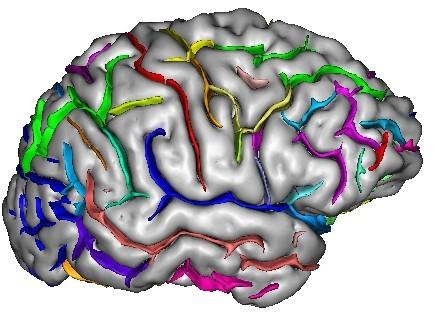
9 Superior and Inferior Temporal Sulci Superior Temporal Sulcus (red) -divides superior temporal gyrus (peach) from middle temporal gyrus (lime) Inferior Temporal Sulcus (blue) -not usually very continuous -divides middle temporal gyrus from inferior temporal gyrus (lavender)
10 Central, Postcentral and Precentral Sulci Precentral Sulcus (red) Central Sulcus (red) -usually freestanding (no intersections) -just anterior to ascending cingulate -often in two parts (superior and inferior) -intersects with superior frontal sulcus (T-junction) -marks anterior end of precentral gyrus (motor strip, yellow) Postcentral Sulcus (red) -often in two parts (superior and inferior) -often intersects with intraparietal sulcus -marks posterior end of postcentral gyrus (somatosensory strip, purple) ascending band of the cingulate fmri for Dummies
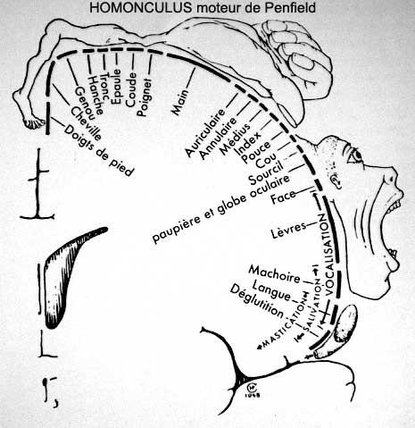
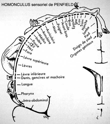

11 Penfield homonculus
 orbital gyrus (green) and frontal pole (gray) also shown Frontal Eye fields lie at this junction fmri for")
12 Superior and Inferior Frontal Sulci Superior Frontal Sulcus (red) -divides superior frontal gyrus (mocha) from middle frontal gyrus (pink) Inferior Frontal Sulcus (blue) -divides middle frontal gyrus from inferior frontal gyrus (gold) orbital gyrus (green) and frontal pole (gray) also shown Frontal Eye fields lie at this junction fmri for Dummies

13 Early Visual Areas
 Parieto-occipital fissure (red) -very deep")

 -visual areas on medial side below calcarine")
 -contains V1 POS CS lg ua ng Li")
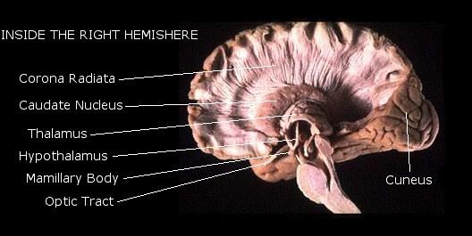
14 Parieto-occipital Fissure and Sulcus Cuneus Calcarine (pink) Parieto-occipital fissure (red) -very deep -often Y-shaped from sagittal view, X-shaped in horizontal and coronal views -visual areas on medial side above calcarine (lower visual field) Lingual gyrus (yellow) -visual areas on medial side below calcarine and above collateral sulcus (upper visual field) Calcarine sulcus (blue) -contains V1 POS CS lg ua ng Li us yr fmri for Dummies
15 Atlases on the web


16 Brains are Heterogeneous Slide from Duke course fmri for Dummies

17 Gyral variability Cachia, Magin, Riviere et al., MICCAI 2002, Medical Image Anal., 2003

18 Variability of Sulci Source: Szikla et al., 1977 in Tamraz & Comair, 2000 fmri for Dummies


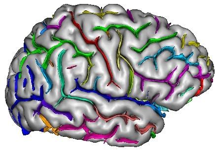
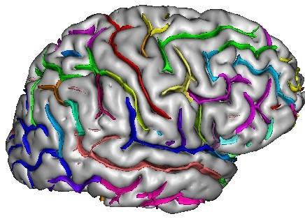
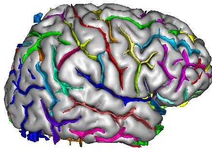
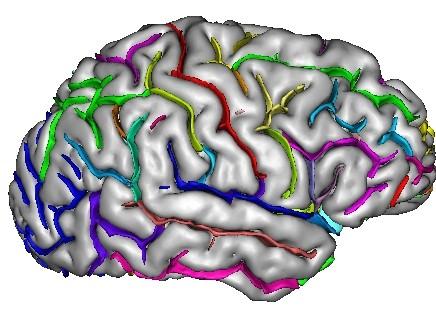

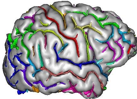
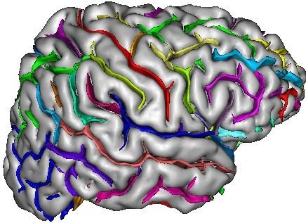
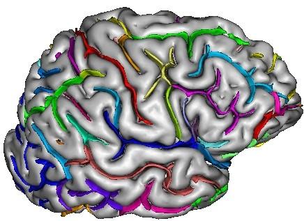
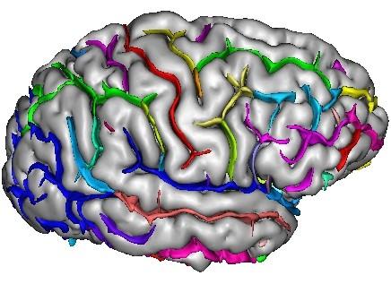
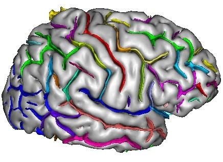
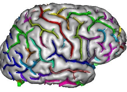
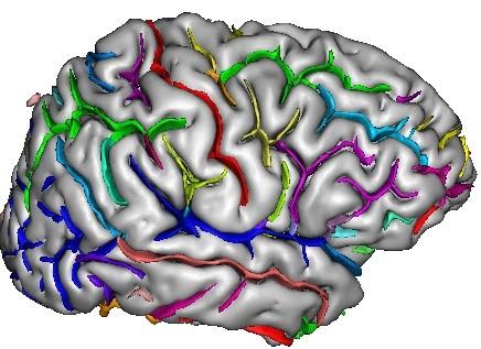
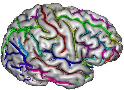

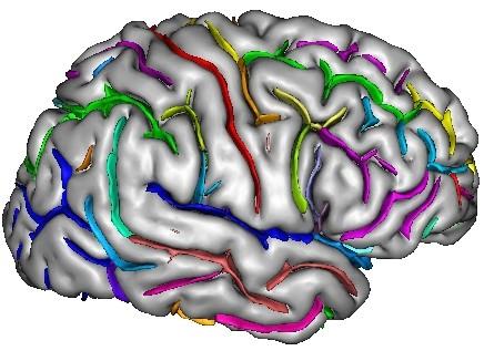
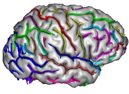
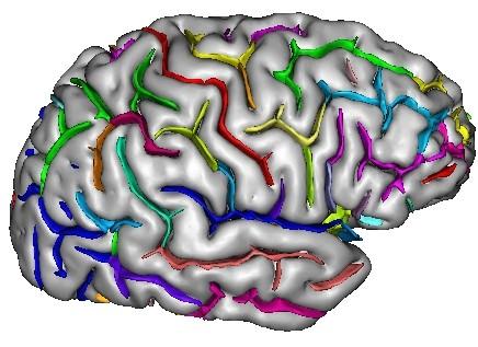
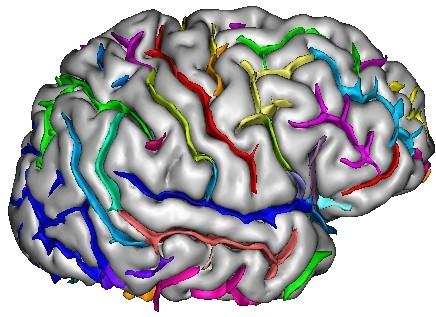
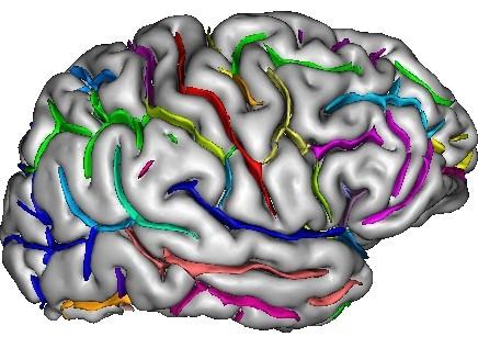


19 A database of manually labelled sulci Jean-Francois Mangin & Denis Riviere, SHFJ, Orsay
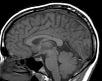
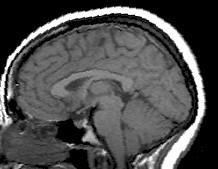
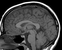
20 Exercices Open an anatomical scan with Anatomist and/or mricro. Find the corpus callosum, the cerebellum, the thalamus.

21 Exercice 2: Find heschl gyrus
: au dessus dugyrus cingulaire")
22 Exercice: Repérer le sillon central? 1 Sur une coupe sagittale 2 Repérer le sillon calloso-marginal (1): au dessus dugyrus cingulaire (2), Encoche en avant : sillon central (3). IRM: coupe sagittale

23 Comment repérer le sillon central? - repérer le sillon précentral qui croise à sa partie supérieure le sillon frontal supérieur sillon central : 1ère scissure verticale située en arrière du sillon pré central. IRM: coupe axiale Sur une coupe axiale
24 Localiser le cortex moteur primaire
Brain anatomy tutorial. Dr. Michal Ben-Shachar 459 Neurolinguistics
 Brain anatomy tutorial Dr. Michal Ben-Shachar 459 Neurolinguistics The human brain Left hemisphere Right hemisphere http://www.brainmuseum.org/ Zoom out Zoom in Types of Brain Tissue Gray Matter: Cell
Neuroanatomy lecture (1)
") Neuroanatomy lecture (1) Introduction: Neuroanatomy has two parts: the central and peripheral nervous system. The central nervous system is composed of brain and spinal cord. The brain has the following
Neuroanatomy lecture (1) Introduction: Neuroanatomy has two parts: the central and peripheral nervous system. The central nervous system is composed of brain and spinal cord. The brain has the following
Regional and Lobe Parcellation Rhesus Monkey Brain Atlas. Manual Tracing for Parcellation Template
 Regional and Lobe Parcellation Rhesus Monkey Brain Atlas Manual Tracing for Parcellation Template Overview of Tracing Guidelines A) Traces are performed in a systematic order they, allowing the more easily
CEREBRUM. Dr. Jamila EL Medany
 CEREBRUM Dr. Jamila EL Medany Objectives At the end of the lecture, the student should be able to: List the parts of the cerebral hemisphere (cortex, medulla, basal nuclei, lateral ventricle). Describe
FRONTAL LOBE. Central Sulcus. Ascending ramus of the Cingulate Sulcus. Cingulate Sulcus. Lateral Sulcus
 FRONTAL LOBE Central Ascending ramus of the Cingulate Cingulate Lateral Lateral View Medial View Motor execution and higher cognitive functions (e.g., language production, impulse inhibition, reasoning
Medical Neuroscience Tutorial Notes
 Medical Neuroscience Tutorial Notes Finding the Central Sulcus MAP TO NEUROSCIENCE CORE CONCEPTS 1 NCC1. The brain is the body's most complex organ. LEARNING OBJECTIVES After study of the assigned learning
Telencephalon (Cerebral Hemisphere)
") Telencephalon (Cerebral Hemisphere) OUTLINE The Cortex - Lobes, Sulci & Gyri - Functional Subdivisions - Limbic Lobe & Limbic System The Subcortex - Basal Ganglia - White Matter (Internal Capsule) - Relations
Telencephalon (Cerebral Hemisphere) OUTLINE The Cortex - Lobes, Sulci & Gyri - Functional Subdivisions - Limbic Lobe & Limbic System The Subcortex - Basal Ganglia - White Matter (Internal Capsule) - Relations
PROPERTY OF ELSEVIER SAMPLE CONTENT - NOT FINAL. Gross Anatomy and General Organization of the Central Nervous System
 3 Gross Anatomy and General Organization of the Central Nervous System C h a p t e r O u t l i n e The Long Axis of the CNS Bends at the Cephalic Flexure Hemisecting a Brain Reveals Parts of the Diencephalon,
Announcement. Danny to schedule a time if you are interested.
 Announcement If you need more experiments to participate in, contact Danny Sanchez (dsanchez@ucsd.edu) make sure to tell him that you are from LIGN171, so he will let me know about your credit (1 point).
P. Hitchcock, Ph.D. Department of Cell and Developmental Biology Kellogg Eye Center. Wednesday, 16 March 2009, 1:00p.m. 2:00p.m.
 Normal CNS, Special Senses, Head and Neck TOPIC: CEREBRAL HEMISPHERES FACULTY: LECTURE: READING: P. Hitchcock, Ph.D. Department of Cell and Developmental Biology Kellogg Eye Center Wednesday, 16 March
Cerebrum-Cerebral Hemispheres. Cuneyt Mirzanli Istanbul Gelisim University
 Cerebrum-Cerebral Hemispheres Cuneyt Mirzanli Istanbul Gelisim University The largest part of the brain. Ovoid shape. Two incompletely separated cerebral hemispheres. The outer surface of the cerebral
For more information about how to cite these materials visit
 Author(s): Peter Hitchcock, PH.D., 2009 License: Unless otherwise noted, this material is made available under the terms of the Creative Commons Attribution Non-commercial Share Alike 3.0 License: http://creativecommons.org/licenses/by-nc-sa/3.0/
CEREBRUM Dr. Jamila Elmedany Dr. Essam Eldin Salama
 CEREBRUM Dr. Jamila Elmedany Dr. Essam Eldin Salama Objectives At the end of the lecture, the student should be able to: List the parts of the cerebral hemisphere (cortex, medulla, basal nuclei, lateral
Supplementary Online Material Supplementary Table S1 to S5 Supplementary Figure S1 to S4
 Supplementary Online Material Supplementary Table S1 to S5 Supplementary Figure S1 to S4 Table S1: Brain regions involved in the adapted classification learning task Brain Regions x y z Z Anterior Cingulate
CISC 3250 Systems Neuroscience
 CISC 3250 Systems Neuroscience Levels of organization Central Nervous System 1m 10 11 neurons Neural systems and neuroanatomy Systems 10cm Networks 1mm Neurons 100μm 10 8 neurons Professor Daniel Leeds
The Nervous system is divided into 2 major divisions: 1) Central Nervous System (CNS): found within bones & consists of:
 Central Nervous System (CNS): found within bones & consists of:") The Nervous system is divided into 2 major divisions: 1) Central Nervous System (CNS): found within bones & consists of: - The Brain: within the skull, composed of cerebrum, cerebellum and brain stem.
The Nervous system is divided into 2 major divisions: 1) Central Nervous System (CNS): found within bones & consists of: - The Brain: within the skull, composed of cerebrum, cerebellum and brain stem.
Nervous System, Neuroanatomy, Neurotransmitters
 Nervous System, Neuroanatomy, Neurotransmitters Neurons Structure of neurons Soma Dendrites Spines Axon Myelin Nodes of Ranvier Neurons Structure of neurons Axon collaterals 1 Neurons Structure of neurons
Medical Neuroscience Tutorial Notes
 Medical Neuroscience Tutorial Notes Lateral Surface of the Brain MAP TO NEUROSCIENCE CORE CONCEPTS 1 NCC1. The brain is the body's most complex organ. LEARNING OBJECTIVES After study of the assigned learning
Dissection of the Sheep Brain
 Dissection of the Sheep Brain Laboratory Objectives After completing this lab, you should be able to: 1. Identify the main structures in the sheep brain and to compare them with those of the human brain.
Anatomy Lab (1) Theoretical Part. Page (2 A) Page (2B)
 Theoretical Part. Page (2 A) Page (2B)") Anatomy Lab (1) This sheet only includes the extra notes for the lab handout regarding the theoretical part, as for the practical part it includes everything the doctor mentioned. Theoretical Part Page
Anatomy Lab (1) This sheet only includes the extra notes for the lab handout regarding the theoretical part, as for the practical part it includes everything the doctor mentioned. Theoretical Part Page
PARIETAL LOBE. Vasilios A. Zerris MD, MPH, MSc, FAANS
 PARIETAL LOBE Vasilios A. Zerris MD, MPH, MSc, FAANS Diplomate of the American Board of Neurological Surgery Fellow of the American Association of Neurological Surgeons Professor of Neurosurgery, European
A Dozen Neuroanatomical Landmarks Every Radiologist Should Know
 A Dozen Neuroanatomical Landmarks Every Radiologist Should Know Poster No.: R-0037 Congress: 2015 ASM Type: Educational Exhibit Authors: C. Gan, B. Di Muzio, F. Gaillard; Melbourne/AU Keywords: Neuroradiology
Slide 1. Slide 2. Slide 3. Tomography vs Topography. Computed Tomography (CT): A simplified Topographical review of the Brain. Learning Objective
: A simplified Topographical review of the Brain. Learning Objective") Slide 1 Computed Tomography (CT): A simplified Topographical review of the Brain Jon Wheiler, ACNP-BC Slide 2 Tomography vs Topography Tomography: A technique for displaying a representation of a cross
Slide 1 Computed Tomography (CT): A simplified Topographical review of the Brain Jon Wheiler, ACNP-BC Slide 2 Tomography vs Topography Tomography: A technique for displaying a representation of a cross
BIOL Dissection of the Sheep and Human Brain
 BIOL 2401 Dissection of the Sheep and Human Brain Laboratory Objectives After completing this lab, you should be able to: Identify the main structures in the sheep brain and to compare them with those
Exam 1 PSYC Fall 1998
 Exam 1 PSYC 2022 Fall 1998 (2 points) Briefly describe the difference between a dualistic and a materialistic explanation of brain-mind relationships. (1 point) True or False. George Berkely was a monist.
Outline of the next three lectures
 Outline of the next three lectures Lecture 35 Anatomy of the human cerebral cortex gross and microscopic cell types connections Vascular supply of the cerebral cortex Disorders involving the cerebral cortex
Cerebral Cortex 1. Sarah Heilbronner
 Cerebral Cortex 1 Sarah Heilbronner heilb028@umn.edu Want to meet? Coffee hour 10-11am Tuesday 11/27 Surdyk s Overview and organization of the cerebral cortex What is the cerebral cortex? Where is each
Brain ميهاربا لض اف دمح ا د The Meninges 1- Dura Mater of the Brain endosteal layer does not extend meningeal layer falx cerebri tentorium cerebelli
 .احمد د فاضل ابراهيم Lecture 15 Brain The Meninges Three protective membranes or meninges surround the brain in the skull: the dura mater, the arachnoid mater, and the pia mater 1- Dura Mater of the Brain
Gross Organization I The Brain. Reading: BCP Chapter 7
 Gross Organization I The Brain Reading: BCP Chapter 7 Layout of the Nervous System Central Nervous System (CNS) Located inside of bone Includes the brain (in the skull) and the spinal cord (in the backbone)
Principles Arteries & Veins of the CNS LO14
 Principles Arteries & Veins of the CNS LO14 14. Identify (on cadaver specimens, models and diagrams) and name the principal arteries and veins of the CNS: Why is it important to understand blood supply
ARTICLE IN PRESS YNIMG-03722; No. of pages: 13; 4C: 4, 7, 9, 10
 YNIMG-03722; No. of pages: 13; 4C: 4, 7, 9, 10 DTD 5 www.elsevier.com/locate/ynimg NeuroImage xx (2006) xxx xxx An automated labeling system for subdividing the human cerebral cortex on MRI scans into
Department of Cognitive Science UCSD
 Department of Cognitive Science UCSD Verse 1: Neocortex, frontal lobe, Brain stem, brain stem, Hippocampus, neural node, Right hemisphere, Pons and cortex visual, Brain stem, brain stem, Sylvian fissure,
Text to brain: predicting the spatial distribution of neuroimaging observations from text reports (submitted to MICCAI 2018)
") 1 / 22 Text to brain: predicting the spatial distribution of neuroimaging observations from text reports (submitted to MICCAI 2018) Jérôme Dockès, ussel Poldrack, Demian Wassermann, Fabian Suchanek, Bertrand
1 / 22 Text to brain: predicting the spatial distribution of neuroimaging observations from text reports (submitted to MICCAI 2018) Jérôme Dockès, ussel Poldrack, Demian Wassermann, Fabian Suchanek, Bertrand
Anatomy and Physiology (Bio 220) The Brain Chapter 14 and select portions of Chapter 16
 The Brain Chapter 14 and select portions of Chapter 16") Anatomy and Physiology (Bio 220) The Brain Chapter 14 and select portions of Chapter 16 I. Introduction A. Appearance 1. physical 2. weight 3. relative weight B. Major parts of the brain 1. cerebrum 2.
Anatomy and Physiology (Bio 220) The Brain Chapter 14 and select portions of Chapter 16 I. Introduction A. Appearance 1. physical 2. weight 3. relative weight B. Major parts of the brain 1. cerebrum 2.
Gross Morphology of the Brain
 Gross Morphology of the Brain Done by : Marah Marahleh & Razan Krishan *slides in bold Principal Parts of the Brain Cerebrum : largest part of the brain Diencephalon Thalamus & hypothalamus Cerebellum
Anatomy & Physiology Central Nervous System Worksheet
 1. What are the two parts of the CNS? 2. What are the four functions of the CNS Anatomy & Physiology Central Nervous System Worksheet 3. What are the four functions of the meninges? (p430) 4. Starting
Model 3-50B or 3-88 III VIII. Olfactory Nerve. Optic Nerve. Oculomotor Nerve. Trochlear Nerve. Trigeminal Nerve. Abducens Nerve.
 Model 3-50B or 3-88 I Olfactory Nerve II Optic Nerve Oculomotor Nerve III IV Trochlear Nerve Trigeminal Nerve V VI Abducens Nerve Glossopharyngeal Nerve IX VII Facial Nerve VIII Vestibocochlear Nerve or
Topographical functional connectivity patterns exist in the congenitally, prelingually deaf
 Supplementary Material Topographical functional connectivity patterns exist in the congenitally, prelingually deaf Ella Striem-Amit 1*, Jorge Almeida 2,3, Mario Belledonne 1, Quanjing Chen 4, Yuxing Fang
CEREBRUM & CEREBRAL CORTEX
 CEREBRUM & CEREBRAL CORTEX Seonghan Kim Dept. of Anatomy Inje University, College of Medicine THE BRAIN ANATOMICAL REGIONS A. Cerebrum B. Diencephalon Thalamus Hypothalamus C. Brain Stem Midbrain Pons
Supplementary Digital Content
 Supplementary Digital Content Contextual modulation of pain in masochists: involvement of the parietal operculum and insula Sandra Kamping a, Jamila Andoh a, Isabelle C. Bomba a, Martin Diers a,b, Eugen
Gives few collaterals, it is mainly a single process surrounded by a myelin sheath
 Lecture 1 - Nerve fiber refers to both axons and dendrites, the dendrites are the afferent fibers (sensory); they receive impulses from neighbouring neurons, and the axon is the efferent fiber (motor);
Lecture title: cerebral cortex Lecture number: 4 Doctor: Eizz Elden meqdadi
 Nervous system Lecture title: cerebral cortex Lecture number: 4 Doctor: Eizz Elden meqdadi Raghad ibrahim فقل لمرج معال االمور بغ ر اجتهاد: رجوت لمحاال *slides included in the sheet Cerebral Hemispheres
How to report my result using REST slice viewer?
 How to report my result using REST slice viewer? Han Zhang Center for Cognition and Brain Disorders, Hangzhou Normal University napoleon1982@gmail.com 2013/12/30 Commonly, you got an activation for functional
Overview of the Nervous System (some basic concepts) Steven McLoon Department of Neuroscience University of Minnesota
 Steven McLoon Department of Neuroscience University of Minnesota") Overview of the Nervous System (some basic concepts) Steven McLoon Department of Neuroscience University of Minnesota 1 Coffee Hour Tuesday (Sept 11) 10:00-11:00am Friday (Sept 14) 8:30-9:30am Surdyk s
Overview of the Nervous System (some basic concepts) Steven McLoon Department of Neuroscience University of Minnesota 1 Coffee Hour Tuesday (Sept 11) 10:00-11:00am Friday (Sept 14) 8:30-9:30am Surdyk s
2017, Joule Inc. or its licensors Online appendices are unedited and posted as supplied by the authors.
 Results Validation: Reproducibility Figure S1. Reproducibility of the results of small-world parameters. Differences in topological properties of functional brain networks between bulimia nervosa (BN)
Professor Dr.Muhammad Ajmal Dr.Tehmina Nazir. HOLY FAMILY HOSPITAL Rawalpindi
 Professor Dr.Muhammad Ajmal Dr.Tehmina Nazir HOLY FAMILY HOSPITAL Rawalpindi SCHEME OF PRESENTATION PLAIN X-RAYS CT SCAN MRI CONCLUSION IMAGING MODALITIES PLAIN X-RAYS CT SCAN MRI OCCIPITOMENTAL/WATER
Chapter 3. Structure and Function of the Nervous System. Copyright (c) Allyn and Bacon 2004
 Allyn and Bacon 2004") Chapter 3 Structure and Function of the Nervous System 1 Basic Features of the Nervous System Neuraxis: An imaginary line drawn through the center of the length of the central nervous system, from the
Chapter 3 Structure and Function of the Nervous System 1 Basic Features of the Nervous System Neuraxis: An imaginary line drawn through the center of the length of the central nervous system, from the
XIXth Century: Localization of Functions to Different Parts of the Brain
 XIXth Century: Localization of Functions to Different Parts of the Brain Studies by Bell and Magendie initiated an extremely important scientific procedure,, where a specific part of the nervous system
Copy Right- Hongqi ZHANG-Department of Anatomy-Fudan University. Systematic Anatomy. Nervous system Telencephalon. Dr.Hongqi Zhang ( 张红旗 )
") Systematic Anatomy Nervous system Telencephalon Dr.Hongqi Zhang ( 张红旗 ) Email: zhanghq58@126.com 1 The Telencephalon Gray matter Cortex Basilar nuclei White matter-medulla Lateral ventricles General introduction
Systematic Anatomy Nervous system Telencephalon Dr.Hongqi Zhang ( 张红旗 ) Email: zhanghq58@126.com 1 The Telencephalon Gray matter Cortex Basilar nuclei White matter-medulla Lateral ventricles General introduction
Supplemental Information. Triangulating the Neural, Psychological, and Economic Bases of Guilt Aversion
 Neuron, Volume 70 Supplemental Information Triangulating the Neural, Psychological, and Economic Bases of Guilt Aversion Luke J. Chang, Alec Smith, Martin Dufwenberg, and Alan G. Sanfey Supplemental Information
Bogomolets National Medical University. Department of Human Anatomy. GUIDELINES for students
 Bogomolets National Medical University Department of Human Anatomy GUIDELINES for students Academic discipline Human Anatomy module number 2 Semantic module 1 1 number Study subject Relief of pallium.
Supporting online material for: Predicting Persuasion-Induced Behavior Change from the Brain
 1 Supporting online material for: Predicting Persuasion-Induced Behavior Change from the Brain Emily Falk, Elliot Berkman, Traci Mann, Brittany Harrison, Matthew Lieberman This document contains: Example
XIXth Century: Localization of Functions to Different Parts of the Brain
 XIXth Century: Localization of Functions to Different Parts of the Brain Studies by Bell and Magendie initiated an extremely important scientific procedure,, where a specific part of the nervous system
Supplementary Material S3 Further Seed Regions
 Supplementary Material S3 Further Seed Regions Figure I. Changes in connectivity with the right anterior insular cortex. (A) wake > mild sedation, showing a reduction in connectivity between the anterior
-Zeina Assaf. -Omar Odeh. - Maha Beltagy
 -3 -Zeina Assaf -Omar Odeh - Maha Beltagy 1 P a g e The Inferior Surface Of The Brain The inferior surface of the brain is divide by the stem of the lateral fissure into 2 parts : The orbital surface and
The human brain. of cognition need to make sense gives the structure of the brain (duh). ! What is the basic physiology of this organ?
. ! What is the basic physiology of this organ?") The human brain The human brain! What is the basic physiology of this organ?! Understanding the parts of this organ provides a hypothesis space for its function perhaps different parts perform different
The human brain The human brain! What is the basic physiology of this organ?! Understanding the parts of this organ provides a hypothesis space for its function perhaps different parts perform different
BASIC ANATOMICAL STRUCTURES. The Cerebrum
 BASIC ANATOMICAL STRUCTURES The Cerebrum Development of the Cerebral Vesicles Primary Vesicle secondary Vesicles CNS structures Ventricle telencephalon Cerebral Hemispheres Lateral Ventricles Prosencephalon
The Central Nervous System I. Chapter 12
 The Central Nervous System I Chapter 12 The Central Nervous System The Brain and Spinal Cord Contained within the Axial Skeleton Brain Regions and Organization Medical Scheme (4 regions) 1. Cerebral Hemispheres
Neuroanatomy. Dr. Maha ELBeltagy. Assistant Professor of Anatomy Faculty of Medicine The University of Jordan 2018
 Neuroanatomy Dr. Maha ELBeltagy Assistant Professor of Anatomy Faculty of Medicine The University of Jordan 2018 THE NERVOUS SYSTEM (NS) It is divided into 2 major divisions: 1) Central Nervous System
Use of Multimodal Neuroimaging Techniques to Examine Age, Sex, and Alcohol-Related Changes in Brain Structure Through Adolescence and Young Adulthood
 American Psychiatric Association San Diego, CA 24 May 2017 Use of Multimodal Neuroimaging Techniques to Examine Age, Sex, and Alcohol-Related Changes in Brain Structure Through Adolescence and Young Adulthood
Fig.1: A, Sagittal 110x110 mm subimage close to the midline, passing through the cingulum. Note that the fibers of the corpus callosum run at a
 Fig.1 E Fig.1:, Sagittal 110x110 mm subimage close to the midline, passing through the cingulum. Note that the fibers of the corpus callosum run at a slight angle are through the plane (blue dots with
Essentials of Human Anatomy & Physiology. Seventh Edition. The Nervous System. Copyright 2003 Pearson Education, Inc. publishing as Benjamin Cummings
 Essentials of Human Anatomy & Physiology Seventh Edition The Nervous System Copyright 2003 Pearson Education, Inc. publishing as Benjamin Cummings Functions of the Nervous System 1. Sensory input gathering
Disorders affecting region: depression anxiety
 Amygdala Involved in learning, and the processing of emotional memories. Measures sensory input for potential threat level, then hypothalamus Regulates volatile emotions like fear and anger. Disorders
Hemispherically-Unified Surface Maps of Human Cerebral Cortex: Reliability and Hemispheric Asymmetries
 Hemispherically-Unified Surface Maps of Human Cerebral Cortex: Reliability and Hemispheric Asymmetries Xiaojian Kang 1,2 *, Timothy J. Herron 1, Anthony D. Cate 1, E. William Yund 1, David L. Woods 1,2,3
Effects of Cerebellar Damage
 Effects of Cerebellar Damage Impaired rapid alternation movements Directed movements overshoot their mark (past-pointing on finger-tonose). Diencephalon COMPOSED OF THREE THALAMIC STRUCTURES: Thalamus
M555 Medical Neuroscience Lab 1: Gross Anatomy of Brain, Crainal Nerves and Cerebral Blood Vessels
 M555 Medical Neuroscience Lab 1: Gross Anatomy of Brain, Crainal Nerves and Cerebral Blood Vessels Anatomical Directions Terms like dorsal, ventral, and posterior provide a means of locating structures
Homework Week 2. PreLab 2 HW #2 Synapses (Page 1 in the HW Section)
") Homework Week 2 Due in Lab PreLab 2 HW #2 Synapses (Page 1 in the HW Section) Reminders No class next Monday Quiz 1 is @ 5:30pm on Tuesday, 1/22/13 Study guide posted under Study Aids section of website
Homework Week 2 Due in Lab PreLab 2 HW #2 Synapses (Page 1 in the HW Section) Reminders No class next Monday Quiz 1 is @ 5:30pm on Tuesday, 1/22/13 Study guide posted under Study Aids section of website
ACTIVITY 7: NERVOUS SYSTEM HISTOLOGY, BRAIN, CRANIAL NERVES
 ACTIVITY 7: NERVOUS SYSTEM HISTOLOGY, BRAIN, CRANIAL NERVES LABORATORY OBJECTIVES: 1. Histology: Identify structures indicated on three different slides or images of nervous system tissue. These images
The Nervous System PART B
 7 The Nervous System PART B PowerPoint Lecture Slide Presentation by Jerry L. Cook, Sam Houston University ESSENTIALS OF HUMAN ANATOMY & PHYSIOLOGY EIGHTH EDITION ELAINE N. MARIEB The Reflex Arc Reflex
Pediatric MS MRI Study Methodology
 General Pediatric MS MRI Study Methodology SCAN PREPARATION axial T2-weighted scans and/or axial FLAIR scans were obtained for all subjects when available, both T2 and FLAIR scans were scored. In order
Visual Rating Scale Reference Material. Lorna Harper Dementia Research Centre University College London
 Visual Rating Scale Reference Material Lorna Harper Dementia Research Centre University College London Background The reference materials included in this document were compiled and used in relation to
LIMBIC SYSTEM. Dr. Amani A. Elfaki Associate Professor Department of Anatomy
 LIMBIC SYSTEM Dr. Amani A. Elfaki Associate Professor Department of Anatomy Learning Objectives Define the limbic system Identify the parts of the limbic system Describe the circulation of the limbic system
Lecture - Chapter 13: Central Nervous System
 Lecture - Chapter 13: Central Nervous System 1. Describe the following structures of the brain, what is the general function of each: a. Cerebrum b. Diencephalon c. Brain Stem d. Cerebellum 2. What structures
Student Lab #: Date. Lab: Gross Anatomy of Brain Sheep Brain Dissection Organ System: Nervous Subdivision: CNS (Central Nervous System)
") Lab: Gross Anatomy of Brain Sheep Brain Dissection Organ System: Nervous Subdivision: CNS (Central Nervous System) Student Lab #: Date 1 Objectives: 1. Learn the main components making up a motor neuron.
Lab: Gross Anatomy of Brain Sheep Brain Dissection Organ System: Nervous Subdivision: CNS (Central Nervous System) Student Lab #: Date 1 Objectives: 1. Learn the main components making up a motor neuron.
Theory of mind skills are related to gray matter volume in the ventromedial prefrontal cortex in schizophrenia
 Theory of mind skills are related to gray matter volume in the ventromedial prefrontal cortex in schizophrenia Supplemental Information Table of Contents 2 Behavioral Data 2 Table S1. Participant demographics
The human brain weighs roughly 1.5 kg and has an average volume of 1130 cm 3. A sheep s brain weighs in however at kg.
 Sheep Brain Dissection Objectives: 1. List and describe the principal structures of the sheep brain 2. Identify important parts of the sheep brain in a preserved specimen Materials: Dissection tools, lab
Validation of non- REM sleep stage decoding from resting state fmri using linear support vector machines
 Validation of non- REM sleep stage decoding from resting state fmri using linear support vector machines Altmann A. 1,2,7 *, Schröter M.S. 1,3 *, Spoormaker V.I. 1, Kiem S.A. 1, Jordan D. 4, Ilg R. 5,6,
THE cerebral surfaces are molded into several irregular. The occipital lobe convexity sulci and gyri. Laboratory investigation
 DOI: 10.3171/2012.1.JNS11978 The occipital lobe convexity sulci and gyri Laboratory investigation RAPHAEL V. ALVES, M.D., 1 GUILHERME C. RIBAS, M.D., PH.D., 1 3 RICHARD G. PÁRRAGA, M.D., 1 AND EVANDRO
Automated Whole Brain Segmentation Using FreeSurfer
 Automated Whole Brain Segmentation Using FreeSurfer https://surfer.nmr.mgh.harvard.edu/ FreeSurfer (FS) is a free software package developed at the Martinos Center for Biomedical Imaging used for three
Inferior Surface of the Brain:-
 The following sheet s sources: -Recording Section 2 -Slides All slides not mentioned by the doctor will also be included in the sheet. -Wikipedia Inferior Surface of the Brain:- The inferior surface of
Biological Bases of Behavior. 3: Structure of the Nervous System
 Biological Bases of Behavior 3: Structure of the Nervous System Neuroanatomy Terms The neuraxis is an imaginary line drawn through the spinal cord up to the front of the brain Anatomical directions are
A New Cortical Surface Parcellation Model and Its Automatic Implementation
 A New Cortical Surface Parcellation Model and Its Automatic Implementation Cédric Clouchoux 1, Olivier Coulon 1,Jean-LucAnton 2, Jean-François Mangin 3,andJeanRégis 4 1 Laboratoire LSIS, UMR 6168, CNRS,
Sheep Brain Dissection
 Sheep Brain Dissection Mammalian brains have many features in common. Human brains may not be available, so sheep brains often are dissected as an aid to understanding the mammalian brain since he general
Human Paleoneurology and the Evolution of the Parietal Cortex
 PARIETAL LOBE The Parietal Lobes develop at about the age of 5 years. They function to give the individual perspective and to help them understand space, touch, and volume. The location of the parietal
Human Anatomy. Brain and Cranial Nerves
 Human Anatomy Brain and Cranial Nerves 1 Brain and Cranial Nerves An adult brain weighs between 1.35 and 1.4 kilograms (kg) (around 3 pounds) and has a volume of about 1200 cubic centimeters (cc). Brain
Brain-Behavior Network. Central Nervous System. Cerebral Cortex Gyrus and Sulcus. Nervous System
 Brain-Behavior Network Nervous System Sensory information comes into and decisions come out of the central nervous system (CNS) Central Nervous System The nerves outside the CNS are called the peripheral
Supplementary Online Content
 Supplementary Online Content Devenney E, Bartley L, Hoon C, et al. Progression in behavioral variant frontotemporal dementia: a longitudinal study. JAMA Neurol. Published online October 26, 2015. doi:10.1001/jamaneurol.2015.2061.
Neocortex. Hemispheres 9/22/2010. Psychology 472 Pharmacology of Psychoactive Drugs. Structures are divided into several section or lobes.
 Neocortex Psychology 472 Pharmacology of Psychoactive Drugs 1 Is the most developed in Humans Has many folds and fissures The folds of tissue are called gyri or a gyrus (single) The fissures or valleys
doi: /brain/aws295 Brain 2013: 136;
 doi:10.1093/brain/aws295 Brain 2013: 136; 1304 1327 1304 BRAIN A JOURNAL OF NEUROLOGY OCCASIONAL PAPER The cerebral cortex of Albert Einstein: a description and preliminary analysis of unpublished photographs
THE ESSENTIAL BRAIN INJURY GUIDE
 THE ESSENTIAL BRAIN INJURY GUIDE Neuroanatomy & Neuroplasticity Section 2 Contributors Erin D. Bigler, PhD Michael R. Hoane, PhD Stephanie Kolakowsky-Hayner, PhD, CBIST, FACRM Dorothy A. Kozlowski, PhD
Sectional Anatomy Head Practice Problems
 1. Which of the following is illustrated by #3? (Fig. 5-42) A) maxillary sinus B) vomer C) septal cartilage D) perpendicular plate of ethmoid bone 2. What number illustrates the cornea? (Fig. 5-42) A)
Pathologies of postchiasmatic visual pathways and visual cortex
 Pathologies of postchiasmatic visual pathways and visual cortex Optic radiation: anatomy Pathologies of the postchiamsatic visual pathways and visual cortex Characterized by homonymous hemianopsia. This
BIO 210 CHAPTER 13. The Central Nervous System SUPPLEMENT 2. PowerPoint by John McGill Supplemental Notes by Beth Wyatt CEREBELLUM
 BIO 210 CHAPTER 13 The Central Nervous System SUPPLEMENT 2 PowerPoint by John McGill Supplemental Notes by Beth Wyatt CEREBELLUM Second Largest Division of the Brain Lies Below the Posterior Portion of
Psyc 311A, fall 2008 Conference week 3 TA: Jürgen Germann
 Psyc 311A, fall 2008 Conference week 3 TA: Jürgen Germann e-mail: jurgen.germann@mcgill.ca Overview: 1. Meninges 2. Cerebral cortex-cytoarchitecture 3. Diencephalon (thalamus/hypothalamus) (this replaces
Leah Militello, class of 2018
 Leah Militello, class of 2018 Objectives 1. Describe the general organization of cerebral hemispheres. 2. Describe the locations and features of the different functional areas of cortex. 3. Understand
OBJECTIVES. At the end of the lecture, students should be able to: List the cerebral arteries.
 DR JAMILA EL MEDANY OBJECTIVES At the end of the lecture, students should be able to: List the cerebral arteries. Describe the cerebral arterial supply regarding the origin, distribution and branches.
a) Central sulcus- shallow groove that runs across brain sagitally
 Central sulcus- shallow groove that runs across brain sagitally") KEY BRAIN Brain Gross Anatomy Terms 1) Explain each of the following in terms of structure of the brain a) Central sulcus- shallow groove that runs across brain sagitally b) Lateral fissure- deep groove
KEY BRAIN Brain Gross Anatomy Terms 1) Explain each of the following in terms of structure of the brain a) Central sulcus- shallow groove that runs across brain sagitally b) Lateral fissure- deep groove
Parts of the Brain. Hindbrain. Controls autonomic functions Breathing, Heartbeat, Blood pressure, Swallowing, Vomiting, etc. Upper part of hindbrain
 Parts of the Brain The human brain is made up of three main parts: 1) Hindbrain (or brainstem) Which is made up of: Myelencephalon Metencephalon 2) Midbrain Which is made up of: Mesencephalon 3) Forebrain
Review of Week 2. COGS1 Spring 2019
 Review of Week 2 COGS1 Spring 2019 Quiz B in section during week 3 Quiz B will be on week 2 reading and lecture material. Sign up on Piazza!!! Coulson Lateralization and Aphasia What does lateralization
Prof. Greg Francis 5/23/08
 Brain parts The brain IIE 269: Cognitive Psychology Greg Francis Lecture 02 The source of cognition (consider transplant!) Weighs about 3 pounds Damage to some parts result in immediate death or disability