DIAGNOSIS AND INTERVENTIONAL TREATMENT OF CHRONIC FACIAL PAIN
|
|
|
- Jade Riley
- 5 years ago
- Views:
Transcription

1 DIAGNOSIS AND INTERVENTIONAL TREATMENT OF CHRONIC FACIAL PAIN MILES DAY MD, DABA-PM, FIPP, DABIPP TRAWEEK-RACZ ENDOWED PROFESSOR IN PAIN RESEARCH MEDICAL DIRECTOR THE PAIN CENTER AT GRACE CLINIC PAIN MEDICINE FELLOWSHIP DIRECTOR DEPARTMENT OF ANESTHESIOLOGY TEXAS TECH UNIVERSITY HEALTH SCIENCES CENTER LUBBOCK, TEXAS USA
2 HOW TO MAKE $70 PLACING NEEDLES INTO SMALL HOLES WHILE MAINTAINING EXCELLENT RECTAL TONE
3 DISCLOSURES PRODUCT ROYALTY FROM EPIMED INTERNATIONAL FORMER ADVISORY BOARD MEMBER OF AIS
4 OBJECTIVES REVIEW THE WORK UP FOR FACIAL PAIN DIFFERENTIAL DIAGNOSIS FOR FACIAL PAIN REVIEW THE TRIGEMINAL NERVE BLOCK REVIEW THE SPHENOPALATINE GANGLION BLOCK REVIEW THE GLOSSOPHARYNGEAL NERVE BLOCK
5 PAIN IN FACE TRIGEMINAL NEURALGIA
6 FACIAL PAIN PAIN IN THE HEAD AND NECK IS MEDIATED BY AFFERENT FIBERS IN THE TRIGEMINAL NERVE, NERVUS INTERMEDIUS, GLOSSOPHARYNGEAL AND VAGUS NERVES AND THE UPPER CERVICAL ROOTS VIA THE OCCIPITAL NERVES STIMULATION OF THESE NERVES BY COMPRESSION, DISTORTION, EXPOSURE TO COLD OR OTHER FORMS OF IRRITATION OR BY A LESION IN CENTRAL PATHWAYS MAY GIVE RISE TO STABBING OR CONSTANT PAIN FELT IN THE AREA INNERVATED CAUSE MAY BE CLEAR, BUT IN SOME CASES THERE MAY BE NO CAUSE APPARENT FOR NEURALGIC PAIN
7 EVALUATION THOROUGH HISTORY SOMETIMES THE PAIN THE PATIENT PRESENTS WITH IS NOT THE SAME PAIN THE PATIENT STARTED WITH PHYSICAL EXAM EVALUATE THE FACE AS WELL AS THE NECK PAIN ORIGINATING IN THE NECK CAN PRESENT ITSELF AS PAIN IN THE FACE CRANIAL NERVES AS WELL AS C 2 AND C 3 RADIOLOGICAL EVALUATION USUALLY MRI
8 Haldeman S, Dagenais S. Cervicogenic headaches: a critical review. Spine J 2001;1:31 46.

9

10 IHS CLASSIFICATION ICHD-3

11 IHS CLASSIFICATION ICHD-3
12 CLASSICAL TRIGEMINAL NEURALGIA PAROXYSMAL ATTACKS OF PAIN LASTING FROM A FRACTION OF A SECOND TO 2 MINUTES, AFFECTING ONE OR MORE DIVISIONS OF THE TRIGEMINAL NERVE AND FULFILLING CRITERIA B AND C PAIN HAS AT LEAST ONE OF THE FOLLOWING CHARACTERISTICS: INTENSE, SHARP, SUPERFICIAL OR STABBING PRECIPITATED FROM TRIGGER AREAS OR BY TRIGGER FACTORS ATTACKS ARE STEREOTYPED IN THE INDIVIDUAL PATIENT THERE IS NO CLINICALLY EVIDENT NEUROLOGICAL DEFICIT NOT ATTRIBUTED TO ANOTHER DISORDER
13 CLASSICAL TRIGEMINAL NEURALGIA INCIDENCE 4-5/100,OOO/YEAR HIGHEST INCIDENCE AGES FEMALE : MALE : 1 DISTRIBUTION V2 + V3-32% V2 ONLY - 17% V1 + V2 + V3-17% V3 ONLY - 15% V1 + V2-14% V1 ONLY - 4%
14 ETIOLOGY OF CLASSICAL TRIGEMINAL NEURALGIA VASCULAR COMPRESSION OF THE TRIGEMINAL ROOT AT THE DORSAL ROOT ENTRY ZONE BY ARTERY OR VEIN USUALLY THE SUPERIOR CEREBELLAR ARTERY TUMORS FOUND IN 2% OF PATIENTS WITH TN USUALLY POSTERIOR FOSSA MENINGIOMAS OR NEUROMAS AT THE CEREBELLOPONTINE ANGLE MULTIPLE SCLEROSIS DIAGNOSED IN 2-5% OF PATIENTS WITH MS 20-FOLD INCREASED RISK OF TRIGEMINAL NEURALGIA IDIOPATHIC 11% OF PATIENTS WITH TN
15 PATHOPHYSIOLOGY OF TN SEVERAL LOCATIONS INVOLVED: TRIGEMINAL GANGLION IS NOT NORMAL DEGENERATIVE HYPERMYELINATION AND FORMATION OF MICRONEUROMITA MECHANISM OF HOW THIS OCCURS IS UNKNOWN TRIGEMINAL DORSAL ROOT ENTRY ZONE COMPRESSION OR MS PLAQUES LEADS WITH TIME TO HYPEREXCITABILITY IN TRIGEMINAL AFFERENTS TRIGEMINAL ROOT SITE OF COMPRESSION SHOWS DEMYELINATION AND REMYELINATION IN THE PRIMARY AFFERENTS AT THE ENTRY OF THE ROOT INTO THE PONS HYPERACTIVITY OF PRIMARY AFFERENTS SECONDARILY INDUCES CENTRAL SENSITIZATION OF WIDE DYNAMIC RANGE NEURONS IN THE SPINAL TRIGEMINAL NUCLEUS
16 CLASSICAL TRIGEMINAL NEURALGIA ContinuumJournal.com April 2017
17 SYMPTOMATIC TRIGEMINAL NEURALGIA PAROXYSMAL ATTACKS OF PAIN LASTING FROM A FRACTION OF A SECOND TO 2 MINUTES, WITH OR WITHOUT PERSISTENCE OF ACHING BETWEEN PAROXYSMS, AFFECTING ONE OR MORE DIVISIONS OF THE TRIGEMINAL NERVE AND FULFILLING CRITERIA B AND C PAIN HAS AT LEAST ONE OF THE FOLLOWING CHARACTERISTICS: INTENSE, SHARP, SUPERFICIAL OR STABBING PRECIPITATED FROM TRIGGER AREAS OR BY TRIGGER FACTORS ATTACKS ARE STEREOTYPED IN THE INDIVIDUAL PATIENT A CAUSATIVE LESION, OTHER THAN VASCULAR COMPRESSION, HAS BEEN DEMONSTRATED BY SPECIAL INVESTIGATIONS AND/OR POSTERIOR FOSSA EXPLORATION
18 TRIGEMINAL NEUROPATHIC PAIN ONSET 3-6 MONTHS AFTER A TRAUMATIC EVENT PAIN CONSTANT WITH MINOR FLUCTUATIONS ALTHOUGH SOME MAY HAVE INTERMITTENT EPISODES PAIN IN THE DISTRIBUTION OF THE INJURED NERVE PE: ALLODYNIA AND HYPOESTHESIA
19 INTERVENTIONAL TREATMENT OPTIONS FOR TRIGEMINAL PAINS INTERVENTIONAL THERAPIES TRIGEMINAL NERVE/GANGLION BLOCK/RFTC/PULSED RF SURGICAL MICROVASCULAR DECOMPRESSION (MVD) STERIOTACTIC RADIATION THERAPY, GAMMA KNIFE BALLOON MICROCOMPRESSION PERCUTANEOUS GLYCEROL RHIZOLYSIS GASSERIAN GANGLION STIMULATION/PERIPHERAL NERVE STIMULATORS
20 TRIGEMINAL NERVE/GANGLION BLOCK INDICATED FOR TREATMENT OF: TRIGEMINAL NEURALGIA TYPICAL ATYPICAL MIGRAINE HEADACHES CLUSTER HEADACHES
21 TRIGEMINAL NERVE ANATOMY THE TRIGEMINAL NERVE IS THE LARGEST AND MOST COMPLEX OF THE 12 CRANIAL NERVES. IT SUPPLIES SENSATIONS TO THE FACE, MUCOUS MEMBRANES, AND OTHER STRUCTURES OF THE HEAD. IT IS THE MOTOR NERVE FOR THE MUSCLES OF MASTICATION AND CONTAINS PROPRIOCEPTIVE FIBERS. IT EXITS THE BRAIN BY A LARGE SENSORY ROOT AND A SMALLER MOTOR ROOT COMING OUT OF THE PONS AT ITS JUNCTION WITH THE MIDDLE CEREBRAL PEDUNCLE. IT PASSES LATERALLY TO JOIN THE GASSERIAN GANGLION IN THE MECKEL CAVE
22 TRIGEMINAL NERVE ANATOMY THERE ARE 3 MAJOR BRANCHES: OPHTHALMIC SENSORY BRANCHES: FRONTAL, LACRIMAL, AND NASOCILLIARY NERVES MAXILLARY SENSORY BRANCHES: MIDDLE MENINGEAL, ZYGOMATIC, PTERYGOPALATINE, POSTERIOR SUPERIOR ALVEOLAR MANDIBULAR SENSORY AND MOTOR BRANCHES: RECURRENT MENINGEAL, MEDIAL PTERYGOID, MASSETERIC, DEEP TEMPORAL, LATERAL PTERYGOID, BUCCAL, AURICULOTEMPORAL, LINGUAL, INFERIOR ALVEOLAR

23

24

25 Foramen ovale Sella Turcica
26

27
28 TRIGEMINAL GANGLION BLOCK COMPLICATIONS: BLEEDING INTRACRANIAL EXTRACRANIAL MENINGITIS HIGH SPINAL DECREASED CORNEAL REFLEX HYPOESTHESIA ANESTHESIA DOLOROSA INJURY TO ADJACENT CRANIAL NERVES
29 Review Article Var ious sur gical modalities for tr igeminal neur algia: liter atur e study of r espective long-ter m outcomes M. Tatli 1, O. Satici 2, Y. Kanpolat 3, M. Sindou 1 1 Department of Neurosurgery, Hospital Neurologique Pierre Wertheimer Hospital, University of Lyon, Lyon, France 2 Department of Biostatistics, Faculty of Medicine, University of Dicle, Diyarbakir, Turkey 3 Department of Neurosurgery, Faculty of Medicine, University of Ankara, Ankara, Turkey Received 22 May 2007; Accepted 6 November 2007; Published online 14 January 2008 # Springer-Verlag 2008 Summary Acta Neurochir (Wien) (2008) 150: DOI /s Background. The literature contains many varying, often conflicting surgical results. However, there is no study comparing long-term effectiveness of all surgical procedures for trigeminal neuralgia (TN). The aim of the present analysis is to report the long-term outcomes of surgical options of TN since the development of electronic databases, to evaluate them with the same clinical and statistical criteria and determine the most appropriate treatment. Method. All studies that had a minimum 5 years or more ( 5 years) mean duration of follow-up were included in the review. The identified studies were evaluated independently by two authors for quality using a modified inclusion criteria. The evaluated outcome measures of this study were, the initial acute pain relief (APR), follow-up pain free period and recurrence rates as well as complications. In comparisons of the data, the Student s t-test, Chi-square followed by Pearson s risk analysis tests were used. Kaplan Meier actuarial analysis of pain free-survival curves were constructed for each surgical option that had enough data. Findings. Twenty-eight studies, mostly including microvascular decompression (MVD) and radiofrequency thermorhizotomy (RF-TR), that met the inclusion criteria were included in the review. The efficacy of MVD and percutaneous balloon microcompression (PBC) were similar (Odds ratio ¼0.15, P> 0.05), and their effects were superior to those of the other modalities (P< 0.001). Although RF-TR provided a high initial pain relief, its average pain free rate was 50.4% for a mean follow-up of 5 years. The recurrence rate was high after RF-TR (46%), while the lowest recurrence rate (18.3%) was after MVD (P< 0.001). Within the long-term followup period recurrence of pain affects at least 19% of patients who undergo any surgical treatment for TN. Conclusions. The study suggests that each surgical technique for treatment of trigeminal neuralgia has merits and limitations. However, MVD provides the highest rate of long-term patient satisfaction with the lowest rate of pain recurrence. Keywords: Glycerol rhizotomy; long-term outcome; microvascular decompression; partial sensory rhizotomy; percutaneous balloon microcompression; radiofrequency thermorhizotomy; stereotactic radiosurgery; trigeminal neuralgia. I ntr oduction Trigeminal neuralgia (TN) is a well known facial pain
30 Acta Neurochir (Wien) (2008) 150:
31 PERIPHERAL NERVE STIMULATION TRIGEMINAL NERVE CENTRALLY, I.E. GANGLION PERIPHERALLY SUBRAORBITAL / SUPRATROCHLEAR NERVES FOR NEURALGIAS AND MIGRAINES (USUALLY WITH ONS) INFRAORBITAL NERVE NEURALGIAS AURICULOTEMPORAL NERVE INTRACTABLE HEADACHES

32


33


34

35 Don t get carried away!
36 PERSISTENT IDIOPATHIC FACIAL PAIN PREVIOUSLY USED TERM: ATYPICAL FACIAL PAIN. DESCRIPTION: PERSISTENT FACIAL AND/OR ORAL PAIN, WITH VARYING PRESENTATIONS BUT RECURRING DAILY FOR MORE THAN 2 HOURS/DAY OVER MORE THAN 3 MONTHS, IN THE ABSENCE OF CLINICAL NEUROLOGICAL DEFICIT. DIAGNOSTIC CRITERIA: FACIAL AND/OR ORAL PAIN FULFILLING CRITERIA B AND C RECURRING DAILY FOR >2 HOURS/DAY FOR >3 MONTHS PAIN HAS BOTH OF THE FOLLOWING CHARACTERISTICS: POORLY LOCALIZED, AND NOT FOLLOWING THE DISTRIBUTION OF A PERIPHERAL NERVE DULL, ACHING OR NAGGING QUALITY CLINICAL NEUROLOGICAL EXAMINATION IS NORMAL A DENTAL CAUSE HAS BEEN EXCLUDED BY APPROPRIATE INVESTIGATIONS NOT BETTER ACCOUNTED FOR BY ANOTHER ICHD-3 DIAGNOSIS.
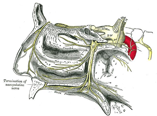
37 SPHENOPALATINE GANGLION SPG IS ONE OF FOUR AUTONOMIC GANGLIA INSIDE THE HEAD LOCATED IN THE PTERYGOPALATINE FOSSA WHICH IS LOCATED POSTERIOR TO THE MIDDLE TURBINATE AND IS 2 TO 7 MM DEEP TO THE LATERAL NASAL MUCOSA ANTERIOR BORDER: MAXILLARY SINUS POSTERIOR BORDER: MEDIAL PTERYGOID PLATE SUPERIOR BORDER: SPHENOID SINUS MEDIAL: PALATINE BONE
38 SPHENOPALATINE GANGLION AUTONOMIC COMPONENTS DEEP PETROSAL NERVE: POSTGANGLIONIC SYMPATHETICS FROM THE SUPERIOR CERVICAL GANGLION AND UPPER THORACIC SPINAL CORD GREATER PETROSAL NERVE: PREGANGLIONIC PARASYMPATHETICS FROM THE SUPERIOR SALIVATORY NUCLEUS SYNAPSE IN THE SPG WITH POSTGANGLIONIC AXONS, VASODILATOR, AND SECRETORY FIBERS SENSORY FIBERS FROM TWO SPHENOPALATINE BRANCHES OF THE MAXILLARY NERVE PASS THROUGH THE SPG AND FORM THE PALATINE NERVES WHICH INNERVATE THE UPPER TEETH, NASAL MEMBRANES, SOFT PALATE AND SOME PARTS OF THE PHARYNX. ALSO HAS SECRETOMOTOR NERVES TO THE NASAL GLANDS FROM THE GREATER PETROSAL NERVE.

39
40 SPHENOPALATINE GANGLION BLOCK INDICATED FOR TREATMENT OF: PERSISTENT IDIOPATHIC FACIAL PAIN AKA ATYPICAL FACIAL PAIN TRIGEMINAL NEURALGIA SPHENOPALATINE NEURALGIA MIGRAINE HEADACHES CLUSTER HEADACHES POST-TRAUMATIC HEADACHES CANCER PAIN OF FACIAL AND OROFACIAL STRUCTURES
41 TREATMENT ALGORITHM
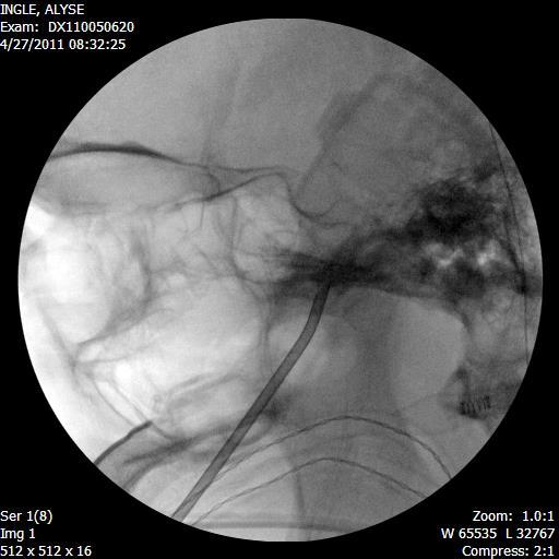
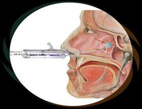
42 SPHENOPALATINE GANGLION BLOCK TECHNIQUES: INTRANASAL COTTON SWABS SOAKED IN LOCAL ANESTHETIC 4% COCAINE 2% LIDOCAINE VISCOUS LIDOCAINE INTRAORAL VIA THE PALATINE FORAMEN LOCATED MEDIAL TO THE 2 ND MOLAR FORAMEN ENTERED WITH A CURVED NEEDLE INFRAZYGOMATIC THROUGH THE MANDIBULAR NOTCH RFTC PULSED RF


43 INTRANASAL APPLICATORS TX360 ALLEVIO
44 TECHNIQUE FOR RFA, APPLY SENSORY STIMULATION AT 50 HZ TO LOCALIZE THE GANGLION PLACEMENT OF THE NEEDLE AT THE GANGLION WILL GENERATE A PARESTHESIA AT THE ROOT OF THE NOSE THE LOWER THE STIMULATION THE BETTER. BEST TO USE A 2-3 MM ACTIVE TIP TO DECREASE THE POSSIBILITY OF LESIONING THE MAXILLARY NERVE OR PALATINE NERVES LESION AT DEGREES CELSIUS PULSE RF CAN BE DONE AT 45 VOLTS

45 Zygoma Mandibular notch Ramus of mandible

46 Base of inverted vase Pterygomaxillary fissure

47

48
49 SPHENOPALATINE GANGLION BLOCK COMPLICATIONS HEMATOMA LARGE VENOUS PLEXUS MAXILLARY ARTERY EPISTAXIS NERVE INJURY EYE INJURY RETROBULBAR HEMATOMA VIA THE INFRAORBITAL FISSURE INTRAVASCULAR INJECTION MAXILLARY SINUS FRACTURE
50 Day M. Sympathetic blocks: the evidence. Pain Practice 2008;8:98-109

51
52 GLOSSOPHARYNGEAL NEURALGIA DESCRIPTION: A DISORDER CHARACTERIZED BY UNILATERAL BRIEF STABBING PAIN, ABRUPT IN ONSET AND TERMINATION, IN THE DISTRIBUTIONS NOT ONLY OF THE GLOSSOPHARYNGEAL NERVE BUT ALSO OF THE AURICULAR AND PHARYNGEAL BRANCHES OF THE VAGUS NERVE. PAIN IS EXPERIENCED IN THE EAR, BASE OF THE TONGUE, TONSILLAR FOSSA AND/OR BENEATH THE ANGLE OF THE JAW. IT IS COMMONLY PROVOKED BY SWALLOWING, TALKING OR COUGHING AND MAY REMIT AND RELAPSE IN THE FASHION OF TRIGEMINAL NEURALGIA. DIAGNOSTIC CRITERIA: RECURRING PAROXYSMAL ATTACKS OF UNILATERAL PAIN IN THE DISTRIBUTION OF THE GLOSSOPHARYNGEAL NERVE 1 AND FULFILLING CRITERION B PAIN HAS ALL OF THE FOLLOWING CHARACTERISTICS: LASTING FROM A FEW SECONDS TO 2 MINUTES SEVERE INTENSITY ELECTRIC SHOCK-LIKE, SHOOTING, STABBING OR SHARP IN QUALITY PRECIPITATED BY SWALLOWING, COUGHING, TALKING OR YAWNING NOT BETTER ACCOUNTED FOR BY ANOTHER ICHD-3 DIAGNOSIS.
53 GLOSSOPHARYNGEAL NEURALGIA CLASSICAL GLOSSOPHARYNGEAL NEURALGIA DESCRIPTION: GLOSSOPHARYNGEAL NEURALGIA DEVELOPING WITHOUT APPARENT CAUSE OTHER THAN NEUROVASCULAR COMPRESSION. SECONDARY GLOSSOPHARYNGEAL NEURALGIA DESCRIPTION: GLOSSOPHARYNGEAL NEURALGIA CAUSED BY AN UNDERLYING DISEASE. IDIOPATHIC GLOSSOPHARYNGEAL NEURALGIA DESCRIPTION: GLOSSOPHARYNGEAL NEURALGIA WITH NO EVIDENCE EITHER OF NEUROVASCULAR COMPRESSION OR OF CAUSATIVE UNDERLYING DISEASE.
54 GLOSSOPHARYNGEAL NERVE GLOSSOPHARYNGEAL NERVE RECEIVES SENSORY INFORMATION FROM THE POSTERIOR THIRD OF THE TONGUE, SOFT PALATE, TONSILS, PHARYNX, AND THE AUDITORY CANAL. INNERVATES THE STYLOPHARYNGEUS MUSCLE
55 GLOSSOPHARYNGEAL NERVE GLOSSOPHARYNGEAL NERVE ARISES FROM THE MEDULLA AND RUNS ANTERIORLY UNDER THE PETROUS PORTION OF THE TEMPORAL BONE. EXITS THE SKULL VIA THE JUGULAR FORAMEN AND RUNS BETWEEN THE INTERNAL CAROTID ARTERY AND THE INTERNAL JUGULAR VEIN POSTERIOR TO THE STYLOID PROCESS.
56 GLOSSOPHARYNGEAL NERVE BLOCK INDICATIONS GLOSSOPHARYNGEAL NEURALGIA PERSISTENT IDIOPATHIC FACIAL PAIN PAIN SECONDARY PHARYNGEAL CANCER DIAGNOSTIC BLOCK WITH LOCAL ANESTHETIC DEFINITIVE TREATMENT WITH RF/PULSED RF OR CRYONEUROLYSIS

57

58 Styloid process

59 Styloid Process


60
61 GLOSSOPHARYNGEAL NERVE BLOCK COMPLICATIONS INTRAVASCULAR INJECTION UNINTENTIONAL BLOCKADE OF OTHER CRANIAL NERVES DIFFICULTIES SWALLOWING AND HOARSENESS SECONDARY TO CN IX AND CN X BLOCKADE, RESPECTIVELY. TACHYCARDIA AND HYPERTENSION SECONDARY TO BLOCKADE OF PARASYMPATHETIC OUTFLOW
62
63 QUESTIONS
PTERYGOPALATINE FOSSA
 PTERYGOPALATINE FOSSA Outline Anatomical Structure and Boundaries Foramina and Communications with other spaces and cavities Contents Pterygopalatine Ganglion Especial emphasis on certain arteries and
By : Prof Saeed Abuel Makarem & Dr.Sanaa Alshaarawi
 By : Prof Saeed Abuel Makarem & Dr.Sanaa Alshaarawi OBJECTIVES By the end of the lecture, students shouldbe able to: List the nuclei of the deep origin of the trigeminal and facial nerves in the brain
Mohammad Hisham Al-Mohtaseb. Lina Mansour. Reyad Jabiri. 0 P a g e
 2 Mohammad Hisham Al-Mohtaseb Lina Mansour Reyad Jabiri 0 P a g e This is only correction for the last year sheet according to our record. If you already studied this sheet just read the yellow notes which
Temporal region. temporal & infratemporal fossae. Zhou Hong Ying Dept. of Anatomy
 Temporal region temporal & infratemporal fossae Zhou Hong Ying Dept. of Anatomy Temporal region is divided by zygomatic arch into temporal & infratemporal fossae. Temporal Fossa Infratemporal fossa Temporal
Parotid Gland, Temporomandibular Joint and Infratemporal Fossa
 M1 - Anatomy Parotid Gland, Temporomandibular Joint and Infratemporal Fossa Jeff Dupree Sanger 9-057 jldupree@vcu.edu Parotid gland: wraps around the mandible positioned between the mandible and the sphenoid
Cranial Nerve VII - Facial Nerve. The facial nerve has 3 main components with distinct functions
 Cranial Nerve VII - Facial Nerve The facial nerve has 3 main components with distinct functions Somatic motor efferent Supplies the muscles of facial expression; posterior belly of digastric muscle; stylohyoid,
Dr.Ban I.S. head & neck anatomy 2 nd y. جامعة تكريت كلية طب االسنان املرحلة الثانية أ.م.د. بان امساعيل صديق 6102/6102
 جامعة تكريت كلية طب االسنان التشريح مادة املرحلة الثانية أ.م.د. بان امساعيل صديق 6102/6102 Parotid region The part of the face in front of the ear and below the zygomatic arch is the parotid region. The
Omran Saeed. Luma Taweel. Mohammad Almohtaseb. 1 P a g e
 2 Omran Saeed Luma Taweel Mohammad Almohtaseb 1 P a g e I didn t include all the photos in this sheet in order to keep it as small as possible so if you need more clarification please refer to slides In
Trigeminal Nerve (V)
") Trigeminal Nerve (V) Lecture Objectives Discuss briefly how the face is developed. Follow up the course of trigeminal nerve from its point of central connections, exit and down to its target areas. Describe
Trigeminal Nerve (V) Lecture Objectives Discuss briefly how the face is developed. Follow up the course of trigeminal nerve from its point of central connections, exit and down to its target areas. Describe
Dr.Ban I.S. head & neck anatomy 2 nd y جامعة تكريت كلية طب االسنان مادة التشريح املرحلة الثانية أ.م.د. بان امساعيل صديق 6102/6102
 جامعة تكريت كلية طب االسنان مادة التشريح املرحلة الثانية أ.م.د. بان امساعيل صديق 6102/6102 Pterygopalatine fossa: The pterygopalatine fossa is a cone-shaped depression, It is located between the maxilla,
Infratemporal fossa: Tikrit University college of Dentistry Dr.Ban I.S. head & neck Anatomy 2 nd y.
 Infratemporal fossa: This is a space lying beneath the base of the skull between the lateral wall of the pharynx and the ramus of the mandible. It is also referred to as the parapharyngeal or lateral pharyngeal
Lec [8]: Mandibular nerve:
![Lec [8]: Mandibular nerve:](/thumbs/94/121295776.jpg "Lec [8]: Mandibular nerve:") Lec [8]: Mandibular nerve: The mandibular branch from the trigeminal ganglion lies in the middle cranial fossa lateral to the cavernous sinus. With the motor root of the trigeminal nerve [motor roots lies
Lec [8]: Mandibular nerve: The mandibular branch from the trigeminal ganglion lies in the middle cranial fossa lateral to the cavernous sinus. With the motor root of the trigeminal nerve [motor roots lies
Trigeminal Nerve Anatomy. Dr. Mohamed Rahil Ali
 Trigeminal Nerve Anatomy Dr. Mohamed Rahil Ali Trigeminal nerve Largest cranial nerve Mixed nerve Small motor root and large sensory root Motor root Nucleus of motor root present in the pons and medulla
Temporal fossa Infratemporal fossa Pterygopalatine fossa Terminal branches of external carotid artery Pterygoid venous plexus
 Outline of content Temporal fossa Infratemporal fossa Pterygopalatine fossa Terminal branches of external carotid artery Pterygoid venous plexus Boundary Content Communication Mandibular division of trigeminal
Parotid Gland. Parotid Gland. Largest of 3 paired salivary glands (submandibular; sublingual) Ramus of Mandible. Medial pterygoid.
 Ramus of Mandible. Medial pterygoid.") Parotid region Parotid Gland Largest of 3 paired salivary glands (submandibular; sublingual) Ramus of Mandible Medial pterygoid Cross section of mandible Masseter D S SCM Parotid Gland Mastoid Process
Parotid region Parotid Gland Largest of 3 paired salivary glands (submandibular; sublingual) Ramus of Mandible Medial pterygoid Cross section of mandible Masseter D S SCM Parotid Gland Mastoid Process
Maxilla, ORBIT and infratemporal fossa. Neophytos C Demetriades MD, DDS, MSc Associate professor European University of Cyprus School of Medicine
 Maxilla, ORBIT and infratemporal fossa Neophytos C Demetriades MD, DDS, MSc Associate professor European University of Cyprus School of Medicine MAXILLA Superior, middle, and inferior meatus Frontal sinus
Trigeminal Nerve Worksheets, Distributions Page 1
 Trigeminal Nerve Worksheet #1 Distribution by Nerve Dr. Darren Hoffmann Dental Gross Anatomy, Spring 2013 We have drawn out each of the branches of CN V in lecture and you have an idea now for their basic
Functional components
 Facial Nerve VII cranial nerve Emerges from Pons Two roots Functional components: 1. GSA (general somatic afferent) 2. SA (Somatic afferent) 3. GVE (general visceral efferent) 4. BE (Special visceral/branchial
Anatomic Relations Summary. Done by: Sohayyla Yasin Dababseh
 Anatomic Relations Summary Done by: Sohayyla Yasin Dababseh Anatomic Relations Lecture 1 Part-1 - The medial wall of the nose is the septum. - The vestibule lies directly inside the nostrils (Nares). -
Cranial nerves.
 Cranial nerves eaglezhyxzy@163.com Key Points of Learning Name Components Passing through Peripheral distribution Central connection Function Cranial nerves Ⅰ olfactory Ⅱ optic Ⅲ occulomotor Ⅳ trochlear
Anatomy of the Trigeminal Nerve
 19 Anatomy of the Trigeminal Nerve.1 Introduction 0. The Central Part of the Trigeminal Nerve 1..1 Origin 1.. Trigeminal Nuclei.3 The Peripheral Part of the Trigeminal Nerve 4.3.1 Ophthalmic Nerve 4.3.
*in general the blood supply of the nose comes from branches of the internal and external carotid arteries.
 In the previous lecture we talked about the anatomy of the nasal cavity, today we will talk about its blood supply, venous drainage, innervations, and finally about the paranasal sinuses. When we describe
Introduction to Local Anesthesia and Review of Anatomy
 5-Sep Introduction and Anatomy Review 12-Sep Neurophysiology and Pain 19-Sep Physiology and Pharmacology part 1 26-Sep Physiology and Pharmacology part 2 Introduction to Local Anesthesia and Review of
MAXILLA, ORBIT & PTERYGOPALATINE FOSSA. Neophytos C Demetriades MD, DDS, MSc Associate professor European University of Cyprus School of Medicine
 MAXILLA, ORBIT & PTERYGOPALATINE FOSSA Neophytos C Demetriades MD, DDS, MSc Associate professor European University of Cyprus School of Medicine Maxilla MAXILLA Superior, middle, and inferior meatus Frontal
For the following questions, indicate the letter that corresponds to the SINGLE MOST APPROPRIATE ANSWER
 GROSS ANATOMY EXAMINATION May 15, 2000 For the following questions, indicate the letter that corresponds to the SINGLE MOST APPROPRIATE ANSWER 1. Pain associated with an infection limited to the middle
Dr. Sami Zaqout, IUG Medical School
 The skull The skull is composed of several separate bones united at immobile joints called sutures. Exceptions? Frontal bone Occipital bone Vault Cranium Sphenoid bone Zygomatic bones Base Ethmoid bone
Bony orbit Roof The orbital plate of the frontal bone Lateral wall: the zygomatic bone and the greater wing of the sphenoid
 Bony orbit Roof: Formed by: The orbital plate of the frontal bone, which separates the orbital cavity from the anterior cranial fossa and the frontal lobe of the cerebral hemisphere Lateral wall: Formed
Skull-2. Norma Basalis Interna Norma Basalis Externa. Dr. Heba Kalbouneh Associate Professor of Anatomy and Histology
 Skull-2 Norma Basalis Interna Norma Basalis Externa Dr. Heba Kalbouneh Associate Professor of Anatomy and Histology Norma basalis interna Base of the skull- superior view The interior of the base of the
Tikrit University collage of dentistry Dr.Ban I.S. head & neck anatomy 2 nd y. Lec [5] / Temporal fossa :
![Tikrit University collage of dentistry Dr.Ban I.S. head & neck anatomy 2 nd y. Lec [5] / Temporal fossa :](/thumbs/88/115294566.jpg "Tikrit University collage of dentistry Dr.Ban I.S. head & neck anatomy 2 nd y. Lec [5] / Temporal fossa :") Lec [5] / Temporal fossa : Borders of the Temporal Fossa: Superior: Superior temporal line. Inferior: gap between zygomatic arch and infratemporal crest of sphenoid bone. Anterior: Frontal process of the
Lec [5] / Temporal fossa : Borders of the Temporal Fossa: Superior: Superior temporal line. Inferior: gap between zygomatic arch and infratemporal crest of sphenoid bone. Anterior: Frontal process of the
Veins of the Face and the Neck
 Veins of the Face and the Neck Facial Vein The facial vein is formed at the medial angle of the eye by the union of the supraorbital and supratrochlear veins. connected through the ophthalmic veins with
Cranial Nerves IX-X (Glossopharyngeal & Vagus Nerves)
") Cranial Nerves IX-X (Glossopharyngeal & Vagus Nerves) Please view our Editing File before studying this lecture to check for any changes. Color Code Important Doctors Notes Notes/Extra explanation Objectives
Cranial Nerves IX-X (Glossopharyngeal & Vagus Nerves) Please view our Editing File before studying this lecture to check for any changes. Color Code Important Doctors Notes Notes/Extra explanation Objectives
Laith Sorour. Facial nerve (vii):
:") Laith Sorour Cranial nerves 7 & 8 Hello, there are edited slides please go back to them to see pictures, they are not that much important in this lecture but still, and yes slides are included :p Let s
Laith Sorour Cranial nerves 7 & 8 Hello, there are edited slides please go back to them to see pictures, they are not that much important in this lecture but still, and yes slides are included :p Let s
Trigeminal nerve. Slide in bold and please go back to see the pictures, if I skipped any part of record that because it wasn t clear to me
 Trigeminal nerve Slide in bold and please go back to see the pictures, if I skipped any part of record that because it wasn t clear to me Hala nsour 2/26/2018 P a g e 1 this lecture contain two topics
Cranial Nerve VII & VIII
 Cranial Nerve VII & VIII Lecture Objectives Follow up the course of facial nerve from its point of central connections, exit and down to its target areas. Follow up the central connections of the facial
University of Palestine. Midterm Exam 2013/2014 Total Grade:
 Course No: DNTS2208 Course Title: Head and Neck Anatomy Date: 09/11/2013 No. of Questions: (50) Time: 1hour Using Calculator (No) University of Palestine Midterm Exam 2013/2014 Total Grade: Instructor
Chapter 7: Head & Neck
 Chapter 7: Head & Neck Osteology I. Overview A. Skull The cranium is composed of irregularly shaped bones that are fused together at unique joints called sutures The skull provides durable protection from
Biology 323 Human Anatomy for Biology Majors Week 10; Lecture 1; Tuesday Dr. Stuart S. Sumida. Cranial Nerves and Soft Tissues of the Skull
 Biology 323 Human Anatomy for Biology Majors Week 10; Lecture 1; Tuesday Dr. Stuart S. Sumida Cranial Nerves and Soft Tissues of the Skull FOREBRAIN MIDBRAIN HINDBRAIN Forebrain: Cerebrum Perception,
C h a p t e r PowerPoint Lecture Slides prepared by Jason LaPres North Harris College Houston, Texas
 C h a p t e r 15 The Nervous System: The Brain and Cranial Nerves PowerPoint Lecture Slides prepared by Jason LaPres North Harris College Houston, Texas Copyright 2009 Pearson Education, Inc., publishing
human anatomy 2016 lecture fifteen Dr meethak ali ahmed neurosurgeon
 Cranial Nerves Organization of the Cranial Nerves The cranial nerves are named as follows: I. Olfactory II. Optic III. Oculomotor IV. Trochlear V. Trigeminal VI. Abducent VII. Facial VIII. Vestibulocochlear
Major Anatomic Components of the Orbit
 Major Anatomic Components of the Orbit 1. Osseous Framework 2. Globe 3. Optic nerve and sheath 4. Extraocular muscles Bony Orbit Seven Bones Frontal bone Zygomatic bone Maxillary bone Ethmoid bone Sphenoid
SCHOOL OF ANATOMICAL SCIENCES Mock Run Questions. 4 May 2012
 SCHOOL OF ANATOMICAL SCIENCES Mock Run Questions 4 May 2012 1. With regard to the muscles of the neck: a. the platysma muscle is supplied by the accessory nerve. b. the stylohyoid muscle is supplied by
Face. Definition: The area between the two ears and from the chin to the eye brows. The muscles of the face
 Face Definition: The area between the two ears and from the chin to the eye brows. The muscles of the face The muscle of facial expression (include the muscle of the face and the scalp). All are derived
Anatomy #9. Rashed AL-Jomared. The Cranial Nerves IX. Amneh Hazaimeh & Alanood Bostanji
 Anatomy #9 The Cranial Nerves IX Rashed AL-Jomared Amneh Hazaimeh & Alanood Bostanji السالم عليكم This lecture talks about the cranial nerves IX & X:: *Glossopharyngeal nerve : The nerve gets out of the
Introduction to Head and Neck Anatomy
 Introduction to Head and Neck Anatomy Nervous Tissue Controls and integrates all body activities within limits that maintain life Three basic functions 1. sensing changes with sensory receptors 2. interpreting
The Pharynx. Dr. Nabil Khouri MD. MSc, Ph.D
 The Pharynx Dr. Nabil Khouri MD. MSc, Ph.D Introduction The pharynx is the Musculo-fascial halfcylinder that links the oral and nasal cavities in the head to the larynx and esophagus in the neck Common
Structure Location Function
 Frontal Bone Cranium forms the forehead and roof of the orbits Occipital Bone Cranium forms posterior and inferior portions of the cranium Temporal Bone Cranium inferior to the parietal bone forms the
Tracing the Cranial Nerves Osteologically
 CN I II III IV V 1 Supra-orbital ethmoidal nn. Ext. nasal V 2 Tracing the Cranial Nerves Osteologically Nucleus of Origin Olfactory tracts of frontal lobe of cerebrum Optic tracts from optic chiasma and
Skull-2. Norma Basalis Interna. Dr. Heba Kalbouneh Assistant Professor of Anatomy and Histology
 Skull-2 Norma Basalis Interna Dr. Heba Kalbouneh Assistant Professor of Anatomy and Histology Norma basalis interna Base of the skull- superior view The interior of the base of the skull is divided into
Brain and spinal nerve. By: shirin Kashfi
 Brain and spinal nerve By: shirin Kashfi Nervous system: central nervous system (CNS) peripheral nervous system (PNS) Brain (cranial) nerves Spinal nerves Ganglions (dorsal root ganglions, sympathetic
The sebaceous glands (glands of Zeis) open directly into the eyelash follicles, ciliary glands (glands of Moll) are modified sweat glands that open
 open directly into the eyelash follicles, ciliary glands (glands of Moll) are modified sweat glands that open") The Orbital Region The orbits are a pair of bony cavities that contain the eyeballs; their associated muscles, nerves, vessels, and fat; and most of the lacrimal apparatus upper eyelid is larger and more
The Orbital Region The orbits are a pair of bony cavities that contain the eyeballs; their associated muscles, nerves, vessels, and fat; and most of the lacrimal apparatus upper eyelid is larger and more
lecture #2 Done by : Tyma'a Al-zaben
 lecture #2 Done by : Tyma'a Al-zaben ** Hello SERTONIN! note:: the slide included within the sheet but make sure back to slide for pictures in the previous lecture we talk about ascending tract and its
Bisection of Head & Nasal Cavity 頭部對切以及鼻腔. 解剖學科馮琮涵副教授 分機
 Bisection of Head & Nasal Cavity 頭部對切以及鼻腔 解剖學科馮琮涵副教授 分機 3250 E-mail: thfong@tmu.edu.tw Outline: The structure of nose The concha and meatus in nasal cavity The openings of paranasal sinuses Canals, foramens
Nose & Mouth OUTLINE. Nose. - Nasal Cavity & Its Walls. - Paranasal Sinuses. - Neurovascular Structures. Mouth. - Oral Cavity & Its Contents
 Dept. of Human Anatomy, Si Chuan University Zhou hongying eaglezhyxzy@163.com Nose & Mouth OUTLINE Nose - Nasal Cavity & Its Walls - Paranasal Sinuses - Neurovascular Structures Mouth - Oral Cavity & Its
Anatomy and Physiology. Bones, Sutures, Teeth, Processes and Foramina of the Human Skull
 Anatomy and Physiology Chapter 6 DRO Bones, Sutures, Teeth, Processes and Foramina of the Human Skull Name: Period: Bones of the Human Skull Bones of the Cranium: Frontal bone: forms the forehead and the
General Sensory Pathways of the Face Area, Taste Pathways and Hearing Pathways
 General Sensory Pathways of the Face Area, Taste Pathways and Hearing Pathways Lecture Objectives Describe pathways for general sensations (pain, temperature, touch and proprioception) from the face area.
Anterior Ethmoidal Nerve Overview
 Anterior Ethmoidal Nerve Overview Name Anterior Ethmoidal Nerve Latin Nervus Ethmoidalis anterior Etymology Pain Differential Diagnosis - Enrico Dellacà M.D Ph.D. Nerve from Latin nervus meaning sinew,
Bones of the skull & face
 Bones of the skull & face Cranium= brain case or helmet Copyright The McGraw-Hill Companies, Inc. Permission required for reproduction or display. The cranium is composed of eight bones : frontal Occipital
Human Anatomy and Physiology - Problem Drill 07: The Skeletal System Axial Skeleton
 Human Anatomy and Physiology - Problem Drill 07: The Skeletal System Axial Skeleton Question No. 1 of 10 Which of the following statements about the axial skeleton is correct? Question #01 A. The axial
AXIAL SKELETON SKULL
 AXIAL SKELETON SKULL CRANIAL BONES (8 total flat bones w/ 2 paired) 1. Frontal forms forehead & upper portion of eyesocket (orbital) 2. Parietal paired bones; form superior & lateral walls of cranium 3.
Prevertebral Region, Pharynx and Soft Palate
 Unit 20: Prevertebral Region, Pharynx and Soft Palate Dissection Instructions: Step1 Step 2 Step 1: Insert your fingers posterior to the sternocleidomastoid muscle, vagus nerve, internal jugular vein,
Dr.Ban I.S. head & neck anatomy 2 nd y. جامعة تكريت كلية طب االسنان املرحلة الثانية
 جامعة تكريت كلية طب االسنان التشريح مادة املرحلة الثانية أ.م.د. بان امساعيل صديق 6102-6102 1 The Palate The palate forms the roof of the mouth and the floor of the nasal cavity. It is divided into two
function - sensory & postganglionic sympathetic [communication from the internal carotid plexus in the cavernous sinus] innervation of the mucosa of
![function - sensory & postganglionic sympathetic [communication from the internal carotid plexus in the cavernous sinus] innervation of the mucosa of](/thumbs/74/71276096.jpg "function - sensory & postganglionic sympathetic [communication from the internal carotid plexus in the cavernous sinus] innervation of the mucosa of") Nerves I. Cranial nerves A. Olfactory (CN I) 1. Olfactory bulb 2. Olfactory tract B. Optic n. (CNII) function - carries visual sensory information from the neural retina to the diencephalon & midbrain
Nerves I. Cranial nerves A. Olfactory (CN I) 1. Olfactory bulb 2. Olfactory tract B. Optic n. (CNII) function - carries visual sensory information from the neural retina to the diencephalon & midbrain
3-Deep fascia: is absent (except over the parotid gland & buccopharngeal fascia covering the buccinator muscle)
") The Face 1-Skin of the Face The skin of the face is: Elastic Vascular (bleed profusely however heal rapidly) Rich in sweat and sebaceous glands (can cause acne in adults) It is connected to the underlying
The Face 1-Skin of the Face The skin of the face is: Elastic Vascular (bleed profusely however heal rapidly) Rich in sweat and sebaceous glands (can cause acne in adults) It is connected to the underlying
Cranial Cavity REFERENCES: OBJECTIVES OSTEOLOGY. Stephen A. Gudas, PT, PhD
 Stephen A. Gudas, PT, PhD Cranial Cavity REFERENCES: Moore and Agur, Essential Clinical Anatomy (ECA), 3rd ed., pp. 496 498; 500 507; 512 514 Grant s Atlas 12 th ed., Figs 7.6; 7.19 7.30. Grant s Dissector
PERIPHERAL NERVOUS SYSTEM
 CHAPTER 13 PERIPHERAL NERVOUS SYSTEM Functional division of nervous system = afferent info to the CNS ascending spinal cord = efferent info from CNS descending spinal cord somatic skin, muscles visceral
HBA THE BODY Head & Neck Written Examination October 23, 2014
 HBA 531 - THE BODY Head & Neck Written Examination October 23, 2014 Name: NOTE 2: When asked to trace nerve, artery, or vein pathways, do so by using arrows, e.g., structure a structure b structure c...
Oral cavity : consist of two parts: the oral vestibule and the oral cavity proper. Oral vestibule : is slit like space between.
 Oral cavity Oral cavity : consist of two parts: the oral vestibule and the oral cavity proper Oral vestibule : is slit like space between the teeth, buccal gingiva, lips, and cheeks 1 Oral cavity Oral
Dr. Sami Zaqout Faculty of Medicine IUG
 Auricle External Ear External auditory meatus The Ear Middle Ear (Tympanic Cavity) Auditory ossicles Internal Ear (Labyrinth) Bony labyrinth Membranous labyrinth External Ear Auricle External auditory
Perineural Tumor Spread (PNS) Perineural Tumor Spread (PNS) PNS Anatomic Considerations. Perineural Tumor Spread-Imaging
 Perineural Tumor Spread (PNS) PNS Anatomic Considerations. Perineural Tumor Spread-Imaging") Imaging of Perineural Tumor Spread in Head and Neck Cancer Lawrence E. Ginsberg, MD Departments of Diagnostic Radiology and Head and Neck Surgery University of Texas M.D. Anderson Cancer Center Houston,
Imaging of Perineural Tumor Spread in Head and Neck Cancer Lawrence E. Ginsberg, MD Departments of Diagnostic Radiology and Head and Neck Surgery University of Texas M.D. Anderson Cancer Center Houston,
3. The Jaw and Related Structures
 Overview and objectives of this dissection 3. The Jaw and Related Structures The goal of this dissection is to observe the muscles of jaw raising. You will also have the opportunity to observe several
Cranial Nerves. Steven McLoon Department of Neuroscience University of Minnesota
 Cranial Nerves Steven McLoon Department of Neuroscience University of Minnesota 1 Course News Change in Lab Sequence Week of Oct 2 Lab 5 Week of Oct 9 Lab 4 2 Sensory and Motor Systems Sensory Systems:
INTRODUCTION: ANATOMY UNDERLYING CLINICAL TESTS OF CRANIAL NERVES
 INTRODUCTION: ANATOMY UNDERLYING CLINICAL TESTS OF CRANIAL NERVES CRANIAL NERVE I - OLFACTORY I - OLFACTORY NERVE - SMELL TEST: SMELL ODORS (note: not ammonia; pain in nasal cavity CN5 DAMAGE: LOSS OF
HEAD AND NECK ANATOMY PRACTICE QUESTIONS
 HEAD AND NECK ANATOMY PRACTICE QUESTIONS 1. A patient complains that he has lost sensation on his face and that the skin of his face feels numb. The physician tests tactile acuity by touching the forehead
The Seventh Cranial Nerve The Facial By Prof. Dr. Muhammad Imran Qureshi
 The Seventh Cranial Nerve The Facial By Prof. Dr. Muhammad Imran Qureshi Functional Components: SVE: Fibers originate from nucleus of facial nerve, and supply facial muscles GVE: Fibers derived from superior
Unit 18: Cranial Cavity and Contents
 Unit 18: Cranial Cavity and Contents Dissection Instructions: The calvaria is to be removed without damage to the dura mater which is attached to the inner surface of the calvaria. Cut through the outer
CN I Olfactory. CN II Optic. CN III Oculomotor. Special Sensory Efferent fibers to Olfactory Bulb. Cribiform Plate of Ethmoid
 CN I Olfactory Efferent fibers to Olfactory Bulb Olfactory Tract Olfactory Bulb Cribiform Plate of Ethmoid Anosmia Loss of sense of smell Uncinate Fits olfactory hallucinations To Olfactory Epithelium
often the opposing teeth will manifest symptoms as well, due to extrusion of the tooth from increased pressure from the cyst.
 Mucous Retention Cysts of the Maxillary Sinus and Superiority of 3D Cone Beam CT Scans versus Traditional Panoramic Imaging Rebecca L Griffiths, BS, DMD Mucous retention cysts of the maxillary sinus are
ParasymPathetic Nervous system. Done by : Zaid Al-Ghnaneem
 ParasymPathetic Nervous system Done by : Zaid Al-Ghnaneem In this lecture we are going to discuss Parasympathetic, in the last lecture we took sympathetic and one of the objectives of last lecture was
The Skull and Temporomandibular joint II Prof. Abdulameer Al-Nuaimi. E. mail:
 The Skull and Temporomandibular joint II Prof. Abdulameer Al-Nuaimi E-mail: a.al-nuaimi@sheffield.ac.uk E. mail: abdulameerh@yahoo.com Temporal fossa The temporal fossa is a depression on the temporal
Brain ميهاربا لض اف دمح ا د The Meninges 1- Dura Mater of the Brain endosteal layer does not extend meningeal layer falx cerebri tentorium cerebelli
 .احمد د فاضل ابراهيم Lecture 15 Brain The Meninges Three protective membranes or meninges surround the brain in the skull: the dura mater, the arachnoid mater, and the pia mater 1- Dura Mater of the Brain
Lab 16: PNS: Nerves and Autonomic NS Hamilton Answers to Pre- Lab Assignments
 Lab 16: PNS: Nerves and Autonomic NS Hamilton Answers to Pre- Lab Assignments Pre-Lab Activity 1: 1. a. olfactory nerve b. optic nerve c. oculomotor nerve d. abducens nerve e. trochlear nerve f. trigeminal
Trigeminal Neuralgia Involving All Three Branches Of Trigeminal Nerve Treated By Peripheral Neurectomy: An Interesting Case Report
 ISPUB.COM The Internet Journal of Dental Science Volume 10 Number 2 Trigeminal Neuralgia Involving All Three Branches Of Trigeminal Nerve Treated By Peripheral Neurectomy: An Interesting Case Report K
University of Palestine. Midterm Exam 2013/2014 Total Grade:
 [ Course No: DNTS2208 Course Title: Head and Neck Anatomy Date: 17/11/1024 No. of Questions: (52) Time: 2hours Using Calculator (No) University of Palestine Midterm Exam 2013/2014 Total Grade: Instructor
Head and Face Anatomy
 Head and Face Anatomy Epicranial region The Scalp The soft tissue that covers the vault of skull. Extends from supraorbital margin to superior nuchal line. Layers of the scalp S C A L P = skin = connective
Chapter 7 Part A The Skeleton
 Chapter 7 Part A The Skeleton Why This Matters Understanding the anatomy of the skeleton enables you to anticipate problems such as pelvic dimensions that may affect labor and delivery The Skeleton The
Primary Headache Prevalence % (95% CI) Migraine without aura 9 (7-9) Migraine with aura 6 (5-8)
 Migraine without aura 9 (7-9) Migraine with aura 6 (5-8)") Primary Headache Prevalence % (95% CI) Migraine without aura 9 (7-9) Migraine with aura 6 (5-8) Episodic tension-type headache 66 (62-69) Chronic tension-type headache 3 (2-5) Cluster headache 0.1 (0 1)
Primary Headache Prevalence % (95% CI) Migraine without aura 9 (7-9) Migraine with aura 6 (5-8) Episodic tension-type headache 66 (62-69) Chronic tension-type headache 3 (2-5) Cluster headache 0.1 (0 1)
Classification of Facial Pain. Surgical Treatment of Facial Pain. Typical trigeminal neuralgia. Atypical trigeminal neuralgia
 Surgical Treatment of Facial Pain Nicholas M. Barbaro, MD University of California at San Francisco Classification of Facial Pain Trigeminal neuralgia Atypical trigeminal neuralgia Neuropathic facial pain
Learning Outcomes. The Carotid 20/02/2013. Scalp, Face, Parotid. Layers of the Scalp. The Parotid Gland. The Scalp. The Carotid The Facial Artery
 Learning Outcomes The Scalp Layers of the Scalp Bleeding from the Scalp The Carotid The Facial Artery Major Muscles of the Face and Jaw(s) Muscles of Mastication Muscles of Facial Expression The Parotid
Biology 218 Human Anatomy. Adapted from Martini Human Anatomy 7th ed. Chapter 6 The Skeletal System: Axial Division
 Adapted from Martini Human Anatomy 7th ed. Chapter 6 The Skeletal System: Axial Division Introduction The axial skeleton: Composed of bones along the central axis of the body Divided into three regions:
Cranial Nerves and Spinal Cord Flashcards
 1. Name the cranial nerves and their Roman numeral. 2. What is Cranial Nerve I called, and what does it 3. Scientists who are trying to find a way to make neurons divide to heal nerve injuries often study
Tympanic Bulla Temporal Bone. Digastric Muscle. Masseter Muscle
 Superior view Hyoid Bone The hyoid bone does not articulate with any other bones. It is held in place by ligaments to the styloid process of the temporal bone and the thyroid cartilage of the larynx. It
Trigeminal Neuralgia (facial pain)
") Trigeminal Neuralgia (facial pain) Overview Trigeminal neuralgia is an inflammation of the trigeminal nerve, causing extreme pain and muscle spasms in the face. Attacks of intense, electric shock-like
Trigeminal Neuralgia (facial pain) Overview Trigeminal neuralgia is an inflammation of the trigeminal nerve, causing extreme pain and muscle spasms in the face. Attacks of intense, electric shock-like
Unit VIII Problem 3 Neuroanatomy: Brain Stem, Cranial Nerves and Scalp
 Unit VIII Problem 3 Neuroanatomy: Brain Stem, Cranial Nerves and Scalp - Brain stem: It is connected to the cerebellum and cerebral hemispheres. Rostral end of brain stem: diencephalon is the area which
Face and Scalp 解剖學科鄭授德
 Face and Scalp 解剖學科鄭授德 本教材之圖片取自於 1 Gray s Anatomy for Students, 3rd ed, 2015, by Drake, Vogl, and Mitchell 2 Clinically Oriented Anatomy, 7th ed, 2014, by Moore, Dalley, and Agur 3 Clinically Oriented
The Ear The ear consists of : 1-THE EXTERNAL EAR 2-THE MIDDLE EAR, OR TYMPANIC CAVITY 3-THE INTERNAL EAR, OR LABYRINTH 1-THE EXTERNAL EAR.
 The Ear The ear consists of : 1-THE EXTERNAL EAR 2-THE MIDDLE EAR, OR TYMPANIC CAVITY 3-THE INTERNAL EAR, OR LABYRINTH 1-THE EXTERNAL EAR Made of A-AURICLE B-EXTERNAL AUDITORY MEATUS A-AURICLE It consists
REVIEW/PREVIEW OF HEAD AND NECK ANATOMY FOR ENT EXAM
 REVIEW/PREVIEW OF HEAD AND NECK ANATOMY FOR ENT EXAM - 2017 PALPATE CAROTID ARTERY: AT LEVEL OF CAROTID BIFURCATION VERTEBRAL LEVEL C4 Sternocleidomastoid Muscle INTERNAL CAROTID EXTERNAL CAROTID COMMON
Trigeminal Neuralgia > 1
 Trigeminal Neuralgia Overview Trigeminal neuralgia is an inflammation of the trigeminal nerve causing extreme pain and muscle spasms in the face. Attacks of intense, electric shock-like facial pain can