When you see this diagram, remember that you are looking at the embryo from above, through the amniotic cavity, where the epiblast appears as an oval
|
|
|
- Griffin Chambers
- 5 years ago
- Views:
Transcription


1 When you see this diagram, remember that you are looking at the embryo from above, through the amniotic cavity, where the epiblast appears as an oval disc
2 2
3 Why the embryo needs the vascular system? When it appears? Where it appears? with later contributions from neural crest mesenchyme

4 The Cardiac progenitor cells migrate from the Epiblast Through Primitive streak In a Cranial direction on each side of the notochordal process and around the prechordal plate 4

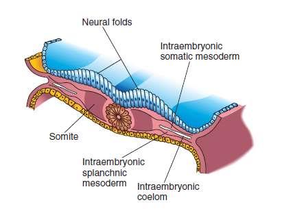
5 5 into the splanchnic layer of the lateral plate mesoderm

6 6 into the splanchnic layer of the lateral plate mesoderm
7 The cells from both sides meet cranially to form the Primary Heart Field (PHF) These cells will form : The atria Left ventricle Part of right ventricle The remainder of the right ventricle outflow tract (conus cordis and truncus arteriosus) Are derived from the Secondary Heart Field (SHF) 7
8 8 ONE-SOMITE AND TWO-SOMITE STAGES
9 Induction They form 9


10 10 Paired endothelial strands ANGIOBLASTIC CORDS appear in the cardiogenic mesoderm during the third week of development
11 11


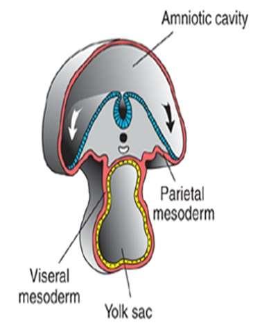
12 These cords canalize to form two heart tubes that soon fuse as embryo folds laterally to form a single heart tube late in the third week Two hearts tubs The two tubs are Fused Single heart tube is formed 12 As the embryo folds laterally
13 13
14 The mesodermal tissue surrounding the endothelial heart (endocardial) tube, has differentiated into three layers 1-The inner layer immediately around the endothelium is initially thick, gelatinous connective tissue called the cardiac jelly. (The cardiac jelly disappears later) 2-The next layer is the cellular primitive myocardium. (elaborates and matures to become the muscular wall of the heart, the myocardium) 3- The third (outer) layer consists of flat mesothelial cells that also line the remaining pericardial cavity. The endothelial tube becomes the internal endothelial lining of the heart the Endocardium 14

15 15 In addition to the cardiogenic region, other blood islands appear bilaterally, parallel and close to the midline of the embryonic shield. These islands form a pair of longitudinal vessels, the dorsal aortae.
16 16
17 AS you can see, the cells that will form the heart are located in a cranial position!!!!!!! 17 From cranial to caudal before folding 1- Septum transversum 2- Pericardium and ventral to it the Heart tube 4- oropharyngeal membrane. How will the heart move to its normal position in the thorax?????
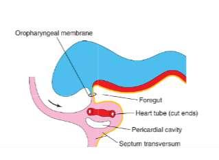
18 Cephalocaudal folding

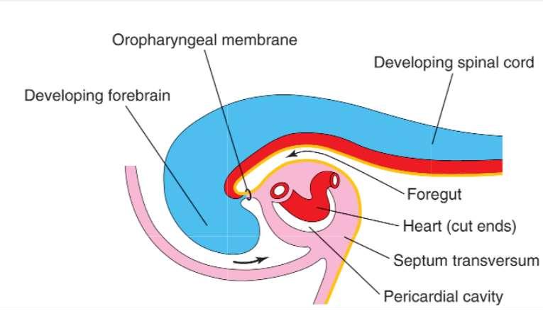
19 The result of cephalocaudal folding From cranial to caudal: 1- Buccopharyngeal membrane. 2- Pericardium. 3- Heart tube becomes dorsal to paricardial Cavity and ventral to foregut and invaginates the pericardial cavity 4- Septum transversum. 19
20 20 As the primitive, bilaterally symmetric cardiovascular system appears, shaping of the embryo during the fourth week profoundly influences the relative position of the cardiac portion of this system. The trilaminar embryonic disc folds into a cylinder, and the amnion tucks around the embryo on each side. The amnion also envelops the head end of the embryo as the ectodermal tube of the forebrain rapidly increases in size in a cranial and ventral direction. The result is a 180-degree sagittal plane rotation of the cardiogenic mesoderm and oropharyngeal membrane, which were originally cranial to the neural plate and the developing neural tube. The heart is now caudal to the oropharyngeal membrane rather than cranial, and the heart locates dorsal to the developing pericardial cavity
21 21 As indicated earlier, the heart tube is dorsal to the developing pericardial cavity As the tube enlarges and bends, it bugles into the underlying coelom As the heart tube comes to rest entirely within the pericardial cavity, it is suspended by the two opposing epithelial layers of the pericardial sac, The dorsal mesocardium. A ventral mesocardium never develops
22 22 No known cardiac anomaly can be attributed to the developmental phases described thus far
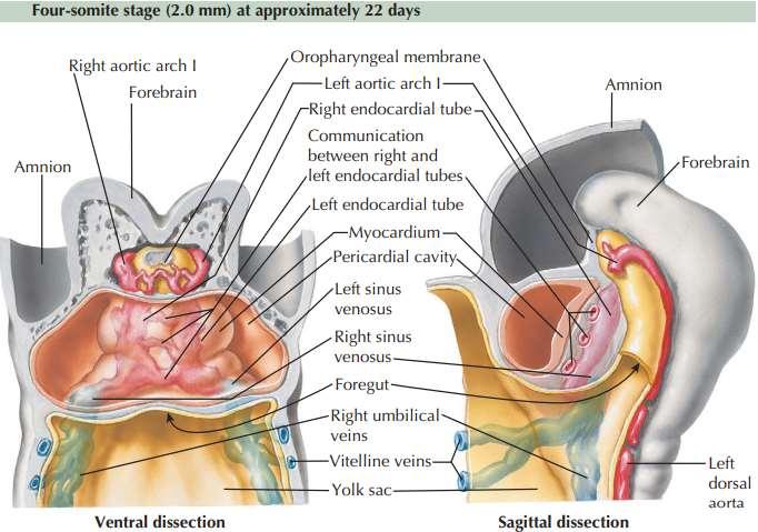
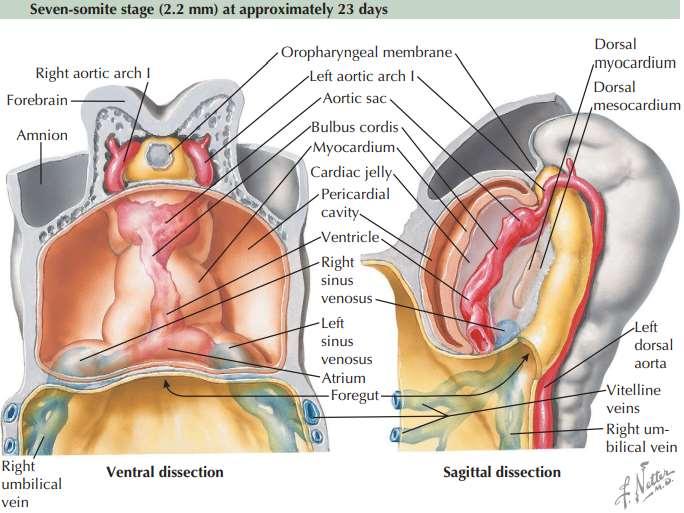
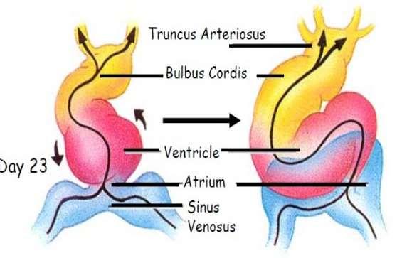
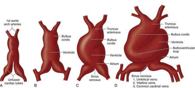
23 Formation of the cardiac loop What we have by now The heart is essentially a straight tube with a caudal venosus end and cranial arterial end It lies within the pericardial cavity is attached posteriorly only by the dorsal mesocardium The embryo now has seven somites is about 2.2 mm long is approximately 23 days old begins to beat About 3 days have elapsed between the appearance of intraembryonic vasculogenesis and the formation of the endocardial tube 23
 1- Sinus venosus 2- Primitive atrium. 3- primitive ventricle. 4- Bulbus cordis (conus).")
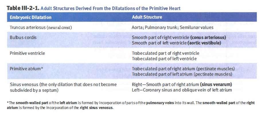
24 Differential growth defines five segments of the heart tube: (from caudal to cephalic or according to direction of blood flow) 1- Sinus venosus 2- Primitive atrium. 3- primitive ventricle. 4- Bulbus cordis (conus). 5- truncus arteriosus. 24
25 25
 is continuous cranially with the aortic")
26 The sinus venosus represent the venous end of the heart It receives 3 veins: 1- Common cardinal vein body wall 2- Umbilical vein from placenta 3- Vitelline vein from yolk sac The tubular truncus arteriosus (TA) is continuous cranially with the aortic sac

27 The arterial end of the heart is fixed by the pharyngeal arches 27 The venous end of the heart is fixed by the septum transversum

28 28
 lying anterior and to the right of the primitive ventricle) forming a U-shaped bulboventricular loop")
29 The part of the tube lying within the pericardial cavity is made up of bulbus cordis and ventricle the heart bends on itself (usually bends to the right, thus the proximal bulbus cordis (RV) lying anterior and to the right of the primitive ventricle) forming a U-shaped bulboventricular loop 29
30 As the atrium and sinus venosus are freed from the septum transverum they come to lie behind and above the ventricle and the heart tube is now S-shaped 30
31 31
32 32
33 33 At this stage

34 At this stage the bulbus cordis and ventricle are separated by a deep bulbo-ventricular sulcus. 34

35 This sulcus gradually becomes shallower so that the bulbus cordis and the ventricle come to form one chamber which communicates with the truncus arteriosus. The primary interventricular foramen 35

36 The atrial chamber expands so that parts of it come to project forwards on either side of the truncus 36

37 37 The atrial chamber expands so that parts of it come to project forwards on either side of the truncus

38 38 As a result of these changes the exterior of the heart assumes its definitive shape

39 Abnormalities of Cardiac Looping Dextrocardia, in which the heart lies on the right side of the thorax instead of the left, is caused because the heart loops to the left instead of the right. Dextrocardia may coincide with situs inversus, a complete reversal of asymmetry in all organs. Situs inversus, which occurs in 1/7000 individuals, usually is associated with normal physiology, although there is a slight risk of heart defects 39
40 It is often stated that looping of the tube is the first visual evidence of asymmetry in the embryo, although careful examination reveals that the atrioventricular canal has become asymmetric prior to the start of looping. Although the sense of laterality of the developing organs of the body, including the atrial appendages, develops during gastrulation, the pathway of signalling that governs rightward looping of the heart tube remains unknown However, it is now well established that signalling pathways including Pitx2, nodal, lefty, and cited-2, determine the formation of the morphologically left-sided or right-sided features seen in organs such as the lungs, the bronchial tree, the liver and spleen, and the atrial appendages 40
Development of the Heart
 Development of the Heart Thomas A. Marino, Ph.D. Temple University School of Medicine Stages of Development of the Heart 1. The horseshoe-shaped pericardial cavity. 2. The formation of the single heart
Embryology of the Heart
 *Page 1A: Embryology of the Heart Human embryonic disc is divided into three layers: ectoderm, intraembryonic mesoderm, and endoderm. The embryonic disc lies between the amniotic cavity and the primary
DEVELOPMENT OF THE CIRCULATORY SYSTEM L E C T U R E 5
 DEVELOPMENT OF THE CIRCULATORY SYSTEM L E C T U R E 5 REVIEW OF CARDIAC ANATOMY Heart 4 chambers Base and apex Valves Pericardial sac 3 layers: epi, myo, endo cardium Major blood vessels Aorta and its
Heart & vascular system I. Dawei Dong
 Heart & vascular system I Dawei Dong Lecture goal Learn the basics of heart and vascular development. Development of Heart, Blood, and Blood Vessels LEARNING GOALS: 1. explain the early development of
Development of the heart
 Development of the heart Prof. Abdulameer Al-Nuaimi E-mail: a.al-nuaimi@sheffield.ac.uk abdulameerh@yahoo.com Early Development of the Circulatory System Appears in the middle of the third week, when the
W.S. O The University of Hong Kong
 W.S. O The University of Hong Kong Objectives: Describe early angiogenesis. Describe the heart tube formation. Describe the partitioning into a 4- chambered heart. List the formation of heart valves and
6. HEART AND CIRCULATORY SYSTEM I
 6. HEART AND CIRCULATORY SYSTEM I Dr. Taube P. Rothman P&S 12-520 Tpr2@columbia.edu 212-305-7930 RECOMMENDED READING: Larsen Human Embryology, 3rd Edition, pp. 195-199; 157-169 top left; 172-174; bottom
Development and teratology of cardiovascular and lymphatic systems. Repetition: Muscle tissue
 Development and teratology of cardiovascular and lymphatic systems Repetition: Muscle tissue Beginning of the cardiovascular system development the 3rd week: Hemangiogenesis (day 15 16) blood islets (insulae
W.S. O. School of Biomedical Sciences, University of Hong Kong
 W.S. O School of Biomedical Sciences, University of Hong Kong Objectives: Describe early angiogenesis. Describe the heart tube formation. Describe the partitioning into a 4- chambered heart. List the formation
Development of the Heart *
 OpenStax-CNX module: m46673 1 Development of the Heart * OpenStax This work is produced by OpenStax-CNX and licensed under the Creative Commons Attribution License 3.0 By the end of this section, you will
Heart Development and Congenital Heart Disease
 Heart Development and Congenital Heart Disease Sally Dunwoodie s.dunwoodie@victorchang.edu.au Developmental and Stem Cell Biology Division Victor Chang Cardiac Research Institute for the heart of Australia...
6. Development of circulatory system II. Cardiac looping. Septation of atria and ventricles. Common heart malformations.
 6. Development of circulatory system II. Cardiac looping. Septation of atria and ventricles. Common heart malformations. Formation of heart tube paired endothelial-lined heart tube is formed from blood
Organogenesis Part 2. V. Lateral Plate Mesoderm VI. Endoderm VII. Development of the Tetrapod Limb VIII. Sex Determination. V. Lateral Plate Mesoderm
 Organogenesis Part 2 V. Lateral Plate Mesoderm VI. Endoderm VII. Development of the Tetrapod Limb VIII. Sex Determination V. Lateral Plate Mesoderm chordamesoderm paraxial mesoderm intermediate mesoderm
Circulatory system. Lecture #2
 Circulatory system Lecture #2 The essential components of the human cardiovascular system: Heart Blood Blood vessels Arteries - blood vessels that conduct arterial blood from heart ventricle to organs
The Cardiovascular System (Part I) 黃敏銓 解剖學暨細胞生物學研究所
 黃敏銓 解剖學暨細胞生物學研究所") The Cardiovascular System (Part I) 黃敏銓 解剖學暨細胞生物學研究所 1 Congenital heart defects (CHDs) 台灣兒童心臟學會 Sinus venarum Membranous septum Conus arteiosus (infundibulum) Aortic vestibule The Cardiovascular System
The Cardiovascular System (Part I) 黃敏銓 解剖學暨細胞生物學研究所 1 Congenital heart defects (CHDs) 台灣兒童心臟學會 Sinus venarum Membranous septum Conus arteiosus (infundibulum) Aortic vestibule The Cardiovascular System
CARDIAC DEVELOPMENT CARDIAC DEVELOPMENT
 CARDIAC DEVELOPMENT CARDIAC DEVELOPMENT Diane E. Spicer, BS, PA(ASCP) University of Florida Dept. of Pediatric Cardiology Curator Van Mierop Cardiac Archive This lecture is given with special thanks to
Notes: 1)Membranous part contribute in the formation of small portion in the septal cusp.
Membranous part contribute in the formation of small portion in the septal cusp.") Embryology 9 : Slide 16 : There is a sulcus between primitive ventricular and bulbis cordis that will disappear gradually and lead to the formation of one chamber which is called bulboventricular chamber.
Embryology 9 : Slide 16 : There is a sulcus between primitive ventricular and bulbis cordis that will disappear gradually and lead to the formation of one chamber which is called bulboventricular chamber.
The sinus venosus represent the venous end of the heart It receives 3 veins: 1- Common cardinal vein body wall 2- Umbilical vein from placenta 3-
 1 2 The sinus venosus represent the venous end of the heart It receives 3 veins: 1- Common cardinal vein body wall 2- Umbilical vein from placenta 3- Vitelline vein from yolk sac 3 However!!!!! The left
Chapter 4: The thoracic cavity and heart. The Heart
 Chapter 4: The thoracic cavity and heart The thoracic cavity is divided into right and left pleural cavities by a central partition, the mediastinum. The mediastinum is bounded behind by the vertebral
IN THE NAME OF GOD. Development of the Heart and Vasculature
 IN THE NAME OF GOD Development of the Heart and Vasculature Overview vascular system appears (middle of 3 rd week) when the embryo is not able to satisfy its nutrition by diffusion Heart is the first functional
Development of the Great Vessels and Conduc6on Tissue
 Development of the Great Vessels and Conduc6on Tissue Development of the heart fields h:p://php.med.unsw.edu.au/embryology/ index.php?6tle=advanced_- _Heart_Fields! 2 Septa6on of the Bulbus Cordis Bulbus
Figure 10.1A Transparency Master 79
 Brain Carotid arteries Jugular vein Right front leg Lungs (inflated) Cranial Right atrium To left front leg Left subclavian Bronchus capillaries Brachiocephalic vein Left atrium Dorsal aorta Right ventricle
Pharyngeal apparatus. - At the third week, it is a 3 layered structure: ectoderm, mesoderm and endoderm. This is called trilaminar disc
 Pharyngeal apparatus Remember from the first year embryology - The embryo was disc shaped in the second week of development (this is called embryonic disc) and it is a 2 layered disc (composed of two layers)---bilaminar
2/2/2011. Primitive Gut Tube Proctodeum and Stomodeum Stomach Duodenum Pancreas Liver and Biliary Apparatus Spleen Midgut
 DEVELOPMENT OF THE DIGESTIVE SYSTEM Development of Endodermal Organs Primitive Gut Tube Proctodeum and Stomodeum Stomach Duodenum Pancreas Liver and Biliary Apparatus Spleen Midgut Wednesday, February
Transcription for Narration of Embryology of the Great Arteries
 Transcription for Narration of Embryology of the Great Arteries Slide 1: In this presentation I am going to describe for you the development of what are known as the great arteries. The great arteries
The embryonic endoderm initially is widely connected with the yolk sac. As a consequence of cephalocaudal and lateral folding, a portion of the
 DIGESTIVE SYSTEM The embryonic endoderm initially is widely connected with the yolk sac. As a consequence of cephalocaudal and lateral folding, a portion of the endoderm-lined yolk sac cavity is incorporated
CJ Shuster A&P2 Lab Addenum Beef Heart Dissection 1. Heart Dissection. (taken from Johnson, Weipz and Savage Lab Book)
") CJ Shuster A&P2 Lab Addenum Beef Heart Dissection 1 Heart Dissection. (taken from Johnson, Weipz and Savage Lab Book) Introduction When you have finished examining the model, you are ready to begin your
CJ Shuster A&P2 Lab Addenum Beef Heart Dissection 1 Heart Dissection. (taken from Johnson, Weipz and Savage Lab Book) Introduction When you have finished examining the model, you are ready to begin your
human anatomy 2016 lecture thirteen Dr meethak ali ahmed neurosurgeon
 Heart The heart is a hollow muscular organ that is somewhat pyramid shaped and lies within the pericardium in the mediastinum. It is connected at its base to the great blood vessels but otherwise lies
LECTURE 5. Anatomy of the heart
 LECTURE 5. Anatomy of the heart Main components of the CVS: Heart Blood circulatory system arterial compartment haemomicrocirculatory (=microvascular) compartment venous compartment Lymphatic circulatory
Anatomy of the Heart. Figure 20 2c
 Anatomy of the Heart Figure 20 2c Pericardium & Myocardium Remember, the heart sits in it s own cavity, known as the mediastinum. The heart is surrounded by the Pericardium, a double lining of the pericardial
Development of the Liver and Pancreas
 Development of the Liver and Pancreas Professor Alfred Cuschieri Department of Anatomy University of Malta Three glandular buds arise from the distal end of the foregut during the fourth week Day 22 -The
The heart & Cardiovascular system
 The heart & Cardiovascular system The heart s continuous pulse create a base for our understanding of rhythms in everyday life. Bonnie Bainbridge Cohen The heart constantly beats throughout our lives never
Cardiovascular System Anatomy & Embryology
 بسم رلاهللا Cardiovascular System Anatomy & Embryology Portal circulation: Portal vessel: a vessel or a vein running between two sets of capillaries, the portal vein of the liver starts from a set of capillaries
Read Me. covering the Heart Anatomy. Labs. textbook. use. car: you
 Heart Anatomy Lab Pre-Lab Exercises Read Me These exercises should be done before coming to lab, after watching the videos covering the Heart Anatomy Labs. Answer the questions in this guide using the
Development of Blood Vessels and Fetal Circulation *
 OpenStax-CNX module: m46610 1 Development of Blood Vessels and Fetal Circulation * OpenStax This work is produced by OpenStax-CNX and licensed under the Creative Commons Attribution License 3.0 By the
Remember from the first year embryology Trilaminar disc has 3 layers: ectoderm, mesoderm, and endoderm
 Development of face Remember from the first year embryology Trilaminar disc has 3 layers: ectoderm, mesoderm, and endoderm The ectoderm forms the neural groove, then tube The neural tube lies in the mesoderm
Development of Spinal Cord & Vertebral Column. Dr. Sanaa Alshaarawi & Prof. Ahmed Fathalla
 Development of Spinal Cord & Vertebral Column Dr. Sanaa Alshaarawi & Prof. Ahmed Fathalla OBJECTIVES At the end of the lecture, students should be able to: q Describe the development of the spinal cord
Development of the Digestive System. W.S. O The University of Hong Kong
 Development of the Digestive System W.S. O The University of Hong Kong Plan for the GI system Then GI system in the abdomen first develops as a tube suspended by dorsal and ventral mesenteries. Blood
LAB 12-1 HEART DISSECTION GROSS ANATOMY OF THE HEART
 LAB 12-1 HEART DISSECTION GROSS ANATOMY OF THE HEART Because mammals are warm-blooded and generally very active animals, they require high metabolic rates. One major requirement of a high metabolism is
Through the 20th century, knowledge of the events occurring during cardiac development was
 806 * Anatomy DEVELOPMENT OF THE HEART: (1) FORMATION OF THE CARDIAC CHAMBERS AND ARTERIAL TRUNKS Antoon Moorman, Sandra Webb, Nigel A Brown, Wouter Lamers, Robert H Anderson See end of article for authors
Early Development of Neural Tube Development of Medulla Spinalis and Peripheral Nervous System. Assoc.Prof. E.Elif Güzel, M.D.
 Early Development of Neural Tube Development of Medulla Spinalis and Peripheral Nervous System Assoc.Prof. E.Elif Güzel, M.D. Third week of Embryogenesis Primitive streak/pit appears on the epiblast (day
Segmental Analysis. Gautam K. Singh, M.D. Washington University School of Medicine St. Louis
 Segmental Analysis Gautam K. Singh, M.D. Washington University School of Medicine St. Louis Segmental Analysis Segmental Analysis: From Veins to Ventricles Segmental Approach to Evaluation of Congenital
SLIDES 6 AND 10 MM PIG SLIDES; TRANSVERSE SECTIONS AND SAGITAL; FETAL PIGS-1-8INCH; HUMAN SAGITAL DIAGRAMS:DRAWINGS OF THE PIG SECTIONS TO BE
 SLIDES 6 AND 10 MM PIG SLIDES; TRANSVERSE SECTIONS AND SAGITAL; FETAL PIGS-1-8INCH; HUMAN SAGITAL SECTION DIAGRAMS:DRAWINGS OF THE PIG SECTIONS TO BE LABELLED, AND FOUR DRAWINGS TO BE MADE. REFERENCES:PATTEN:
Biology 340 Comparative Embryology Lecture 10 Dr. Stuart Sumida. Further Development of the Mesoderm (and Endoderm)
") Biology 340 Comparative Embryology Lecture 10 Dr. Stuart Sumida Further Development of the Mesoderm (and Endoderm) Further Development: Digestive System Foregut, Midgut, Hindgut Heart and Aortic Arches
Biology 340 Comparative Embryology Lecture 10 Dr. Stuart Sumida Further Development of the Mesoderm (and Endoderm) Further Development: Digestive System Foregut, Midgut, Hindgut Heart and Aortic Arches
Circulatory System: Introduction. Dr. Carmen E. Rexach Anatomy 35 Mt. San Antonio College
 Circulatory System: Introduction Dr. Carmen E. Rexach Anatomy 35 Mt. San Antonio College Components Cardiovascular system Lymphatic system Cardiovascular system Heart, blood vessels, blood Functions: transport
The Foregut. At first the esophagus is short. but with descent of the heart and lungs it lengthens rapidly
 GI embryology 2 The Foregut At first the esophagus is short but with descent of the heart and lungs it lengthens rapidly The muscular coat, which is formed by surrounding splanchnic mesenchyme, is striated
THE HEART OBJECTIVES: LOCATION OF THE HEART IN THE THORACIC CAVITY CARDIOVASCULAR SYSTEM
 BIOLOGY II CARDIOVASCULAR SYSTEM ACTIVITY #3 NAME DATE HOUR THE HEART OBJECTIVES: Describe the anatomy of the heart and identify and give the functions of all parts. (pp. 356 363) Trace the flow of blood
Heart and Lungs. LUNG Coronal section demonstrates relationship of pulmonary parenchyma to heart and chest wall.
 Heart and Lungs Normal Sonographic Anatomy THORAX Axial and coronal sections demonstrate integrity of thorax, fetal breathing movements, and overall size and shape. LUNG Coronal section demonstrates relationship
the Cardiovascular System I
 the Cardiovascular System I By: Dr. Nabil A Khouri MD, MsC, Ph.D MEDIASTINUM 1. Superior Mediastinum 2. inferior Mediastinum Anterior mediastinum. Middle mediastinum. Posterior mediastinum Anatomy of
Human Anatomy and Physiology Chapter 19 Worksheet 1- The Heart
 Human Anatomy and Physiology Chapter 19 Worksheet 1- The Heart Name Date Period 1. The "double pump" function of the heart includes the right side, which serves as the circuit pump, while the left side
Development of the Digestive System. W.S. O School of Biomedical Sciences, University of Hong Kong.
 Development of the Digestive System W.S. O School of Biomedical Sciences, University of Hong Kong. Organization of the GI tract: Foregut (abdominal part) supplied by coeliac trunk; derivatives include
Spleen. Vertebrate hearts Pericardial cavity division in coelum. Vessel walls. Endocardium = endothelium of blood vessels. Artery elastic tissue
 Spleen White pulp macrophages, monocyte storage Red pulp - (RBC) storage, and prod n (in nonmammals) Vertebrate hearts Pericardial cavity division in coelum Endocardium = endothelium of blood vessels Fig.
Fig General Structural Features of a kidney
 Chapter 11 Development of the Mesodermal Organs in Vertebrates 11.1. Subdivisions of the Mesoderm There are 4 subdivisions of the mesoderm: 1. The Notochord develops into the centrum of the vertebral column.
Anatomy of the Heart
 Biology 212: Anatomy and Physiology II Anatomy of the Heart References: Saladin, KS: Anatomy and Physiology, The Unity of Form and Function 8 th (2018). Required reading before beginning this lab: Chapter
Midgut. Over its entire length the midgut is supplied by the superior mesenteric artery
 Gi Embryology 3 Midgut the midgut is suspended from the dorsal abdominal wall by a short mesentery and communicates with the yolk sac by way of the vitelline duct or yolk stalk Over its entire length the
"Lecture Index. 1) Heart Progenitors. 2) Cardiac Tube Formation. 3) Valvulogenesis and Chamber Formation. 4) Epicardium Development.
 Heart Progenitors. 2) Cardiac Tube Formation. 3) Valvulogenesis and Chamber Formation. 4) Epicardium Development.") "Lecture Index 1) Heart Progenitors. 2) Cardiac Tube Formation. 3) Valvulogenesis and Chamber Formation. 4) Epicardium Development. 5) Septation and Maturation. 6) Changes in Blood Flow during Development.
"Lecture Index 1) Heart Progenitors. 2) Cardiac Tube Formation. 3) Valvulogenesis and Chamber Formation. 4) Epicardium Development. 5) Septation and Maturation. 6) Changes in Blood Flow during Development.
Cardiovascular Anatomy Dr. Gary Mumaugh
 Cardiovascular Anatomy Dr. Gary Mumaugh Location of Heart Approximately the size of your fist Location o Superior surface of diaphragm o Left of the midline in mediastinum o Anterior to the vertebral column,
THE HEART. A. The Pericardium - a double sac of serous membrane surrounding the heart
 THE HEART I. Size and Location: A. Fist-size weighing less than a pound (250 to 350 grams). B. Located in the mediastinum between the 2 nd rib and the 5 th intercostal space. 1. Tipped to the left, resting
Chapter 20 (1) The Heart
 The Heart") Chapter 20 (1) The Heart Learning Objectives Describe the location and structure of the heart Describe the path of a drop of blood from the superior vena cava or inferior vena cava through the heart out
Chapter 20 (1) The Heart Learning Objectives Describe the location and structure of the heart Describe the path of a drop of blood from the superior vena cava or inferior vena cava through the heart out
Cardiovascular Respiratory Renal/ Urinary. VOLUME 111 Nervous System. Organ ~ Systems ~
 Cardiovascular Respiratory Renal/ Urinary VOLUME 111 Nervous System Organ ~ Systems ~ Contents Section I: Cardiovascular System Chapter 1. Embryology... 3 Chapter 2. Histology... 13 Chapter 3. Anatomy...
Approximately the size of your fist Location Superior surface of diaphragm Left of the midline in mediastinum Anterior to the vertebral column,
 Dr. Gary Mumaugh Approximately the size of your fist Location Superior surface of diaphragm Left of the midline in mediastinum Anterior to the vertebral column, posterior to the sternum Posteriorly the
Development of Respiratory System. Dr. Sanaa Alshaarawy& Dr. Saeed Vohra
 Development of Respiratory System Dr. Sanaa Alshaarawy& Dr. Saeed Vohra OBJECTIVES At the end of the lecture the students should be able to: Identify the development of the laryngeotracheal (respiratory)
Supplementary Figure 1. A microarray screen of organizers compared to non-organizer tissue reveals a putative organizer gene set.
 Supplementary Figure 1. A microarray screen of organizers compared to non-organizer tissue reveals a putative organizer gene set. (a, b) Venn diagrams of 31 enriched (a) and 17 depleted (b) genes significantly
Embryonic Development of the Human Heart
 Embryonic Development of the Human Heart 1. Embryonic development of From formation of the zygote up until the third week of development the embryo s demand for O 2 and nutrients is met by simple diffusion.
ULTRASOUND OF THE FETAL HEART
 ULTRASOUND OF THE FETAL HEART Cameron A. Manbeian, MD Disclosure Statement Today s faculty: Cameron Manbeian, MD does not have any relevant financial relationships with commercial interests or affiliations
Chapter 6. Villous Growth
 Core Curriculum in Perinatal Pathology Chapter 6 Villous Growth Overview of vasculogenesis and angiogenesis Vasculogenesis Extraembryonic Vasculogenesis Angiogenesis Branching angiogenesis Sprouting angiogenesis
A. Incorrect! Think of a therapy that reduces prostaglandin synthesis. B. Incorrect! Think of a therapy that reduces prostaglandin synthesis.
 USMLE Step 1 - Problem Drill 02: Embryology Question No. 1 of 10 1. A premature infant is born with a patent ductus arteriosis. Which of the following treatments may be used as part of the treatment regimen?
Heart Anatomy. 7/5/02 Stephen G Davenport 1
 Heart Anatomy Copyright 1999, Stephen G. Davenport, No part of this publication may be reproduced, stored in a retrieval system, or transmitted, in any form without prior written permission. 7/5/02 Stephen
Chapter 14. The Cardiovascular System
 Chapter 14 The Cardiovascular System Introduction Cardiovascular system - heart, blood and blood vessels Cardiac muscle makes up bulk of heart provides force to pump blood Function - transports blood 2
Mediastinum and pericardium
 Mediastinum and pericardium Prof. Abdulameer Al-Nuaimi E-mail: a.al-nuaimi@sheffield.ac.uk E. mail: abdulameerh@yahoo.com The mediastinum: is the central compartment of the thoracic cavity surrounded by
V the fifth week of life early branchial arch and limb bud period
 V the fifth week of life early branchial arch and limb bud period 1. EXTERNAL APPEARANCE Fig. 5-1A 1. The specimens measure approximately 4 to 8 mm in length and have up to 42 to 44 pairs of somites. 2.
The Physiology of the Fetal Cardiovascular System
 The Physiology of the Fetal Cardiovascular System Jeff Vergales, MD, MS Department of Pediatrics Division of Pediatric Cardiology jvergales@virginia.edu Disclosures I serve as the medical director for
Heart and Soul Evaluation of the Fetal Heart
 Heart and Soul Evaluation of the Fetal Heart Ivana M. Vettraino, M.D., M.B.A. Clinical Associate Professor, Michigan State University College of Human Medicine Objectives Review the embryology of the formation
An Illustrated 1. Dissection Guide. To The... Mammalian. rr= Heart. Right ventricle+---, by David H. Hall
 An Illustrated 1. Dissection Guide. To The... Mammalian rr= Heart ventricle+---, by David H. Hall The Mam.malian Heart Because mammals are warm blooded (endothermic) and generally very active animals,
THE VESSELS OF THE HEART
 1 THE VESSELS OF THE HEART The vessels of the heart include the coronary arteries, which supply the heart and the veins and lymph vessels, which drain the heart. THE CORONARY ARTERIES These are the blood
CARDIOVASCULAR SYSTEM
 CARDIOVASCULAR SYSTEM Overview Heart and Vessels 2 Major Divisions Pulmonary Circuit Systemic Circuit Closed and Continuous Loop Location Aorta Superior vena cava Right lung Pulmonary trunk Base of heart
Middle mediastinum---- heart & pericardium. Dep. of Human Anatomy Zhou Hongying
 Middle mediastinum---- heart & pericardium Dep. of Human Anatomy Zhou Hongying eaglezhyxzy@163.com Subdivisions of the mediastinum Contents of Middle mediastinum Heart Pericardium: a serous sac enclosing
- anterior endoderm produces Wnt inhibitors (e.g. Cerberus, Dickkopf, Crescent); prevent Wnts from binding to their receptors
; prevent Wnts from binding to their receptors") Biology 4361 Developmental Biology Gilbert Chapter 15. Lateral Plate Mesoderm and Endoderm December 12, 2006 LATERAL PLATE MESODERM Two bands of lateral plate mesoderm lie peripheral to the intermediate
Biology 4361 Developmental Biology Gilbert Chapter 15. Lateral Plate Mesoderm and Endoderm December 12, 2006 LATERAL PLATE MESODERM Two bands of lateral plate mesoderm lie peripheral to the intermediate
Heart Dissection. 5. Locate the tip of the heart or the apex. Only the left ventricle extends all the way to the apex.
 Heart Dissection Page 1 of 6 Background: The heart is a four-chambered, hollow organ composed primarily of cardiac muscle tissue. It is located in the center of the chest in between the lungs. It is the
Chapter 14. Circulatory System Images. VT-122 Anatomy & Physiology II
 Chapter 14 Circulatory System Images VT-122 Anatomy & Physiology II The mediastinum Dog heart Dog heart Cat heart Dog heart ultrasound Can see pericardium as distinct bright line Pericardial effusion Fluid
- Tamara Wahbeh. - Fareed Khdair. 0 P a g e
 -1 - Tamara Wahbeh - - Fareed Khdair 0 P a g e GI Embryology Note: I included everything in the records and slides; anything in the slide not included in this sheet was not mentioned by the doctor during
- what other structures, besides the heart, does the mediastinum contain?
 Basic A & P II Dr. L. Bacha Chapter Outline (Martini & Nath 2010) An Introduction to the Cardiovascular System - read the paragraphs under this heading on page 580 The Heart is a Four Chambered Organ describe
Congenital Heart Defects
 Normal Heart Congenital Heart Defects 1. Patent Ductus Arteriosus The ductus arteriosus connects the main pulmonary artery to the aorta. In utero, it allows the blood leaving the right ventricle to bypass
Lab Photo Review Sheet
 9 8 0. Posterior Median Sulcus. Central Canal. Dorsal (Posterior) Horn. Ventral (Anterior) Horn. Grey Matter. White Matter. Anterior Median Fissure 8. Ventral (Anterior) Root (ramus) 9. Dorsal (Posterior)
This lab activity is aligned with Visible Body s A&P app. Learn more at visiblebody.com/professors
 1 This lab activity is aligned with Visible Body s A&P app. Learn more at visiblebody.com/professors 2 PRE-LAB EXERCISES: A. Watch the video 29.1 Heart Overview and make the following observations: 1.
Department of Cognitive Science UCSD
 Department of Cognitive Science UCSD Verse 1: Neocortex, frontal lobe, Brain stem, brain stem, Hippocampus, neural node, Right hemisphere, Pons and cortex visual, Brain stem, brain stem, Sylvian fissure,
Slide 1. Slide 2. Slide 3 CONGENITAL HEART DISEASE. Papworth Hospital NHS Trust INTRODUCTION. Jakub Kadlec/Catherine Sudarshan INTRODUCTION
 Slide 1 CONGENITAL HEART DISEASE Jakub Kadlec/Catherine Sudarshan NHS Trust Slide 2 INTRODUCTION Most common congenital illness in the newborn Affects about 4 9 / 1000 full-term live births in the UK 1.5
Human Anatomy, First Edition
 Human Anatomy, First Edition McKinley & O'Loughlin Chapter 22 : Heart 1 Functions of the Heart Center of the cardiovascular system, the heart. Connects to blood vessels that transport blood between the
CV Anatomy Quiz. Dr Ella Kim Dr Pip Green
 CV Anatomy Quiz Dr Ella Kim Dr Pip Green Q1 The location of the heart is correctly described as A) lateral to the lungs. B) medial to the sternum. C) superior to the diaphragm. D) posterior to the spinal
The cardiovascular system is composed of the heart and blood vessels that carry blood to and from the body s organs. There are 2 major circuits:
 1 The cardiovascular system is composed of the heart and blood vessels that carry blood to and from the body s organs. There are 2 major circuits: pulmonary and systemic. The pulmonary goes out to the
Lectures of Human Embryology
 Lectures of Human Embryology "Body Cavities & GIT" By DR. ABDEL-MONEM AWAD HEGAZY M.B. with honor 1983, Dipl."Gynecology and Obstetrics "1989, Master "Anatomy and Embryology" 1994, M.D. "Anatomy and Embryology"
Ligaments of the vertebral column:
 In the last lecture we started talking about the joints in the vertebral column, and we said that there are two types of joints between adjacent vertebrae: 1. Between the bodies of the vertebrae; which
Ch.15 Cardiovascular System Pgs {15-12} {15-13}
 Ch.15 Cardiovascular System Pgs {15-12} {15-13} E. Skeleton of the Heart 1. The skeleton of the heart is composed of rings of dense connective tissue and other masses of connective tissue in the interventricular
BRAIN DEVELOPMENT I: ESTABLISHMENT OF BASIC ARCHITECTURE. Thomas Marino, Ph.D.
 BRAIN DEVELOPMENT I: ESTABLISHMENT OF BASIC ARCHITECTURE Thomas Marino, Ph.D. Development of the Brain I. Competencies: Upon completion of this section of the course, the student must be able to: 1. Understand
HUMAN HEART. Learn the following structures on the heart models.
 HUMAN HEART Learn the following structures on the heart models. The human heart has four chambers that consist of the right atrium, left atrium, right ventricle, and left ventricle. The atria are smaller
2. right heart = pulmonary pump takes blood to lungs to pick up oxygen and get rid of carbon dioxide
 A. location in thorax, in inferior mediastinum posterior to sternum medial to lungs superior to diaphragm anterior to vertebrae orientation - oblique apex points down and to the left 2/3 of mass on left
ANATDMY. lecture # : Date : Lecturer : Maher Hadidi
 ANATDMY 27 lecture # : Date : Lecturer : Maher Hadidi Pericardium A double-walled fibroserous conical-shaped sac, within middle mediastinum. Enclose the heart and roots of its large vessels. Vagus nerves
Part 1. Copyright 2011 Pearson Education, Inc. Figure Copyright 2011 Pearson Education, Inc.
 PowerPoint Lecture Slides prepared by Leslie Hendon University of Alabama, Birmingham C H A P T E R The Heart 19 Part 1 The Heart A muscular double pump circuit vessels transport blood to and from the