IVU ((INTRAVENOUSUROGRAM))
|
|
|
- Rodger Scott
- 5 years ago
- Views:
Transcription
1 IVU ((INTRAVENOUSUROGRAM))
2

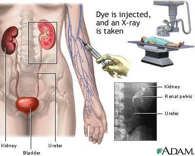
3 Anatomy The urinary system consists of : 2 kidneys, 2 ureters,1 bladder, 1 urethra Renal pelvis Minor calyx Major calyx Renal parenchyma Proximal ureter Pelvi-uretric junction Distal ureter Vesico-uretric junction
4 CONTAST MEDIA: Is a pharmaceutical given to the patient to enhance the original organ or tissue, e.g. kidneys, ureters, bladder. There are two types of contrast media : positive contrast & negative contrast 1- positive contrast with atomic number (Ƶ), e.g. barium sulphate and iodine compounds 2-negative contrast with atomic number (Ƶ), e.g. air, oxygen & carbondioxide. Administration of contrast media: 1-injcted intravascularly as in IVU exam. 2-ingested as in barium meal examinations 3-injected into the (CSF) cerebrospinal fluid.
5 Iodinated Contrast Media Iodinated C.M Water-insoluble Water-soluble Oily C.M HOCM LOCM Ionic Non-ionic
6 1- (HOCM) : high osmular contrast media : osmolality is 4-7 times that of human blood. This HOCM consists of a benzene ring with 3 iodine atoms and a positive cation (usually sodium or meglumin) and a negative anion (usually carboxyl group). Once injected into the plasma the cation dissociates from the compound creating two separate ions in the blood,these free ions cause an imbalance in homeostasis and an increase in plasma osmolality.this increase in osmolality causes the body to have a reaction. COO- Monomer l R l R /Na+ l so by reducing the number of free cations which do not contribute to the diagnostic image and are responsible for 50% of reactions, it was possible to the toxicity of the contrast.
7 2-(LOCM) : low osmular contrast media: osmolality is 1/3 of HOCM. By adding another benzene ring with an organic side chain, they were able to increase the iodine concentration with less number of cations Ratio=6/1 and this will reduce the body reaction,but still cause some reaction (ionic) DIMER R COO- Na+ I I I I R R R I Then they were able to modify the contrast even further by eliminating the cation and replacing the (coo-) with an amide or glucose (nondisassociated group) so when injected into the blood the C.M does not dissociate into two separate ions but remains intact (nonionic). e,g LOCM Niopam,Omnipaque,Isovist,Ultravist,Hexabrix. They are all Low osmolar non-ionic contrast media except for Hexabrix it is Low osolar ionic contrast media WHEN SHOULD WE USE (LOCM)? 1-infants & small children 2-pt with renal and /or cardiac failure 3-poorly hydrated pt 4-pt with diabetes and sickle-cell aniemia 5-pt who have had previous reactions to CM or have a strong allergic history. I
8 Adverse Reactions to C.M: CONTRAST REACTIONS MILD No need for medication Just reassure the pt e.g. flushing,urticaria nausia,vomating,pruritis MODERATE Needs medication & observation e.g. facial oedema, Bronchospasm hypotension SEVER Needs immediate response or CPR e.g.convulsions. cardiac arrest
9 IN CASE OF REACTION TO C.M : Adrenaline,Aminophylline,Atropine,Hydrocortisone,Lignocaine,Antihistamine, Dopamine CONTRAST DOSE : adults : 1ml per kg - minimum dose is 50 ml paediatrics :2 ml per kg Neonates :4ml per kg PT PREPARATION : -Creatinine levels : levels of creatinine indicates renal disease.(can not do the exam ) - If the pt have any allergy to egg or bannana,or have any history of asthma (need special praparation ) Preparation for allergic pt :prednisone tablet 2x25mg twice a day, one day before the exam and the day of the exam. Then 2x5mg twice a day for the 2 days after the exam. Then 5mg one tablet twice a day then stop. - pt should be NPO for 8 hours. and the last meal should be at 7:30 without any dairy products.
10 -instruct the pt to take 60 mg of caster oil the night before the Exam around 8 :30 pm. -In case of female pt do a pegnancy test or use the 10 days rule. -cleansing enema before the exam to reduce bowel gas. -pt must micturate immediately before the examination and wear the hospital gown. -Consent must be signed. -take the pt weight (to determ C.M dose) -after that the nurse will make the IV line for the patient EXPOUSER FACTOR: KV mas FFD (SID)
verify pt position. b)verify exposure factors. c)check pt preparation.")
11 Procedure of the exam: 1-Preliminary film,(scout film), (control film): patient position patient supine, with pillow for head, arms at side away from body, support under knees full length AP(KUB) of the abdomen, it should include the symphysis pubis. Center point to level of iliac crest the control film is taken to: a)verify pt position. b)verify exposure factors. c)check pt preparation. d) rule out any calcifications or stones.


12 Calcification appearing better in plain film stone plain film 5 Min film
13 the scout film should be shown to the radiologist before injection of C.M and if the patient has a catheter it should clamp before injection then we can start inject C.M and the exact starting time should be noted 2.Nephrogram(immediate film) Is taken immediately after completion of injection AP supine of the kidney area (the sam position) Center point Midway between xiphoid tip and iliac crest this film is taken to: Capture the early stages of the C.M entering the collecting system and to reveal any difference in function between the two kidneys.

14 Immediate (kidney area)

15 3-5 MIN FILM : patient position AP supine of the kidney area (the sam position) Center point Midway between xiphoid tip and iliac crest This film is taken to reveal any difference in function or excretion between the two kidneys.
 of the abdomen, it should include the symphysis pubis. Center point to level of iliac crest")
16 4-10 OR 15 MIN FILM patient position patient supine, with pillow for head, arms at side away from body, support under knees full length AP(KUB) of the abdomen, it should include the symphysis pubis. Center point to level of iliac crest

17 5-FULL BLADDER FILM patient position It is AP supine of the bladder area, coned view of the bladder (same position of KUB). Center point 5 cm superior to symphysis pubis with 10 º to 15 º cauded This film is taken to show if there are any abnormalities in the bladder. If the pt is not full bladder you may ask him/her to wait in the waiting area until he/she is full then take the full bladder film.
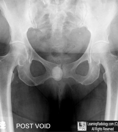
18 If this film is satisfactory,the pt is asked to empty his/her bladder. 6-POST VOID FILM Patient position Based on the clinical findings & the radiological findings on the earlier films, this will be either a full-length KUB abdominal film or a coned view of the bladder area with the tube angled 15 º caudad. This film is taken to assess bladder emptying, to demonstrate a return to normal dilated upper tract after the relief of bladder pressure,to aid in diagnosis of bladder tumours or diverticulms,to confirm vesicouretric calculi, to R/O reflux


19 Post void film

20 Additional Film 1-20 minute obliques Patient position Rotate the body into a 30 º posterior oblique for both R and L oblique position,flex elevated side knee,raise arm on elevated side Center point Level of iliac crest This film is taken to Provid a different perspective of the kidneys and project the ureters away from the spine

21 2- COMPRESSION FILM PATIENT POSITION AP supine of the kidney area, A compression band is now applied and the balloon is positioned midway between the A.S.I.S. ( over the ureters as they cross the pelvic brim). Center point Midway between xiphoid and iliac crest This film is taken to to enhance the filling of the pelvi-calyceal system and upper ureters.
 after abdominal")
22 If satisfactory demonstration of the pelvicalyceal system has been achieved, compression is released. 4-RELEASE FILM PATIENT POSITION It is a full length AP supine film taken to show the whole urinary tract Compression is contraindicated in the following cases : a) after abdominal trauma. b)after recent abdominal surgery c) abdominal mass. d) uretric stone.

23 e) infants and small children. f) severe abdominal pain. 5-PRONE VIEW it may provide better visualization of the ureters by making them more dependent. 6-DELAYED FILMS in cases of obstruction delayed films may be necessary for up to 24h. THINGS TO REMEMBER 1-DON`T FORGET TO PUT THE MARKER BEFORE EXPOSURE. 2-MAKE SURE YOU HAVE THE CORRECT (SID) BEFORE EXPOSURE. 3-ALWAYS PAY ATTENTION TO THE TIME OF INJECTION, AND MAKE SURE THAT THE EXPOSURE IS DONE AT THE CORRECT TIME IT SHOULD BE DONE. 4-IF THE PATIENT IS FEMALE ALWAYS MAKE SURE SHE IS NOT PREGNANT!


24 Abnormal Cases Horseshoe kidney

25 Transitional cell carcinoma. image shows multiple filling defects in the left renal pelvis and ureter. This finding is typical of lesions that grow slowly into the lumen of the ureter. Multifocal transitional cell carcinoma was confirmed in this case

26 Bladder transitional cell carcinoma.

27 Urethral diverticulum Bladder diverticulum

28 Renal cyst
29 Duplicate collecting system

30 Urethral calculus
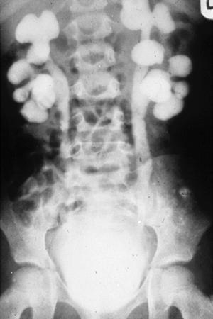
31 hydronephrosis kidney
32 Report What type of C.M used for adult and pediatric in KKUH? What the different between them? Why there is different?
33 Case study: -Pt history. -Symptoms and signs. -Pt preperation. -Procedure + technique. -Images taken. -Additional images and why. -Show anatomy and pathology in images -Talk briefly about pts disease.
IVU ((INTRAVENOUSUROGRAM))
)") IVU ((INTRAVENOUSUROGRAM)) Anatomy The urinary system consists of the following : 2 kidneys, 2 ureters,1 bladder, 1 urethra Renal pelvis Minor calyx Major calyx Proximal ureter Pelvi-uretric junction
IVU ((INTRAVENOUSUROGRAM)) Anatomy The urinary system consists of the following : 2 kidneys, 2 ureters,1 bladder, 1 urethra Renal pelvis Minor calyx Major calyx Proximal ureter Pelvi-uretric junction
Radiographic Procedures III (RAD 228)
") Radiographic Procedures III (RAD 228) Urinary System RADIOGRAPHIC EXAMINATIONS Urinary System Antegrade Exam IVU Functional test Hypertensive evaluation as per protocol Retrograde Exams Retrograde Urography
Radiographic Procedures III (RAD 228) Urinary System RADIOGRAPHIC EXAMINATIONS Urinary System Antegrade Exam IVU Functional test Hypertensive evaluation as per protocol Retrograde Exams Retrograde Urography
PROFESSIONAL SKILLS 1 3RD YEAR SEMESTER 6 RADIOGRAPHY. THE URINARY SYSTEM Uz. Fatema shmus aldeen Tel
 PROFESSIONAL SKILLS 1 3RD YEAR SEMESTER 6 RADIOGRAPHY THE URINARY SYSTEM Uz. Fatema shmus aldeen Tel. 0925111552 Professional skills-2 THE URINARY SYSTEM The urinary system (review anatomy and physiology)
R adio logical investigations of urinary system
 R adio logical investigations of urinary system There are 4 main radiological Ix: 1 IVU: Intravenous urography. 2- U/S 3-CT scan 4-Radioisotope scan. Others (not frequently used): MRI, arteriography, antegrade
Excretory urography (EU) or IVP US CT & radionuclide imaging
 or IVP US CT & radionuclide imaging") Excretory urography (EU) or IVP US CT & radionuclide imaging MRI arteriography studies requiring catherization or direct puncture of collecting system EU & to a lesser extent CT provide both functional
Excretory urography (EU) or IVP US CT & radionuclide imaging MRI arteriography studies requiring catherization or direct puncture of collecting system EU & to a lesser extent CT provide both functional
Uroradiology For Medical Students
 Uroradiology For Medical Students Lesson 4: Cystography & Urethrography - Part 2 American Urological Association Review Cystography is useful in evaluating the bladder, the urethra and the competence of
Uroradiology Tutorial For Medical Students
 Uroradiology Tutorial For Medical Students Lesson 3: Cystography & Urethrography Part 1 American Urological Association Introduction Conventional radiography of the urinary tract includes several diagnostic
Hydronephrosis. Nephrosis. Refers to the kidney
 What is hydronephrosis? Hydro Nephrosis Refers to water or fluid Refers to the kidney A build-up of fluid (urine) in the kidney is the medical term for a build-up of urine in the kidney. As the urine builds
Paediatric Fluoroscopy
 Paediatric Fluoroscopy It s a Small World Robyn Crapp Nurse Unit Manager Medical Imaging CHW The most requested studies in our paediatric fluoroscopy are: MCU Contrast Meal MCU MCU s are requested when
URINARY SYSTEM I. Kidneys II. Nephron Unit and Urine Formation
 URINARY SYSTEM I. Kidneys A. Location and Structure 1. Retroperitoneal 2. Between T12 and L3 3. Rt. kidney slightly lower 4. Two bean shaped organs 5. Adrenal gland 6. Internal construction a. Renal cortex
Lecture 56 Kidney and Urinary System
 Lecture 56 Kidney and Urinary System The adrenal glands are located on the superomedial aspect of the kidney The right diagram shows a picture of the kidney with the abdominal walls and organs removed
Contents. Review anatomy of the urinary tract Imaging modalities
 Contents Review anatomy of the urinary tract Imaging modalities The Urinary Tract Kidneys ตาแหน งไต (position) อย ใน retroperitoneum ระด บ T12-L3 โดยไต ขวาจะม ระด บตากว าไตซ ายเล กน อย ร ปร าง (shape)
Intravenous Pyelogram (IVP)
") Scan for mobile link. Intravenous Pyelogram (IVP) Intravenous pyelogram (IVP) is an x-ray exam that uses an injection of contrast material to evaluate your kidneys, ureters and bladder and help diagnose
Scan for mobile link. Intravenous Pyelogram (IVP) Intravenous pyelogram (IVP) is an x-ray exam that uses an injection of contrast material to evaluate your kidneys, ureters and bladder and help diagnose
Acute renal colic Radiological investigation in patients with renal colic
 Acute renal colic Radiological investigation in patients with renal colic Mikael Hellström Professor Department of Radiology Sahlgrenska University Hospital Göteborg University 0.9-1.8/1.000 inhabitants
Children's (Pediatric) Voiding Cystourethrogram
 Voiding Cystourethrogram") Scan for mobile link. Children's (Pediatric) Voiding Cystourethrogram A children s (pediatric) voiding cystourethrogram uses fluoroscopy a form of real-time x-ray to examine a child s bladder and lower
Scan for mobile link. Children's (Pediatric) Voiding Cystourethrogram A children s (pediatric) voiding cystourethrogram uses fluoroscopy a form of real-time x-ray to examine a child s bladder and lower
Continuous Bladder Irrigation
 Continuous Bladder Irrigation Introduction Continuous bladder irrigation, or CBI, is the infusion of a sterile solution into the urinary bladder. The purpose of CBI is to prevent the formation of blood
W/ (2) (3) (4) (5) (5) (6) (6) CTA
 (3) (4) (5) (5) (6) (6) CTA") Index Abdomen W/ and W/Out (2) Abdomen Pelvis W/Out (3) Abdomen Pelvis W/ (4) Pelvis W/ (5) Chest W/Out (5) Chest/Abdomen/Pelvis W/ (6) Chest W/ (6) CTA ( 7-8) Neuro (8-9) Musculoskeletal (10) Trauma (11)
Index Abdomen W/ and W/Out (2) Abdomen Pelvis W/Out (3) Abdomen Pelvis W/ (4) Pelvis W/ (5) Chest W/Out (5) Chest/Abdomen/Pelvis W/ (6) Chest W/ (6) CTA ( 7-8) Neuro (8-9) Musculoskeletal (10) Trauma (11)
Barium Enema RD Sheet
 SIMS POSITION Instruct patient to turn onto the left side, lean forward and about 35 to 40 degree, and knees are flexed right knee on the table, above and in front of the slightly flexed left knee This
Abdominal Ultrasound : Aorta, Kidneys, Bladder
 Abdominal Ultrasound : Aorta, Kidneys, Bladder Nilam J. Soni, MD, MSc Associate Professor of Medicine Divisions of Hospital Medicine and Pulmonary/Critical Care Medicine Department of Medicine University
Pediatric Ure-Radiology*
 Pediatric Ure-Radiology* HERMAN GROSSMAN, M.D. Professor of Radiology and Pediatrics, Duke University Medical Center, Durham, North Carolina "Routine" radiologic studies do not, often enough, concentrate
Obstetrics Content Outline Obstetrics - Fetal Abnormalities
 Obstetrics Content Outline Obstetrics - Fetal Abnormalities Effective February 2007 10 16% renal agenesis complete absence of the kidneys occurs when ureteric buds fail to develop Or degenerate before
Bio 322 Human Anatomy Objectives for the laboratory exercise Urinary System Filtration Reabsorption Secretion Concentration
 Bio 322 Human Anatomy Objectives for the laboratory exercise Urinary System Required reading before beginning this lab: Saladin, KS: Human Anatomy 5 th ed (2017) Chapter 25 For this lab you will use parts
Outline. Introduction to imaging modalities of the urinary system. Case base learning of common diseases in urinary tract
 Outline Introduction to imaging modalities of the urinary system Case base learning of common diseases in urinary tract Outline Introduction to imaging modalities of the urinary system Case base learning
Outline. Introduction to imaging modalities of the urinary system. Case base learning of common diseases in urinary tract
 Outline Introduction to imaging modalities of the urinary system Case base learning of common diseases in urinary tract Diagnostic Investigations in Urinary System PLAIN KUB EXCRETORY UROGRAPHY RETROGRADE
URINARY SYSTEM. Lecturer Dr.Firdous M.Jaafar Department of anatomy/histology section Lecture 3
 URINARY SYSTEM Lecturer Dr.Firdous M.Jaafar Department of anatomy/histology section Lecture 3 Objectives 1- Describe the structure of the urinary bladder, 2- Describe the structure of the ureters, bladder,
Gross Anatomy of the Urinary System
 Gross Anatomy of the Urinary System Lecture Objectives Overview of the urinary system. Describe the external and internal anatomical structure of the kidney. Describe the anatomical structure of the ureter
ASSESSING THE PLAIN ABDOMINAL RADIOGRAPH M A A M E F O S U A A M P O F O
 ASSESSING THE PLAIN ABDOMINAL RADIOGRAPH M A A M E F O S U A A M P O F O Introduction The abdomen (less formally called the belly, stomach, is that part of the body between the thorax (chest) and pelvis,
Case MDCT 3D reconstructed features of posterior urethral valve
 Case 12688 MDCT 3D reconstructed features of posterior urethral valve Hidayatullah Hamidi Third year Resident of Radiology French medical institute for children Radiology Department; Kabul, Afghanistan;
X-ray (Radiography) - Lower GI Tract
 - Lower GI Tract") Scan for mobile link. X-ray (Radiography) - Lower GI Tract Lower gastrointestinal tract radiography or lower GI uses a form of real-time x-ray called fluoroscopy and a barium-based contrast material to
Scan for mobile link. X-ray (Radiography) - Lower GI Tract Lower gastrointestinal tract radiography or lower GI uses a form of real-time x-ray called fluoroscopy and a barium-based contrast material to
Contrast Materials Patient Safety: What are contrast materials and how do they work?
 Contrast Materials Patient Safety: What are contrast materials and how do they work? Which imaging exams use contrast materials? How safe are contrast materials? How should I prepare for my imaging procedure
Radiographer Performed Paediatric Micturating Cystograms
 Radiographer Performed Paediatric Micturating Cystograms A Solution to a Radiology Service Provision Problem Rosalind Waugh PgC. DCR.R Clinical Lead Fluoroscopy / Advanced Practitioner James Cook University
RENAL SCINTIGRAPHY IN THE 21 st CENTURY
 RENAL SCINTIGRAPHY IN THE 21 st CENTURY 99m Tc- MAG 3 with zero time injection of Furosemide (MAG 3 -F 0 ) : A Fast and Easy Protocol, One for All Indications Clinical Experience Congenital Disorders PROTOCOL
Prenatal Hydronephrosis
 Prenatal Hydronephrosis What is hydronephrosis? Hydronephrosis is dilation of the kidney, specifically the renal pelvis (place where urine is stored after its production). This can be the result of an
Hydronephrosis. What is hydronephrosis?
 What is hydronephrosis? Hydronephrosis Hydronephrosis describes the situation where the urine collecting system of the kidney is dilated. This may be a normal variant or it may be due to an underlying
Urinary. Smooth, collapsible, muscular sac stores urine. Figure Slide 15.21a
 Urinary Smooth, collapsible, muscular sac stores urine Figure 15.6 Slide 15.21a Urinary Bladder Wall Walls are and folded in an empty bladder Bladder can significantly without increasing internal pressure
Congenital Pediatric Anomalies: A Collection of Abdominal Scintigraphy Findings: An Imaging Atlas
 ISPUB.COM The Internet Journal of Nuclear Medicine Volume 5 Number 1 Congenital Pediatric Anomalies: A Collection of Abdominal Scintigraphy Findings: An Imaging Atlas V Vijayakumar, T Nishino Citation
RADIOLOGY REQUEST MANUAL. (615)
") RADIOLOGY REQUEST MANUAL www.vanderbiltchildrens.com RADIOLOGY REQUEST MANUAL EXAM PROTOCOL QUESTIONS? Please call: DIAGNOSTIC RADIOLOGY (X-RAY) Pager (615) 835-1714 CT (615) 936-4920 MRI (615) 936-4933
RADIOLOGY REQUEST MANUAL www.vanderbiltchildrens.com RADIOLOGY REQUEST MANUAL EXAM PROTOCOL QUESTIONS? Please call: DIAGNOSTIC RADIOLOGY (X-RAY) Pager (615) 835-1714 CT (615) 936-4920 MRI (615) 936-4933
DEPARTMENT OF IMAGING SERVICES TESTS AND PREPARATION
 DEPARTMENT OF IMAGING SERVICES S AND PREPARATION LUNG BIOPSY COMPUTERIZED TOMOGRAPHY (CT SCAN) You may have clear liquids after midnight. Do not eat any food. It is very important that you let the doctor
General Anatomy of Urinary System
 General Anatomy of Urinary System URINARY SYSTEM ORGANS Kidneys (2) Ureters (2) Urinary bladder Urethra KIDNEY FUNCTIONS Control blood volume and composition KIDNEY FUNCTIONS Filter blood plasma, eliminate
Abdomen and Pelvis CT (1) By the end of the lecture students should be able to:
 By the end of the lecture students should be able to:") RAD 451 Abdomen and Pelvis CT (1) By the end of the lecture students should be able to: State the common indications for Abdomen and pelvis CT exams Identify possible contra indications for Abdomen and
RAD 451 Abdomen and Pelvis CT (1) By the end of the lecture students should be able to: State the common indications for Abdomen and pelvis CT exams Identify possible contra indications for Abdomen and
Lec-8 جراحة بولية د.نعمان
 4th stage Lec-8 جراحة بولية د.نعمان 11/10/2015 بسم هللا الرحمن الرحيم Ureteric, Vesical, & urethral stones Ureteric Calculus Epidemiology like renal stones Etiology like renal stones Risk factors like
Obstructive Uropathy. PATHOPHYSIOLOGIC CHANGES UUO vs BUO. Arry Rodjani Urology Department Ciptomangunkusumo Hospital Jakarta
 Obstructive Uropathy PATHOPHYSIOLOGIC CHANGES UUO vs BUO Arry Rodjani Urology Department Ciptomangunkusumo Hospital Jakarta INTRODUCTION Obstructive uropathy refers to the functional or anatomic obstruction
Fluoroscopy Protocols. Upper GI with Barium Swallow-Combination UGIBS
 Fluoroscopy Protocols Upper GI with Barium Swallow-Combination UGIBS Fluoro Time Target Limit: 4.0 minutes Scheduling and Prep: Supplies: *The patient should be NPO from midnight before their exam Until
Chapter 6: Genitourinary and Gastrointestinal Systems 93
 Chapter 6: Genitourinary and Gastrointestinal Systems 93 Chapter 6 Genitourinary and Gastrointestinal Systems Embryology Three sets of excretory organs or kidneys develop in human embryos: Pronephros:
DISCHARGE DIAGNOSES: End stage renal disease secondary to rapidly progressive glomerulonephritis.
 DISCHARGE SUMMARY DISCHARGE DIAGNOSES: End stage renal disease secondary to rapidly progressive glomerulonephritis. OPERATIONS/PROCEDURES: Living related renal transplantation. HISTORY: For full details
Ureters, Urinary Bladder & Urethra
 Ureters, Urinary Bladder & Urethra Please check our Editing File هذا العمل ال يغني عن المصدر األساسي للمذاكرة Lecture 2 } و م ن ي ت و ك ع ل ا لل ه ف ه و ح س ب ه { Objectives o Describe the course of ureter
Objectives: To analyze various factors predicting success of retrograde ureteric stenting in managing patients with ureteric obstruction.
 ISPUB.COM The Internet Journal of Urology Volume 14 Number 1 Factors Predicting Success Rate Of Retrograde Ureteric Stenting In Managing Patients With Ureteric Obstruction- Our Experiences In A South Indian
US in non-traumatic acute abdomen. Lalita, M.D. Radiologist Department of radiology Faculty of Medicine ChiangMai university
 US in non-traumatic acute abdomen Lalita, M.D. Radiologist Department of radiology Faculty of Medicine ChiangMai university Sagittal Orientation Transverse (Axial) Orientation Coronal Orientation Intercostal
Urinary System VASTACCESS, INC.
 Urinary System www.vastaccess.com 2 Urinary Tract Kidney Ureter Urinary Bladder Urethra Prostate (male) Membranous (male) Spongy (male) 3 Kidney Relations Suprarenal (Adrenal) Glands Liver Duodenum Transverse
LESSON ASSIGNMENT. Positioning for Exams of the Spine. After completing this lesson, you should be able to identify:
 LESSON ASSIGNMENT LESSON 4 Positioning for Exams of the Spine. LESSON ASSIGNMENT Paragraphs 4-1 through 4-15. LESSON OBJECTIVES After completing this lesson, you should be able to identify: 4-1. Identify
Abdominal ultrasound:
 Abdominal ultrasound: Non-traumatic acute abdomen Wittanee Na-ChiangMai, MD Department of Radiology ChiangMai University 26/04/2017 Contents Technique of examination Normal anatomy Emergency conditions
Radiographic Procedures III (RAD 228)
") Radiographic Procedures III (RAD 228) Barium Enema Dual Contrast Barium Enema or BE (Lower GI Series, Colon) Purpose: Radiographic examination of the large intestine Double-contrast study using air and
Radiographic Procedures III (RAD 228) Barium Enema Dual Contrast Barium Enema or BE (Lower GI Series, Colon) Purpose: Radiographic examination of the large intestine Double-contrast study using air and
Chapter 17: Urinary System
 Introduction Chapter 17: Urinary System Organs of the Urinary System REFERENCE FIGURE 17.1 2 kidneys filters the blood 2 ureters transport urine from the kidneys to the urinary bladder Urinary bladder
Day 1 Bell Work We will be discussing one of FIVE excretory organs in the human body. We have already studied four of them. The kidneys are considered
 URINARY SYSTEM 1 Day 1 Bell Work We will be discussing one of FIVE excretory organs in the human body. We have already studied four of them. The kidneys are considered the main organ in the excretory system.
Focused Assessment Sonography of Trauma (FAST) Scanning Protocol
 Scanning Protocol") Focused Assessment Sonography of Trauma (FAST) Scanning Protocol Romolo Gaspari CHAPTER 3 GOAL OF THE FAST EXAM Demonstrate free fluid in abdomen, pleural space, or pericardial space. EMERGENCY ULTRASOUND
Focused Assessment Sonography of Trauma (FAST) Scanning Protocol Romolo Gaspari CHAPTER 3 GOAL OF THE FAST EXAM Demonstrate free fluid in abdomen, pleural space, or pericardial space. EMERGENCY ULTRASOUND
Abdomen and Retroperitoneum Ultrasound Protocols
 Abdomen and Retroperitoneum Ultrasound Protocols Reviewed By: Anna Ellermeier, MD Last Reviewed: March 2018 Contact: (866) 761-4200, Option 1 **NOTE for all examinations: 1. If documenting possible flow
CYSTIC DISEASES of THE KIDNEY. Dr. Nisreen Abu Shahin
 CYSTIC DISEASES of THE KIDNEY Dr. Nisreen Abu Shahin 1 Types of cysts 1-Simple Cysts 2-Dialysis-associated acquired cysts 3-Autosomal Dominant (Adult) Polycystic Kidney Disease 4-Autosomal Recessive (Childhood)
Nephrology - the study of the kidney. Urology - branch of medicine dealing with the male and female urinary systems and the male reproductive system
 Urinary System Nephrology - the study of the kidney Urology - branch of medicine dealing with the male and female urinary systems and the male reproductive system Functions of the Urinary System 1. Regulation
Mosby s PATHOLOGY for Massage Therapists. Lesson 12.1 Objective. Chapter 12 Urinary Pathologies. Urinary System (cont. Urinary System. (cont d.
 Mosby s PATHOLOGY for Massage Therapists Lesson 12.1 Objective Discuss anatomic structures and physiologic processes related to the urinary system. Chapter 12 Urinary Pathologies 2 Urinary System Urinary
Urinary 1 Checklist Gross Anatomy of the Urinary System
 Urinary 1 Checklist Gross Anatomy of the Urinary System Urinary system Kidneys Parietal peritoneum Retroperitoneal Renal fascia The urinary system consists of two kidneys, two ureters, the urinary bladder,
Urinary System. consists of the kidneys, ureters, urinary bladder and urethra
 Urinary System 1 Urinary System consists of the kidneys, ureters, urinary bladder and urethra 2 Location of Kidneys The kidneys which are positioned retroperitoneally lie on either side of the vertebral
Abdominal Ultrasonography
 Abdominal Ultrasonography David A. Masneri, DO, FACEP, FAAEM Assistant Professor of Emergency Medicine Assistant Director, Emergency Medicine Residency Medical Director, Operational Medicine Division Center
Therapeutic Enema for Intussusception
 Scan for mobile link. Therapeutic Enema for Intussusception Therapeutic enema is used to help identify and diagnose intussusception, a serious disorder in which one part of the intestine slides into another
URINARY TRACT IMAGING - BASIC PRINCIPLES
 URINARY TRACT IMAGING - BASIC PRINCIPLES Clinical Radiology Every physician needs a basic understanding of diagnostic imaging to understand how to order the appropriate studies and to understand the resulting
Nuclear medicine in renal scarring
 Paediatric NM General principles Need to image in a child friendly department Maybe use of play therapist to help explain the procedure Reduce activities as weight a proportion of 70kg adult with 10% minimum
ASDIN 10th Annual Scientific Meeting Final. COI Disclosure Statement. Intravenous Contrast Media: Basics
 COI Disclosure Statement There are no financial relationships or conflicts of interest to disclose with this presentation Melissa Hicks, BA, RT(R)(VI) Vascular Interventional Technologist University of
Patient and Family Education. Bladder Exstrophy. What is bladder exstrophy? How common is bladder exstrophy? What causes bladder exstrophy?
 Patient and Family Education Bladder Exstrophy What is bladder exstrophy? Bladder exstrophy (x-tro-fee) is a bladder that is not formed right. The bladder and genitals are split in half, turned inside
Upper Gastrointestinal (GI) Tract X-ray (Radiography)
 Tract X-ray (Radiography)") Upper Gastrointestinal (GI) Tract X-ray (Radiography) What is Upper Gastrointestinal (GI) Tract Radiography? What are some common uses of the procedure? How should I prepare? What does the equipment look
Upper Gastrointestinal (GI) Tract X-ray (Radiography) What is Upper Gastrointestinal (GI) Tract Radiography? What are some common uses of the procedure? How should I prepare? What does the equipment look
MAKING THE BEST USE OF CLINICAL RADIOLOGY SERVICES. Dr Martina Paetzel Consultant Radiologist
 MAKING THE BEST USE OF CLINICAL RADIOLOGY SERVICES Dr Martina Paetzel Consultant Radiologist LEARNING OBJECTIVES To be aware of guidelines regulating ionising radiation & radiation dose To introduce how
Recommendations. Management of Renal Calculi PCNL. Complications of PCNL: How to avoid and manage them 2/8/2008
 Complications of PCNL: How to avoid and manage them Recommendations An exhaustive meta-analysis completed by the panel revealed a lower complication rate and higher stone free rate when PCNL was used as
Bladder Management. A guide for patients. Key points
 Bladder Management A guide for patients Key points Urinary issues remain one of the highest causes of readmission to hospital following Spinal Cord Injury (SCI). Following SCI most patients experience
FHS Appendicitis US Protocol
 FHS Appendicitis US Protocol Reviewed By: Shireen Khan, MD; Sarah Farley, MD; Anna Ellermeier, MD Last Reviewed: May 2018 Contact: (866) 761-4200 **NOTE for all examinations: 1. If documenting possible
JEFFERSON COLLEGE. Radiographic Positioning II
 JEFFERSON COLLEGE COURSE SYLLABUS RAD125 Radiographic Positioning II 3 Credit Hours Revised by: Janet E. Akers BS RT (R)(M) Date: September 25, 2013 Kenny Wilson, Director, Health Occupation Programs Dena
My Patient Has Abdominal Pain PoCUS of the Biliary Tract and the Urinary Tract
 My Patient Has Abdominal Pain PoCUS of the Biliary Tract and the Urinary Tract Objectives PoCUS for Biliary Disease PoCUS for Renal Colic PoCUS for Urinary Retention Biliary Disease A patient presents
Information for Patients
 Having a CT Intravenous Urogram (CT IVU) Information for Patients In this leaflet: Introduction 2 What is a CT IVU?...2 How does it work?. 2 Are there any risks?.3 What do I need to do to prepare for my
IMAGING OF THE UROGENITAL TRACT
 IMAGING OF THE UROGENITAL TRACT 1 A) URINARY TRACT There are many methods of imaging the urinary tract but plain abdominal X-ray and ultrasound scan are usually done first in most cases, especially in
Kidney & Urinary Tract Ultrasound. Fatina Fadel Hafez Bazaraa
 Kidney & Urinary Tract Ultrasound Fatina Fadel Hafez Bazaraa Ultrasonography Ultrasound Available Rapid Inexpensive Painless & no sedation needed No adverse effects/ complications Can be repeated Useful
Radiology of the abdomen Lecture -1-
 Radiology of the abdomen Lecture -1- Objectives To know radiology modalities used in abdomen imaging mainly GI tract. To know advantages and disadvantages of each modality. To know indications and contraindications
Children's (Pediatric) Contrast-enhanced Voiding Urosonography
 Contrast-enhanced Voiding Urosonography") Scan for mobile link. Children's (Pediatric) Contrast-enhanced Voiding Urosonography Pediatric contrast-enhanced voiding urosonography uses ultrasound to examine a child's bladder and urinary tract. It
Scan for mobile link. Children's (Pediatric) Contrast-enhanced Voiding Urosonography Pediatric contrast-enhanced voiding urosonography uses ultrasound to examine a child's bladder and urinary tract. It
8/14/2017. Kidney location & visualization. Brief Review with tips & Case Based Illustrations. Size = x L2. Size =
 Dr. Russell Tucker, DACVR Brief Review with tips & Case Based Illustrations Kidney location & visualization K9 Kidneys: Rt @ T13-L1 Lt @ L2-L4 Kidney visualization K9 Kidneys: Rt @ T13-L1 Lt @ L2-L4 Size
Find Medical Solutions to Your Problems HYDRONEPHROSIS. (Distension of Renal Calyces & Pelvis)
") HYDRONEPHROSIS (Distension of Renal Calyces & Pelvis) Hydronephrosis is the distension of the renal calyces and pelvis due to accumulation of the urine as a result of the obstruction to the outflow of
HYDRONEPHROSIS (Distension of Renal Calyces & Pelvis) Hydronephrosis is the distension of the renal calyces and pelvis due to accumulation of the urine as a result of the obstruction to the outflow of
Functions of the Urinary System
 The Urinary System Functions of the Urinary System Elimination of waste products Nitrogenous wastes Toxins Drugs Regulate aspects of homeostasis Water balance Electrolytes Acid-base balance in the blood
Imaging spectrum of genitourinary tuberculosis: Our experience at a tertiary care centre of a third world country
 Imaging spectrum of genitourinary tuberculosis: Our experience at a tertiary care centre of a third world country Poster No.: C-361 Congress: ECR 2009 Type: Educational Exhibit Topic: Genitourinary Authors:
Development of the Urinary System
 Development of the Urinary System Lecture Objectives Understand the development of the kidney and related organs of the urinary system. Define the pronephrons, mesonephrons and metanephrons. Understand
BIOL2030 Human A & P II -- Exam 6
 BIOL2030 Human A & P II -- Exam 6 Name: 1. The kidney functions in A. preventing blood loss. C. synthesis of vitamin E. E. making ADH. B. white blood cell production. D. excretion of metabolic wastes.
Kaiser Oakland Urology
 Kaiser Oakland Urology What is Laparoscopy? Minimally invasive surgical alternative to standard surgery How is Laparoscopy Performed? A laparoscope and video camera are used to visualize internal organs
Controversies around antenatally detected PUJ syndrom. Amy Piepsz, CHU St Pierre, Brussels, Belgium
 Controversies around antenatally detected PUJ syndrom Amy Piepsz, CHU St Pierre, Brussels, Belgium Editors : Anthony Caldamone, USA Pierre Mouriquand, France Newborn boy History of prenatally diagnosed
Request Card Task ANSWERS
 Request Card Task ANSWERS Medical Student Workbook Author: Dr Sam Leach, SpR Case 1 What differential diagnoses are most likely? Which investigation is most appropriate? Case 1 The most likely diagnosis
URINARY SYSTEM. These organs lie posterior or inferior to the. (membrane).
.") URINARY SYSTEM I. INTRODUCTION Each kidney is made up of about a million tiny tubules called nephrons. Each nephron individually filters the blood and makes urine and it does the job completely, from start
URINARY SYSTEM I. INTRODUCTION Each kidney is made up of about a million tiny tubules called nephrons. Each nephron individually filters the blood and makes urine and it does the job completely, from start
Penis and Prostate. Holly White Jennifer Zang September 7, Penis and Prostate. 1) Other Names None
 Other Names None") Penis and Prostate Penis and Prostate Holly White Jennifer Zang September 7, 2006 1) Other Names None 2) Definition/ Location The prostate is a doughnut-like gland that lies inferior to the urinary bladder
Penis and Prostate Penis and Prostate Holly White Jennifer Zang September 7, 2006 1) Other Names None 2) Definition/ Location The prostate is a doughnut-like gland that lies inferior to the urinary bladder
Inserting an antegrade ureteric stent. Brought to you in association with EIDO Healthcare and endorsed by the Royal College of Surgeons England.
 Inserting an antegrade ureteric stent Brought to you in association with EIDO Healthcare and endorsed by the Royal College of Surgeons England. Discovery has made every effort to ensure that we obtained
PAEDIATRIC RENAL IMAGING. Dr A Brink
 PAEDIATRIC RENAL IMAGING Dr A Brink Causes of hydronephrosis includes: Pelvi-ureteric obstruction Vesico-ureteric reflux Vesico-ureteric obstruction Posterior uretral valves Duplex kidneys Radiopharmaceutical
An overview of Extracorporeal shock wave lithotripsy (ESWL) and the role of Radiographers in ESWL. Tse Ka Wai, Sam (Rad II, TMH)
 and the role of Radiographers in ESWL. Tse Ka Wai, Sam (Rad II, TMH)") An overview of Extracorporeal shock wave lithotripsy (ESWL) and the role of Radiographers in ESWL Tse Ka Wai, Sam (Rad II, TMH) What is ESWL? ESWL Machine Body Stone Renal Stone Incidence rate in HK population
An overview of Extracorporeal shock wave lithotripsy (ESWL) and the role of Radiographers in ESWL Tse Ka Wai, Sam (Rad II, TMH) What is ESWL? ESWL Machine Body Stone Renal Stone Incidence rate in HK population
Perineal Sonography in Diagnosis of an Ectopic Ureteric Opening Into the Urethra
 Case Series Perineal Sonography in Diagnosis of an Ectopic Ureteric Opening Into the Urethra S. Boopathy Vijayaraghavan, MD, DMRD Objective. To study the role of perineal sonography in the diagnosis of
Urinary System. Chapter 17 7/19/11. Introduction
 7/19/11 Chapter 17 Urinary System Introduction A. The urinary system consists of two kidneys that filter the blood, two ureters, a urinary bladder, and a urethra to convey waste substances to the outside.
Genitourinary Trauma Introduction GU Trauma overlooked
 Genitourinary Trauma Introduction GU Trauma overlooked 10-20% of all injured patients Long term morbidity Impotence Incontinence Life-threatening injuries first Urethral Injury Plan Bladder Injury Kidney
Getting Ready. My Child Is Having A VCUG
 Getting Ready My Child Is Having A VCUG Welcome to Inova Fairfax Hospital for Children. Your child is going to have a voiding cystourethrogram,a diagnostic test commonly referred to as a VCUG. It is very
Intrarenal reflux and the scarred kidney
 Archives of Disease in Childhood, 1974, 49, 531. Intrarenal reflux and the scarred kidney G. L. ROLLESTON, T. M. J. MALING, and C. J. HODSON* From the Department of Radiology, Christchurch Hospital and
A. Incorrect! The urinary system is involved in the regulation of blood ph. B. Correct! The urinary system is involved in the synthesis of vitamin D.
 Human Anatomy - Problem Drill 22: The Urinary System Question No. 1 of 10 1. Which of the following statements about the functions of the urinary system is not correct? Question #01 (A) The urinary system
Human Anatomy Unit 3 URINARY SYSTEM
 Human Anatomy Unit 3 URINARY SYSTEM In Anatomy Today Components Kidneys Ureters Urinary bladder Urethra Functions Storage of urine Bladder stores up to 1 L of urine Excretion of urine Transport of urine