Department of General Surgery
|
|
|
- Randolph Cannon
- 5 years ago
- Views:
Transcription
 Under the Guidance of Dr. V. BALAKRISHNAN M.S. Department of General Surgery THANJAVUR MEDICAL COLLEGE, THANJAVUR.")
1 STUDY OF CLINICAL AND FUNCTIONAL RESULTS OF ABDOMINAL MESH RECTOPEXY FOR COMPLETE RECTAL PROLAPSE By Dr. JINREEVE S.W. DANIEL Dissertation Submitted to The Tamilnadu DR.MGR Medical university, Chennai. In partial fulfillment of the requirements for the degree of MASTER OF SURGERY (GENERAL) Under the Guidance of Dr. V. BALAKRISHNAN M.S. Department of General Surgery THANJAVUR MEDICAL COLLEGE, THANJAVUR. 2014
2 DECLARATION BY THE CANDIDATE I hereby declare that this dissertation entitled STUDY OF CLINICAL AND FUNCTIONAL RESULTS OF ABDOMINAL MESH RECTOPEXY FOR COMPLETE RECTAL PROLAPSE is a bonafide and genuine research work carried out by me under the guidance of Dr. V. BALAKRISHNAN M.S., Professor and head, Department of General Surgery, THANJAVUR MEDICAL COLLEGE, TAMILNADU. Date: Place: THANJAVUR Dr. JINREEVE S.W. DANIEL
3 CERTIFICATE BY THE GUIDE This is to certify that the dissertation entitled STUDY OF CLINICAL AND FUNCTIONAL RESULTS OF ABDOMINAL MESH RECTOPEXY FOR COMPLETE RECTAL PROLAPSE is a bonafide research work done by Dr. JINREEVE S.W. DANIEL in partial fulfillment of the requirement for the degree of MASTER OF GENERAL SURGERY. DR.V.BALAKRISHNAN M.S PROFESOR AND HEAD DEPARTMENT OF GENERAL SURGERY, THANJAVUR MEDICAL COLLEGE,THANJAVUR Date: Place:
4 ENDORSEMENT BY THE HOD, PRINCIPAL/HEAD OF THE INSTITUTION This is to certify that the dissertation entitled STUDY OF CLINICAL AND FUNCTIONAL RESULTS OF ABDOMINAL MESH RECTOPEXY FOR COMPLETE RECTAL PROLAPSE is a bonafide research work done by Dr. JINREEVE S.W. DANIEL under the guidance of DR.V.BALAKRISHNAN M.S,PROFESSOR AND HEAD DEPARTMENT OF GENERAL SURGERY, THANJAVUR MEDICAL COLLEGE,THANJAVUR. DR.V.BALAKRISHNAN M.S PROFESSOR AND HEAD DEPARTMENT OF GENERAL SURGERY, THANJAVUR MEDICAL COLLEGE,THANJAVUR. DEAN THANJAVUR MEDICAL COLLEGE THANJAVUR. Date: Place: Thanjavur Date: Place: Thanjavur
5 COPY RIGHT DECLARATION BY THE CANDIDATE I hereby declare that The Tamilnadu DR.MGR Medical University, Chennai. Shall have the rights to preserve, use and disseminate this dissertation/thesis in print or electronic format for academic/research purpose. Date: Place:Thanjavur Dr. JINREEVE S.W. DANIEL
6 ACKNOWLEDGEMENT It gives me immense pleasure to express my deep sense of gratitude and sincere thanks to my beloved teacher and guide DR.V.BALAKRISHNAN M.S,PROFESSOR AND HEAD DEPARTMENT OF GENERAL SURGERY, THANJAVUR MEDICAL COLLEGE,THANJAVUR for the kind guidance, suggestions, advice and constant encouragement during the course of my study and preparation of this dissertation. I am extremely grateful and highly indebted to Dr. A. MICHAEL M.S. MCh, Asst Professor, Department of General Surgery, for the valuable suggestions, advice and constant encouragement during the period of my study. My sincere thanks to my respected Professors DR.M.ELANGOVAN, DR.YEGANATHAN, DR.SHANTHINI, DR.THOMASKARUNAKARAN and DR.RAJENDERAN for allowing to collect cases from their units and for their valuable guidance. I express my sincere thanks to my respected teachers Dr.S.JAGATHESAN, DR.V.MARIMUTHU, DR.A.MUTHUVINAYAGAM and DR.K.ANBARASAN,for the strength & support they provided during the period of study. My special thanks to Prof.Dr.ARAVINDHAN M.S, MCh, head of department, SURGICAL GASTROENTEROLOGY, for his guidance, suggestion and advice during the course and preparation of this dissertation.
7 I also express my thanks to Prof.DR.K.MAHADEVAN M.S, DEAN THANJAVUR MEDICAL COLLEGE,THANJAVUR. I own my deep sense of gratitude to my parents, Shri M. DANIEL RAJ and Smt. SAROJINI DANIEL my wife Dr.NAGANIKKA and my son Master ABISHEK, for their support and constant encouragement extended to me during my entire post graduate career. Above all I express my deep gratitude towards THE ALMIGHTY for all the blessings. Lastly I thank everyone concerned, my colleagues, including the patients for their co-operation, without which this dissertation would have never materialized. Place:THANJAVUR Date: Dr. JINREEVE S.W. DANIEL.
8 LIST OF ABBREVIATION BP mmhg DC ECG ESR FBS Blood Pressure millimeter of Mercury Differential Count Electrocardiography Erythrocyte Sedimentation Rate Fasting Blood Sugar Hb% Haemoglobin % HBsAg HIV TC Hepatitis B Surface Antigen Human Immunodeficiency Virus Total Count
9 S.NO CONTENTS PAGE NO 1 INTRODUCTION 1 2 OBJECTIVES 4 3 REVIEW OF LITERATURE 5 HISTORICAL EVALUATION OF TREATMENT 5 SURGICAL ANATOMY OF ANAL CANAL 10 AND RECTUM RECTAL PROLAPSE 35 CLASSIFICATION OF RECTAL PROLAPSE 47 CLINICAL FEATURES 54 TESTING AND EVALUVATION 58 DIFFERENTIAL DIAGNOSIS 60 MANAGEMENT 61 4 METHODOLOGY 64 5 OBSERVATION AND RESULTS 75 6 DISCUSSION 92 7 CONCLUSION SUMMARY BIBLIOGRAPHY ANNEXURE 105 PROFORMA 105 CONSENT FORM 111 MASTER CHART 112 KEY TO MASTER CHART 116
10
11 Digital Receipt This receipt acknowledges that Turnitin received your paper. Below you will find the receipt information regarding your submission. The first page of your submissions is displayed below. Submission author: Assignment title: Submission title: File name: File size: Page count: Word count: Character count: Submission date: Submission ID: ms General Surgery JIN TNMGRMU EXAMINATIONS STUDY OF CLINICAL AND FUNCTIO EDITED_COPY_THESIS.docx 12.16M ,613 84, Sep :43PM Copyright 2014 Turnitin. All rights reserved.
12 ABSTRACT Background and Objectives: There are many often confusing, surgical procedures available for treating complete rectal prolapse ranging from complex abdominal interventions to simple perineal procedures with varying results. The ideal procedure suitable in all cases is still an enigma. Many surgeons today believe that abdominal rectopexy has become the operation of choice not only in the young but even in the elderly patients, resulting in a low recurrence rate and restoration of continence in significant number of patients. The present study attempted to evaluate clinical and functional results of abdominal rectopexy using prolene mesh for complete rectal prolapse in our centre. Methods: Thirty patients with complete rectal prolapse underwent abdominal rectopexy using prolene mesh after investigations. They were followed up for a minimum period of 6 months. Each case was analysed for postoperative complications like haemorrhage, operative mortality, wound infection, infection around prolene mesh, bladder and erectile dysfunction.
13 During follow up, recurrence of rectal prolapse, changes in bowel frequency and restoration or deterioration in continence were particularly noted. Results: There was no mortality or recurrence in any patient. There was significant (100%) improvement in continence in two patients with incontinence. There was decrease in bowel frequency in 4 patients (13%), postoperatively but this did not result in clinical constipation in any of the patient. There were no major postoperative complications. Interpretation and Conclusion: Abdominal rectopexy using prolene mesh is a simple operation with satisfactory low recurrence rate, good functional out come with low morbidity and mortality. This procedure can be considered in all patients who are considered fit to undergo an abdominal procedure. KEY WORDS: Complete Rectal Prolapse; Abdominal Rectopexy; Prolene Mesh; Incontinence; Constipation.
14 INTRODUCTION When an internal organ persists in an endeavor to become an external organ, it generally causes a great deal of trouble. The rectum is occasionally an offender in this respect 1. W. Ernest Miles, Complete rectal prolapse or procidentia is a distressing and demoralizing condition. Patients are troubled by a protrusion beyond the anal verge which secretes mucus and may bleed. It is frequently associated with incontinence either because there is an underlying weakness in the sphincter mechanism which allows the prolapse to occur, or because of the presence of the prolapse protruding through the anal canal leads to poor sphincter function 2,3. Full thickness rectal prolapse is the complete eversion of the rectum through the anal canal. Although complete rectal prolapse occur at any age the mean age of incidence being in the fourth to seventh decades. The sex distribution ranges from 10 to 6:1, women to men in west. In Asia slight male preponderance is seen 2,4. 1
15 Complete rectal prolapse is such a problematic condition for which in the past century at least 100 operations have been advocated for its correction 3,5. Complete prolapse of the rectum enjoys an enviable reputation for intractability to treatment and additional evidence to this effect is provided by the multitude of methods that have been devised for its relief. In the past recurrence of the prolapse was all too frequent, though there have been considerable improvements in the results in recent years with the adoption of more rational if also more elaborate surgical procedures. Another cause of disappointment is the persistently poor state of rectal function of many of these patients, even after successful cure of the actual prolapse. Unfortunately there has been little if any advance in this respect with newer methods of treatment 2,4,6. The most common and successful operations are those that either use a synthetic material to fix the rectum to the sacrum, as described by Ripstein and Lanter and Wells, or those that resect a portion of the rectosigmoid, as described by Theuerkauf and others 5,7. 2
16 Among abdominal procedures of rectopexy the most frequently used is some form of posterior rectopexy which involves mobilization of the rectum from the sacrum and fixation either directly or by the use of an artificial material such as Marlex mesh, Ivalon sponge (Well s operation) or an absorbable mesh such as vicryl. Abdominal rectopexy has a low morbidity and mortality rate. The recurrence rate is usually less than 4% with recorded improvement in incontinence and constipation 6,7. Hence the present study is undertaken in THANJAVUR MEDICAL COLLEGE,THANJAVUR to analyse the effectiveness of abdominal rectopexy for complete rectal prolapse using prolene mesh in terms of functional and clinical results. 3
17 OBJECTIVES This clinical study of abdominal rectopexy using prolene mesh was done. 1. To study recurrence and post operative complications of abdominal rectopexy using prolene mesh for complete rectal prolapse. 2. To study the functional results (bowel frequency and incontinence following abdominal rectopexy) 4
18 REVIEW OF LITERATURE HISTORICAL EVALUATION OF TREATMENT Ballantyne summarized the management of the rectal prolapse throughout recorded history. Prolapse was described in the Ebers, Papyrus, written in 1500 BC. The Bible also alludes to a great sickness by disease of thy bowels, until the bowels fall out by reason of the sickness day by day when recording the reign of Jehoram, son of Jehoshapat 4. In ancient times Hippocrates suggested that the patient hanging by his heels, be shaken so the gut by that shaking will return to its place. Several treatment modalities of Hippocrates also include the application of a caustic poultice to the rectal mucosa while binding the thighs together for 3 days. The pathophysiology underlying rectal prolapse remained poorly understood in the sixteenth century, largely because of lack of accurate anatomic knowledge. After Vesalius in 1543 published a detailed description of the anorectum and its surrounding muscular support, it was suggested that the underlying mechanism was a weakness of the levator ani, the anal sphincter or both. 5
19 In 1889 Mikulicz first introduced the perineal procedure of amputation of the rectal prolapse from below known as Rectosigmoidectomy which was coined by miles in 1933 and used by Gabriel to treat patients. In 1939 Pemberton and Stalker introduced the suspension fixation operation. In 1942 Roscoe Graham recommended a purely abdominal approach to mobilize the rectum, expose and suture the levator muscles in front of the rectum and remove the deep pouch of Douglas. In 1947 Orr introduced rectopexy with two strips of fascia lata or nylon mesh. In 1948 Dunphy incorporated rectosigmoidectomy in the perineal phase. In 1955 Wells of Liverpool advocated rectopexy using polyvinyl alcohol (Ivalon) sponge wrap which is very popular in the United Kingdom. In 1955 Muir introduced close fixation of rectum to sacrum after anterior resection. In the eighteenth century, surgeons such as Morgagni, Vonhall, and Hunter suggested that rectal prolapse was an intussusception of the colon a view that was the precursor of the modern theory. 6
20 Moschcowitz, in the early twentieth century introduced the idea that prolapse was in fact a sliding perineal hernia, based on the observation that many with prolapse have a deep rectovaginal or rectovesical cul-desac. This above concept provided the basis for his repair and for modifications still used today. Ripstein espoused this theory as recently as 1963, also noting the loss of the normal posterior curvature of the rectum. He suggested that laxity of the suspensory ligaments, allowing anterior rectal displacement, was congenital in young patients and acquired in older ones. In 1962 Jenkins and Thomas developed technique for repair of complete rectal prolapse through a sacral or kraske type of approach. In 1965 Ripstein devised a method of fixing the mobilized rectum to the front of the sacrum by means of a sling of Teflon mesh. The technique being similar in principle to the polyvinyl alcohol sponge operation. This operation has become very popular in United States. Since 1976, Hoffman and Others have used a modification of the Ripstein procedure to achieve retrorectal sacral fixation of the rectum using marlex mesh requiring minimal pararectal dissection. 7
21 In 1980 Atri S.P. introduced treatment of complete rectal prolapse by graciloplasty. In 1981 Fergusson EF, Houston CH gave a preliminary report of a new method of omental pedicle graft rectopexy for rectal procidentia. Keighley et al (1983) in their series of 100 cases of abdominal rectopexy using polypropylene (Marlex) mesh reported no recurrence and no mortality 19. Hilsabeck (1981) reported no recurrence or mortality in his series of 17 cases 20. G.S. Duthie and D.C.C. Bartolo in their series of 20 patients who underwent abdominal rectopexy using Marlex mesh reported improved continence in 67% of cases and there was no evidence of significant postoperative constipation when they were followed up for a period of 6 months (median) 21. Boccasauta P. et al (1999) reported a recurrence rate of 8% in their series of 19 cases of abdominal (laparotomy or laparoscopy) rectopexy after a follow up of 38 months (mean) 22. 8
22 Kim D.S. et al (1999) found recurrence in 5% of 161 patients who underwent abdominal rectopexy with bowel resection with a follow up of 64 months (median) 23. Hiltunen K.M. and Matikainen M. (1991) found recurrence in 1.8% improvement in continence in 75% and postoperative constipation in 31% of cases in their series of 54 consecutive Marlex mesh abdominal rectopexy 24. 9
23 ANATOMY OF ANAL CANAL AND RECTUM Embryology 13,14 : The proximal anal canal lining derives from hindgut endoderm and the distal portion from ectoderm. Early in embryologic life, the gastrointestinal tract (terminal portion of the hindgut) and urinary tract (allantois) empty into a common endoderm lined cavity, the cloaca, which is bound ventrally by the cloacal membrane. A urogenital septum develops between the allantois and hindgut and descends caudally to divide the cloaca into an anterior primitive urogenital sinus and a posterior anorectal canal. By 7 weeks, the septum reaches the cloacal membrane and divides into a posterior anal membrane and an anterior urogenital membrane. Mesenchymal swellings, which ultimately form the external anal sphincter (EAS), surround the anal membrane. An ectodermal depression (anal pit or proctodeum) forms in the anal membrane and migrates dorsally towards the rectum. It eventually fuses with it to establish continuity between the rectum and the outside. 10
24 Epithelium: The anal canal is cm in length and is encircled by internal and external sphincters. The area below the dentate line, the pecten, has a modified squamous epithelium lacks skin appendages and is pale. Beyond the anal verge, the anal margin is keratinized squamous epithelium bearing normal appendages. Pecten mucosa becomes more cuboidal as it approaches the dentate line, where keratin is absent. Above this point, the mucosa is plum-colored, has transitional cell histology over some 2cm and is known as the anal transitional zone. Here squamous elements intermix with transitional cellular elements and, more proximally, with columnar rectal mucosa. The anal transitional zone contains transitional, squamous, columnar, endocrine and glandular cells, plus melanocytes. Mucosa above the dentate line is supplied by autonomic nerves and is insensitive to pain. Below the dentate line, the supply is provided by spinal nerves, which are highly sensitive to pain. The upper anal canal contains longitudinal anal cushions, which are joined distally by small horizontal anal valves. Anal sinuses form above these valves, and into these sinuses drain 3-12 mucus-secreting anal glands, the bodies of which lie submucosally within the internal anal sphincter (IAS) or in the intersphincteric space. 11
25 The anatomic definition of the anal canal as opposed to the anal margin is debatable. The puborectalis defines the proximal boundary of the anal canal. Different sources define the junction between the canal and margin as the dentate line or the anal verge. The anal margin is the region distal to the dividing line that incorporates perianal skin out to 5cm from the anal verge. Anal musculature: Perianal and anal musculature includes the internal and external anal sphincters and the levator ani. 12

26 13
27 Internal anal sphincter: The internal anal sphincter is an inferior continuation of the inner circular fibers of the rectum, is made up of smooth muscle and is innervated by the autonomic nervous system. It maintains 80% of the resting tone of the anal canal and is responsible for continence during periods of unconsciousness. It relaxes in response to parasympathetic stimuli and also secondary to rectal distension in an act called the rectoanal inhibitory reflex (RAIR). External anal sphincter: The external anal sphincter is composed of striated muscle under voluntary control and is innervated by somatic nerve fibers. It is a continuation of the outer longitudinal fibers of the rectal wall, and its uppermost portion is contiguous with puborectalis. The external anal sphincter is responsible for anal squeeze pressure and is essential for normal continence. Levator ani: The levator ani form the pelvic floor that separates the pelvic structures above and the anal structures below. It is funnel shaped and traditionally has been considered to contain three muscles, the iliococcygeus, the pubococcygeus and the puborectalis: 14
28 The iliococcygeus is the most lateral of the three and originates on the ischial spine and the obturator internus fascia it travels medially and posteriorly around the rectum to insert on the lower sacrum, coccyx and anococcygeal ligament; The pubococcygeus lies medial to the iliococcygeus and originates on the posterior pubis and obturator internus fascia it decussates with its mirror image in the midline and also extends posteriorly to the coccyx; an opening within it forms the levator hiatus through which the rectum, urethra and vagina pass. The puborectalis originates posteriorly on the pubis and encircles the rectum in the shape of a U contraction deflects the anorectal junction anteriorly, decreasing the anorectal angle, a mechanism thought to be important in continence. Some authors believe that the puborectalis is a continuation of the superior portion of the external sphincter and under voluntary control. Rectum: This part of the large bowel commences where the pelvic colon loses its mesentery opposite the third piece of the sacrum and ends at the tip of the apex of the prostate, 1 to 1.25 inches (2.5 to 3.1 cm) below the tip of the coccyx, where it changes direction to become part of the anal canal. Approximately 5 inches (12.5 cm) long, its name belies its configuration, 15
29 for it is anything but straight, curving in two directions. It is curved in an anteroposteior direction so as to fit into the hollow of the sacrum, a most important point in preventing descent or prolapse. In the sagittal plane, the upper and lower thirds are straight, but the middle third curves to the left. The latter curve results in indentations into the rectum, two of which lie to the left and one to the right. The rectum is much straighter in the child and also relatively larger. 16

30 Structure: The upper rectum and sigmoid are of a similar and narrow caliber, but near its lower end, the rectum dilates into an ampulla, as do most ducts in the body when they approach their external opening. The taenia coli spread out to provide the rectum with a complete outer longitudinal muscular coat that is thicker anteriorly and posteriorly than laterally. The walls of the rectum are composed of the same layers as the colon. 17
31 Immediately above its transition to the anal canal, the smooth muscle is reflected anteriorly to insert into the urogenital diaphragm and perineal body as the rectourethralis muscle and posteriorly to the coccyx as the rectococcygeus muscle. Both of these structures require division during mobilization of the rectum. The indentations, plicae transversalis recti (transverse folds, Houston s valves) are usually three in number, although there may be one less or more. Unlike the small bowel, where the valvulae conniventes are folds of mucous membrane only, the rectal folds contain submucosa and circular muscle. The levels of these semilunar folds are important. The upper fold lies near the upper end of the rectum and projects from the right and left wall. The middle fold the largest and most constant, lies at the upper end of the rectal ampulla and projects from the anterior and right wall. The lowest fold which is constant lies 2.5cm below the middle fold and projects from the left wall. The function of these folds is intriguing; they are held by Houston to delay the passage of stool and by others to help distinguish flatus from feces. The peritoneum gradually passes onto the bladder and uterus so that the upper third of the rectum is covered in front and on either side and the middle third anteriorly only, whereas the lower third lies below the peritoneal reflection. 18
32 Rectal fascia 13,14 : The rectum is supported by several fascial structures: The endopelvic fascia is a pelvic continuation of the transversalis fascia and lines the walls and the floor of the pelvis several of its condensations provide important support for the rectum; The fascia propria of the rectum is a visceral continuation of the endopelvic fascia and surrounds the extraperitoneal rectal wall clinically, it is not a very distinct fascial layer; Immediately anterior to the sacrum lies the presacral fascia, which is an important marker because rectal dissection should remain anterior to this layer large presacral veins and autonomic nerve fibers important in genitourinary function are located immediately posterior to the presacral fascia, and improper dissection posterior to this plane can result in major haemorrhage and nerve injury. The rectosacral fascia extends from the presacral fascia at the level of S4 and runs posterior to the rectum, angling both anterior and inferior to meet the fascia propria of the rectum just superior to the anorectal junction it is readily visible during rectal dissection, and transecting this fascia allows entry into the supralevator space. 19
33 Denonvilliers fascia (in males) begins below the anterior peritoneal reflection at the base of the bladder and extends inferiorly to the superior surface of the levator ani it separates the prostate and seminal vesicles anteriorly from the fascia propria of the rectum posteriorly and acts as an anatomic barrier, often preventing the anterior spread of rectal cancer and posterior spread of prostate cancer an unnamed corresponding rectovaginal septum exists in females. The term Waldeyer s fascia has been used to describe many aspects of the endopelvic fascia. Since Waldeyer originally described all the pelvic fascia without particular attention to one component, Waldeyer s fascia is now considered synonymous with the presacral fascia in many texts, but properly refers only to the most distal portion of the presacral fascia that joins the anorectal junction. The lateral ligaments are described in all surgical texts in sections that discuss mobilization and resection of the rectum, yet anatomists debate their existence. Surgically, they represent connective tissue loosely attached to the lateral wall of the rectum that must be transected to perform resection. 20
34 They are often incorrectly described as containing the middle rectal (hemorrhoidal) arteries. Anatomic studies show that the middle rectal arteries enter the rectum anterior and inferior to the lateral stalks. In 25% of cases an accessory middle rectal artery may course through the lateral stalks. The mesorectum is another term used by surgeons but not by anatomists. It defines the loose areolar tissue posterior to the rectum that contains the terminal branches of the superior rectal artery. Excision of the mesorectum is essential when removing the rectum for cancer because it is the earliest site of lymphatic spread. The retrorectal space: The retrorectal space is the most superior space. It lies posterior to the rectum and is bordered by the presacral fascia posteriorly, the peritoneum superiorly, the fascia propria of the rectum anteriorly, the retrosacral fascia inferiorly, and the lateral ligaments of the rectum laterally. The retrorectal space contains loose areolar tissue, but is avascular, and entrance into this space allows proper posterior dissection of the rectum. 21
35 Below the retrosacral fascia, inferior to the retrorectal space, lies the supralevator space. This space is bound anteriorly by the rectum, laterally by the pelvic side walls and inferiorly by the levator ani. It is an unusual site for spread of anorectal infection. The rectovesical pouch in males is located anterior to the distal rectum, below the peritoneal reflection; anteriorly, it is bordered by Denonvillers fascia, the prostate and seminal vesicles. The peritoneum forms the superior border and the levator ani the inferior border. The fascia propria of the rectum closes the space posteriorly. 22

36 23
37 BLOOD SUPPLY: 15,18 Inferior mesenteric artery: Inferior mesenteric artery arises from the aorta just proximal to its bifurcation, quickly giving off a left colic artery and then descending at the base of the sigmoid mesentery to terminate as the superior rectal artery. The left colic artery immediately branches into ascending and descending branches to supply the left colon. The ascending branch forms an anastomosis between the inferior and superior mesenteric circulations by meeting the left branch of the middle colic at the splenic flexure. The descending branch supplies the distal left colon and often supplies sigmoidal arteries. The left colic artery is absent in 6% of individuals. Sigmoidal arteries (2 6 in number) arise from both the left colic artery and inferior mesenteric artery and supply the sigmoid colon. The inferior mesenteric artery becomes the superior rectal (hemorrhoidal) artery as it crosses over the left common iliac vessels. It continues through the base of the sigmoid colon mesentery and terminates in the mesorectum. The superior rectal artery bifurcates into two right sided branches and one left sided branch. 24
38 The paired middle rectal arteries arise from the anterior division of the internal iliac arteries and enter the rectum anteromedially, slightly proximal to the levators. (They do not travel in the lateral stalks as commonly described). In 25% of cases an accessory middle rectal artery exists as a branch of the internal pudendal artery in Alclock s (pudendal) canal and travels through the ischiorectal fossa and supply the anus and sphincter muscles. They contribute little to rectal perfusion. The middle sacral artery arises inferiorly off the aortic bifurcation, travels posterior to the rectum and provides additional perfusion to the distal rectum. 25

39 26
40 Venous drainage: Rectal Venous Plexus: 15,18 The rectal venous plexus surrounds the rectum, and connects anteriorly with the vesical plexus in males or the uterovaginal plexus in females. It consists of an internal part, beneath the rectal and anal epithelium, and an external part outside the muscular wall. In the anal canal the internal plexus displays longitudinal dilatations, connected by transverse branches in circles immediately above the anal valves. The dilatations are most prominent in the left lateral, right anterolateral and right posterolateral sectors. The internal plexus drains mainly to the superior rectal vein but connects widely with the external plexus. The inferior portion of the external plexus is drained by the inferior rectal vein into the internal pudendal vein, the middle portion by a middle rectal vein into the internal iliac vein, and its superior part by the superior rectal vein, which is the start of the inferior mesenteric vein. Communication between portal and systemic venous system is thus established in the rectal plexus. Superior Rectal Veins: The superior rectal veins are formed from the internal rectal plexus. The tributaries of the superior rectal vein ascend in the rectal submucosa as about six vessels of considerable size which pierce the rectal wall c 7.5cm above the anus. The branches unite to form the superior rectal 27
41 vein, which runs along the superior rectal artery in the root of the mesorectum and mesosigmoid, passes to the left of the midline and continues as the inferior mesenteric vein. Middle Rectal Veins: The middle rectal veins pass alongside the middle rectal arteries to drain into the anterior division of the internal iliac vein on the lateral wall of the pelvis. Lymphatics: The proximal and middle part of the rectum are drained via superior rectal nodes, which lead to inferior mesenteric artery nodes. The distal rectum drains into both the inferior mesenteric nodes via superior rectal nodes and laterally via middle rectal nodes into the internal iliac nodes. Innervations: 17 The colon and rectum are innervated by the parasympathetic, sympathetic and enteric nervous systems. The enteric neurons: Lie within Auerbach s myenteric plexuses and Meissner s submucosal plexus; Regulate intrinsic intestinal activity; and Are under the influence of both the autonomic nervous system and intestinal hormones. 28

42 Hindgut innervation originates from S2, 3 and 4 via the nervi erigentes, which then splits into superior and inferior hypogastric plexus. The rectum receives fibers via the inferior hypogastric plexus located anterior to the sacrum. These parasympathetic fibers then join the sympathetic fibers to form the pelvic plexus and travel laterally along the pelvic side walls before innervating the rectum, bladder, and genital organs. 29
43 The pelvic plexus are formed by the union of sympathetic fibers from the superior hypogastric plexus (via the hypogastric nerves) and the pelvic parasympathetic fibers (via the nervi erigentes). This arrangement has been described as resembling the shape of a Y, and it occurs at the level of sacral promontory just medial to each ureter. The lateral branch of the Y represents the hypogastric nerves and the medial branch the nervi erigentes. The pelvic plexus leaves inferiorly as the stem of the Y. As the fibers descend into the pelvis, they migrate laterally. These nerves carry fibers to the rectum, bladder and genital organs and occupy an area posterior to the rectum, anterior to the iliac vessels and medial to the ureters. 30
44 Physiology of defecation: 13,14,15,16 A complex combination of anatomy and physiology allows the cortically controlled act of passage of stool from the rectum. The many factors that contribute to continence are: Factors contributing to continence Contributing factors Feature Stool Volume Viscosity Velocity Rectum Reservoir volume Distensibility Neurologic factors Autonomic Sensory Motor Rectoanal inhibitory reflex Muscular Internal anal sphincter External anal sphincter Puborectalis 31
45 Stool characteristics are important because marginal continence may allow leakage of liquid stool, yet solid stool may be controlled without difficulty.the rectum acts as a reservoir for stool. In addition to its reservoir volume its distensible walls can expand to increase its capacity. This process allows from small increases in intra luminal pressure in response to large changes in rectal volume. This adaptation is essential because defecation or incontinence occurs if rectal pressure increases rapidly and exceeds anal pressure. Sensation consists of both intrinsic and extrinsic components Intrinsic sensory components lie within the distal anal canal and are thought to be important in distinguishing liquid from solid and gas somatic nerve endings have been identified upto 1.5 cm proximal to the dentate line; and Extrinsic receptor exists either within the rectal wall or, more probably within the puborectalis these fibers sense rectal distension and initiate the events that lead to defaecation. The internal sphincter, external sphincter and puborectalis muscle maintain an anal pressure gradient that is important in maintaining fecal control. The internal sphincter is innervated by the autonomic nervous system and is responsible for 80% of resting anal tone (keeping the anus closed without cortical control). 32
46 The external sphincter, under voluntary control provides squeeze pressure. The puborectalis is tonically contracted and maintains the ano rectal angle, but is also under voluntary control and contributes to anal squeeze pressure. The neural components come from both the autonomic and somatic systems and contribute to involuntary reflexes and voluntary actions that lead to defecation and normal continence. Defecation begins with rectal distension as stool passes distally from the left colon. This distension leads to recto anal inhibitory reflex or sampling reflex. As the rectum distends, the internal sphincter involuntarily relaxes, simultaneously external sphincter contracts and allows rectal contents to reach the upper anal canal. Here the sensory fibers distinguish between liquid, solid and gas, and send a signal that the rectum is full and defecation should proceed. Contraction of the external sphincter maintains continence during this sampling reflex 23,24. At this point the individual determines whether defecation or passage of flatus is socially appropriate. If so, the following events occur the puborectalis muscle relaxes, straightening the anorectal angle, and the external sphincter relaxes, with decrease in anal pressure. 33
47 With Valsalva s maneuver intra-abdominal pressure and (secondarily) intra rectal pressure increase. Defecation or passage of flatus occurs as intra rectal pressure overcomes anal canal pressure 26. If an individual chooses not to defecate, he/she voluntarily contracts the external sphincter and puborectalis muscle, increasing the anal pressure, narrowing the anorectal angle and preventing evacuation of rectal contents. The internal sphincter then regains its resting tone, rectal contents move proximally to the mid and upper rectum, and the urge to defecate ceases. 34
48 RECTAL PROLAPSE 17,18,19 Incidence: Rectal prolapse has been described in all ages. Peaks of frequency are obtained in the fourth and seventh decades of life. The sex incidence is equal or slightly weighted toward male in the pediatric population. In the western adult population, however, females are much more commonly afflicted representing 80-90% of patients. A familial association has been reported. Between 5% and 50% of patients with prolapse have a history of psychiatric illness. Definition of Complete Rectal Prolapse: Complete rectal prolapse is a protrusion of all layers of rectal wall through the anal verge. Etiology: The following factors have been alleged to be important in the etiology of complete prolpase 12, An abnormally deep rectovaginal or rectovesical pouch of peritoneum: A remarkable constant finding in the cases of complete prolapse is the presence of an especially deep and broad pouch of 35
49 Douglas. This was first pointed out by Jeanel (1896) and Quenu and Duval (1910) and especially by Moschhowitz (1912) and Roscoe Graham (1942) who suggested complete prolapse as simply a sliding hernia of this pouch. 2. Idiopathic intussusception of the upper rectum: Using a special cineradiographic technique with a radiopaque medium in the rectum and distal colon, in the small bowel lying in the pouch Douglas, in the vagina and in the bladder, Broden and Snellman (1968) of Stockholm analyzed the visceral displacements that take place when patients with complete rectal prolapse strain and produce their prolapse. The initial step in the genesis of the prolapse appears to be intussusception of the rectum, with its starting point usually about 6 to 8 cm from the anal verge and affecting the bowel not just anteriorly but circumferentially. The apex passes down into the lower rectum and through the anus to the exterior. When the prolapse is fully descended it often may be shown to contain pouch of Douglas with loops of small bowel protruding through the anus, but in the early stages of the descent of the prolapse there may be no small bowel discernible in it. Devahdhar (1965) also believes that prolapse of the rectum is basically a symmetrical intussusception of the bowel. 36
50 3. A loosening of the normal fixation of the rectum to its bed: It has been claimed by Jeannel (1896), Pemberton and Stalker (1939) and Muir (1955) that abnormal mobility of the rectum in its bed is a factor in the causation of prolapse, but it seems not unlikely that such increased mobility is a consequence rather than a cause of the recurrent prolapse. 4. A lax and Atonic State of the Anal Sphincters and Pelvic Diaphragm: Although there are a few striking exceptions to this generalization, in the great majority of patients with complete rectal prolapse the anal sphincter exhibits abnormal laxity, which in many cases is so profound that the anus is quite patulous and admits several fingers or the whole hand with ease. There has always been much controversy whether this state of affairs is the cause or effect of the prolapse. The clearest demonstration of the possible primary role of muscular laxity in the production of rectal prolapse is afforded by patients with paralysis of these muscles due to lesions of the cauda equina, for, as Butler (1954), Muir (1955) and Todd (1959) have pointed out, complete prolapse of the rectum may arise as a complication of these conditions. However, in the average case of rectal prolapse no clinically obvious neurologic abnormality is present to account for the relaxed state of the musculature of the anal canal 37
51 and pelvic floor. But Porter (1962a) demonstrated by electromyographic studies that these muscle behave somewhat differently in patients with complete prolapse from the way they do in normal individuals. He found that the reflex suppression of the resting activity of the external sphincter and levator muscles that occurs normally on distention of the rectum with a balloon presumably also with feces immediately prior to and during defecation is more profound and prolonged in case of complete rectal prolapse. It may thus be that some subtle disorder of the sphincteric mechanism in connection with defecation is the primary etiologic factor, to which the laxity of the muscles, the development of the deep pelvic peritoneal pouch and the increased mobility of the rectum are all secondary, with the ultimate formation of complete rectal prolapse. However, the intussusception of the rectum, observed cineradiographically by Broden and Snellman (1968) as the start of complete rectal prolapse, commences well above the pelvic floor. This finding would suggest that laxity of the pelvic musculature cannot be the primary factor in the production of this condition. 38
52 Predisposing and associated factors for procidentia: 1. Chronic constipation 2. Neurologic disease 3. Female sex 4. MultiparityRedundant rectosigmoid 5. Deep pouch of Douglas 6. Patulous anus (weak internal sphincter) 7. Diastasis of levator ani muscle (defect in pelvic floor) 8. Lack of fixation of rectum to sacrum 9. Operative procedure such as hemorrhoidectomy or fistulectomy Pathogenesis: 10,21 After years of debate concerning the nature of the pathologic condition of rectal prolapse the cause is unknown. A number of physiologic and anatomic abnormalities are associated with prolapse, but it is unclear whether these abnormalities are primary, and therefore causative, or secondary to the prolapse. 39
53 Functional Changes in Complete Rectal Prolapse: 10,12 Moschcowitz, in 1912, found that many of these patients have a deep rectovaginal or rectovesicular culde-sac and suggested that the prolapse was a sliding hernia through a defect in the pelvic floor. This theory remained popular during the following half century. Ripstein and Lanter believed that the pelvic floor defect primarily occurred in multiparous women in whom stretching and atrophy of the levators and rectal suspensory ligaments had occurred. Devadhar, in 1967, suggested that rectal prolapse was an intussuception. He noted that patients with prolapse had diminished rectal sensation and believed that this change was maximal at a point 2cm below the sacral promontory. Increased rectal motility focused on this point in response to the diminished sensation, resulting in intussuception 12. Broden and Snellman and subsequently Theuerkauf et al, using videodefecography, confirmed that prolapse begins as an intussusception. They identified both internal and complete prolapses starting 6 to 8 cm proximal to the anal verge on the anterior wall. The proximal rectum is pulled down with the lead point. In Broden and 40
54 Snellman s series, only 2 of 33 cases of complete prolapse appeared to originate as anal or rectal mucosal prolapse. Enteroceles were identified in 11 cases of complete prolapse but usually occurred only after the intussusception, suggesting that the pelvic floor defect identified by Moschcowitz is a result of prolapse rather than a cause 16. It has been suggested that chronic straining at stool may result in prolapse of the rectal mucosa. During normal defecation, the anterior rectal wall is forced into the upper anal canal and is subsequently returned to its normal position by the pelvic floor. With persistent straining, the pelvic floor loses its resilience and the anterior rectal wall remains in the anal canal, causing a sensation of incomplete evacuation leading to further straining. This theory is supported by the manometric study by Spencer, who found that the rectoanal inhibitory reflex is absent or decreased in patients with prolapse. He suggested that this may be due to continuous stimulation of the rectum by the intussusceptum, ultimately leading to a diminished ability to sense a stimulus. Others have also found that a significant number of patients with rectal prolapse have prolonged colonic transit, suggesting that constipation, leading to straining, may be causative in some cases. 41
55 Weakness of the pelvic floor has been suggested as a pathogenetic mechanism owing to the association of fecal incontinence with rectal prolapse. Anorectal physiologic studies have demonstrated that impairment of sphincter pressure is invariably decreased. It seems most likely that this is a secondary change and may represent inhibition of the internal sphincter secondary to rectal stimulation by the prolapse, or it may be a result of repeated direct sphincter injury as the prolapse dilates the anus. Maximum voluntary contraction is variably affected. Patients who have a prolapse and who are continent have normal maximum voluntary contraction pressures, whereas incontinent patients, with or without prolapse, have decreased pressures. This most likely reflects damage to the innervation of the external anal sphincter in incontinent patients. Despite sphincter dysfunction, fecal incontinence is not invariably present in patients with rectal prolapse. The reported incidence ranges from 35% to 80%. 27,29 It has been suggested that the mechanism underlying prolapse differs between continent and incontinent patients, because the duration of prolapse in the two groups was similar in one study and longer in the continent group in another. If incontinence is due to the prolapse, it would be logical for the duration of prolapse to be longer in the 42
56 incontinent group. Therefore, pelvic floor weakness may precede the prolapse in incontinent patients, and internal sphincter dysfunction is a result of the prolapse. Continent patients likely have no prior pelvic floor weakness, although they develop internal sphincter dysfunction. Constipation with chronic straining may be causative in the latter group. Constipation is a concomitant complaint of approximately 50% of patients with prolapse. Interestingly, the majority of patients with severe constipation are women, as are most patients with rectal prolapse. Primary sphincter dysfunction, as occurs after obstetric injury, does not seem to predispose to prolapse. In one study, complete prolapse was found in 20% of women with neuropathic fecal incontinence, and if cystoceles and uterine prolapse were included, more than 50% had some form of pelvic visceral prolapse. None of the patients with sphincter injuries had prolapse, although it is likely that the ages of these two groups differed. It has been suggested that childbirth may be responsible for the damage to the pelvic floor and nerves that results in prolapse. The predominance of this condition in women supports this hypothesis. Approximately 35% of women with rectal prolapse are nulliparous, however, again demonstrating that the underlying pathophysiology is complex and varied. 43
57 Anatomic Changes in Rectal Prolapse: 16,26 The primary support of the rectum is the levator ani. The puborectalis pulls the rectum anteriorly, and its fibers intermingle with those of the external anal sphincter to provide further support. Conformation of the rectum to the posteriorly curved sacrum also ensures that abdominal pressure is not directed in a straight line towards the rectum. Anatomic features associated with prolapse include: 1. deep cul-de-sac (Moschcowitz s sliding hernia theory) 2. Poor posterior fixation of the rectum, and 3. Redundant rectosigmoid. Whether these features are primary or secondary remains unresolved. Ripstein and other have suggested that a long mesorectum, congenital or acquired, results in straightening of the rectum and allows abdominal forces to be transmitted directly to the rectum. This theory provides the basis for Ripstein s repair, which fixes the rectum posteriorly to the sacrum. 44
58 An anatomic basis for prolapse is supported by the increased familial incidence of prolapse. Prolapse presenting within 6 weeks in identical twins has been reported. Between 30% and 50% of women presenting with rectal prolapse have had previous gynecologic surgery. In one study, 14% related the onset of prolapse to the date of hysterectomy. In addition, 10% to 25% of female patients also have uterine prolapse and upto 35% have a cystocele. This suggests that an anatomic abnormality of the pelvic floor affecting all pelvic viscera may exist or may be created by pelvic surgery, thus predisposing to prolapse. Infestations such as amoebiasis or schistosomiasis have been identified as causative factors in Middle Eastern cultures. Differential diagnosis of mass per anus: 1. Extensive prolapsing internal hemorrhoids 2. Submucosal prolapse 3. Complete rectal prolapse 4. Rectal polyps 5. Intussusception 45
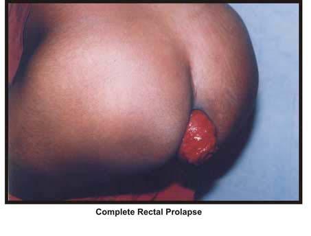
59 Differentiating features: Extensive prolapsing internal hemorrhoids appear pink or bluish in colour globular in shape with a definite sulcus between the protruding mass and the perianal skin. Submucosal prolapse appears pink in colour. Radial folds of mucosa are seen. Size is 2-3cms contains only partial thickness of mucosa. Lumen of the bowel is centrally placed. Complete rectal prolapse appears plum coloured with concentric rings of mucosa lining the prolapse through out the entire circumference with a sulcus between the anal canal and rectum and varies in size > 4 cms. Lumen of the bowel is posteriorly or eccentrically placed. The prolapse segment contains all three layers of intestine. Rectal polyp is usually mobile can be separated from outer part of rectum and anal canal by digital examination. Intussuception can be differentiated when we are able to insert a finger in the deep groove between the anal canal and the prolapsing tissue. 46
60 CLASSIFICATION OF RECTAL PROLAPSE A. Clinical Classification: This is based on clinical presentation of the prolapse like Reducible prolapse Irreducible prolapse Obstructed or incarcerated prolapse Strangulated prolapse Reducible prolapse: In this type of prolapse it gets completely reduced in to the anal canal on its own or on manipulation. A reducible prolapse appears at the anus on coughing or straining. Irreducible prolapse: In this type of prolapse it cannot be completely emptied into the anal canal. This can happen due to adhesions. Obstruction or incarcerated prolapse: Irreducibility + features of intestinal obstruction The features are: a. The prolapse is irreducible but painless b. The prolapse is non tender c. No variation on coughing d. Features of intestinal obstruction are present 47
61 In this type of prolapse there is no arrest of blood flow to the prolapsed rectum. Strangulated prolapse: Irreducibility + features of strangulated intestinal obstruction The features are: a. Severely painful irreducible prolapse. b. The prolapsed tissue is very tender c. Features of intestinal obstruction is evident d. Necrosis and the rupture of the prolapse with gangrenous bowel In this type of prolapse there is as arrest of the blood supply to the prolapsed rectum. B. Anatomical Classification: According to the anatomical classification,prolapse is classified as a. Anterior mucosal prolapse b. Internal rectal prolapse c. External full thickness rectal prolapse Anterior mucosal prolapse: In this type of prolapse the underlying rectal musculature remains in situ, with only overlying mucosa prolapsing distally. Sometimes beyond the anal verge. It presents with minor 48
62 soiling, pruritis ani, or bright rectal bleeding secondary to trauma. Internal rectal prolapse (Occult, hidden or concealed prolapse): This type of prolapse is an intussusception of the upper rectum that does not reach the anal verge and may be a precursor of external prolapse. External full thickness rectal prolapse: This type of prolapse is also known as complete rectal prolapse or procidentia. The intussusception extends beyond the anal verge with full thickness circumferential rectal protrusion. Anatomical findings in complete rectal prolapse: 1. Diastasis of the levators 2. Deep pouch of Douglas 3. Redundant rectosigmoid 4. Lax lateral and posterior attachments 5. loss of horizontal line 6. Elongated mesorectum Anorectal Physiology in Rectal Prolapse: 25,28,30 Patients with Full Thickness Rectal Prolapse (FTRP) show high pressure, rectal waves combined with Internal Anal Sphincter (IAS) inhibition and a fall in anal pressures. The rectoanal inhibitory reflex is frequently absent. These high pressure rectal waves are often abolished by sutures, 49
63 meshed or resection rectopexy, resulting in increased Internal Anal Sphincter activity, increased resting anal pressures, at least partial restoration of the rectoanal inhibitory reflex, and improvement in continence to solid and liquid stool. A plausible explanation is that the presence of the intussusceptum induces reversible rectoanal reflex, Internal Anal Sphincter relaxation and a fall in anal pressures. In the preoperative setting the high pressure rectal waves exceed the weak anal pressures. After operation this pressure gradient reverses as the anal pressure rises in association with Internal Anal Sphincter recovery. Although anal pressures remain relatively low in patients, it may not exceed the lower postoperative rectal pressures. This fact may explain continence recovery in up to 75% of cases after prolapse operations. History of Surgical Meshes: 3,1 The history of surgical meshes is an outstanding example for the progress in hernia surgery, and the possible benefit for patients from the use of biomaterials in general. The idea of strengthening the abdominal wall and, in particular, surgical hernia repair with autologous materials has been claimed in the last century, although meshes were first commercially available 40 years ago. 50
64 It was Billroth who at the end of the last century dreamed of strengthening the repair if we would artificially produce tissues of the density and toughness of fascia, the secret of the radical cure of hernia would be discovered. Artificial material was introduced in 1889 by Witzel, who used a mesh of silver wire for abdominal wall hernias; Busse in 1901 even used meshes of gold wire. In 1931 Fieschi proposed the implantation of rubber sponges. In 1940, Ogilvie published the use of cloth meshes of metallic wire to treat hernia patients. The triumphant progress of meshes had its beginning after World War II, with the development of synthetic polymers for medical purposes, particularly with the construction of the polyester mesh mersilene in 1954 and the polypropylene mesh Marlex in In 1958, Horwich used a prosthesis made of elasticated nylon in patients with large or recurrent inguinal hernias. He recognized that any recurrence would occur at edge of the prosthesis and that an implant of sufficient size to widely overlap the deficiency is required. In 1959, Usher et al reported the successful implantation of surgical meshes at first in 13 dogs and afterward in patients with abdominal wall hernias. They used relatively thin strip (2.5cm x 7cm) of marlex as an additional buttress to reinforce conventional repairs and his initial experience was favorable with no infective complication. He also 51
65 commented on the benign post operative course of these patients, who had remarkably little post operative pain. In 1963, an improved version of Marlex was introduced by Usher based on a new knitted mesh of polypropylene monofilament fiber, used initially as a suture material, and this remains the prosthesis in use today. Polypropylene mesh has had an enormous impact on surgery over the past 35 years, and countless patients have had their lives extended or improved by its application to numerous surgical problems. It is quite clearly and justifiably the most popular prosthetic mesh available today for surgical implantation. In 1970s mainly French surgeons elaborated further technical details to cure various hernias with the help of mesh prosthesis, but it was in the 1990s that the use of meshes spread unimaginably due to simple Lichtenstein tension-free repair and the newly developed laparoscopic technique, which are based on the obligate use of meshes. 52
66 Properties of Polypropylene Mesh: Polypropylene ( CH2 CH (CH3) ) is a thermoplast based on propane with a molecular weight of 100,000. It is supposed to resist physical decay even after years of being implanted. Filaments made of polypropylene have a similar strength to steel, although they are only one eighth the density of iron. A disadvantage is the high bending stiffness of the monofilaments, being susceptible to increase even during incorporation. Nevertheless, most of the current meshes are built of monofilaments, after implantation, this polymer initiates a (sub) acute inflammatory reaction of the host tissue with a consecutive fibrosis and high mechanical stability. Direct contact with the intestine has to be prevented very carefully because polypropylene meshes tend to form intense adhesions and later fistulas. As a consequence of the physiologic wound contraction, depending largely on the extent of inflammation, the polypropylene meshes show a considerable shrinkage of about 20% in length, and 40% of the original area, sometime folding and forming sharp edges. Polypropylene regularly causes, as do many other materials, the development of edema around the implant, so that drainage for 2-7 days is usually advisable. 53
67 CLINICAL FEATURES: 10 Age: Complete rectal prolapse occurs at all ages. More common in adults than in children. Peaks of frequency are in the fourth and seventh decades of life. Sex: Males and females are equally affected with a male preponderance in Asia. Occupation: Heavy work especially those lifting heavy weights causing strain on abdominal muscles increases the abdominal pressure causing prolapse. Hard labour workers, weight lifters are prone. Associated factors: Prolapse may at time be due to weakness of the pelvic floor muscles like in repeated parturition (child birth), episiotomy, forceps delivery. Perianal pelvic or sphincteric operations like sphincterotomy, fissurectomy, fistulectomy, hemorroidectomy, etc., predispose to complete rectal prolapse Disease which lead to increase in abdominal pressure such as 54
68 prostatic enlargement, stricture urether, chronic cough and chronic constipation, rectocoele, genital prolapse in females and cystocoele, predispose to complete rectal prolapse. Symptoms: The patient gives history of a mass protruding through the anus during or after defecation. Anal Bleeding Difficulty with evacuation, feeling of incomplete evacuation Tenesmus or rectal pain during or after defecation Dragging pain in the pelvic regions and low backache Increased anal pressure in the form of straining during defecation Distension and change in bowel habits Incontinence to solid, liquids and gas The patient may initially feel some discomfort in the anal region and notice a mass protruding when coughing, straining or on defecation that immediately subsides all by itself. As it progresses to increase in size it may become irreducible and patient learns to manually reduce it. 55
69 Later when the prolapse is established the complaints are usually concerned with the mass protruding out of the anus interfering with walking, social activities and defecation. The dragging sensation in the perineum, backache and most of the time soiling of under garments, bed linen, incontinence to solid, liquids and gas causing psychological disturbances. Bleeding from the mass due to repeated trauma, pain due to pull on the supports of the rectum are also complained. Rarely the prolapse may be incarcerated or strangulated leading to severe pain due to necrosis or rupture and manifests as an emergency with extreme pain and signs of intestinal obstruction and later signs of gangrenous bowel. Signs: Clinical examination with the patient in squatting position will reveal a red coloured mass that protrudes on straining/coughing beyond the anal verge. Size of the protruding mass is > 4cm 56
70 Circular folds of mucosa are evident Eccentric position or posteriorly placed bowel lumen is significant Bidigital examination reveals a patulous anus through which the rectum prolapses. On palpation the presence of all the layers of the rectal wall are noted. 57
71 TESTING AND EVALUATION Anorectal physiology testing refers to evaluation of anal canal resting pressure, squeeze pressure anal reflexes, pudendal nerve conduction velocities and electromyographic muscle fibre recruitment. Manometry involves use of water filled balloons attached to catheters and transducers placed in the anal canal.normal resting pressure is 40 mmhg and that of squeeze pressure is80 mmhg.resting pressure indicates the function of internal anal sphincter while squeeze pressure measures external anal sphincter function. These are useful in evaluation of conditions ranging from incontinence to obstructive defecation. Pudendal nerve terminal motor latency (PNTML) times are measured with transducer attached to the glove light apparatus designed to be worn on the finger and hand. Per-rectal examination is required with application of finger electrode to the right and left leavtor ani complex. normal value ranges from 1.8 to 2.4msecs. Prolonged values are seen in traumatic injuries of anal canal, sacral nerve root damage and diabetic autonomic neuropathy. 58
72 Defecography: Defeography is an useful modality to determine the nature of various pelvic floor abnormalities. Barium paste is placed in the rectum after the patient ingests water soluble contrast to opacify the small bowel. As the patient evacutes the rectal barium paste abnormalities occurring during defecation can be recorded with fluoroscopic video typing. By this method various anatomical abnormalities such as rectocele enterocele and vaginal wall prolapse can be evaluated. 59
73 DIFFERENTIAL DIAGNOSIS: Prolapsed incarcerated internal hemorrhoids Intussusception Proctitis Rectal polyps 60
74 MANAGEMENT: In adult patients treatment of rectal prolapse is mainly surgical. No specific medical management is available. More than 50 types of procedures have been documented but most of them are of historical interest only. Surgical management is broadly classified into Abdominal approach and Perineal approach. Choice of procedure is decided by the co-morbidities, presence or absence of constipation, patient s choice and surgeon s skills. Some of the abdominal surgical procedures are -RIPSTEIN REPAIR -WELLS PROCEDURE -ANTERIOR RESECTION -RESECTION RECTOPEXY Some of the perineal surgical procedures are -THIERSCH ANAL ENCIRCLEMENT -DELORME MUCUSAL SLEEVE RESECTION -ALTEMEIER PERINEAL RECTOSIGMOIDECTOMY -PERINEAL STAPLED PROLAPSED REECTION. 61
75 Of all the above mentioned procedures abdominal mesh rectopexy is the choice of treatment for patients with no co-morbidities as this procedure has the least recurrence rate. Now-a-days laparascopic surgical rectopexy procedures are gaining importance as they have good outcomes as that of open abdominal procedures. Choice of Treatment: Abdominal rectopexy using prolene mesh. Principles and Advantages of Rectopexy: 9 It restores normal anatomy by fixing the rectum to the sacrum Prolene mesh used in rectopexy favours fibrosis and fixes the rectum to the sacrum It is a simplest procedure and access to rectum is better and easier It has low risk of perioperative death or morbidity and an acceptable rate of recurrence It corrects the functional disturbances such as incontinence and constipation It can be safely operated in elderly and young alike 62
76 Outcome: As reported by more than 50 authors in the United Kingdom and United States of America the results of abdominal rectopexy using prolene mesh has been very successful enough to be considered the treatment of choice for complete rectal prolapse both in young and elderly because it allows fixation of the freed rectum and the rectosigmoid to the sacrum in such a manner that it can not slip down, thereby restoring anatomy. Incontinence has been corrected to some extent in 60-80% of patients. The reported recurrence is less than 4%. Complications of surgery are very minimal. 63
77 METHODOLOGY This prospective clinical study included 30 cases of complete rectal prolapse who underwent abdominal rectopexy using prolene mesh. These patients were admitted at THANJAVUR MEDICAL COLLEGE, THANJAVUR, during the period from march 2013 to august The patients coming with a history of protrusion of mass per anus were interviewed and a diagnosis of complete rectal prolapse was made essentially on clinical examination. For assessing functional results, continence was classified after Browning and Park s as follows 8 : Classification of continence (Browning and parks) Grade 1 Grade 2 Fully continent for flatus & stool Continent for stools but not for flatus Grade 3 Grade 4 Incontinent for liquid stool Incontinent for solid stool Grade 3 and 4 were considered unacceptable. No distinction was made between occasional and regular episodes of incontinence. 64
78 For assessing bowel function, constipation was defined as passage of hard stools with frequency less than once a day or marked straining at stools. Fecal frequency was more than 3 bowel actions a day. Following investigations were done in all cases. Blood: Hb%, TC, DC and ESR. Urine: sugar, albumin, microscopy. FBS, serum creatine, HIV, HbsAg. Chest x-ray and ECG. Sigmoidoscopy. All patients were subjected to abdominal rectopexy using prolene mesh. Operative details: Patients after preoperative mechanical bowel preparation received inj. Ceftriaxone 1gm iv 8 th hourly (3 doses) and inj. Metronidazole iv 500mg 8 th hourly (3 doses) as antibiotic prophylaxis from the starting of the operation. Operative steps: Technique of Abdominal Rectopexy: This procedure was done under spinal anaesthesia with the patient in supine position. 65
79 The parts painted and draped, a head down tilt was made to make easy access to pelvic structures By a lower left midline skin incision of 10-12cm, the abdomen was opened in layers The rectosigmoid was identified and the small bowel packed off and hitched up by a ribbon gauze and held aside The peritoneal cuts were made in the mesorectum to mobilize the rectum starting on the right side of the base of the mesosigmoid but a little way up the mesentery (so that the peritoneum could be preserved and closed over the repair). The incision was carried down to the bottom of the pouch of Douglas crossing anteriorly and joining a similar cut on the left side. The presacral space was opened identifying and preserving the presacral nerves posterolaterally and the left ureter The posterior dissection was carried right down to bottom of pelvic floor and preserving presacral nerves posterolaterally and the left ureter. Anteriorly the plain between the bladder and the rectum was dissected with blunt and sharp dissection Lateral ligaments were preserved A cm prolene mesh cut to a appropriate size if required was fixed to the presacral fascia with 1-0 prolene over the sacral 66
80 promontory and S3 vertebra posteriorly with a series of 3-4 sutures, care being taken not to injure middle sacral vessels and pelvic veins. The implanted mesh was partially wrapped around the lateral sides of the mobilized rectum leaving anterior half of circumference of rectum free to avoid risk of stenosis.. Sutures were then placed to fix the mesh in position through the seromuscular layer of the rectum laterally. Pelvic peritoneum was covered over and abdomen closed with a redivac drain in presacral space to prevent hematoma. Postoperatively patients were treated with: Intravenous fluids for 48 hours (due to paralytic ileus). Laxative from 3 rd and 4 th postoperative day to prevent constipation and straining at stool for a period of one month. Drain removed after 48 hours. Liquid and semi solid diet allowed from the 3 rd postoperative day Normal diet advised from the 4 th to 5 th postoperative day Sutures were removed on the 7 th postoperative day. Each case was followed up in outpatient department once in a month for a minimum period of 6 months. Each case was assessed clinically for postoperative complications like haemorrhage, operative mortality, 67
81 wound infection, infection around prolene mesh, bladder and erectile dysfunction. During follow up, recurrence of rectal prolapse, effect on bowel frequency and improvement or deterioration of preexisting incontinence were particularly noted down. Inclusion Criteria: 1. Patients with complete rectal prolapse who underwent abdominal rectopexy using prolene mesh. Exclusion Criteria: 1. Cases of complete rectal prolapse where sigmoidectomy or colectomy was combined with abdominal rectopexy. 2. Cases which could not be followed up for a minimum period of 6 months. Overall, 36 cases of complete rectal prolapse were admitted during the period. Only 30 of them underwent abdominal rectopexy using prolene mesh. 6 patients refused to undergo this operation and were excluded from this study. All the 30 cases who underwent abdominal rectopexy were followed up for a minimum period of 6 months and were included in the study. 68
82 69

83 70

84 71

85 72

86 73

87 74
Summary and conclusion. Summary And Conclusion
 Summary And Conclusion Summary and conclusion Rectal prolapse remain a disorder for which no single ideal treatment was approved for all cases. Complete rectal prolapse (procidentia) is the circumferential
Dana Alrafaiah. - Amani Nofal. - Ahmad Alsalman. 1 P a g e
 - 2 - Dana Alrafaiah - Amani Nofal - Ahmad Alsalman 1 P a g e This lecture will discuss five topics as follows: 1- Arrangement of pelvic viscera. 2- Muscles of Pelvis. 3- Blood Supply of pelvis. 4- Nerve
-15. -Alaa Albandi. -Dr. Mohammad Almohtasib. 0 P a g e
 -15 -Alaa Albandi - -Dr. Mohammad Almohtasib 0 P a g e In this last lecture, we will talk about the sigmoid colon, rectum, and anal canal. Sigmoid colon It has a mesentery called pelvic mesocolon or sigmoidal
Inferior Pelvic Border
 Pelvis + Perineum Pelvic Cavity Enclosed by bony, ligamentous and muscular wall Contains the urinary bladder, ureters, pelvic genital organs, rectum, blood vessels, lymphatics and nerves Pelvic inlet (superior
Urinary Bladder. Prof. Imran Qureshi
 Urinary Bladder Prof. Imran Qureshi Urinary Bladder It develops from the upper end of the urogenital sinus, which is continuous with the allantois. The allantois degenerates and forms a fibrous cord in
Preview from Notesale.co.uk Page 1 of 34
 Abdominal viscera and digestive tract Digestive tract Abdominal viscera comprise majority of the alimentary system o Terminal oesophagus, stomach, pancreas, spleen, liver, gallbladder, kidneys, suprarenal
Pelvis MCQs. Block 1. B. Reproductive organs. C. The liver. D. Urinary bladder. 1. The pelvic diaphragm includes the following muscles: E.
 Pelvis MCQs Block 1 1. The pelvic diaphragm includes the following muscles: A. The obturator internus B. The levator ani C. The coccygeus D. The external urethral sphincter E. The internal urethral sphincter
Perineum. done by : zaid al-ghnaneem
 Perineum done by : zaid al-ghnaneem Hello everyone, this sheet will talk about 2 nd Lecture which is Perineum but there are some slides and info from 1 st Lecture. Everything included Slides + Pics Let
2. List the 8 pelvic spaces: list one procedure or dissection which involves entering that space.
 Name: Anatomy Quiz: Pre / Post 1. In making a pfannensteil incision you would traverse through the following layers: a) Skin, Camper s fascia, Scarpa s fascia, external oblique aponeurosis, internal oblique
TME and autonomic nerve preservation techniques: based on Video and Cadaveric anatomy
 TME and autonomic nerve preservation techniques: based on Video and Cadaveric anatomy Nam Kyu Kim M.D., Ph.D., FACS, FRCS, FASCRS Professor Department of Surgery Yonsei University College of Medicine Seoul,
LAPAROSCOPIC REPAIR OF PELVIC FLOOR
 LAPAROSCOPIC REPAIR OF PELVIC FLOOR Dr. R. K. Mishra Elements comprising the Pelvis Bones Ilium, ischium and pubis fusion Ligaments Muscles Obturator internis muscle Arcus tendineus levator ani or white
Midgut. Over its entire length the midgut is supplied by the superior mesenteric artery
 Gi Embryology 3 Midgut the midgut is suspended from the dorsal abdominal wall by a short mesentery and communicates with the yolk sac by way of the vitelline duct or yolk stalk Over its entire length the
Pelvis Perineum MCQs. Block 1.1. A. Urinary bladder. B. Rectum. C. Reproductive organs. D. The thigh
 Pelvis Perineum MCQs Block 1.1 1. The pelvic diaphragm includes the following muscles: A. The coccygeus B. The levator ani C. The external urethral sphincter D. The internal urethral sphincter E. The obturator
Embryology of the Midgut and Hind gut
 Embryology of the Midgut and Hind gut Prof. Abdulameer Al-Nuaimi E-mail: a.al-nuaimi@sheffield.ac.uk E-mail: abdulameerh@yahoo.com Abdominal organs www.google.co.uk/search? Development of Duodenum The
REPRODUCTIVE SYSTEM By Dr.Ahmed Salman
 The University Of Jordan Faculty Of Medicine Anatomy Department REPRODUCTIVE SYSTEM By Dr.Ahmed Salman Assistant Professor of Anatomy &embryology Perineum It is the diamond-shaped lower end of the trunk
Anatomy of the Large Intestine
 Large intestine Anatomy of the Large Intestine 2 Large Intestine Extends from ileocecal valve to anus Length = 1.5-2.5m = 5 feet Regions Cecum = 2.5-3 inch Appendix= 3-5 inch Colon Ascending= 5 inch Transverse=
THE ORAL CAVITY
 THE ORAL CAVITY WALL OF ABDOMEN (ANTERIOR) The paraumbilical vein drains into the portal vein and then through the liver. This is an important clinical connection. THE ABDOMINAL VISCERA The small
STRUCTURAL BASIS OF MEDICAL PRACTICE EXAMINATION 3. October 17, 2014
 STRUCTURAL BASIS OF MEDICAL PRACTICE EXAMINATION 3 October 17, 2014 PART l. Answer in the space provided. (12 pts) 1. Identify the structures. (2 pts) A. B. A B C. D. C D 2. Identify the structures. (2
STRUCTURAL BASIS OF MEDICAL PRACTICE EXAMINATION 3. October 16, 2015
 STRUCTURAL BASIS OF MEDICAL PRACTICE EXAMINATION 3 October 16, 2015 PART l. Answer in the space provided. (12 pts) 1. Identify the structures. (2 pts) A. B. A B C. D. C D 2. Identify the structures. (2
Gynecology Dr. Sallama Lecture 3 Genital Prolapse
 Gynecology Dr. Sallama Lecture 3 Genital Prolapse Genital(utero-vaginal )prolapse is extremely common, with an estimated 11% of women undergoing at least one operation for this condition. Definition: A
Femoral Triangle and Adductor Canal. Dr. Heba Kalbouneh Associate Professor of Anatomy and Histology
 Femoral Triangle and Adductor Canal Dr. Heba Kalbouneh Associate Professor of Anatomy and Histology Femoral Triangle and Adductor Canal Femoral triangle Is a triangular depressed area located in the upper
Biology Human Anatomy Abdominal and Pelvic Cavities
 Biology 351 - Human Anatomy Abdominal and Pelvic Cavities Please place your name and I.D. number on the back of the last page of this exam. You must answer all questions on this exam. Because statistics
Function of the anal sphincters in patients with
 Function of the anal sphincters in patients with intussusception of the rectum B. FRENCKNER AND T. IHRE Gut, 1976,17, 147-151 From the Department of Clinical Physiology and the Department of Surgery, Serafimer
Physiologic Anatomy and Nervous Connections of the Bladder
 Micturition Objectives: 1. Review the anatomical organization of the urinary system from a physiological point of view. 2. Describe the micturition reflex. 3. Predict the lines of treatment of renal failure.
Gross Anatomy of the Urinary System
 Gross Anatomy of the Urinary System Lecture Objectives Overview of the urinary system. Describe the external and internal anatomical structure of the kidney. Describe the anatomical structure of the ureter
NOTES FROM GUTMAN LECTURE 10/26 Use this outline to study from. As you go through Gutman s lecture, fill in the topics.
 NOTES FROM GUTMAN LECTURE 10/26 Use this outline to study from. As you go through Gutman s lecture, fill in the topics. Anatomy above the arcuate line Skin Camper s fascia Scarpa s fascia External oblique
Table 2. First Generated List of Expert Responses. Likert-Type Scale. Category or Criterion. Rationale or Comments (1) (2) (3) (4)
 (2) (3) (4)") Table 2. First Generated List of Expert Responses. Likert-Type Scale Category or Criterion Anatomical Structures and Features Skeletal Structures and Features (1) (2) (3) (4) Rationale or Comments 1. Bones
Table 2. First Generated List of Expert Responses. Likert-Type Scale Category or Criterion Anatomical Structures and Features Skeletal Structures and Features (1) (2) (3) (4) Rationale or Comments 1. Bones
Small Plicae Circularis. Short Closely packed together. Sparse, completely absent at distal part Lymphoid Nodule
 Intestines Differences Between Jejunum and Ileum Types Jejunum Ileum Color Deeper red Paler pink Calibre Bigger Smaller Thickness of wall Thick and Heavy Thin and Lighter Vascularity Highly vascularised
Robot Assisted Rectopexy
 1. Abdominal cavity approach 1A Trocars Introduce Introduce five trocars to gain access to the abdominal cavity (in da Vinci Si type; In Xi type the trocar placement may differ slightly). First the camera
Exploring Anatomy: the Human Abdomen
 Exploring Anatomy: the Human Abdomen PERITONEUM AND PERITONEAL CAVITY PERITONEUM The peritoneum is a thin serous membrane that lines the abdominal cavity and covers, in variable amounts, the viscera within
Bony ypelvis. Composition: formed by coccyx, and their articulations Two portions
 Pelvis Bony ypelvis Composition: formed by paired hip bones, sacrum, coccyx, and their articulations Two portions Greater pelvis Lesser pelvis Terminal line ( pelvic inlet): formed by promontory of sacrum,
Pancreas & Biliary System. Dr. Vohra & Dr. Jamila
 Pancreas & Biliary System Dr. Vohra & Dr. Jamila 1 Objectives At the end of the lecture, the student should be able to describe the: Location, surface anatomy, parts, relations & peritoneal reflection
SUBJECTS 2nd year, 1st semester I. 1. Primitive gut - limits, derivatives 2. Foregut -limits, evolution, derivatives 3. Midgut -limits, evolution,
 SUBJECTS 2nd year, 1st semester I. 1. Primitive gut - limits, derivatives 2. Foregut -limits, evolution, derivatives 3. Midgut -limits, evolution, derivatives 4. Hindgut- limits, evolution, derivatives
Rectal Prolapse: A 10-Year Experience
 24 The Ochsner Journal Volume 7, Number 1, Spring 2007 25 Rectal Prolapse: A 10-Year Experience Figure 2. Physical examination. A. Concentric folds of prolapsed rectum. B. Radial folds of hemorrhoids (mucosal
The abdominal Esophagus, Stomach and the Duodenum. Prof. Oluwadiya KS
 The abdominal Esophagus, Stomach and the Duodenum Prof. Oluwadiya KS www.oluwadiya.com Viscera of the abdomen Abdominal esophagus: Terminal part of the esophagus The stomach Intestines: Small and Large
Dr. Zahiri. In the name of God
 Dr. Zahiri In the name of God small intestine = small bowel is the part of the gastrointestinal tract Boundaries: Pylorus Ileosecal junction Function: digestion and absorption of food It receives bile
Anorectal Diagnostic Overview
 Anorectal Diagnostic Overview 11-25-09 3.11.2010 2009 2010 Anorectal Manometry Overview Measurement of pressures and the annotation of rectal sensation throughout the rectum and anal canal to determine:
Ureters, Urinary Bladder & Urethra
 Ureters, Urinary Bladder & Urethra Please check our Editing File هذا العمل ال يغني عن المصدر األساسي للمذاكرة Lecture 2 } و م ن ي ت و ك ع ل ا لل ه ف ه و ح س ب ه { Objectives o Describe the course of ureter
Embryology, anatomy and physiology of the oesophagus. Sarah Forsyth
 Embryology, anatomy and physiology of the oesophagus Sarah Forsyth Embryology Basics Endoderm forms scaffolding of GIT Endoderm forms the lining of the yolk sac Derivative of foregut Wk 4 - Foregut develops
The anatomy of the rectum and anal canal
 The anatomy of the rectum and anal canal Vishy Mahadevan Abstract Diseases of the rectum and anal canal, both benign and malignant, account for a very large part of colorectal surgical practice in the
ANATOMY OF PELVICAYCEAL SYSTEM -DR. RAHUL BEVARA
 1 ANATOMY OF PELVICAYCEAL SYSTEM -DR. RAHUL BEVARA 2 KIDNEY:ANATOMY OVERVIEW Kidneys are retroperitoneal, in posterior abdominal region, extending from T12 L3 Bean-shaped Right kidney is lower than left
ORIENTING TO BISECTED SPECIMENS ON THE PELVIS PRACTICAL
 ORIENTING TO BISECTED SPECIMENS ON THE PELVIS PRACTICAL The Pelvis is just about as complicated as head and neck and considerably more mysterious. You have to be able to visualize (imagine) the underlying
Slide Read the tables it is about the difference between male & female pelvis.
 I didn t include the slides, this is only what the doctor read or said because he skipped a lot of things because we took it previously, very important to go back to the slides (*there is an edited version)
Principles of Anatomy and Physiology
 Principles of Anatomy and Physiology 14 th Edition CHAPTER 24 The Digestive System Introduction The purpose of this chapter is to Identify the anatomical components of the digestive system as well as their
By:Dr:ISHRAQ MOHAMMED
 By:Dr:ISHRAQ MOHAMMED Protrusion of an organ or structure beyond its normal confines. Prolapses are classified according to their location and the organs contained within them. 1-Anterior vaginal wall
The peritoneum. Prof. Oluwadiya KS, MBBS, FMCS(Orthop) Website:
 Website:") The peritoneum Prof. Oluwadiya KS, MBBS, FMCS(Orthop) Website: http://oluwadiya.com The peritoneum Serous membrane that lines the abdominopelvic cavity and invests the viscera The largest serous membrane
The peritoneum Prof. Oluwadiya KS, MBBS, FMCS(Orthop) Website: http://oluwadiya.com The peritoneum Serous membrane that lines the abdominopelvic cavity and invests the viscera The largest serous membrane
The posterior abdominal wall. Prof. Oluwadiya KS
 The posterior abdominal wall Prof. Oluwadiya KS www.oluwadiya.sitesled.com Posterior Abdominal Wall Lumbar vertebrae and discs. Muscles opsoas, quadratus lumborum, iliacus, transverse, abdominal wall
Functional anatomy of the female pelvic floor and lower urinary tract Stefano Floris, MD, PhD Department of Obstetrics and Gynaecology
 Functional anatomy of the female pelvic floor and lower urinary tract Stefano Floris, MD, PhD Department of Obstetrics and Gynaecology Ospedale San Giovanni di Dio, Gorizia, Italy ANATOMY URINARY CONTINENCE
RECTAL INJURY IN UROLOGIC SURGERY. Inadvertent rectal injury from a urologic procedure is often subtle but has serious postoperative consequences.
 RECTAL INJURY IN 27 UROLOGIC SURGERY Inadvertent rectal injury from a urologic procedure is often subtle but has serious postoperative consequences. With good mechanical bowel preparation plus antibiotic
Development of the Digestive System. W.S. O The University of Hong Kong
 Development of the Digestive System W.S. O The University of Hong Kong Plan for the GI system Then GI system in the abdomen first develops as a tube suspended by dorsal and ventral mesenteries. Blood
Hemorrhoids. Carlos R. Alvarez-Allende PGY-III Colorectal Surgery
 Hemorrhoids Carlos R. Alvarez-Allende PGY-III Colorectal Surgery Overview Anatomy Classification Etiology Incidence Symptoms Differential Diagnosis Medical Management Surgical Management Anatomy Anal canal
The Urinary System Pearson Education, Inc.
 26 The Urinary System Introduction The urinary system does more than just get rid of liquid waste. It also: Regulates plasma ion concentrations Regulates blood volume and blood pressure Stabilizes blood
THE ABDOMEN SUPRARENAL GLANDS KIDNEY URETERS URINARY BLADDER
 THE ABDOMEN SUPRARENAL GLANDS KIDNEY URETERS URINARY BLADDER THE SUPRARENAL GLANDS The suprarenal (adrenal) glands lie immediately superior and slightly anterior to the upper pole of either kidney. Golden
THE SACRAL PARASYMPATHETIC INNERVATION OF THE COLON
 THE SACRAL PARASYMPATHETIC INNERVATION OF THE COLON RUSSELL T. WOODBURNE Department of Anatomy, University of Michigan Hedical School, Ann Arbofi TWO FIGURES Autonomic nerves distribute by a variety of
Duodenum retroperitoneal
 Duodenum retroperitoneal C shaped Initial region out of stomach into small intestine RETROperitoneal viscus Superior 1 st part duodenal cap ; moves upwards and backwards to lie on the R crura medial to
An Evaluation of the Cause and Mechanism of Complete Rectal Prolapse
 PRESIDENTIAL ADDRESS An Evaluation of the Cause and Mechanism of Complete Rectal Prolapse NORMAN D. NIGRO, M.D. Detroit, Michigan I appreciate deeply the honor which the American Proctologic Society conferred
disfunzioni sessuali ed urinarie: come evitarle? D. Mascagni
 disfunzioni sessuali ed urinarie: come evitarle? D. Mascagni Cattedra di Chirurgia Generale Direttore: Prof. A. Filippini Verona, 2010 CHIRURGIA RADICALE PER CANCRO DEL RETTO SOTTOPERITONEALE 5cm 2 cm
MCOMPASS ANAL MANOMETRY AN OVERVIEW
 MCOMPASS ANAL MANOMETRY AN OVERVIEW ANAL MANOMETRY MEASURES PRESSURE ALLOWS INTERPRITATION SENSATION RAIR RECTAL COMPLIANCE MOTIVATION OF THE PATIENT FUNCTION OF THE PUDENDAL NERVE WHEN TO USE ANAL MANOMETRY
MCOMPASS ANAL MANOMETRY AN OVERVIEW
 MCOMPASS ANAL MANOMETRY AN OVERVIEW ANAL MANOMETRY MEASURES PRESSURE ALLOWS INTERPRITATION SENSATION RAIR RECTAL COMPLIANCE MOTIVATION OF THE PATIENT FUNCTION OF THE PUDENDAL NERVE WHEN TO USE ANAL MANOMETRY
Review Article Pelvic Floor Dysfunction, Body Excreta Incontinence and Continence
 Cronicon OPEN ACCESS GYNAECOLOGY Review Article Pelvic Floor Dysfunction, Body Excreta Incontinence and Continence Abdel Karim M El Hemaly 1 * and Laila ASE Mousa 1 1 Professor of Obstetrics and gynaecology,
POSTERIOR LEVATOR REPAIR AND RECTAL SUSPENSION FOR RECURRENT RECTAL PROLAPSE: AUTHOR'S EXPERIENCE
 POSTERIOR LEVATOR REPAIR AND RECTAL SUSPENSION FOR RECURRENT RECTAL PROLAPSE: AUTHOR'S EXPERIENCE *Shyam B. Sharma 1&2 and Rahul Gupta 2 1 Department of Pediatric Surgery SMS Medical College 2 Pediatric
INTRODUCTION TO GASTROINTESTINAL FUNCTIONS
 1 INTRODUCTION TO GASTROINTESTINAL FUNCTIONS 2 Learning outcomes List two main components that make up the digestive system Describe the 6 essential functions of the GIT List factors (neurological, hormonal
IMAGING GUIDELINES - COLORECTAL CANCER
 IMAGING GUIDELINES - COLORECTAL CANCER DIAGNOSIS The majority of colorectal cancers are diagnosed on colonoscopy, with some being diagnosed on Ba enema, ultrasound or CT. STAGING CT chest, abdomen and
Anatomy: Know Your Abdomen
 Anatomy: Know Your Abdomen Glossary Abdomen - part of the body below the thorax (chest cavity); separated by the diaphragm. Anterior - towards the front of the body. For example, the umbilicus is anterior
Prediction and prevention of stress urinary incontinence after prolapse surgery van der Ploeg, J.M.
 UvA-DARE (Digital Academic Repository) Prediction and prevention of stress urinary incontinence after prolapse surgery van der Ploeg, J.M. Link to publication Citation for published version (APA): van
3D Dynamic Ultrasound In Obstructed Defecation
 3D Dynamic Ultrasound In Obstructed Defecation By Ramy Salahudin Abdelkader Assist. Lecturer of General Surgery Cairo University Introduction Pelvic floor is complex system, with passive and active components
Gastrointestinal Motility 2: Intestinal and Colonic Motility Jack Grider, Ph.D.
 Gastrointestinal Motility 2: Intestinal and Colonic Motility Jack Grider, Ph.D. OBJECTIVES: 1. Contrast the types of motility in the small intestine. 2. Describe the neural circuits that mediate peristalsis.
consists of: Muscular, hollow tube (= digestive tract ) + Various accessory organs
 + Various accessory organs") DIGESTIVE SYSTEM consists of: Muscular, hollow tube (= digestive tract ) + Various accessory organs FUNCTION Individual parts function in: ingestion mechanical digestion chemical and enzymatic digestion
DIGESTIVE SYSTEM consists of: Muscular, hollow tube (= digestive tract ) + Various accessory organs FUNCTION Individual parts function in: ingestion mechanical digestion chemical and enzymatic digestion
ANATOMY OF THE SMALL & LARGE INTESTINES. Semester 1, 2011 A. Mwakikunga
 ANATOMY OF THE SMALL & LARGE INTESTINES Semester 1, 2011 A. Mwakikunga LEARNING OBJECTIVES 1. List the parts and anatomical regions of the small and large intestines 2. State anatomical relations of the
Development of the Digestive System. W.S. O School of Biomedical Sciences, University of Hong Kong.
 Development of the Digestive System W.S. O School of Biomedical Sciences, University of Hong Kong. Organization of the GI tract: Foregut (abdominal part) supplied by coeliac trunk; derivatives include
Lecture 56 Kidney and Urinary System
 Lecture 56 Kidney and Urinary System The adrenal glands are located on the superomedial aspect of the kidney The right diagram shows a picture of the kidney with the abdominal walls and organs removed
Lab Monitor Images Dissection of the Abdominal Vasculature + Lower Digestive System
 Lab Monitor Images Dissection of the Abdominal Vasculature + Lower Digestive System Stomach & Duodenum Frontal (AP) View Nasogastric tube 2 1 3 4 Stomach Pylorus Duodenum 1 Duodenum 2 Duodenum 3 Duodenum
DISSECTION 8: URINARY AND REPRODUCTIVE SYSTEMS
 8546d_c01_1-42 6/25/02 4:32 PM Page 38 mac48 Mac 48: 420_kec: 38 Cat Dissection DISSECTION 8: URINARY AND REPRODUCTIVE SYSTEMS Typically, the urinary and reproductive systems are studied together, because
GI module Lecture: 9 د. عصام طارق. Objectives:
 GI module Lecture: 9 د. عصام طارق Objectives: To list structures forming posterior abdominal wall. To follow aorta & its main branches. To describe IVC & its main tributaries. To list nerves of posterior
Anatomy of the SMALL INTESTINE. Dr. Noman Ullah Wazir PMC
 Anatomy of the SMALL INTESTINE Dr. Noman Ullah Wazir PMC SMALL INTESTINE The small intestine, consists of the duodenum, jejunum, and illium. It extends from the pylorus to the ileocecal junction were the
د. عصام طارق. Objectives:
 GI anatomy Lecture: 5 د. عصام طارق Objectives: To describe anatomy of stomach, duodenum & pancreas. To list their main relations. To define their blood & nerve supply. To list their lymph drainage. To
Perineum. Dept. of Human Anatomy Zhou Hong Ying
 Perineum Dept. of Human Anatomy Zhou Hong Ying OUTLINE Subdivision The Layers Urogenital Diaphragm Main Structures inside Superficial & Deep Perineal Spaces Ischioanal Fossa Perineum A narrow region Urogenital
World Journal of Colorectal Surgery
 World Journal of Colorectal Surgery Volume 6, Issue 5 2016 Article 8 Sigmoidocele: A Rare Cause Of Constipation In Males Noor Shah MD Milind Kachare MD Craig Rezac MD Rutgers Robert Wood Johnson Medical
BY DR NOMAN ULLAH WAZIR
 BY DR NOMAN ULLAH WAZIR The stomach (from ancient Greek word stomachos, stoma means mouth) is a muscular, hollow and the most dilated part of the GIT. It starts from the point where esophagus ends. It
Laparoscopic Low Anterior Resection of the Rectum
 Laparoscopic Low Anterior Resection of the 4 4.1 Introduction Outcomes of rectal cancer treatment depend on the operative technique adopted. Complications vary, and can occur during mobilisation, with
The jejunum and the Ileum. Prof. Oluwadiya KS
 The jejunum and the Ileum Prof. Oluwadiya KS www.oluwadiya.siteled.com Introduction Introduction The small intestine (SI) comprises of the duodenum, jejunum and the ileum The jejunum is the second part
Overview of the Digestive
 Overview of the Digestive System Bởi: OpenStaxCollege The function of the digestive system is to break down the foods you eat, release their nutrients, and absorb those nutrients into the body. Although
Surgical Management for Defecation Dysfunction
 Defecatory Dysfunction Surgical Management for Defecation Dysfunction JMAJ 46(9): 378 383, 2003 Tatsuo TERAMOTO Professor, 1st Department of Surgery, School of Medicine, Toho University Abstract: Typical
Pelvic Floor Ultrasound Imaging. Prof HP Dietz (Sydney) A/Prof KL Shek (Sydney) Dr R Guzman Rojas (Santiago de Chile) Dr Kamil Svabik (Prague)
 A/Prof KL Shek (Sydney) Dr R Guzman Rojas (Santiago de Chile) Dr Kamil Svabik (Prague)") Pelvic Floor Ultrasound Imaging Workshop IUGA 2015 Nice Faculty: Prof HP Dietz (Sydney) A/Prof KL Shek (Sydney) Dr R Guzman Rojas (Santiago de Chile) Dr Kamil Svabik (Prague) The use of translabial ultrasound
Pelvic Floor Ultrasound Imaging Workshop IUGA 2015 Nice Faculty: Prof HP Dietz (Sydney) A/Prof KL Shek (Sydney) Dr R Guzman Rojas (Santiago de Chile) Dr Kamil Svabik (Prague) The use of translabial ultrasound
TYPES OF RECTAL PROLAPSE
 RECTAL PROLPASE Rectal prolapse describes a condition where either the lining or entire wall of the rectum becomes loose and falls into, or even out of, the rectum through the anus. TYPES OF RECTAL PROLAPSE
Learning objectives. SGD on Functions of Testosterone. Class
 Learning objectives SGD on Functions of Testosterone Class 2016 14-1-2013 Discuss o Process of spermatogenesis. o Sex determination. o Process of maturation of sperms. o Physiology of mature sperms. Discuss
Abdomen. Retroperitoneal space
 Abdomen. Retroperitoneal space Abdominal cavity The space bounded by: Anterolateral abdominal wall Posterior abdominal wall Diaphragm Pelvic walls and pelvic floor. Subdivided into: True abdominal cavity
Done by: Dina Sawadha & Mohammad Abukabeer
 Done by: Dina Sawadha & Mohammad Abukabeer The stomach *the stomach is a dilated part of the gastro intestinal tract, it's "J" shape. *the lower surface of the stomach ( the greater curvature ) reaches
The front of the thigh. Dr.Amjad shatarat
 The front of the thigh Femoral triangle (Scarpa s triangle) Is a triangular depressed area located in the upper part of the medial aspect of the thigh immediately below the inguinal ligament. Superiorly:
Application of Anorectal Manometry in the Treatment of Anorectal Diseases
 Application of Anorectal Manometry in the Treatment of Anorectal Diseases Kai Wang 1, 2, a, Liping Liu 1, 2, b, Yuyan Liu 1, 2, c 3, d* and Xiaoli Zhou 1 Henan Province Hospital of TCM, 450000, Henan,
Block 3: DISSECTION 2 CELIAC TRUNK, JEJUNUM/ILEUM, LARGE INTESTINE, DUODENUM, PANCREAS, PORTAL VEIN; MOBILIZATION OF THE LIVER
 1 Block 3: DISSECTION 2 CELIAC TRUNK, JEJUNUM/ILEUM, LARGE INTESTINE, DUODENUM, PANCREAS, PORTAL VEIN; MOBILIZATION OF THE LIVER Attempt to complete as much as you can of the dissection explained in the
Colorectal procedure guide
 Colorectal procedure guide Illustrations by Lisa Clark Biodesign ADVANCED TISSUE REPAIR cookmedical.com 2 INDEX Anal fistula repair Using the Biodesign plug with no button.... 4 Anal fistula repair Using
Fecal Incontinence. What is fecal incontinence?
 Scan for mobile link. Fecal Incontinence Fecal incontinence is the inability to control the passage of waste material from the body. It may be associated with constipation or diarrhea and typically occurs
Anatomy of the Thorax
 Anatomy of the Thorax A) THE THORACIC WALL Boundaries Posteriorly by the thoracic part of the vertebral column Anteriorly by the sternum and costal cartilages Laterally by the ribs and intercostal spaces
Bushra Arafa Zayed & Hanan Jamal. - Dana AF
 - 10 - Bushra Arafa Zayed & Hanan Jamal - Dana AF - Mohammad Al Muhtaseb Notes: This sheet was written in the same order as the slides, and everything in the slides is mentioned in this sheet. Pictures
ParasymPathetic Nervous system. Done by : Zaid Al-Ghnaneem
 ParasymPathetic Nervous system Done by : Zaid Al-Ghnaneem In this lecture we are going to discuss Parasympathetic, in the last lecture we took sympathetic and one of the objectives of last lecture was
Human Anatomy. Spinal Cord and Spinal Nerves
 Human Anatomy Spinal Cord and Spinal Nerves 1 The Spinal Cord Link between the brain and the body. Exhibits some functional independence from the brain. The spinal cord and spinal nerves serve two functions:
Fareed Khdair, MD Assistant Professor Chief, Section of Pediatric Gastroenterology, Hepatology, and Nutrition University of Jordan School of Medicine
 Fareed Khdair, MD Assistant Professor Chief, Section of Pediatric Gastroenterology, Hepatology, and Nutrition University of Jordan School of Medicine Outline Lecture one : Gut formation Foregut: esophagus,
It passes through the diaphragm at the level of the 10th thoracic vertebra to join the stomach
 The esophagus is a tubular structure (muscular, collapsible tube ) about 10 in. (25 cm) long that is continuous above with the laryngeal part of the pharynx opposite the sixth cervical vertebra The esophagus