Jordan University Faculty Of Medicine. Urinary System. Dr. Ahmed Salman. Assistant professor of anatomy & embryology
|
|
|
- Adelia Carson
- 5 years ago
- Views:
Transcription


1 Jordan University Faculty Of Medicine Urinary System Dr. Ahmed Salman Assistant professor of anatomy & embryology
2 The urinary system is composed of two kidneys,two ureters,urinary bladder and urethra The main function is Excrete most of the waste products of metabolism. Control the water and electrolyte balance within the body. Maintain the acid base balance of the blood.

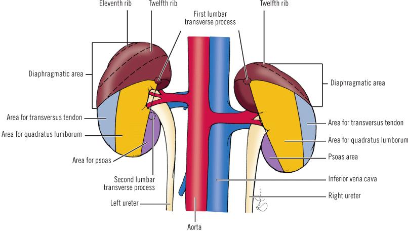
3 Location: Kidneys The kidneys are retroperitoneal organs, on the posterior abdominal wall. They are located at paravertebral gutters opposite T12, L1, L2, L3 vertebrae. The right kidney is about 1.25 cm lower than the left. The upper pole of the right kidney reaches the 12th rib and that of left kidney reaches 11th rib. The hilum of right kidney is just below transpyloric plane (L1), and that of the left kidney is just above it. L1 Dr Ahmed Salman


4 Kidneys Can be palpated in thin people,by press between 11 th and 12 th ribs and iliac crest (posteriorly( and below costal margin (anteriorly)
.")

5 General Features of the Kidneys: The kidneys has :- Two poles (upper and lower) The upper pole is nearer to the midline than the lower pole. The inferior pole of right kidney is about one finger breadth above iliac crest Two borders (lateral and medial). The lateral border is smooth and convex the medial is concave and presents a hilum at its middle. The hilum leads to a space within the kidney, called the sinus of the kidney. The structures passes through the hilum are renal vein, renal artery, renal pelvis. The renal vein is most anterior and renal pelvis is most posterior. Two surfaces (anterior and posterior).

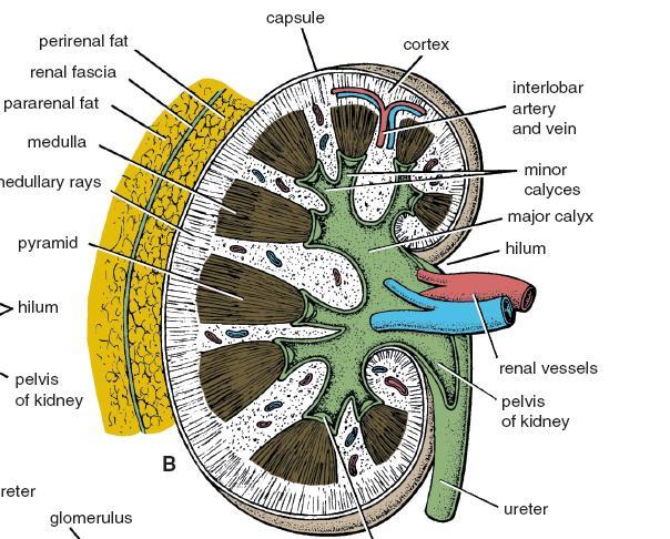
6 Coverings of the kidney :- From the cortex outwards 1- Fibrous capsule: surrounds the kidney all around 2- The perirenal fat: surrounds the kidney all around 3- Renal fascia: it is formed of 2 layers which cover the front and back of the kidneys. The renal fascia is continuous Laterally with fascia transversalis, Medially with the fascia around the renal vessels, aorta and IVC. Superiorly with the diaphragmatic fascia after forming a separate compartment for the suprarenal gland. Inferiorly it remains separate in front and back of the ureter. 4- Pararenal fat : outside the renal fascia, most condensed posterior to the kidney
7


8 Supporting factors of the kidney :- The kidney is kept in situ by Adjacent organs Abdominal pressure Perirenal fat Renal Fascia Pararenal fat Renal Blood vessels and ureters If the fat absorbed, as in rapid weight loss,descent of the kidney occurs (Nephroptosis).
9 Clinical notes : Nephroptosis cause intermittent pain in the renal region, relieved by lying down. This pain as result of traction on the renal vessels. Kidneys Transplantation The site of renal transplantation is the iliac fossa of the greater pelvis,due to lack of inferior support for the kidneys in the lumbar region Perinephric Abscess (pus around the kidney) The attachments of the renal fascia to the renal vessels and ureter, usually preventing the spread of pus to the contralateral side. Pus from an abscess (or blood from an injured kidney) may spread into the pelvis between the loosely attached anterior and posterior layers of the renal fascia.
10
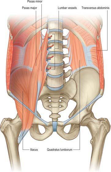
11 Relation of the Kidneys Posterior relations; are nearly similar for both kidneys 1- Four muscles, diaphragm (superiorly), psoas major, quadratus lumborum and transversus abdominis. 2-Four neurovascular structures; subcostal vessels, and subcostal, ilioohypogastric, and ilioinguinal nerves. 3-Pleura and ribs, the diaphragm separates the upper part of each kidney from the costodiaphragmatic recess of the pleura and 12th rib on right side and 11 th and 12 th ribs on left side. Pleura Injury During Renal surgical operations Due to close relation between costodiaphragmatic recess of the pleura and kidney
12
13 Anterior relations Right Kidney Right suprarenal gland Left Kidney Left suprarenal gland Second part of duodenum Right lobe of liver (with hepatorenal pouch in between) Spleen with lienorenal ligament, Body of pancreas with splenic vessels Posterior surface of stomach (with lesser sac in between) Right colic flexure (hepatic flexure) Descending colon Coils of the small intestine Ascending branch of right colic artery Coils of the small intestine ascending branch of left colic artery

14
15 Peritoneal covering of the kidney :- Although, the kidneys are retroperitoneal, the anterior surface of each kidney has 3 bare areas not covered by peritoneum. The other retroperitoneal structures are interposed between front of kidneys and the parietal peritoneum of posterior abdominal wall. Bare areas on right kidney Bare areas on left kidney Suprarenal area Suprarenal area Duodenal area Pancreatic area Colic area (hepatic flexure) Colic area (descending colon)

16
17 Structure of the kidney :- The Kidneys has two zones (outer cortex and inner medulla) surrounding sinus of the kidney. 1- Cortex; pale and adjacent to the capsule. It is divided into; Cortical arches which form caps over the bases of the medullary pyramids. 2- Medulla; is darker, deep to the cortex. It is formed of 7-14 pyramids Each pyramid has a base directed towards the cortex and an apex called renal papilla The part of cortex between the medullary pyramids is called Renal columns Each pyramid with its cap of cortex form a lobe of the kidney (7-14 lobes)

18 The minor calyces are about 5-12 per kidney. Each is a short funnel like tube which receives renal papillae The minor calyces unite to form 2-3 major calyces (in each kidney) and these in turn, unite to form the renal pelvis.

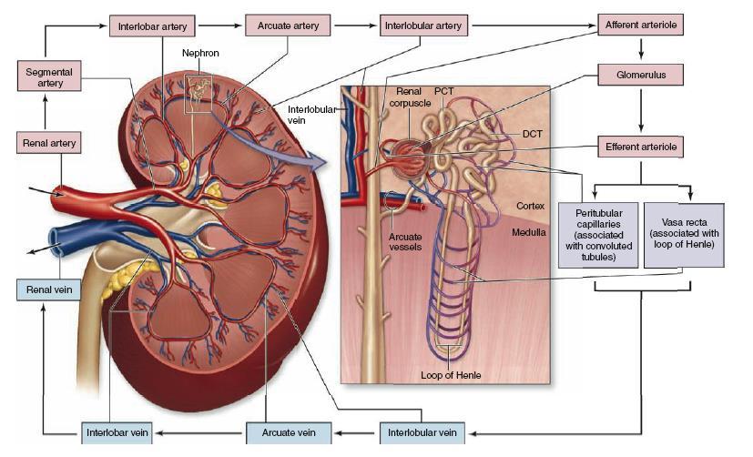
19 Arterial blood supply The renal arteries arise from the side of abdominal aorta, opposite the upper border of L2 vertebra. The right renal artery is longer than the left and passes posterior to IVC The renal artery gives inferior suprarenal artery, It divides into 5 segmental arteries which are end arteries. Collectively, the cortex receives over 10 times more blood than the medulla

20 Renal Artery 5 Segmental arteries Lobar arteries one for each renal pyramid Interlobar arteries run toward the cortex on each side of the renal pyramid arcuate arteries arch over the bases of the pyramids interlobular arteries ascend in the cortex glomerular arterioles
21
22 Venous drainage * Both right and left renal veins open directly into IVC Left renal vein is longer than the right and passes anterior to the aorta below origin of the superior mesenteric artery. The-left vein receives the left suprarenal and left gonadal vein. Lymph drainage To lateral aortic lymph nodes. Nerve supply :- By renal plexus derived from the coeliac plexus and supplemented by the lowest splanchnic nerve. It is mainly vasomotor in function.

23 Renal Vein Entrapment Syndrome (Nutcracker syndrome) Compression of left renal vein between the SMA anteriorly and the abdominal aorta posteriorly. Clinical presentation Haematuria due to renal venous hypertension, rupture of thin-walled veins into the collecting system
24 Renal Pain Renal pain varies from a dull ache to a severe pain in the flank Renal pain can result from stretching of the kidney capsule or spasm of the smooth muscle in the renal pelvis. Afferent nerve renal plexus around the renal artery lowest splanchnic nerve spinal cord at the level of T12 The flank and the anterior abdominal wall Pain is referred along distribution of the subcostal nerve (T12)
25 Surface anatomy of the Kidneys Morris rectangle : Two vertical lines are drawn; one and three inches from the middle line. Two horizontal planes are drawn opposite the spines of T11 and L3. The upper end lies 1 inch from midline opposite upper end of T12 vertebra. The hilum is 2 inches from midline at the transpyloric plane (L1) The lower end is 3 inches from the midline opposite L3 vertebra

26 Kidney Trauma The kidneys are well protected by the lower ribs, the lumbar muscles, and the vertebral column. A severe blunt trauma to the abdomen may crush the kidney against the last rib and the vertebral column. Depending on the severity of the blow, the injury varies from a mild bruising to a complete laceration Because 25% of the cardiac outflow passes through the kidneys, renal injury can result in rapid blood loss
27 The ureters: The ureters are muscular tubes which convey urine from kidneys to the urinary bladder. The ureter lies behind and adheres to the parietal peritoneum of the posterior abdominal wall. The ureter is about 10 inches (25 cm) and has 2 parts; abdominal and pelvic, each is 5 inches long. Dr Ahmed Salman

28 Dr Ahmed Salman
29 Course of the ureter The abdominal part Begins from the lower end of the renal pelvis (it is the pelvi-ureteric junction), It descends downwards and medially on psoas major muscle towards the pelvic brim. It crosses the end of the common or beginning of the external iliac artery to become the pelvic part. The pelvic part Descends downwards and backwards along the anterior margin of greater sciatic foramen till the ischial spine. It forms posterior boundary of the ovarian fossa. It runs forwards on pelvic floor to open in the wall of the urinary bladder. It is crossed by the vas deferens in male and uterine artery in females. It pierces the wall of the bladder obliquely to open at the superolateral angle of the trigone. This oblique termination of the ureter prevents regurgitation of urine from bladder to the ureter
 2.Genitofernoral nerve 3.")
30 Relations of the abdominal part of ureter:- Posterior Relation (BOTH SIDES) 1.Psoas major muscle separating the ureter from the tips of the transverse processes of the lumbar vertebrae (2-5) 2.Genitofernoral nerve 3.Termination of common or beginning of external iliac artery
31 Anterior and medial relations Anterior relation Intestinal structures Peritoneal elements Right ureter 1. Third part of the duodenum at its beginning 2. Terminal ileum near the pelvic brim 1. Parietal peritoneum of the posterior abdominal wall 2. Root of the mesentery Left ureter 1. Sigmoid colon near the pelvic brim 1. Parietal peritoneum of the posterior abdominal wall 2. Apex of sigmoid mesocolon with its intersigmoid recess vessels 1. Right gonadal vessels 2. Superior mesenteric vessels 3. Right colic vessels 4. Ilio-colic vessels Medial relation 1. Left gonadal vessels 2. Left colic vessels 3. Sigmoid vessels Inferior vena cava Inferior mesenteric vein
32
33
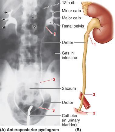
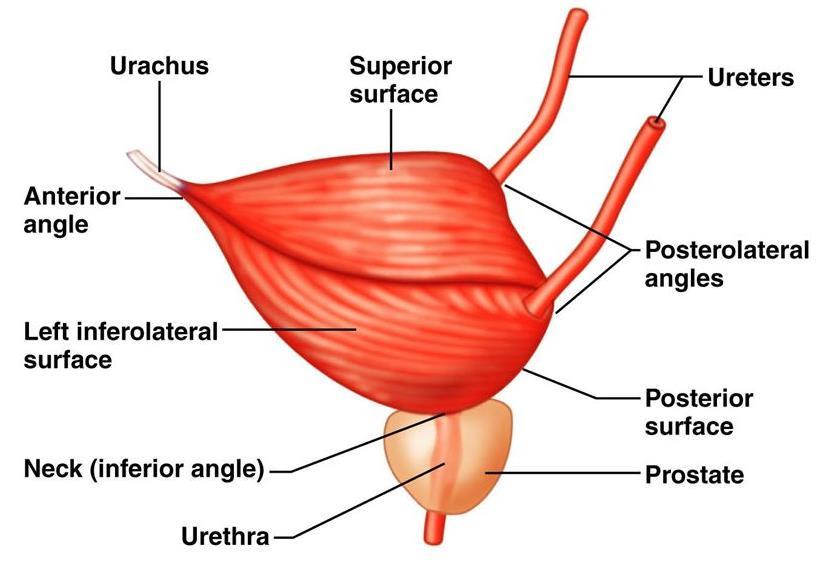
34 Constrictions of the ureters Site of constriction At pelvi-ureteric junction At pelvic brim In the wall of the urinary bladder Corresponding bony Level Near the tip of the transverse process of L2 vertebra In front of sacroiliac joint. Just medial to the ischial spine. (it is the narrowest point of the whole ureter)

35
36 Arterial blood supply :- Abdominal part receives branches from renal artery, abdominal aorta, gonadal and common iliac arteries Pelvic part receives branches from vesical, middle rectal and uterine arteries Lymph drainage:- To lateral aortic, common iliac lymph nodes.
37 Nerve supply :- The ureter receives sympathetic fibers from T11 L2 segments of spinal cord. Sensory fibers from the ureter enter the spinal cord through the same segments. Ureteric colic begins in the loin and is referred to groin, anterior aspect of the thigh through genitofemoral nerve (L1,L2) and scrotum or labium majora Surface markings :- The ureter begins at a point on the transpyloric plane, 5 cm from the midline It enters the bladder at the pubic tubercle.


38 Intra Venous Urogram IVU IVU KUB

39 MRI Abdomen

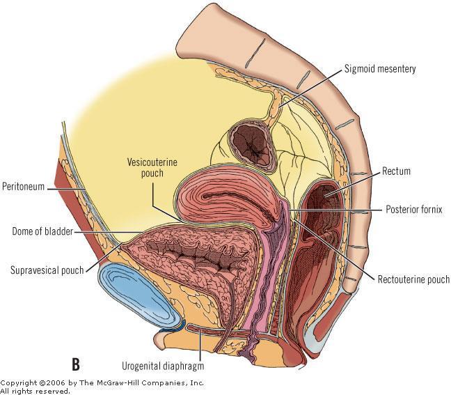
40 MRI Abdomen
41 Urinary Bladder The urinary bladder is a hollow viscus with strong muscular walls which acts as a reservoir for urine. Site of Urinary Bladder In infants: the bladder lies in the abdomen At about 6 years of age : the bladder begins to enter the enlarging pelvis. After puberty : the bladder lies within the lesser pelvis. In the adult: an empty bladder lies in lesser pelvis and as it fills, it ascends to the greater pelvis. Capacity of the Bladder: Average capacity of adult bladder is about 300 ml. Distension of the bladder by 500 ml may be tolerated. Beyond this, distension of the bladder is painful The bladder is enveloped in loose connective tissue called vesical fascia in which vesical venous plexus is embedded. Dr Ahmed Salman
42
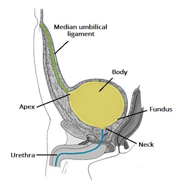
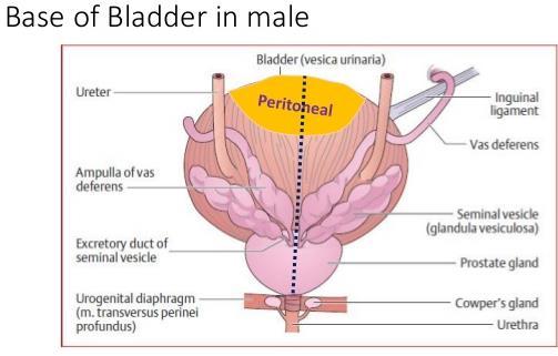
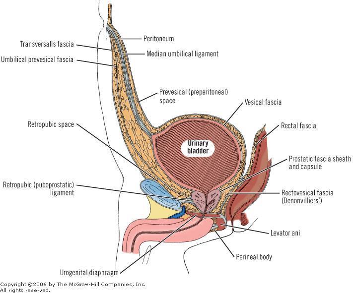
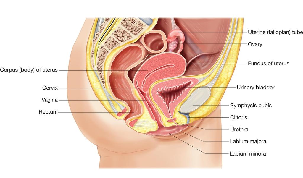
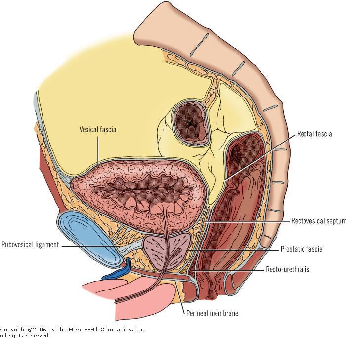
43 Description and Relations of the Urinary Bladder : The empty bladder has; Apex, base, 3 surfaces (superior, right and left inferolateral) and neck. 1- Apex of the bladder: Is continuous with the median umbilical ligament which raises the medianumbilical fold of peritoneum. The ligament is the remnant of the embryonic urachus. 2- Base of the bladder (fundus) : It is directed posteroinferiorly Its superolateral angles receive the ureters Relations : Male Base is related to rectum, but separated from it by Rectovesical pouch 2 seminal vesicles Ampullae of the deferent ducts (vas ) female The base is related to upper part of anterior wall of vagina.
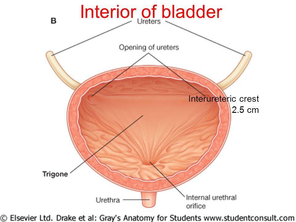
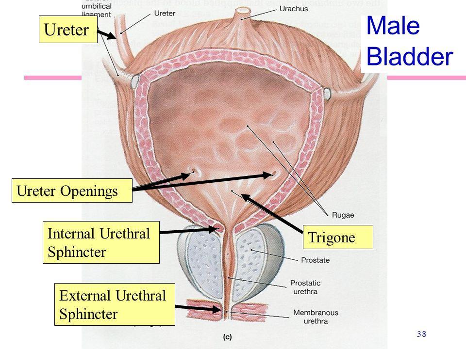
44
45 3-Superior Surface: is covered by peritoneum and is related to Male Sigmoid colon, Loops if ileum female Vesical surface of uterus. Supravaginal part of cervix with uterovesical pouch in between 4-Inferolateral surface: It is not covered by peritoneum. It is related to: Body of pubis with retropubic pad of fat in the retropubic space of Retzius. Levator ani. Obturator internus.
46 5-Neck of the bladder: It is the lowest and most fixed pan of the bladder. In the male: it is continuous with the urethra at the internal ureteral meatus and rests on the upper surface of the prostate. In female: it is continuous with the urethra and rests in the pelvic fascia which surrounds the urethra. At the junction of the neck and urethra, sphincter vesicae is present. Muscular coat of the bladder is composed of smooth muscle and is arranged as three layers known as the detrusor muscle.
47
48

49 Peritoneal Covering of the Bladder : In male, the superior surface and the superior part of its base is covered by peritoneum, In females, only the superior surface is covered by peritoneum. The peritoneum leaving the bladder is loosely attached to the suprapubic part of abdominal wall. The distended bladder lifts this peritoneum from the abdominal wall. In case of suprapubic cystostomy instruments could be introduced into the distended bladder to avoid injury of the peritoneum
50
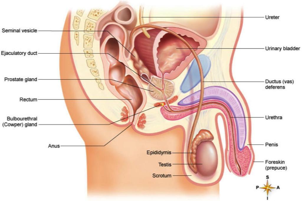
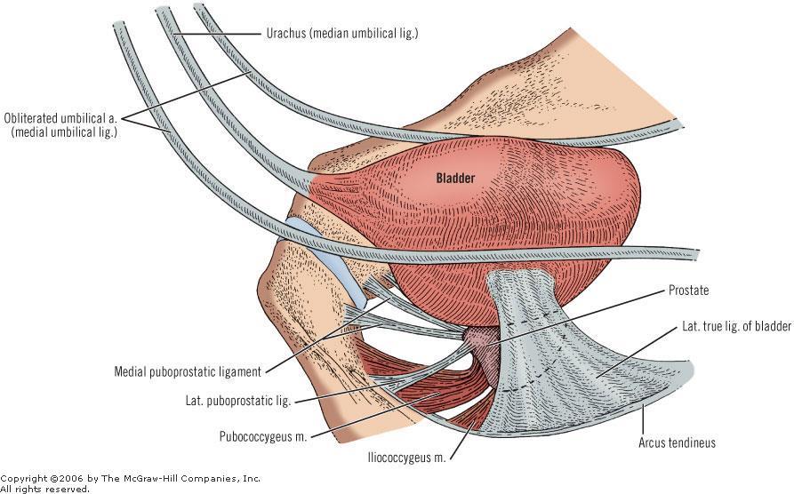
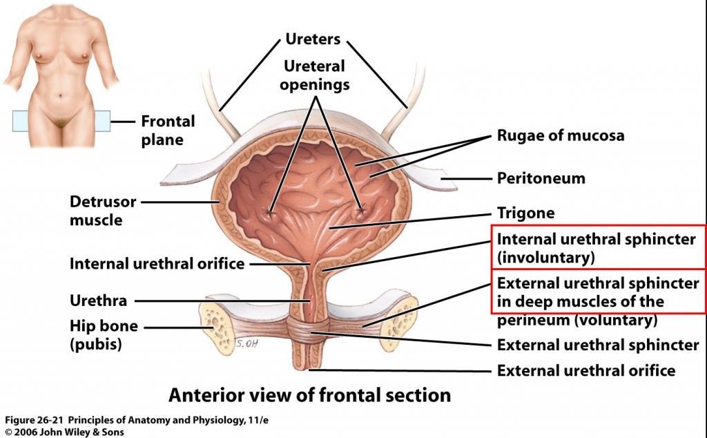
51 The ligaments of the bladder: 1-Median umbilical ligament: Continuous with apex of the bladder (it is the embryonic urachus) 2-Puboprostatic and pubovesical ligaments: In the male, the puboprostatic ligaments extend from back of the bodies of pubic bones to the anterior surface of the sheath of the prostate and neck of the bladder. In the female, the pubovesical ligaments extend from pubic bones to the urethra and neck of the bladder. 3-Lateral ligaments of the bladder: Each extends laterally from the side of the base of the bladder across the pelvic floor to the tendinous arch in side wall of the pelvis. These ligaments enclose arteries and autonomic nerves of the bladder. 4-Posterior ligaments: Each extends backwards from the base of the bladder to the corresponding internal iliac vein. They enclose vesical veins in their way to the internal iliac vein.
52
53 Interior of the Urinary Bladder : The mucous membrane over most of the bladder is loosely attached to the underlying muscular layer (detrusor muscle). The mucous membrane is folded in empty bladder, but in distended bladder, the folds disappear. Trigone of the bladder: It is the small triangular area which lies between the orifices of the ureters and the internal urethral meatus. (it is mesodermal in origin). It has the following special features: Its superior boundary is formed by the interureteric crest (ridge) which connects the two ureteric orifices. Its mucous membrane is always smooth and firmly adherent to the underlying muscle. It is very sensitive and vascular, so that, in cystoscope it appears red violet in colour In the male, the trigone overlies the median lobe of the prostate. After the middle age, the enlarged prostate elevates the mucous membrane behind the internal urethral orifice producing what is known as uvula vesicae of the bladder
54
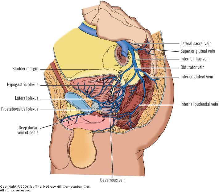
55 Arterial Blood Supply: In the male: superior and inferior vesical arteries. In the female: superior vesical and vaginal arteries. Venous Drainage: Begins by the vesical venous plexus, embedded in the visceral fascia on the inferolateral surfaces of the bladder. Inferiorly: In the male: it communicates with the prostatic venous plexus. In the female: it communicates with the vaginal venous plexus and receives the deep dorsal vein of the clitoris. Posteriorly: the plexus is drained by numerous vesical veins which run in the posterior ligaments of the bladder to end in the internal iliac veins. Vesical venous plexus Posteriorly Internal iliac veins MALE Prostatic venous plexus FEMALE Vaginal venous plexus deep dorsal vein of the clitoris

56
57 Lymphatic Drainage: To internal and external iliac lymph nodes. From the bladder neck, lymphatics drain directly to the sacral lymph nodes. Nerve Supply: By vesical nerve plexus, derived from the inferior hypogastric plexus, it contains t following fibers : Parasympathetic efferents (pelvic splanchnic nerves) (S 2, S 3, S 4 ): motor to the detrusor muscle, inhibitory to sphincter vesicae (they produce micturition). Sympathetic efferents: (L1,L2 ) are inhibitory to detrusor and stimulant to sphincter vesicae. Sensory afferents: Reach central nervous system through pelvic splanchnic nerves or Sympathetic fibers It record bladder distension and pain sensation.

58 Nerve Supply:
59 Bladder Injuries Intraperitoneally Usually involves the superior wall of the bladder Most commonly when the bladder is full Urine and blood escape freely into the peritoneal cavity Extraperitoneally Usually involves the anterior part of the bladder wall below the level of the peritoneal reflection it most commonly occurs in fractures of the pelvis The patient complains of lower abdominal pain and blood in the urine (hematuria)
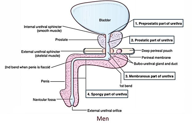
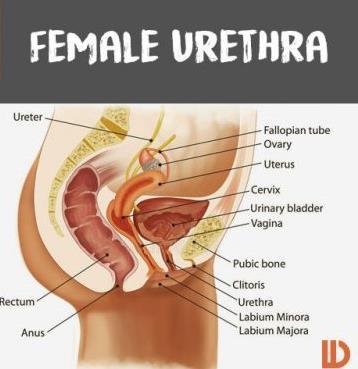
60 Urethra: Male Urethra The male urethra is about 20 cm long. It extends between 2 meatuses. Internal urethral meatus: at its junction with the neck of the bladder External urethral meatus:is a vertical slit, about 6 mm long. It is the narrowest part of urethra, and a calculus may lodge there. It is divided into 4 parts: the first and the second parts are in the pelvis, the third and fourth parts are in the perineum. It has 2 sphincters: Internal urethral sphincter (or sphincter vesicae), surrounds the neck of urinary bladder and the first (preprostatic) part of the urethra. External urethral sphincter (or sphincter urethrae), surrounds the third (membranous) part of the urethra Dr Ahmed Salman
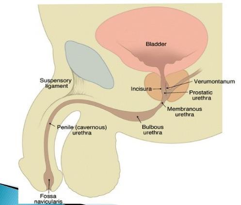
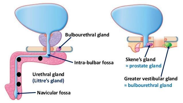
61 Parts of the Urethra First part: preprostatic part of urethra Second part: prostatic part of urethra Third part : membranous part of urethra Fourth : spongy part of urethra Length cm 3 cm 2 cm 15 cm Site between neck of the bladder and the base of the prostate traverses prostate from base to apex runs in deep perineal pouch bulb of penis and corpus spongiosum (Superficial Perineal Pouch) Size it is the widest part of urethra it is the least dilatable part Special features It is surrounded by internal sphincter Urethral crest Seminal colliculus Prostatic sinuses surrounded by external urethral sphincter -Dilated at its beginning to form to form intrabulbar fossa and at termination in glans penis to form the navicular fossa. -The bulbourethral glands open into its beginning
62
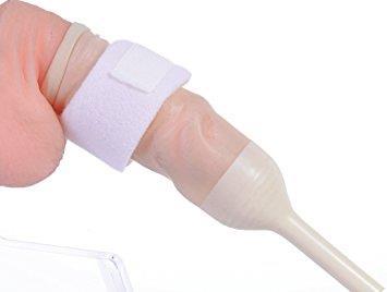
63
: Is a prominence at the middle of the crest.")

64 Special features of prostatic part of urethra Urethral crest: is a median longitudinal elevation in the mucous membrane of its posterior wall. Seminal colliculus (verumontanum): Is a prominence at the middle of the crest. It has three openings ; the opening of the prostatic utricle in its middle, and the openings of the two ejaculatory ducts on the sides. Prostatic sinuses : Each is a shallow depression on the side of the urethral crest. Each receives prostatic ducts.
65 Sphincters of the Urethra Internal Urethral sphincter External Urethral sphincter Site It lies in pelvis around neck of the bladder and preprostatic part of the urethra. It lies in the perineum, surrounds the membranous urethra in the deep perineal pouch. Structures It is formed of smooth muscle fibers It is formed of striated muscle fibers Nerve Supply Autonomic fibers from the inferior hypogastric plexus Functions It acts Involuntarily. It is well-developed in both male and female. It maintains continence of urine. In the male: it has a genital function, it prevents reflux of semen into the urinary bladder during ejaculation Somatic : from the perineal branch of pudendal nerve of the sacral plexus It acts voluntarily. It is well-developed in the male It maintains continence of urine.
66
67 Vessels Nerves and Lymphatics of the urethra : Urethra receives its blood and nerve supply from those of prostate and penis. Lymphatics: From the prostatic and membranous parts to internal and external iliac lymph nodes From the spongy part to deep and superficial inguinal lymph nodes. Urinary Retention It is more common in male due to a benign or malignant enlargement of the prostate or acute urethritis or prostatitis. The only anatomic cause of urinary retention in females is acute inflammation around the urethra (e.g., from herpes).

68 Rupture of the Urethra Rupture of the urethra resulted from a severe blow on the perineum. Site of rupture 1-At bulb of the penis, just below the perineal membrane. The urine extravasates into the superficial perineal pouch and then passes forward over the scrotum beneath the membranous layer of the superficial fascia. 2-Membranous part of the urethra is ruptured, urine escapes into the deep perineal pouch and extravasate upward around the prostate and bladder or downward into the superficial perineal pouch. IN BOOTH CASES The urine cannot passes into thigh because attachment of colles fascia with fasciae lata below inguinal ligament

69 Female Urethra : length : 4 cm. Course. Relations: It begins at the internal urethral meatus at the neck of the bladder. It traverses the deep perineal pouch to end at the external urethral orifice in the vestibule anterior to the vaginal orifice. It is embedded in the anterior wall of the vagina. On each side of the urethra, the mucous membrane of the urethra presents a number of small mucous glands called the paraurethral glands which correspond to the prostate in the male.
70
71 Clinically Significant Differences Between Male and Female Urethrae: The female urethra is distensible because it contains considerable elastic tissue, as well as smooth muscle. It can be easily dilated without injury. Infections of the urethra, and bladder, are more common in women because the female urethra is short, more distensible, and is open to the exterior.
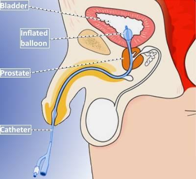
72 Anatomy of the Procedure of Catheterization 1. The patient lies in a supine position. 2. With gentle traction, the penis is held erect at right angles to the anterior abdominal wall. The lubricated catheter is passed through the narrow external urethral meatus. On reaching the membranous part of the urethra, a slight resistance is felt because of the tone of the urethral sphincter and the surrounding rigid perineal membrane. 3. The penis is then lowered toward the thighs, and the catheter is gently pushed through the sphincter 4-Passage of the catheter through the prostatic urethra and bladder neck should be easily.
73
74
Jordan University Faculty Of Medicine. Urinary System. Dr. Ahmed Salman. Assistant professor of anatomy & embryology
 Jordan University Faculty Of Medicine Urinary System Dr. Ahmed Salman Assistant professor of anatomy & embryology The urinary system is composed of two kidneys,two ureters,urinary bladder and urethra The
Gross Anatomy of the Urinary System
 Gross Anatomy of the Urinary System Lecture Objectives Overview of the urinary system. Describe the external and internal anatomical structure of the kidney. Describe the anatomical structure of the ureter
Urinary Bladder. Prof. Imran Qureshi
 Urinary Bladder Prof. Imran Qureshi Urinary Bladder It develops from the upper end of the urogenital sinus, which is continuous with the allantois. The allantois degenerates and forms a fibrous cord in
Rama Nada. - Ensherah Mokheemer. - Ahmed salman. 1 P a g e
 - 5 - Rama Nada - Ensherah Mokheemer - Ahmed salman 1 P a g e We will continue talking about the urinary bladder The ligaments of the bladder: 1-Median umbilical ligament: Continuous with apex of the bladder
THE ABDOMEN SUPRARENAL GLANDS KIDNEY URETERS URINARY BLADDER
 THE ABDOMEN SUPRARENAL GLANDS KIDNEY URETERS URINARY BLADDER THE SUPRARENAL GLANDS The suprarenal (adrenal) glands lie immediately superior and slightly anterior to the upper pole of either kidney. Golden
Ureters, Urinary Bladder & Urethra
 Ureters, Urinary Bladder & Urethra Please check our Editing File هذا العمل ال يغني عن المصدر األساسي للمذاكرة Lecture 2 } و م ن ي ت و ك ع ل ا لل ه ف ه و ح س ب ه { Objectives o Describe the course of ureter
Gross anatomy of the urinary system. Done by : razan krishan. slide in bold and book in green
 Gross anatomy of the urinary system Done by : razan krishan slide in bold and book in green Kidneys, ureters, urinary bladder & urethra Urine flows from each kidney, down its ureter to the bladder and
ANATOMY OF PELVICAYCEAL SYSTEM -DR. RAHUL BEVARA
 1 ANATOMY OF PELVICAYCEAL SYSTEM -DR. RAHUL BEVARA 2 KIDNEY:ANATOMY OVERVIEW Kidneys are retroperitoneal, in posterior abdominal region, extending from T12 L3 Bean-shaped Right kidney is lower than left
Lecture 56 Kidney and Urinary System
 Lecture 56 Kidney and Urinary System The adrenal glands are located on the superomedial aspect of the kidney The right diagram shows a picture of the kidney with the abdominal walls and organs removed
Anatomy of the renal system. Professor Nawfal K. Al-Hadithi
 Anatomy of the renal system Professor Nawfal K. Al-Hadithi Objectives To describe the posterior abdominal wall To identify the main anatomical landmarks of the kidneys & ureters To describe the suprarenal
THE KIDNEY (Fig. 1 & 2) Figure 1
 Figure 1") 1 THE KIDNEY (Fig. 1 & 2) The kidney is a bean-shaped, reddish-brown organ of the urinary system located retroperitoneally in the upper part of the paravertebral gutter of the abdominal cavity, padded
1 THE KIDNEY (Fig. 1 & 2) The kidney is a bean-shaped, reddish-brown organ of the urinary system located retroperitoneally in the upper part of the paravertebral gutter of the abdominal cavity, padded
Inferior Pelvic Border
 Pelvis + Perineum Pelvic Cavity Enclosed by bony, ligamentous and muscular wall Contains the urinary bladder, ureters, pelvic genital organs, rectum, blood vessels, lymphatics and nerves Pelvic inlet (superior
The Urinary System Pearson Education, Inc.
 26 The Urinary System Introduction The urinary system does more than just get rid of liquid waste. It also: Regulates plasma ion concentrations Regulates blood volume and blood pressure Stabilizes blood
د. عصام طارق. Objectives:
 GI anatomy Lecture: 5 د. عصام طارق Objectives: To describe anatomy of stomach, duodenum & pancreas. To list their main relations. To define their blood & nerve supply. To list their lymph drainage. To
Dana Alrafaiah. - Amani Nofal. - Ahmad Alsalman. 1 P a g e
 - 2 - Dana Alrafaiah - Amani Nofal - Ahmad Alsalman 1 P a g e This lecture will discuss five topics as follows: 1- Arrangement of pelvic viscera. 2- Muscles of Pelvis. 3- Blood Supply of pelvis. 4- Nerve
Anatomy of the Large Intestine
 Large intestine Anatomy of the Large Intestine 2 Large Intestine Extends from ileocecal valve to anus Length = 1.5-2.5m = 5 feet Regions Cecum = 2.5-3 inch Appendix= 3-5 inch Colon Ascending= 5 inch Transverse=
The abdominal Esophagus, Stomach and the Duodenum. Prof. Oluwadiya KS
 The abdominal Esophagus, Stomach and the Duodenum Prof. Oluwadiya KS www.oluwadiya.com Viscera of the abdomen Abdominal esophagus: Terminal part of the esophagus The stomach Intestines: Small and Large
Pelvis MCQs. Block 1. B. Reproductive organs. C. The liver. D. Urinary bladder. 1. The pelvic diaphragm includes the following muscles: E.
 Pelvis MCQs Block 1 1. The pelvic diaphragm includes the following muscles: A. The obturator internus B. The levator ani C. The coccygeus D. The external urethral sphincter E. The internal urethral sphincter
REPRODUCTIVE SYSTEM By Dr.Ahmed Salman
 The University Of Jordan Faculty Of Medicine Anatomy Department REPRODUCTIVE SYSTEM By Dr.Ahmed Salman Assistant Professor of Anatomy &embryology Perineum It is the diamond-shaped lower end of the trunk
The functional anatomy of the urinary system. Human Anatomy Department Dr. Anastasia Bendelic
 The functional anatomy of the urinary system Human Anatomy Department Dr. Anastasia Bendelic Plan Development of the kidneys and their abnormalities Development of the urinary ways and their abnormalities
STRUCTURAL BASIS OF MEDICAL PRACTICE EXAMINATION 3. October 16, 2015
 STRUCTURAL BASIS OF MEDICAL PRACTICE EXAMINATION 3 October 16, 2015 PART l. Answer in the space provided. (12 pts) 1. Identify the structures. (2 pts) A. B. A B C. D. C D 2. Identify the structures. (2
Abdomen. Retroperitoneal space
 Abdomen. Retroperitoneal space Abdominal cavity The space bounded by: Anterolateral abdominal wall Posterior abdominal wall Diaphragm Pelvic walls and pelvic floor. Subdivided into: True abdominal cavity
The posterior abdominal wall. Prof. Oluwadiya KS
 The posterior abdominal wall Prof. Oluwadiya KS www.oluwadiya.sitesled.com Posterior Abdominal Wall Lumbar vertebrae and discs. Muscles opsoas, quadratus lumborum, iliacus, transverse, abdominal wall
Benha University. Faculty of Medicine. Anatomy Department Course code (MED 0701) Model answer of Anatomy examination. (Abdomen,Pelvis and Thorax)
 Model answer of Anatomy examination. (Abdomen,Pelvis and Thorax)") 1 Benha University Faculty of Medicine Anatomy Department Course code (MED 0701) Model answer of Anatomy examination (Abdomen,Pelvis and Thorax) 1 st year 2 nd term Date :18 /5 /2013 2 I-Short account
1 Benha University Faculty of Medicine Anatomy Department Course code (MED 0701) Model answer of Anatomy examination (Abdomen,Pelvis and Thorax) 1 st year 2 nd term Date :18 /5 /2013 2 I-Short account
ABDOMINAL WALL & RECTUS SHEATH
 ABDOMINAL WALL & RECTUS SHEATH Learning Objectives Describe the anatomy, innervation and functions of the muscles of the anterior, lateral and posterior abdominal walls. Discuss their functional relations
STRUCTURAL BASIS OF MEDICAL PRACTICE EXAMINATION 3. October 17, 2014
 STRUCTURAL BASIS OF MEDICAL PRACTICE EXAMINATION 3 October 17, 2014 PART l. Answer in the space provided. (12 pts) 1. Identify the structures. (2 pts) A. B. A B C. D. C D 2. Identify the structures. (2
Pancreas & Biliary System. Dr. Vohra & Dr. Jamila
 Pancreas & Biliary System Dr. Vohra & Dr. Jamila 1 Objectives At the end of the lecture, the student should be able to describe the: Location, surface anatomy, parts, relations & peritoneal reflection
Table 2. First Generated List of Expert Responses. Likert-Type Scale. Category or Criterion. Rationale or Comments (1) (2) (3) (4)
 (2) (3) (4)") Table 2. First Generated List of Expert Responses. Likert-Type Scale Category or Criterion Anatomical Structures and Features Skeletal Structures and Features (1) (2) (3) (4) Rationale or Comments 1. Bones
Table 2. First Generated List of Expert Responses. Likert-Type Scale Category or Criterion Anatomical Structures and Features Skeletal Structures and Features (1) (2) (3) (4) Rationale or Comments 1. Bones
Perineum. done by : zaid al-ghnaneem
 Perineum done by : zaid al-ghnaneem Hello everyone, this sheet will talk about 2 nd Lecture which is Perineum but there are some slides and info from 1 st Lecture. Everything included Slides + Pics Let
Peritoneum: Def. : It is a thin serous membrane that lines the walls of the abdominal and pelvic cavities and clothes the viscera.
 Peritoneum: Def. : It is a thin serous membrane that lines the walls of the abdominal and pelvic cavities and clothes the viscera. Layers of the peritoneum: 1. Outer Layer ( Parietal Peritoneum) : lines
URINARY SYSTEM ANATOMY
 URINARY SYSTEM ANATOMY Adapted from Human Anatomy & Physiology Marieb and Hoehn (9 th ed.) OVERVIEW Metabolism of nutrients by the body produces wastes that must be removed from the body. Although excretory
Accessory Glands of Digestive System
 Accessory Glands of Digestive System The liver The liver is soft and pliable and occupies the upper part of the abdominal cavity just beneath the diaphragm. The greater part of the liver is situated under
Duodenum retroperitoneal
 Duodenum retroperitoneal C shaped Initial region out of stomach into small intestine RETROperitoneal viscus Superior 1 st part duodenal cap ; moves upwards and backwards to lie on the R crura medial to
GI module Lecture: 9 د. عصام طارق. Objectives:
 GI module Lecture: 9 د. عصام طارق Objectives: To list structures forming posterior abdominal wall. To follow aorta & its main branches. To describe IVC & its main tributaries. To list nerves of posterior
Pelvis Perineum MCQs. Block 1.1. A. Urinary bladder. B. Rectum. C. Reproductive organs. D. The thigh
 Pelvis Perineum MCQs Block 1.1 1. The pelvic diaphragm includes the following muscles: A. The coccygeus B. The levator ani C. The external urethral sphincter D. The internal urethral sphincter E. The obturator
URINARY SYSTEM ANATOMY PART
 URINARY SYSTEM ANATOMY PART 1 DANIL HAMMOUDI.MD Urinary System Composed of kidneys, ureters, urinary bladder, and urethra Eliminates nitrogenous wastes from the body Regulates water, electrolyte, and ph
The Kidneys. (L., ren; Gk, nephros; hence the adjectives renal and nephric) & Suprarenal (Adrenal) Glands. Dr Maan Al-Abbasi PhD, MBChB
 & Suprarenal (Adrenal) Glands. Dr Maan Al-Abbasi PhD, MBChB") The Kidneys (L., ren; Gk, nephros; hence the adjectives renal and nephric) & Suprarenal (Adrenal) Glands Dr Maan Al-Abbasi PhD, MBChB Functions of Urinary System Regulate electrolytes (K+, Na+, etc) Regulate
The Kidneys (L., ren; Gk, nephros; hence the adjectives renal and nephric) & Suprarenal (Adrenal) Glands Dr Maan Al-Abbasi PhD, MBChB Functions of Urinary System Regulate electrolytes (K+, Na+, etc) Regulate
SUBJECTS 2nd year, 1st semester I. 1. Primitive gut - limits, derivatives 2. Foregut -limits, evolution, derivatives 3. Midgut -limits, evolution,
 SUBJECTS 2nd year, 1st semester I. 1. Primitive gut - limits, derivatives 2. Foregut -limits, evolution, derivatives 3. Midgut -limits, evolution, derivatives 4. Hindgut- limits, evolution, derivatives
Femoral Triangle and Adductor Canal. Dr. Heba Kalbouneh Associate Professor of Anatomy and Histology
 Femoral Triangle and Adductor Canal Dr. Heba Kalbouneh Associate Professor of Anatomy and Histology Femoral Triangle and Adductor Canal Femoral triangle Is a triangular depressed area located in the upper
Dr. Zahiri. In the name of God
 Dr. Zahiri In the name of God small intestine = small bowel is the part of the gastrointestinal tract Boundaries: Pylorus Ileosecal junction Function: digestion and absorption of food It receives bile
Chapter 26. The Urinary System. Lecture Presentation by Steven Bassett Southeast Community College Pearson Education, Inc.
 Chapter 26 The Urinary System Lecture Presentation by Steven Bassett Southeast Community College Introduction The urinary system does more than just get rid of liquid waste. It also: Regulates plasma ion
Urinary System. Chapter 17 7/19/11. Introduction
 7/19/11 Chapter 17 Urinary System Introduction A. The urinary system consists of two kidneys that filter the blood, two ureters, a urinary bladder, and a urethra to convey waste substances to the outside.
Yes, cranially with ovarian, caudally with vaginal. Yes, with uterine artery (collateral circulation between abdominal +pelvic source)
") Blood supply to internal female genitalia: uterine Internal iliac Sup. large branch: uterus, inf. Small branch: cervix+ sup. Vagina Yes, cranially with ovarian, caudally with vaginal Medially in base of
Blood supply to internal female genitalia: uterine Internal iliac Sup. large branch: uterus, inf. Small branch: cervix+ sup. Vagina Yes, cranially with ovarian, caudally with vaginal Medially in base of
Exploring Anatomy: the Human Abdomen
 Exploring Anatomy: the Human Abdomen PERITONEUM AND PERITONEAL CAVITY PERITONEUM The peritoneum is a thin serous membrane that lines the abdominal cavity and covers, in variable amounts, the viscera within
The peritoneum. Prof. Oluwadiya KS, MBBS, FMCS(Orthop) Website:
 Website:") The peritoneum Prof. Oluwadiya KS, MBBS, FMCS(Orthop) Website: http://oluwadiya.com The peritoneum Serous membrane that lines the abdominopelvic cavity and invests the viscera The largest serous membrane
The peritoneum Prof. Oluwadiya KS, MBBS, FMCS(Orthop) Website: http://oluwadiya.com The peritoneum Serous membrane that lines the abdominopelvic cavity and invests the viscera The largest serous membrane
Urinary System VASTACCESS, INC.
 Urinary System www.vastaccess.com 2 Urinary Tract Kidney Ureter Urinary Bladder Urethra Prostate (male) Membranous (male) Spongy (male) 3 Kidney Relations Suprarenal (Adrenal) Glands Liver Duodenum Transverse
BY DR NOMAN ULLAH WAZIR
 BY DR NOMAN ULLAH WAZIR The stomach (from ancient Greek word stomachos, stoma means mouth) is a muscular, hollow and the most dilated part of the GIT. It starts from the point where esophagus ends. It
Copyright 2003 Pearson Education, Inc. publishing as Benjamin Cummings. Dr. Nabil Khouri
 Dr. Nabil Khouri Objectives: General objectives: - to identify the kidney s structures, function and location - to analyze the relationship between microscopic structure and function Specific objectives:
Anatomy of the Thorax
 Anatomy of the Thorax A) THE THORACIC WALL Boundaries Posteriorly by the thoracic part of the vertebral column Anteriorly by the sternum and costal cartilages Laterally by the ribs and intercostal spaces
أحمد رواجبة- محمود الحربي- أحمد السالمان-
 -6 أحمد رواجبة- محمود الحربي- أحمد السالمان- 1 P a g e The Male Reproductive System The male genital system structures are divided into: Internal structures: 1- Prostate 3-Ejaculatory ducts External structures:
Physiologic Anatomy and Nervous Connections of the Bladder
 Micturition Objectives: 1. Review the anatomical organization of the urinary system from a physiological point of view. 2. Describe the micturition reflex. 3. Predict the lines of treatment of renal failure.
2. List the 8 pelvic spaces: list one procedure or dissection which involves entering that space.
 Name: Anatomy Quiz: Pre / Post 1. In making a pfannensteil incision you would traverse through the following layers: a) Skin, Camper s fascia, Scarpa s fascia, external oblique aponeurosis, internal oblique
DISSECTION 8: URINARY AND REPRODUCTIVE SYSTEMS
 8546d_c01_1-42 6/25/02 4:32 PM Page 38 mac48 Mac 48: 420_kec: 38 Cat Dissection DISSECTION 8: URINARY AND REPRODUCTIVE SYSTEMS Typically, the urinary and reproductive systems are studied together, because
Biology Human Anatomy Abdominal and Pelvic Cavities
 Biology 351 - Human Anatomy Abdominal and Pelvic Cavities Please place your name and I.D. number on the back of the last page of this exam. You must answer all questions on this exam. Because statistics
[ANATOMY #12] April 28, 2013
![[ANATOMY #12] April 28, 2013](/thumbs/86/93473883.jpg "[ANATOMY #12] April 28, 2013") Sympathetic chain : Sympathetic chain is each of the pair of ganglionated longitudinal cords of the sympathetic nervous system; extend from level of atlas (base of skull) till coccyx. It is paravertebral
Sympathetic chain : Sympathetic chain is each of the pair of ganglionated longitudinal cords of the sympathetic nervous system; extend from level of atlas (base of skull) till coccyx. It is paravertebral
Anatomy: Know Your Abdomen
 Anatomy: Know Your Abdomen Glossary Abdomen - part of the body below the thorax (chest cavity); separated by the diaphragm. Anterior - towards the front of the body. For example, the umbilicus is anterior
Netter's Anatomy Flash Cards Section 4 List 4 th Edition
 Netter's Anatomy Flash Cards Section 4 List 4 th Edition https://www.memrise.com/course/1577335/ Section 4 Abdomen (31 cards) Plate 4-1 Bony Framework of Abdomen 1.1 Costal cartilages 1.2 Iliac crest 1.3
Mousa Salah. Dr. Mohammad Al. Mohtasib. 1 P a g e
 8 Mousa Salah Dr. Mohammad Al. Mohtasib 1 P a g e In the previous lecture we talked about the peritoneum, and we said that the peritonium is a serous sac, and it consists of two layers, visceral and parietal.
THE THORACIC WALL. Boundaries Posteriorly by the thoracic part of the vertebral column. Anteriorly by the sternum and costal cartilages
 THE THORACIC WALL Boundaries Posteriorly by the thoracic part of the vertebral column Anteriorly by the sternum and costal cartilages Laterally by the ribs and intercostal spaces Superiorly by the suprapleural
General Anatomy of Urinary System
 General Anatomy of Urinary System URINARY SYSTEM ORGANS Kidneys (2) Ureters (2) Urinary bladder Urethra KIDNEY FUNCTIONS Control blood volume and composition KIDNEY FUNCTIONS Filter blood plasma, eliminate
Figure 26.1 An Introduction to the Urinary System
 Chapter 26 Figure 26.1 An Introduction to the Urinary System Components of the Urinary System Kidney Produces urine Ureter Transports urine toward the urinary bladder Urinary Bladder Temporarily stores
LECTURE ON THE URINARY SYSTEM
 LECTURE ON THE URINARY SYSTEM (Uropoetic System) AN OVERVIEW Dr HAMIADJI THE URINARY SYSTEM URINARY SYSTEM The kidneys are responsible for removing wastes from the body, regulating electrolyte balance
In the name ofgod. Abdomen 3. Dr. Zahiri
 In the name ofgod Abdomen 3 Dr. Zahiri Peritoneum Peritoneum It is the serous membrane(a type of loose connective tissue and is covered by mesothelium) that lines the abdominal cavity. Extensions of the
NOTES FROM GUTMAN LECTURE 10/26 Use this outline to study from. As you go through Gutman s lecture, fill in the topics.
 NOTES FROM GUTMAN LECTURE 10/26 Use this outline to study from. As you go through Gutman s lecture, fill in the topics. Anatomy above the arcuate line Skin Camper s fascia Scarpa s fascia External oblique
Urinary 1 Checklist Gross Anatomy of the Urinary System
 Urinary 1 Checklist Gross Anatomy of the Urinary System Urinary system Kidneys Parietal peritoneum Retroperitoneal Renal fascia The urinary system consists of two kidneys, two ureters, the urinary bladder,
The Thoracic wall including the diaphragm. Prof Oluwadiya KS
 The Thoracic wall including the diaphragm Prof Oluwadiya KS www.oluwadiya.com Components of the thoracic wall Skin Superficial fascia Chest wall muscles (see upper limb slides) Skeletal framework Intercostal
Abdomen: Introduction. Prof. Oluwadiya KS
 Abdomen: Introduction Prof. Oluwadiya KS www.oluwadiya.com Abdominopelvic Cavity Abdominal Cavity Pelvic Cavity Extends from the inferior margin of the thorax to the superior margin of the pelvis and the
Inguinal Canal. It is an oblique passage through the lower part of the anterior abdominal wall. Present in both sexes
 Inguinal canal Inguinal Canal It is an oblique passage through the lower part of the anterior abdominal wall Present in both sexes It allows structures to pass to and from the testis to the abdomen in
Preview from Notesale.co.uk Page 1 of 34
 Abdominal viscera and digestive tract Digestive tract Abdominal viscera comprise majority of the alimentary system o Terminal oesophagus, stomach, pancreas, spleen, liver, gallbladder, kidneys, suprarenal
BLOCK IV: OFFICIAL BODY PARTS LIST FOR ANTERIOR ABDOMINAL WALL AND ABDOMINAL CONTENTS
 BLOCK IV: OFFICIAL BODY PARTS LIST FOR ANTERIOR ABDOMINAL WALL AND ABDOMINAL CONTENTS External oblique muscle Muscular portion Aponeurotic portion Superficial inguinal ring Lateral (inferior) crus Medial
-Ensherah Mokheemer. -Shatha Al-Jaberi محمد المحتسب- 1 P a g e
 9-9 -Ensherah Mokheemer -Shatha Al-Jaberi محمد المحتسب- 1 P a g e Small intestine has three regions: ( االثني عشر( The duodenum The jejunum The ileum Small intestine Duodenum: -c-shaped -The concavity
Lecture 02 Anatomy of the LIVER
 Lecture 02 Anatomy of the LIVER BY Dr Farooq Khan Aurakzai Dated: 02.01.2018 Introduction to Liver Largest gland in the body. 2 nd largest organ of the body. Weight approximately 1500 gm, and is roughly
-15. -Alaa Albandi. -Dr. Mohammad Almohtasib. 0 P a g e
 -15 -Alaa Albandi - -Dr. Mohammad Almohtasib 0 P a g e In this last lecture, we will talk about the sigmoid colon, rectum, and anal canal. Sigmoid colon It has a mesentery called pelvic mesocolon or sigmoidal
To describe the liver. To list main structures in porta hepatis.
 GI anatomy Lecture: 6 د. عصام طارق Objectives: To describe the liver. To list main structures in porta hepatis. To define portal system & portosystemic anastomosis. To list parts of biliary system. To
Pancreas and Biliary System
 Pancreas and Biliary System Please view our Editing File before studying this lecture to check for any changes. Color Code Important Doctors Notes Notes/Extra explanation Objectives At the end of the lecture,
Nerves on the Posterior Abdominal Wall
 Nerves on the Posterior Abdominal Wall Lumbar Plexus The lumbar plexus, which is one of the main nervous pathways supplying the lower limb, is formed in the psoasmuscle from the anterior ramiof the upper
The Spleen. Dr Fahad Ullah
 The Spleen BY Dr Fahad Ullah Spleen The spleen is an largest lymphoid organ shaped like a shoe that lies relative to the 9th and 11th ribs and is located in the left hypochondrium. Thus, the spleen is
The jejunum and the Ileum. Prof. Oluwadiya KS
 The jejunum and the Ileum Prof. Oluwadiya KS www.oluwadiya.siteled.com Introduction Introduction The small intestine (SI) comprises of the duodenum, jejunum and the ileum The jejunum is the second part
The Kidneys, the Ureters, the Bladder, and the Urethra
 The Urinary System 21 The Kidneys, the Ureters, the Bladder, and the Urethra Chapter Outline The Kidneys 370 Renal Mobility 370 Kidney Trauma 370 Kidney Tumors 371 Renal Pain 371 Transplanted Kidneys
Bony ypelvis. Composition: formed by coccyx, and their articulations Two portions
 Pelvis Bony ypelvis Composition: formed by paired hip bones, sacrum, coccyx, and their articulations Two portions Greater pelvis Lesser pelvis Terminal line ( pelvic inlet): formed by promontory of sacrum,
Waste. Urinary System Anatomy Urinary Section pages 5-8. Urinary System. Urinary System. Nitrogenous Wastes. Nitrogenous Wastes 4/22/2016
 Waste Urinary System Anatomy Urinary Section pages 5-8 Metabolism produces waste products What is the primary waste product of cellular respiration? How does the body dispose of it? Urinary System Urinary
Anatomy of the SMALL INTESTINE. Dr. Noman Ullah Wazir PMC
 Anatomy of the SMALL INTESTINE Dr. Noman Ullah Wazir PMC SMALL INTESTINE The small intestine, consists of the duodenum, jejunum, and illium. It extends from the pylorus to the ileocecal junction were the
Biology Human Anatomy Abdominal and Pelvic Cavities
 Biology 351 - Human Anatomy Abdominal and Pelvic Cavities You must answer all questions on this exam. Because statistics demonstrate that, on average, between 2-5 questions on every 100-point exam are
Chapter 5: Other mediastinal structures. The Large Arteries. The Aorta. Ascending aorta
 Chapter 5: Other mediastinal structures The Large Arteries The Aorta The aorta is the main arterial trunk of the systemic circulation and in the healthy state its wall contain a large amount of yellow
Surface Anatomy. Location Shape Weight Role of Five Surfaces Borders Fissures Lobes Peritoneal Lig
 The Liver Functions Bile production and secretion Detoxification Storage of glycogen Protein synthesis Production of heparin and bile pigments Erythropoiesis (in fetus) Surface Anatomy Location Shape Weight
It passes through the diaphragm at the level of the 10th thoracic vertebra to join the stomach
 The esophagus is a tubular structure (muscular, collapsible tube ) about 10 in. (25 cm) long that is continuous above with the laryngeal part of the pharynx opposite the sixth cervical vertebra The esophagus
UROGENITAL SYSTEM By Dr.Ahmed Salman
 The University Of Jordan Faculty Of Medicine Anatomy Department UROGENITAL SYSTEM By Dr.Ahmed Salman Assistance Professor of Anatomy &embryology PELVIS Learning Objectives 1. Bony pelvis, its joints and
ABDOMEN. 2. The highest branch of the abdominal aorta is: (a) R suprarenal a (b) Coeliac trunk (c) L renal a (d) L gonadal a (e) SMA
 R suprarenal a (b) Coeliac trunk (c) L renal a (d) L gonadal a (e) SMA") ABDOMEN 1. The duodenum: (a) is a retroperitoneal structure (b) is 25cm long (c) lies between the levels of L2-L4 (d) in its fourth part lies to the R of the aorta (e) all of the above 2. The highest branch
ABDOMEN 1. The duodenum: (a) is a retroperitoneal structure (b) is 25cm long (c) lies between the levels of L2-L4 (d) in its fourth part lies to the R of the aorta (e) all of the above 2. The highest branch
Group of students. - Rawan almujabili د. محمد المحتسب - 1 P a g e
 - 14 - Group of students - Rawan almujabili د. محمد المحتسب - 1 P a g e Nerves of the posterior abdominal wall The spinal cord gives off spinal nerves between the vertebrae. In the abdomen, through the
Lab Activity 31. Anatomy of the Urinary System. Portland Community College BI 233
 Lab Activity 31 Anatomy of the Urinary System Portland Community College BI 233 Urinary System Organs Kidneys Urinary bladder: provides a temporary storage reservoir for urine Paired ureters: transport
Lab Monitor Images Dissection of the Abdominal Vasculature + Lower Digestive System
 Lab Monitor Images Dissection of the Abdominal Vasculature + Lower Digestive System Stomach & Duodenum Frontal (AP) View Nasogastric tube 2 1 3 4 Stomach Pylorus Duodenum 1 Duodenum 2 Duodenum 3 Duodenum
PLEURAE and PLEURAL RECESSES
 PLEURAE and PLEURAL RECESSES By Dr Farooq Aman Ullah Khan PMC 26 th April 2018 Introduction When sectioned transversely, it is apparent that the thoracic cavity is kidney shaped: a transversely ovoid space
Diaphragm and intercostal muscles. Dr. Heba Kalbouneh Associate Professor of Anatomy and Histology
 Diaphragm and intercostal muscles Dr. Heba Kalbouneh Associate Professor of Anatomy and Histology Skeletal System Adult Human contains 206 Bones 2 parts: Axial skeleton (axis): Skull, Vertebral column,
Lab 9 Abdomen MUSCLES
 Lab 9 Abdomen MUSCLES External abdominal oblique continuous with the external intercostal muscle; its fibers point in a caudal direction as it moves anteriorly until it inserts on the linea alba via its
REPRODUCTIVE SYSTEM By Dr.Ahmed Salman
 The University Of Jordan Faculty Of Medicine Anatomy Department REPRODUCTIVE SYSTEM By Dr.Ahmed Salman Assistant Professor of Anatomy &embryology Male genital system Learning Objectives 1. Identify External
The Female and Male External Genitalia. Prof Oluwadiya KS
 The Female and Male External Genitalia Prof Oluwadiya KS www.oluwadiya.com Anatomy of the female external genitalia This consists of : The vulva which is made up of: o The clitoris o Vestibular apparatus
The Anterolateral Abdominal Wall By Prof. Dr. Muhammad Imran Qureshi
 1 P age The Anterolateral Abdominal Wall By Prof. Dr. Muhammad Imran Qureshi Introduction The abdomen is the region of the trunk located between the thorax and the pelvis. It includes the anterolateral
STERNUM. Lies in the midline of the anterior chest wall It is a flat bone Divides into three parts:
 STERNUM Lies in the midline of the anterior chest wall It is a flat bone Divides into three parts: 1-Manubrium sterni 2-Body of the sternum 3- Xiphoid process The body of the sternum articulates above
Bushra Arafa Zayed & Hanan Jamal. - Dana AF
 - 10 - Bushra Arafa Zayed & Hanan Jamal - Dana AF - Mohammad Al Muhtaseb Notes: This sheet was written in the same order as the slides, and everything in the slides is mentioned in this sheet. Pictures
Perineum. Dept. of Human Anatomy Zhou Hong Ying
 Perineum Dept. of Human Anatomy Zhou Hong Ying OUTLINE Subdivision The Layers Urogenital Diaphragm Main Structures inside Superficial & Deep Perineal Spaces Ischioanal Fossa Perineum A narrow region Urogenital