THE KIDNEY (Fig. 1 & 2) Figure 1
|
|
|
- Malcolm Burns
- 5 years ago
- Views:
Transcription
 The kidney is a bean-shaped, reddish-brown organ of the urinary system located retroperitoneally in the upper part of the paravertebral gutter of the abdominal cavity, padded by pararenal")
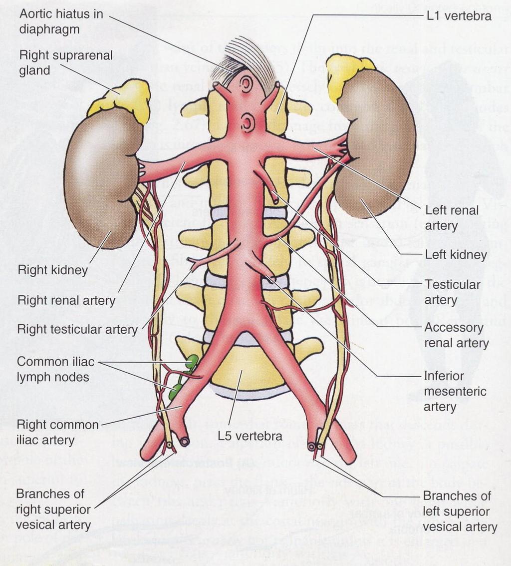
1 1 THE KIDNEY (Fig. 1 & 2) The kidney is a bean-shaped, reddish-brown organ of the urinary system located retroperitoneally in the upper part of the paravertebral gutter of the abdominal cavity, padded by pararenal extraperitoneal fat. There are normally two kidneys per individual, each possessing anterior and posterior surfaces, lateral and medial borders and upper and lower poles. The indented aspect of the medial border, which is called the hilus (hilum), impacts the bean-shaped appearance on the kidney. This area also serves as the route of passage of structures in and out of the organ. Measuring about 10cm in length, 5cm in width and 2.5cm in depth, each kidney lies lateral to the vertebral column between T12 and L3 vertebral bodies. Weighing about 150gms in the male and 135gms in the female, each kidney lies obliquely with the upper pole nearer the midline than the lower pole. Furthermore, the right kidney is lower, wider and shorter than the left one and lies further away from the midline than the latter. Figure 1

2 Figure. 2 2

3 3 Surface Anatomy of the Kidney Fig. 3: The kidneys are located on the posterior abdominal wall and hence the surface anatomy of the kidney is projected to the posterior surface of the body in the abdominal region. The upper pole of each kidney lies 2.5cm from the midline at the level of T12 while the lower pole lies 7.5cm from the midline at the level of L3. The hilar of each kidney lies 5cm from the midline. The transpyloric plane (L1), which passes along the tip of the ninth costal cartilage, traverses the upper part of the right hilum and the lower part of the left hilum. The lower pole of the right kidney is 1.25cm above the iliac crest while that of the left kidney is 2.5cm above the iliac crest. Figure. 3 Structures Closely Associated with the Medial Border of the Kidney: The Renal Sinus: This is the 2.5 cm-deep cavities in the medial aspect of the kidney, which opens into the hilus of the kidney. It is lined by the fibrous renal capsule (The deepest layer of the renal coverings) and contains: 1. Rostral part of the renal pelvis 2. Major calyces (calices) 3. Minor calyces (calices)

4 4 4. Blood vessels 5. Nerves 6. Lymphatics and 7. Adipose tissue The Renal pelvis: This is a funnel-shaped tube located partly in the renal sinus and in the hilum. It projects beyond the hilum to continue caudally with the ureter. In its rostral part within the sinus, it divides to form 2-3 major calyces, which in turn divide to form 7-14 minor calyces Relations of the Kidneys (Fig. 4): The structure entering and leaving the kidney at the hilus constitute its medial relations. These include: 1 The renal vein (Lies anterior) 2 The renal artery (Lies posterior to the vein) 3 The renal pelvis (Lies posterior to the artery) The suprarenal glands are located in the anteromedial aspect of the superior poles of the kidneys. Figure 4 Posterior Relations (Fig. 5): With the exception of the eleventh rib, which is the highest posterior relation of the left kidney, the posterior relations of both kidneys are similar. They include: 1. The muscular bed comprising:

5 5 a. The thoracoabdominal diaphragm, which separates the kidney from the base of the lung, the pleura and the 12 th /11 th rib/11 th intercostal space. b. The psoas major muscle on the medial aspect c. The quadratus lumborum lateral to the psoas muscle and d. The transversus abdominis lateral to the quadratus lumborum muscle e. The medial and lateral arcuate ligaments of the diaphragm above the psoas and quadratus muscles 2. The subcostal nerve (T12) and vessels 3. The iliohypogastric nerve (L1) and 4. The ilioinguinal nerve (L1) Figure 5 Anterior Relations: These are listed in the table below: (See Diagram) RIGHT KIDNEY LEFT KIDNEY Inferior surface, right lobe of the Liver The stomach Hepatorenal recess The pancreas Duodenum The splenic vein Right colic flexure Loops of the small intestine ( Ileum) The splenic artery The spleen Loops of the small intestine (Jejunum) Splenic flexure and descending colon The omental bursa (Lesser sac)
6 6 Blood Supply of the Kidney (Fig: 6): Renal arteries of corresponding sides supply the kidneys. Both are branches of the abdominal aorta arising at the level of L1/L2. The right renal artery which is longer than the left one passes to the right hilum, posterior to the inferior vena cava. The shorter left artery passes directly into the left hilum. In 30% of the population, assessory renal arteries may arise from the aorta above or below the origin of the main renal artery. On entering the kidney, each artery divides to give rise to about 5 segmental arteries within the sinus. The segmental arteries give rise to lobar arteries, which in turn give rise to interlobar arteries. Interlobar arteries enter the column of Bertin and continue as arcuate arteries at the junction of the cortex and the medulla of the kidney. The arcuate arteries give rise to interlobular arteries, which form the basis of the subdivision of the kidney into lobules. Large-diameter afferent arterioles arise from the interlobular arteries to supply the glomeruli (Glomerular capillaries), which are drained by Small-diameter efferent arterioles. These in turn form peritubular capillary network around the proximal and distal convoluted tubules. Peritubular capillaries drain into interlobular veins which, in turn drain into the arcuate vein of the corticomedullay junction. The efferent arterioles associated with Juxtamedullary nephrons form straight capillary network referred to as the Vasa Recta around the corresponding Henle s loops in the renal medulla. The vasa recta invariably drain into the arcuate veins. Venous drainage of the Kidney: The venous drainage of the kidney commences with the stellate veins, which drain the renal capsule and the outer cortex into interlobular veins. From this point on the vein pursue the pattern of the arterial distribution and finally drain into the inferior vena cava via the renal veins. The left renal vein, which passes anterior to the abdominal aorta, is longer than the right renal vein. Lymphatic drainage of the Kidney: Renal lymphatic vessels run with the veins and drain into lumbar (Lateral aortic) lymph nodes. Innervation of the Kidney: The kidney receives sympathetic and parasympathetic fibers from the renal nerve plexus, which is formed from fibers arising from the lesser and least (lowest or renal) splanchnic nerves (T10-L1). The renal plexus is located along the renal arterial tree and it possesses an aorticorenal ganglion on each side of the aorta.

7 Figure 6 7
8 8 THE URETER The ureter is the thick-walled, highly expandable muscular tube, which propels urine in boluses by peristaltic contraction from the renal pelvis to the urinary bladder. It is a direct continuation of the renal pelvis and it terminates by traversing the wall of the base (Fundus) of the urinary bladder. Measuring about 25cm in length, it descends retroperitoneally anterior to the medial aspect of the psoas major muscle along the tips of the lumbar transverse processes and crosses the pelvic brim anterior to the bifurcation of the common iliac artery. The ureter has a narrow lumen, about 3mm** in diameter and is subdivided into two parts, viz. a. The abdominal part (12.5cm), which lies in the abdominal cavity above the pelvic brim and b. The pelvic part (12.5cm), which lies in the pelvic cavity below the pelvic brim. The ureter is constricted in three areas along its course from the abdomen to the pelvis. These areas are: 1. At the pelviureteric region (Not junction) 2. At the pelvic brim and 3. At the part which, traverses the base of the urinary bladder. This is the narrowest part of the ureter, which is also referred to as the intramural (intravesical) part of the ureter. This part also projects into the lumen of the urinary bladder as the Ureteric fold. Course of the Abdominal Ureter (Figure 7): The abdominal ureter passes along the medial aspect of the psoas major muscle, from the tip of the transverse process of the first lumbar vertebra bone to the pelvic brim. Relations of the abdominal ureter The relations of the abdominal ureter are similar in the male and female but differ from one side to the other. Posterior Relations: These are similar on both sides and include: 1. Psoas major muscle 2. Tips of the lumbar transverse processes 3. Genitofemoral nerve Anterior Relations (Right side): 1. Descending part of the duodenum 2. Right colic vessels 3. Iliocolic vessels 4. The root of the mesentery 5. The terminal ileum 6. The gonadal vessels 7. The peritoneum Anterior Relations: Left side: 1. Left colic vessels 2. Sigmoid colon 3. Sigmoid mesocolon 4. Gonadal vessels 5. The peritoneum Medial Relations: These exist only on the right side and include: 1. The inferior vena cava 2. The lumbar lymph nodes 3. The sympathetic trunk

9 9 Figure 7 Course of the Pelvic Ureter The pelvic ureter is retroperitoneal throughout its course. The first segment of the pelvic ureter passes posteroinferiorly from the pelvic brim to the lateral pelvic wall resting on the obturator fascia along the anterior border of the greater sciatic notch. The second segment turns anteromedially at the level of the ischial spine and runs on the levator ani muscle (the pelvic diaphragm) to reach the base of the urinary bladder.
10 10 Relations of the Pelvic Ureter Posterior Relations (First segment): These are similar in both sexes and on both sides. They include: 1. The internal iliac artery and vein 2. The lumbosacral nerve trunk 3. The sacroiliac joint Lateral relations (First segment): 1. Obturator fascia 2. Umbilical artery 3. Obturator vessels 4. Obturator nerve 5. Inferior vesical vessels 6. Middle rectal artery 7. The uterine artery in the Female Anterior relations (First segment): 1. The ovary in the Female Relations of The second Segment In the Male: 1. Vas deferens Anteromedially 2. Seminar vesicle Inferiorly In the Female: 1. Uterine artery superiorly 2. Broad ligament of the Uterus superiorly 3. Lateral fornix of the vagina medially 4. Uterine cervix medially 5. Vagina posteriorlly Blood supply of the ureter The ureter receives blood supply from all the vessels along its course from the abdomen to the pelvis. These arteries establish anastomotic network on the wall of the ureter. The vessels from above downwards are: 1. Renal artery 2. Abdominal aorta 3. Gonadal arteries 4. Common iliac artery 5. Internal iliac artery 6. Superior vesical artery 7. Inferior vesical artery in the Male 8. Uterine artery in the Female Venous Drainage of the ureter
11 11 The veins of the ureter run along corresponding arteries and invariably drain into the inferior vena cava or the renal veins. Nerve Supply of the Ureter The smooth muscles and the blood vessels of the ureter receive autonomic supply from the following plexuses: 1. Renal plexus 2. aortic plexus 3. Superior hypogastric plexus 4. Inferior hypogastric plexus Afferent fiber for pain and awareness of distention traverse T11-L2. Areas of referred pain for the ureter are: Inguinal region Scrotum Medial femoral region and the cremasteric muscle. Histology of the Ureter The mucous membrane which is urothelium is supported by a connective tissue layer of lamina propia. The luminal cells of the urothelium are cuboidal in shape, the middle layer are polyhedral while the basal layer are columnar/cuboidal in shape. In the upper 1/3 the smooth muscle fibers of the tunica muscularis are arranged in helical and longitudinal axis. The middle 1/3 consists of smooth muscles arranged in outer circular and inner longitudinal layers, while the lower 1/3 consists of smooth muscles arranged in inner longitudinal middle circular and outer longitudinal layers. The tunica adventitia consists of fibroelastic and adipose tissue in which are embedded nerve fibers, blood vessels and lymphatics. Applied Anatomy of the Kidney and the Ureter The Kidney: 1. Tract of internal bleeding from a ruptured kidney 2. Pus from a perinephric abscess. 3. The kidney and respiratory movements 4. Surgical exposure of the kidney 5. Nephroptosis dropped kidney 6. Renal transplant The Ureter: 1. The ureter and the peritoneum 2. Constricted sites of the ureter and obstruction by Ureteric stone (Calculus): Consequences of obstruction: a. Colicky pain due to hyperperistalsis of the ureter b. Ureteric distention by urine (Uroureter or Hydroureter) c. Renal Pelvis distensions by urine (Hydronephrosis or Uronephrosis or Nephrohydrosis) 3. Radiological investigations of the ureter: a. Plain X-ray b. Administration of radiopaque materials Pyeloghraphy 4. Endourology Cystoscope insertion 5. Lithotripsy Fragmentation of stones using shock waves 6. Palpation of a Ureteric stone
12 7. Common sites of injury to the ureter in obstetrics practice are At the Pelvic brim where it is close to the ovarian vessels. At the lateral aspect of the cervix where it is crossed by the uterine artery. 12
13 13
14 14
ANATOMY OF PELVICAYCEAL SYSTEM -DR. RAHUL BEVARA
 1 ANATOMY OF PELVICAYCEAL SYSTEM -DR. RAHUL BEVARA 2 KIDNEY:ANATOMY OVERVIEW Kidneys are retroperitoneal, in posterior abdominal region, extending from T12 L3 Bean-shaped Right kidney is lower than left
Anatomy of the renal system. Professor Nawfal K. Al-Hadithi
 Anatomy of the renal system Professor Nawfal K. Al-Hadithi Objectives To describe the posterior abdominal wall To identify the main anatomical landmarks of the kidneys & ureters To describe the suprarenal
Gross Anatomy of the Urinary System
 Gross Anatomy of the Urinary System Lecture Objectives Overview of the urinary system. Describe the external and internal anatomical structure of the kidney. Describe the anatomical structure of the ureter
THE ABDOMEN SUPRARENAL GLANDS KIDNEY URETERS URINARY BLADDER
 THE ABDOMEN SUPRARENAL GLANDS KIDNEY URETERS URINARY BLADDER THE SUPRARENAL GLANDS The suprarenal (adrenal) glands lie immediately superior and slightly anterior to the upper pole of either kidney. Golden
Anatomy of the Large Intestine
 Large intestine Anatomy of the Large Intestine 2 Large Intestine Extends from ileocecal valve to anus Length = 1.5-2.5m = 5 feet Regions Cecum = 2.5-3 inch Appendix= 3-5 inch Colon Ascending= 5 inch Transverse=
The abdominal Esophagus, Stomach and the Duodenum. Prof. Oluwadiya KS
 The abdominal Esophagus, Stomach and the Duodenum Prof. Oluwadiya KS www.oluwadiya.com Viscera of the abdomen Abdominal esophagus: Terminal part of the esophagus The stomach Intestines: Small and Large
The posterior abdominal wall. Prof. Oluwadiya KS
 The posterior abdominal wall Prof. Oluwadiya KS www.oluwadiya.sitesled.com Posterior Abdominal Wall Lumbar vertebrae and discs. Muscles opsoas, quadratus lumborum, iliacus, transverse, abdominal wall
URINARY SYSTEM ANATOMY PART
 URINARY SYSTEM ANATOMY PART 1 DANIL HAMMOUDI.MD Urinary System Composed of kidneys, ureters, urinary bladder, and urethra Eliminates nitrogenous wastes from the body Regulates water, electrolyte, and ph
The Urinary System Pearson Education, Inc.
 26 The Urinary System Introduction The urinary system does more than just get rid of liquid waste. It also: Regulates plasma ion concentrations Regulates blood volume and blood pressure Stabilizes blood
Figure 26.1 An Introduction to the Urinary System
 Chapter 26 Figure 26.1 An Introduction to the Urinary System Components of the Urinary System Kidney Produces urine Ureter Transports urine toward the urinary bladder Urinary Bladder Temporarily stores
Abdomen. Retroperitoneal space
 Abdomen. Retroperitoneal space Abdominal cavity The space bounded by: Anterolateral abdominal wall Posterior abdominal wall Diaphragm Pelvic walls and pelvic floor. Subdivided into: True abdominal cavity
د. عصام طارق. Objectives:
 GI anatomy Lecture: 5 د. عصام طارق Objectives: To describe anatomy of stomach, duodenum & pancreas. To list their main relations. To define their blood & nerve supply. To list their lymph drainage. To
Gross anatomy of the urinary system. Done by : razan krishan. slide in bold and book in green
 Gross anatomy of the urinary system Done by : razan krishan slide in bold and book in green Kidneys, ureters, urinary bladder & urethra Urine flows from each kidney, down its ureter to the bladder and
Chapter 26. The Urinary System. Lecture Presentation by Steven Bassett Southeast Community College Pearson Education, Inc.
 Chapter 26 The Urinary System Lecture Presentation by Steven Bassett Southeast Community College Introduction The urinary system does more than just get rid of liquid waste. It also: Regulates plasma ion
URINARY SYSTEM ANATOMY
 URINARY SYSTEM ANATOMY Adapted from Human Anatomy & Physiology Marieb and Hoehn (9 th ed.) OVERVIEW Metabolism of nutrients by the body produces wastes that must be removed from the body. Although excretory
Urinary System VASTACCESS, INC.
 Urinary System www.vastaccess.com 2 Urinary Tract Kidney Ureter Urinary Bladder Urethra Prostate (male) Membranous (male) Spongy (male) 3 Kidney Relations Suprarenal (Adrenal) Glands Liver Duodenum Transverse
The Kidneys. (L., ren; Gk, nephros; hence the adjectives renal and nephric) & Suprarenal (Adrenal) Glands. Dr Maan Al-Abbasi PhD, MBChB
 & Suprarenal (Adrenal) Glands. Dr Maan Al-Abbasi PhD, MBChB") The Kidneys (L., ren; Gk, nephros; hence the adjectives renal and nephric) & Suprarenal (Adrenal) Glands Dr Maan Al-Abbasi PhD, MBChB Functions of Urinary System Regulate electrolytes (K+, Na+, etc) Regulate
The Kidneys (L., ren; Gk, nephros; hence the adjectives renal and nephric) & Suprarenal (Adrenal) Glands Dr Maan Al-Abbasi PhD, MBChB Functions of Urinary System Regulate electrolytes (K+, Na+, etc) Regulate
Chapter 23. The Nephron. (functional unit of the kidney
 Chapter 23 The Nephron (functional unit of the kidney Renal capsule The Nephron Renal cortex Nephron Collecting duct Efferent arteriole Afferent arteriole (a) Renal corpuscle: Glomerular capsule Glomerulus
GI module Lecture: 9 د. عصام طارق. Objectives:
 GI module Lecture: 9 د. عصام طارق Objectives: To list structures forming posterior abdominal wall. To follow aorta & its main branches. To describe IVC & its main tributaries. To list nerves of posterior
Netter's Anatomy Flash Cards Section 4 List 4 th Edition
 Netter's Anatomy Flash Cards Section 4 List 4 th Edition https://www.memrise.com/course/1577335/ Section 4 Abdomen (31 cards) Plate 4-1 Bony Framework of Abdomen 1.1 Costal cartilages 1.2 Iliac crest 1.3
Jordan University Faculty Of Medicine. Urinary System. Dr. Ahmed Salman. Assistant professor of anatomy & embryology
 Jordan University Faculty Of Medicine Urinary System Dr. Ahmed Salman Assistant professor of anatomy & embryology The urinary system is composed of two kidneys,two ureters,urinary bladder and urethra The
Urinary System Laboratory
 Urinary System Laboratory 1 Adrenal gland Organs of The Urinary System Renal artery and vein Kidney Ureter Urinary bladder Figure 26.1 2 Urethra Functions of the urinary system organs: Urethra expels urine
Lecture 56 Kidney and Urinary System
 Lecture 56 Kidney and Urinary System The adrenal glands are located on the superomedial aspect of the kidney The right diagram shows a picture of the kidney with the abdominal walls and organs removed
Pancreas & Biliary System. Dr. Vohra & Dr. Jamila
 Pancreas & Biliary System Dr. Vohra & Dr. Jamila 1 Objectives At the end of the lecture, the student should be able to describe the: Location, surface anatomy, parts, relations & peritoneal reflection
Lab 9 Abdomen MUSCLES
 Lab 9 Abdomen MUSCLES External abdominal oblique continuous with the external intercostal muscle; its fibers point in a caudal direction as it moves anteriorly until it inserts on the linea alba via its
Biology Human Anatomy Abdominal and Pelvic Cavities
 Biology 351 - Human Anatomy Abdominal and Pelvic Cavities Please place your name and I.D. number on the back of the last page of this exam. You must answer all questions on this exam. Because statistics
URINARY SYSTEM. These organs lie posterior or inferior to the. (membrane).
.") URINARY SYSTEM I. INTRODUCTION Each kidney is made up of about a million tiny tubules called nephrons. Each nephron individually filters the blood and makes urine and it does the job completely, from start
URINARY SYSTEM I. INTRODUCTION Each kidney is made up of about a million tiny tubules called nephrons. Each nephron individually filters the blood and makes urine and it does the job completely, from start
Duodenum retroperitoneal
 Duodenum retroperitoneal C shaped Initial region out of stomach into small intestine RETROperitoneal viscus Superior 1 st part duodenal cap ; moves upwards and backwards to lie on the R crura medial to
Jordan University Faculty Of Medicine. Urinary System. Dr. Ahmed Salman. Assistant professor of anatomy & embryology
 Jordan University Faculty Of Medicine Urinary System Dr. Ahmed Salman Assistant professor of anatomy & embryology The urinary system is composed of two kidneys,two ureters,urinary bladder and urethra The
SUBJECTS 2nd year, 1st semester I. 1. Primitive gut - limits, derivatives 2. Foregut -limits, evolution, derivatives 3. Midgut -limits, evolution,
 SUBJECTS 2nd year, 1st semester I. 1. Primitive gut - limits, derivatives 2. Foregut -limits, evolution, derivatives 3. Midgut -limits, evolution, derivatives 4. Hindgut- limits, evolution, derivatives
[ANATOMY #12] April 28, 2013
![[ANATOMY #12] April 28, 2013](/thumbs/86/93473883.jpg "[ANATOMY #12] April 28, 2013") Sympathetic chain : Sympathetic chain is each of the pair of ganglionated longitudinal cords of the sympathetic nervous system; extend from level of atlas (base of skull) till coccyx. It is paravertebral
Sympathetic chain : Sympathetic chain is each of the pair of ganglionated longitudinal cords of the sympathetic nervous system; extend from level of atlas (base of skull) till coccyx. It is paravertebral
Human Anatomy Unit 3 URINARY SYSTEM
 Human Anatomy Unit 3 URINARY SYSTEM In Anatomy Today Components Kidneys Ureters Urinary bladder Urethra Functions Storage of urine Bladder stores up to 1 L of urine Excretion of urine Transport of urine
STRUCTURAL BASIS OF MEDICAL PRACTICE EXAMINATION 3. October 16, 2015
 STRUCTURAL BASIS OF MEDICAL PRACTICE EXAMINATION 3 October 16, 2015 PART l. Answer in the space provided. (12 pts) 1. Identify the structures. (2 pts) A. B. A B C. D. C D 2. Identify the structures. (2
Lab Activity 31. Anatomy of the Urinary System. Portland Community College BI 233
 Lab Activity 31 Anatomy of the Urinary System Portland Community College BI 233 Urinary System Organs Kidneys Urinary bladder: provides a temporary storage reservoir for urine Paired ureters: transport
Abdomen: Introduction. Prof. Oluwadiya KS
 Abdomen: Introduction Prof. Oluwadiya KS www.oluwadiya.com Abdominopelvic Cavity Abdominal Cavity Pelvic Cavity Extends from the inferior margin of the thorax to the superior margin of the pelvis and the
Kidney Functions Removal of toxins, metabolic wastes, and excess ions from the blood Regulation of blood volume, chemical composition, and ph
 The Urinary System Urinary System Organs Kidneys are major excretory organs Urinary bladder is the temporary storage reservoir for urine Ureters transport urine from the kidneys to the bladder Urethra
The functional anatomy of the urinary system. Human Anatomy Department Dr. Anastasia Bendelic
 The functional anatomy of the urinary system Human Anatomy Department Dr. Anastasia Bendelic Plan Development of the kidneys and their abnormalities Development of the urinary ways and their abnormalities
Dana Alrafaiah. - Amani Nofal. - Ahmad Alsalman. 1 P a g e
 - 2 - Dana Alrafaiah - Amani Nofal - Ahmad Alsalman 1 P a g e This lecture will discuss five topics as follows: 1- Arrangement of pelvic viscera. 2- Muscles of Pelvis. 3- Blood Supply of pelvis. 4- Nerve
19. RENAL PHYSIOLOGY ROLE OF THE URINARY SYSTEM THE URINARY SYSTEM. Components and function. V BS 122 Physiology II 151 Class of 2011
 19. RENAL PHYSIOLOGY THE URINARY SYSTEM Components and function The urinary system is composed of two kidneys, the functionally filtering apparatus, which connect through two tubular structures called
Benha University. Faculty of Medicine. Anatomy Department Course code (MED 0701) Model answer of Anatomy examination. (Abdomen,Pelvis and Thorax)
 Model answer of Anatomy examination. (Abdomen,Pelvis and Thorax)") 1 Benha University Faculty of Medicine Anatomy Department Course code (MED 0701) Model answer of Anatomy examination (Abdomen,Pelvis and Thorax) 1 st year 2 nd term Date :18 /5 /2013 2 I-Short account
1 Benha University Faculty of Medicine Anatomy Department Course code (MED 0701) Model answer of Anatomy examination (Abdomen,Pelvis and Thorax) 1 st year 2 nd term Date :18 /5 /2013 2 I-Short account
BY DR NOMAN ULLAH WAZIR
 BY DR NOMAN ULLAH WAZIR The stomach (from ancient Greek word stomachos, stoma means mouth) is a muscular, hollow and the most dilated part of the GIT. It starts from the point where esophagus ends. It
Ureters, Urinary Bladder & Urethra
 Ureters, Urinary Bladder & Urethra Please check our Editing File هذا العمل ال يغني عن المصدر األساسي للمذاكرة Lecture 2 } و م ن ي ت و ك ع ل ا لل ه ف ه و ح س ب ه { Objectives o Describe the course of ureter
Urinary System. Chapter 17 7/19/11. Introduction
 7/19/11 Chapter 17 Urinary System Introduction A. The urinary system consists of two kidneys that filter the blood, two ureters, a urinary bladder, and a urethra to convey waste substances to the outside.
Anatomy of the SMALL INTESTINE. Dr. Noman Ullah Wazir PMC
 Anatomy of the SMALL INTESTINE Dr. Noman Ullah Wazir PMC SMALL INTESTINE The small intestine, consists of the duodenum, jejunum, and illium. It extends from the pylorus to the ileocecal junction were the
Accessory Glands of Digestive System
 Accessory Glands of Digestive System The liver The liver is soft and pliable and occupies the upper part of the abdominal cavity just beneath the diaphragm. The greater part of the liver is situated under
Bushra Arafa Zayed & Hanan Jamal. - Dana AF
 - 10 - Bushra Arafa Zayed & Hanan Jamal - Dana AF - Mohammad Al Muhtaseb Notes: This sheet was written in the same order as the slides, and everything in the slides is mentioned in this sheet. Pictures
The Thoracic wall including the diaphragm. Prof Oluwadiya KS
 The Thoracic wall including the diaphragm Prof Oluwadiya KS www.oluwadiya.com Components of the thoracic wall Skin Superficial fascia Chest wall muscles (see upper limb slides) Skeletal framework Intercostal
The jejunum and the Ileum. Prof. Oluwadiya KS
 The jejunum and the Ileum Prof. Oluwadiya KS www.oluwadiya.siteled.com Introduction Introduction The small intestine (SI) comprises of the duodenum, jejunum and the ileum The jejunum is the second part
Bio 322 Human Anatomy Objectives for the laboratory exercise Urinary System Filtration Reabsorption Secretion Concentration
 Bio 322 Human Anatomy Objectives for the laboratory exercise Urinary System Required reading before beginning this lab: Saladin, KS: Human Anatomy 5 th ed (2017) Chapter 25 For this lab you will use parts
Anatomy of the Thorax
 Anatomy of the Thorax A) THE THORACIC WALL Boundaries Posteriorly by the thoracic part of the vertebral column Anteriorly by the sternum and costal cartilages Laterally by the ribs and intercostal spaces
Copyright 2003 Pearson Education, Inc. publishing as Benjamin Cummings. Dr. Nabil Khouri
 Dr. Nabil Khouri Objectives: General objectives: - to identify the kidney s structures, function and location - to analyze the relationship between microscopic structure and function Specific objectives:
Chapter 25: Urinary System
 Chapter 25: Urinary System I. Kidney anatomy: retroperitoneal from 12 th thoracic to 3 rd lumbar area A. External anatomy: hilus is the indentation 1. Adrenal gland: in the fat at the superior end of each
It passes through the diaphragm at the level of the 10th thoracic vertebra to join the stomach
 The esophagus is a tubular structure (muscular, collapsible tube ) about 10 in. (25 cm) long that is continuous above with the laryngeal part of the pharynx opposite the sixth cervical vertebra The esophagus
Collin College. BIOL Anatomy & Physiology WEEK 12. Urinary System INTRODUCTION. Main functions of the kidneys are
 Collin College BIOL. 2402 Anatomy & Physiology WEEK 12 Urinary System 1 INTRODUCTION Main functions of the kidneys are regulate blood volume, water content regulate blood composition e..g. Na, Cl, K, ph
Omran Saeed. Mohammad Al-muhtaseb. 1 P a g e
 13 Omran Saeed Mohammad Al-muhtaseb 1 P a g e Posterior abdominal wall - The diaphragm separates between thoracic cavity and abdominal cavity. Structures of posterior abdominal wall: (below diaphragm)
Exploring Anatomy: the Human Abdomen
 Exploring Anatomy: the Human Abdomen PERITONEUM AND PERITONEAL CAVITY PERITONEUM The peritoneum is a thin serous membrane that lines the abdominal cavity and covers, in variable amounts, the viscera within
Urinary system. Urinary system
 INTRODUCTION. Several organs system Produce urine and excrete it from the body Maintenance of homeostasis. Components. two kidneys, produce urine; two ureters, carry urine to single urinary bladder for
musculoskeletal system anatomy nerves of the lower limb 1 done by: dina sawadha & mohammad abukabeer
 musculoskeletal system anatomy nerves of the lower limb 1 done by: dina sawadha & mohammad abukabeer What is the importance of plexuses? plexuses provides us the advantage of a phenomenon called convergence
Peritoneum: Def. : It is a thin serous membrane that lines the walls of the abdominal and pelvic cavities and clothes the viscera.
 Peritoneum: Def. : It is a thin serous membrane that lines the walls of the abdominal and pelvic cavities and clothes the viscera. Layers of the peritoneum: 1. Outer Layer ( Parietal Peritoneum) : lines
LECTURE ON THE URINARY SYSTEM
 LECTURE ON THE URINARY SYSTEM (Uropoetic System) AN OVERVIEW Dr HAMIADJI THE URINARY SYSTEM URINARY SYSTEM The kidneys are responsible for removing wastes from the body, regulating electrolyte balance
YR 1 GROSS ANATOMY/EMBRYOLOGY UNIT EXAM 3 -- November 13, Which of the following statements regarding the pericardium is NOT CORRECT:
 YR 1 GROSS ANATOMY/EMBRYOLOGY UNIT EXAM 3 -- November 13, 1996. CHOOSE THE SINGLE BEST ANSWER FOR QUESTIONS 1-42. 1. Which of the following statements regarding the pericardium is NOT CORRECT: A. The fibrous
BLOCK IV: OFFICIAL BODY PARTS LIST FOR ANTERIOR ABDOMINAL WALL AND ABDOMINAL CONTENTS
 BLOCK IV: OFFICIAL BODY PARTS LIST FOR ANTERIOR ABDOMINAL WALL AND ABDOMINAL CONTENTS External oblique muscle Muscular portion Aponeurotic portion Superficial inguinal ring Lateral (inferior) crus Medial
A. Incorrect! The urinary system is involved in the regulation of blood ph. B. Correct! The urinary system is involved in the synthesis of vitamin D.
 Human Anatomy - Problem Drill 22: The Urinary System Question No. 1 of 10 1. Which of the following statements about the functions of the urinary system is not correct? Question #01 (A) The urinary system
Urinary Bladder. Prof. Imran Qureshi
 Urinary Bladder Prof. Imran Qureshi Urinary Bladder It develops from the upper end of the urogenital sinus, which is continuous with the allantois. The allantois degenerates and forms a fibrous cord in
Anatomy: Know Your Abdomen
 Anatomy: Know Your Abdomen Glossary Abdomen - part of the body below the thorax (chest cavity); separated by the diaphragm. Anterior - towards the front of the body. For example, the umbilicus is anterior
BIOH122 Human Biological Science 2
 BIOH122 Human Biological Science 2 Session 16 Urinary System 1 The Kidneys Bioscience Department Endeavour College of Natural Health endeavour.edu.au Session Plan o Functions of Urinary system o The Kidneys:
Dr. Zahiri. In the name of God
 Dr. Zahiri In the name of God small intestine = small bowel is the part of the gastrointestinal tract Boundaries: Pylorus Ileosecal junction Function: digestion and absorption of food It receives bile
Preview from Notesale.co.uk Page 1 of 34
 Abdominal viscera and digestive tract Digestive tract Abdominal viscera comprise majority of the alimentary system o Terminal oesophagus, stomach, pancreas, spleen, liver, gallbladder, kidneys, suprarenal
STRUCTURAL BASIS OF MEDICAL PRACTICE EXAMINATION 3. October 17, 2014
 STRUCTURAL BASIS OF MEDICAL PRACTICE EXAMINATION 3 October 17, 2014 PART l. Answer in the space provided. (12 pts) 1. Identify the structures. (2 pts) A. B. A B C. D. C D 2. Identify the structures. (2
Nerves on the Posterior Abdominal Wall
 Nerves on the Posterior Abdominal Wall Lumbar Plexus The lumbar plexus, which is one of the main nervous pathways supplying the lower limb, is formed in the psoasmuscle from the anterior ramiof the upper
THE THORACIC WALL. Boundaries Posteriorly by the thoracic part of the vertebral column. Anteriorly by the sternum and costal cartilages
 THE THORACIC WALL Boundaries Posteriorly by the thoracic part of the vertebral column Anteriorly by the sternum and costal cartilages Laterally by the ribs and intercostal spaces Superiorly by the suprapleural
General Anatomy of Urinary System
 General Anatomy of Urinary System URINARY SYSTEM ORGANS Kidneys (2) Ureters (2) Urinary bladder Urethra KIDNEY FUNCTIONS Control blood volume and composition KIDNEY FUNCTIONS Filter blood plasma, eliminate
Lab Monitor Images Dissection of the Abdominal Vasculature + Lower Digestive System
 Lab Monitor Images Dissection of the Abdominal Vasculature + Lower Digestive System Stomach & Duodenum Frontal (AP) View Nasogastric tube 2 1 3 4 Stomach Pylorus Duodenum 1 Duodenum 2 Duodenum 3 Duodenum
ABDOMEN. 2. The highest branch of the abdominal aorta is: (a) R suprarenal a (b) Coeliac trunk (c) L renal a (d) L gonadal a (e) SMA
 R suprarenal a (b) Coeliac trunk (c) L renal a (d) L gonadal a (e) SMA") ABDOMEN 1. The duodenum: (a) is a retroperitoneal structure (b) is 25cm long (c) lies between the levels of L2-L4 (d) in its fourth part lies to the R of the aorta (e) all of the above 2. The highest branch
ABDOMEN 1. The duodenum: (a) is a retroperitoneal structure (b) is 25cm long (c) lies between the levels of L2-L4 (d) in its fourth part lies to the R of the aorta (e) all of the above 2. The highest branch
Pelvis MCQs. Block 1. B. Reproductive organs. C. The liver. D. Urinary bladder. 1. The pelvic diaphragm includes the following muscles: E.
 Pelvis MCQs Block 1 1. The pelvic diaphragm includes the following muscles: A. The obturator internus B. The levator ani C. The coccygeus D. The external urethral sphincter E. The internal urethral sphincter
Waste. Urinary System Anatomy Urinary Section pages 5-8. Urinary System. Urinary System. Nitrogenous Wastes. Nitrogenous Wastes 4/22/2016
 Waste Urinary System Anatomy Urinary Section pages 5-8 Metabolism produces waste products What is the primary waste product of cellular respiration? How does the body dispose of it? Urinary System Urinary
أحمد رواجبة- محمود الحربي- أحمد السالمان-
 -6 أحمد رواجبة- محمود الحربي- أحمد السالمان- 1 P a g e The Male Reproductive System The male genital system structures are divided into: Internal structures: 1- Prostate 3-Ejaculatory ducts External structures:
Kidney Model (Model 3-13)
") Kidney Model (Model 3-13) Kidney Model Blood Vessels (Model 3-29) Segmental Artery Interlobar Artery Renal Artery Renal Vein Interlobular Artery Interlobular Vein Arcuate Artery Interlobar Vein Arcuate
Kidney Model (Model 3-13) Kidney Model Blood Vessels (Model 3-29) Segmental Artery Interlobar Artery Renal Artery Renal Vein Interlobular Artery Interlobular Vein Arcuate Artery Interlobar Vein Arcuate
CHAPTER 25 URINARY. Urinary system. Kidneys 2 Ureters 2 Urinary Bladder 1 Urethra 1. functions
 CHAPTER 25 URINARY Kidneys 2 Ureters 2 Urinary Bladder 1 Urethra 1 fluid waste elimination secretion of wastes control blood volume and BP control blood ph electrolyte levels RBC levels hormone production
THE RENAL / URINARY SYSTEM
 1 THE RENAL / URINARY SYSTEM Definition/Description: The Renal/Urinary system is the body system, which plays a vital role in the maintenance of homeostasis by the following processes: 1. Production of
Pancreas and Biliary System
 Pancreas and Biliary System Please view our Editing File before studying this lecture to check for any changes. Color Code Important Doctors Notes Notes/Extra explanation Objectives At the end of the lecture,
-12. -Renad Habahbeh. -Dr Mohammad mohtasib
 -12 -Renad Habahbeh - -Dr Mohammad mohtasib The Gallbladder -The gallbladder has a body, a fundus (a rounded end), a neck, Hartmann s pouch before the neck and a cystic duct that meets the common hepatic
Histology Urinary system
 Histology Urinary system Urinary system Composed of two kidneys, two ureters, the urinary bladder, and the urethra, the urinary system plays a critical role in: 1- Blood filtration,(filtration of cellular
Table 2. First Generated List of Expert Responses. Likert-Type Scale. Category or Criterion. Rationale or Comments (1) (2) (3) (4)
 (2) (3) (4)") Table 2. First Generated List of Expert Responses. Likert-Type Scale Category or Criterion Anatomical Structures and Features Skeletal Structures and Features (1) (2) (3) (4) Rationale or Comments 1. Bones
Table 2. First Generated List of Expert Responses. Likert-Type Scale Category or Criterion Anatomical Structures and Features Skeletal Structures and Features (1) (2) (3) (4) Rationale or Comments 1. Bones
Macula densa Juxtaglomerular cells (JG cells) Extraglomerular mesangial cells (lacis cell) p.704. gap junction
 Extraglomerular mesangial cells (lacis cell) p.704. gap junction") Macula densa Juxtaglomerular cells (JG cells) Extraglomerular mesangial cells (lacis cell) p.704 gap junction Macula densa: -- tall, narrow, pale cells; sensor for the Na+ and Cl-- in the distal convoluted
Macula densa Juxtaglomerular cells (JG cells) Extraglomerular mesangial cells (lacis cell) p.704 gap junction Macula densa: -- tall, narrow, pale cells; sensor for the Na+ and Cl-- in the distal convoluted
Mousa Salah. Dr. Mohammad Al. Mohtasib. 1 P a g e
 8 Mousa Salah Dr. Mohammad Al. Mohtasib 1 P a g e In the previous lecture we talked about the peritoneum, and we said that the peritonium is a serous sac, and it consists of two layers, visceral and parietal.
ANATOMY OF THE SMALL & LARGE INTESTINES. Semester 1, 2011 A. Mwakikunga
 ANATOMY OF THE SMALL & LARGE INTESTINES Semester 1, 2011 A. Mwakikunga LEARNING OBJECTIVES 1. List the parts and anatomical regions of the small and large intestines 2. State anatomical relations of the
The Urinary System 15PART A. PowerPoint Lecture Slide Presentation by Patty Bostwick-Taylor, Florence-Darlington Technical College
 PowerPoint Lecture Slide Presentation by Patty Bostwick-Taylor, Florence-Darlington Technical College The Urinary System 15PART A Functions of the Urinary System Elimination of waste products Nitrogenous
DISSECTION 8: URINARY AND REPRODUCTIVE SYSTEMS
 8546d_c01_1-42 6/25/02 4:32 PM Page 38 mac48 Mac 48: 420_kec: 38 Cat Dissection DISSECTION 8: URINARY AND REPRODUCTIVE SYSTEMS Typically, the urinary and reproductive systems are studied together, because
Posterior Abdominal wall-
 Structures of posterior abdominal wall: o Bony boundaries: 5 lumber vertebra and their intervertebral disc, iliac fossa and iliac crest. o Muscles: psoas major, quadrates lumborum, transversus abdominis,
THE ORAL CAVITY
 THE ORAL CAVITY WALL OF ABDOMEN (ANTERIOR) The paraumbilical vein drains into the portal vein and then through the liver. This is an important clinical connection. THE ABDOMINAL VISCERA The small
DESCRIPTION: This is the part of the trunk, which is located between the root of the neck and the superior border of the abdominal region.
 1 THE THORACIC REGION DESCRIPTION: This is the part of the trunk, which is located between the root of the neck and the superior border of the abdominal region. SHAPE : T It has the shape of a truncated
Urinary Anatomy. Lab 40. Kidneys. Nephrons. Renal Corpuscle
 Urinary Anatomy Lab 40. Urinary Anatomy and Kidney Dissection Kidneys: filters blood, produces urine Ureters: convey urine to bladder Bladder: holding tank Urethra: carries urine to the outside for elimination
NOTES FROM GUTMAN LECTURE 10/26 Use this outline to study from. As you go through Gutman s lecture, fill in the topics.
 NOTES FROM GUTMAN LECTURE 10/26 Use this outline to study from. As you go through Gutman s lecture, fill in the topics. Anatomy above the arcuate line Skin Camper s fascia Scarpa s fascia External oblique
-Ensherah Mokheemer. -Shatha Al-Jaberi محمد المحتسب- 1 P a g e
 9-9 -Ensherah Mokheemer -Shatha Al-Jaberi محمد المحتسب- 1 P a g e Small intestine has three regions: ( االثني عشر( The duodenum The jejunum The ileum Small intestine Duodenum: -c-shaped -The concavity
Chapter 5: Other mediastinal structures. The Large Arteries. The Aorta. Ascending aorta
 Chapter 5: Other mediastinal structures The Large Arteries The Aorta The aorta is the main arterial trunk of the systemic circulation and in the healthy state its wall contain a large amount of yellow
Module: Foundation Principles of Life Science for Midwifery Practice. WHH1008-N
 Module: Foundation Principles of Life Science for Midwifery Practice. WHH1008-N 2015 Welcome to the Anatomy Workbook. This directed learning has been developed to prepare you for lectures designed to study
بسم االه الرحمن الرحيم
 MAY 3, 2012 [POSTERIOR ABDOMINAL WALL] LECTURE 26 ANATOMY Quick Revision: بسم االه الرحمن الرحيم Last time we started with the anterior abdominal wall and said that: 1. Diaphragm is the root of the abdomen.
Group of students. - Rawan almujabili د. محمد المحتسب - 1 P a g e
 - 14 - Group of students - Rawan almujabili د. محمد المحتسب - 1 P a g e Nerves of the posterior abdominal wall The spinal cord gives off spinal nerves between the vertebrae. In the abdomen, through the
In the name ofgod. Abdomen 3. Dr. Zahiri
 In the name ofgod Abdomen 3 Dr. Zahiri Peritoneum Peritoneum It is the serous membrane(a type of loose connective tissue and is covered by mesothelium) that lines the abdominal cavity. Extensions of the