Digestive System. Presented by: Dr M. Arianmanesh PhD in Reproductive and Developmental Biology Dept. of Anatomical Sciences
|
|
|
- May Beasley
- 5 years ago
- Views:
Transcription

1

2 Digestive System Presented by: Dr M. Arianmanesh PhD in Reproductive and Developmental Biology Dept. of Anatomical Sciences
3 Today we will discuss: Histological layers of alimentary canal Oral cavity Lip Tongue Teeth Esophagus Stomach Small intestine Large intestine

4 Gastrointestinal tract (GIT) tract: Upper GIT: Oral cavity + Esophagus + Stomach Lower GIT: Small intestine + colon + Rectum + anal canal Accessory digestive organs: Liver Pancreas Gall bladder Salivary glands
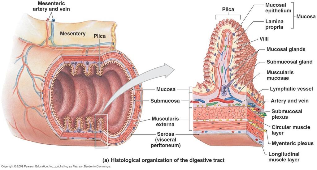
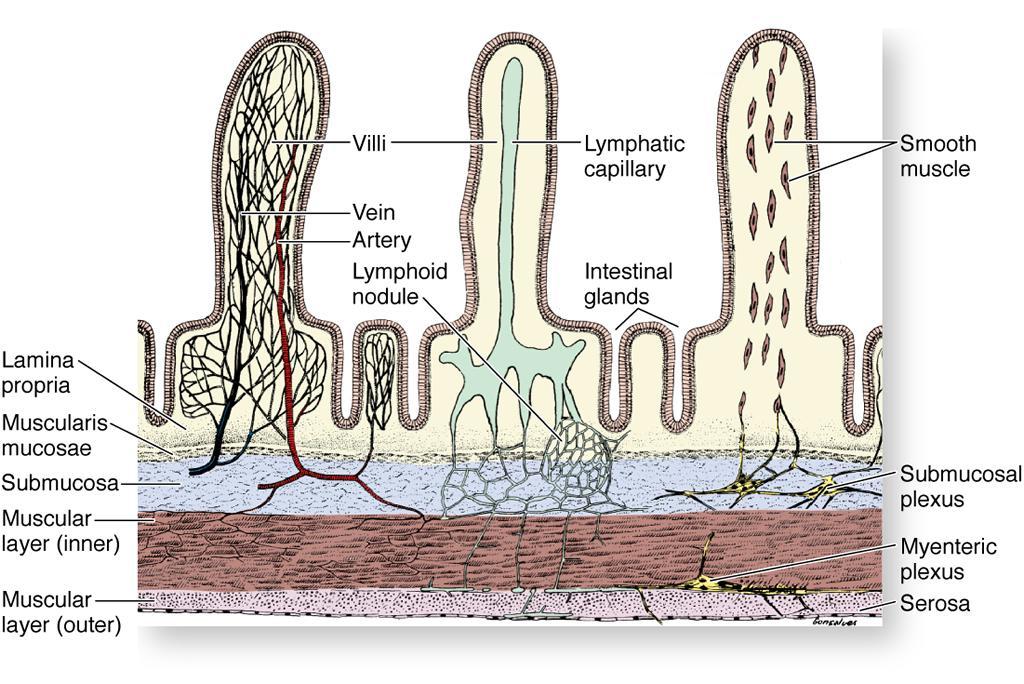
5 Histological Layers of Alimentary Canal Mucosa: Epithelium Lamina propria: Loose CT Contains lymphoid tissue and glands Muscularis mucosa: 1 or 2 layers circular smooth muscle Submucosa: Meissner s/submucosal Plexus Dense CT or loose CT Present in some parts Contains lymphatic follicles in ileum & appendix Contains mucus glands in esophagus & deodenum
 Mesothelium + loose underlying CT From stomach to end Adventitia: CT such as")
6 Continued Histological Layers of Alimentary Canal Muscularis externa: Two layers: Inner circular layer: Sphincters Outer longitudinal layer Myenteric/Auerbach s Plexus Serosa: (Visceral peritoneum) Mesothelium + loose underlying CT From stomach to end Adventitia: CT such as esophagus

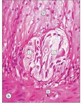
7 Meissner s Plexus Auerbach s Plexus
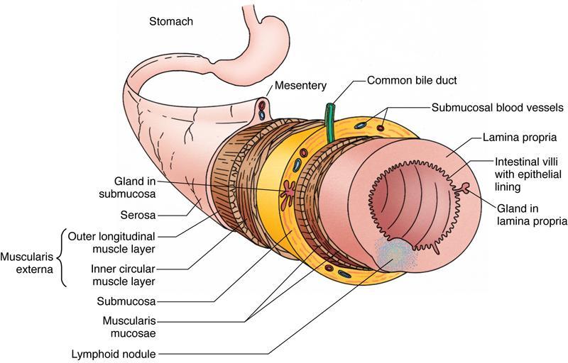
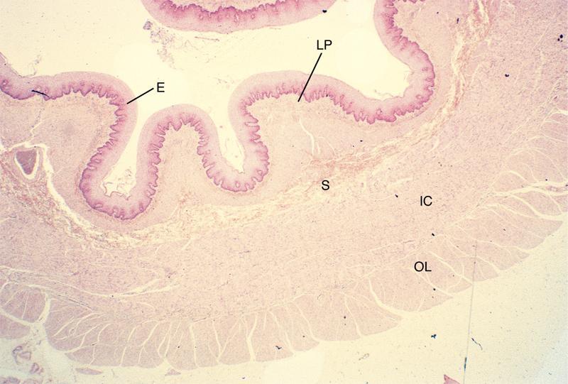
8 Histological Layers of Digestive System Mucosa Submucosa Muscularis Externa Inner Circular Outer Longitudinal Epithelium Lamina Propria (LP) Muscularis Mucosa Serosa (or) Adventitia
9


10 Oral Cavity Epithelium: Cheeks, oral surface of lips and soft palate: Non keratinized stratified squamus Hard palate and gingiva: Keratinized stratified squamus: Orthokeratinized: Squamus cells without nucleus Parakeratinized: Most squamus cells contain pycnotic nucleus Orthokeratinized Ep. Parakeratinized Ep.
11 Vermilion border Lip Skin: Keratinized stratified squamous epithelium Vermilion zone: Very thin keratinized epithelium Oral (Labial): Non keratinized stratified squamous epithelium Fibers of orbicularis oris muscle in submucosa Vermilion Border Vermilion Zone


12 A D C B A: Skin B: Vermillion Zone C: Oral (Labial) Mucosa D: Minor Salivary Glands


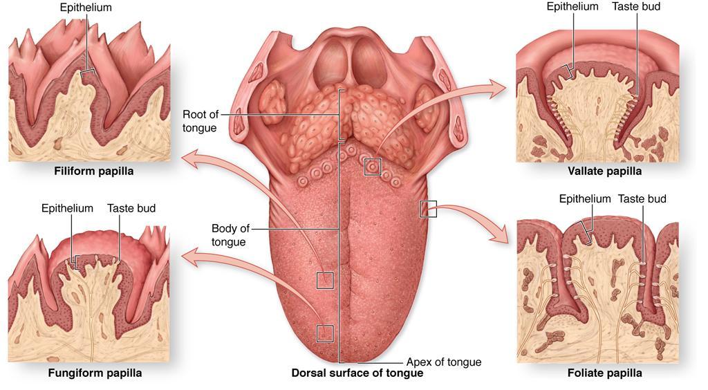
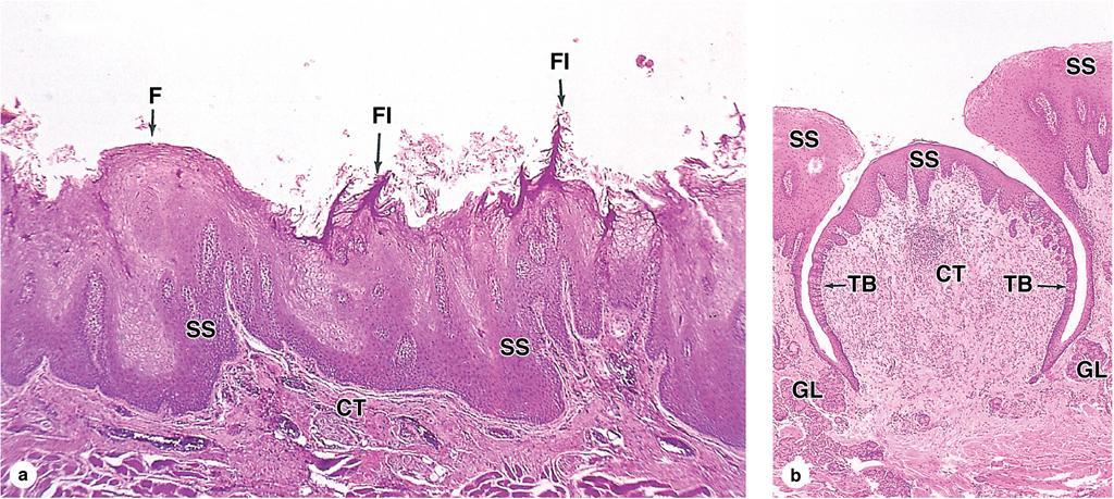
13 Tongue Ventral of tongue: Non keratinized stratified squamus EP. Dorsal of tongue: Median sulcus Terminal sulcus: Base of the tongue: Post. 1/3 Lingual tonsil Body of the tongue: Ant. 2/3 Lingual papillae: Filiform: Keratinized Projections (2-3 mm)


14 Continued Fungiform: Non keratinized Ep. Contains taste buds Tongue C A B A A. Fungiform Papilla B. Filiform Papilla


15 Continued Tongue Foliate: A few taste buds

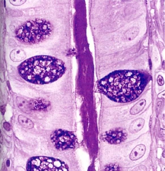
16 Continued Tongue Circumvallate: 8-12 Surface epithelium: Keratinized Wall epithelium: Nonkeratinized, presence of taste buds Von Ebner s Glands (Serous Glands)
17
18


19 Unique sense organ Taste Buds Barrel shaped bodies in EP of circumvallate, foliate & fungiform papillae Found in soft palate, epiglottis, larynx & pharynx Cells of taste bud: Cells Taste pore In LM?
")
 Type IV: Basal cells")
20 Continued Taste Buds In EM: 4 Types of cells: Type I: Supporting cells (60%) Type II: Supporting cells (30%) Type III: Gustatory cells (7%) Type IV: Basal cells (3%)

 Lingual")
21 Lingual: Anterior: Mixed Middle: Serous (Von Ebner) Posterior: Mucous (Weber) Lingual Glands

22 Teeth Deciduous/Milk Teeth Permanent Teeth
 Alveolar bone Woven bone Structure")
23 Crown: Dental pulp Dentin Enamel Neck Root: Dental pulp Dentin Cementum Periodontal ligament (PDL) Alveolar bone Woven bone Structure of Tooth

24 Dental Pulp Pulp Horns Pulp Chamber Located in Crown Accessory Canals/Lateral Canal Root Canal Treatment Failures Root Canal Located in Root Apical Foramen

25 Dental Pulp Specialized loose connective tissue Composition of the dental pulp Extracellular matrix Cells: Fibroblast Macrophage Plasma cell Undifferentiated mesenchyme cell Odontoblasts surrounding pulp Blood vessels and nerves
 6 (OH) 2 ] 20% Organic Material: 90% Collagen: Type I 10%")

26 Composed of: Dentin 70% Mineral Materials: Hydroxyapatite [Ca 10 (PO 4 ) 6 (OH) 2 ] 20% Organic Material: 90% Collagen: Type I 10% glycosaminoglycans and glycoproteins 10% Water Yellow Odontoblast cells: Apical cytoplasmic process Dentinal tubule Predentin and dentin

27 Odontoblast layer Predentin Dentinal tubules
 Thickest at the")
28 Composed of: Enamel 95-96% Mineral Materials: Hydroxyapatite 4% Organic Material & Water: Amelogenins (90%) Non-amelogenins: (10%) Enamelin White Ameloblasts: Tall columnar Tom s process Disappear after amelogenesis Thinnest at the neck (cementoenamel junction) Thickest at the cusps


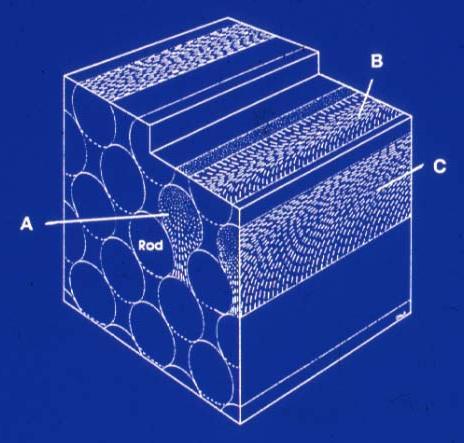
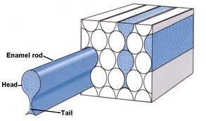
29 Structure of Enamel Rod enamel Interrod enamel Keyhole shape Rod sheath: Ameloblastin protein
30


31 pcw = Proximal Cell Web dcw= Distal Cell Web
 50-55% Organic Material: 90%")

32 Cementum Composed of: 45-50% Mineral Material (Hydroxyapatite) 50-55% Organic Material: 90% collagen type I Acellular cementum: Next to dentin Cellular cementum: Cementoblasts Cementocytes
 Oxytalan Cells: Fibroblasts Cementoblasts Osteoblasts and osteoclasts Ground Substance Vitamin C")
33 Periodontal Ligament (PDL) Dense specialized connective tissue Location: between cementum & alveolar Bone Fibers: Collagen (type I) Oxytalan Cells: Fibroblasts Cementoblasts Osteoblasts and osteoclasts Ground Substance Vitamin C Deficiency


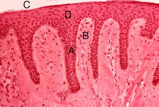
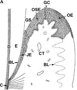
34 Gingiva/Gum Free (Marginal), Attached & Interdental Gingiva Thick Orthokeratinized or Parakeratinized Stratified Squamous EP Attached Gingiva Attached to the Neck of the Tooth by Junctional Ep Gingival sulcus
35
 Adventitia: Loose CT In thorax Serosa in abdominal esophagus Myenteric/Auerbach s")
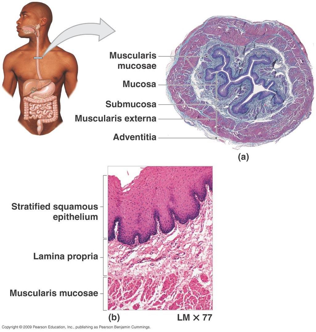
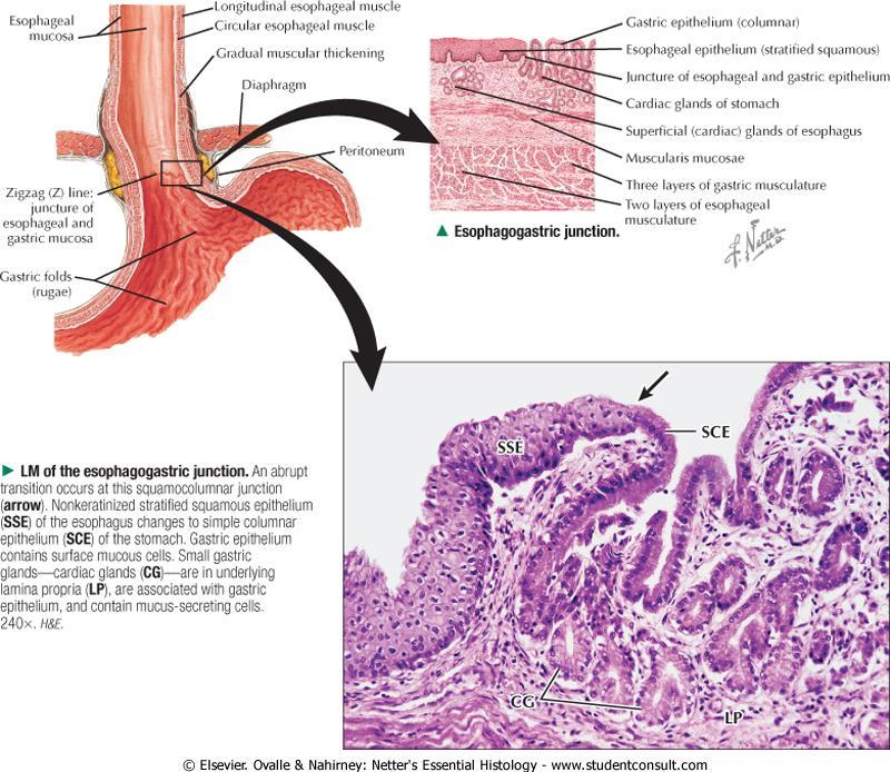
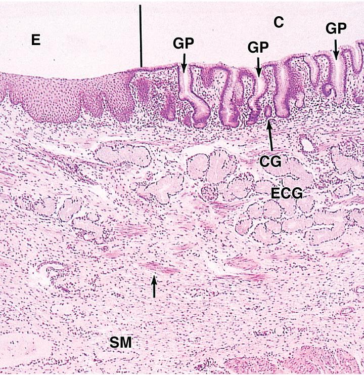
36 Esophagus Mucosa: cm Epithelium: Nonkeratinized stratified squamus At the end: Simple columnar Mucus glands in LP Submucosa: loose CT Mucus glands Muscularis mucosa Muscularis externa: 1/3 superior: Striated 1/3 middle: Striated + Smooth 1/3 inferior: Smooth (inner circular + outer longitudinal) Adventitia: Loose CT In thorax Serosa in abdominal esophagus Myenteric/Auerbach s Plexus
37

38 Longitudinal section of esophagus
39
40
41


42 Stomach Mucosa: Epithelium: Simple columnar Gastric pit Secretes bicarbonate-buffered mucus LP contains glands Muscularis mucosa Submucosa: Fibroelastic CT Rugae Muscularis externa: Innermost oblique Intermediate circular: Pyloric sphincter Outermost longitudinal Serosa


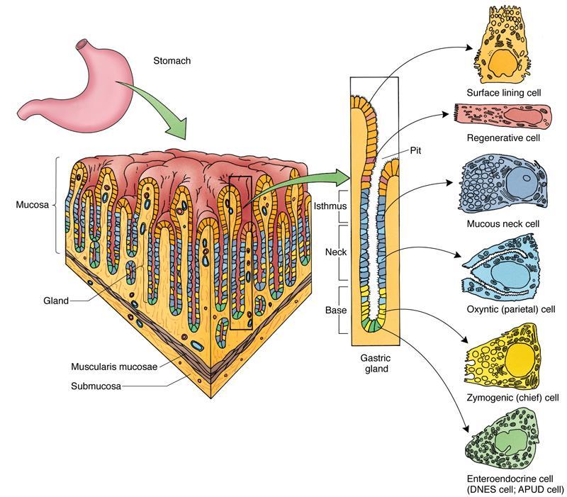
43 Gastric Glands Cardiac & pyloric glands: Mucosal glands Fundic & body glands: Gastric glands High in number of glands Simple & compound tubular glands Three parts in one gland: Pit Neck Base


44 Continued Gastric Glands Gastric glands: Mucous neck cells: Between parietal cells Secretion of acidic mucous Parietal cells/oxyntic: Big pyramidal Acidophilic In all parts of gland High in mitochondria Long microvilli Secretory canaliculi HCL secretion Intrinsic factor: Absorption of vitamin B12 Anemia
45 Continued Gastric Glands Chief/Zymogenic cells: Pyramidal At the base of the gland Basophilic cytoplasm Zymogen granules Secretion of: Pepsinogen Pepsin Lipase Renin Endocrine cells/apud/argentaffin: Between chief cells Small & round Secretion of: APUD in fundus: Seretonin APUD in pyloric: Gastrin Stem cells/regenerative cells: At the neck of the gland Small, basal nucleus
46

47

48 Layers of the Stomach

49 Gastric Pits & Glands

50 Neck of the Glands Gastric Glands Base of the Glands
51
 Ileum (3.")
52 Duodenum (25 cm) Jejunum (2.5 m) Ileum (3.5 m) Small Intestine


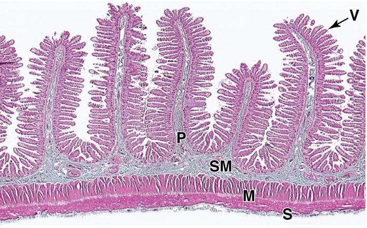
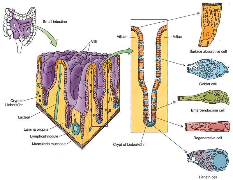
53 Small Intestine Designed for absorption: Great length: 5-7 m Plicae circulares/kerckring s valves: Penetration of submucosa toward mucosa Increasing 3-fold of mucosa Villi: mm in height Penetration of LP toward EP Contains: Blood vessels Lymphatic/lacteal vessels Smooth muscle cells Increasing 10-fold of mucosa Microvilli: Striated border Increasing 20-fold of mucosa Crypts/Lieberkuhn s glands 600-fold
54


55 Continued Small Intestine Mucosa: Epithelium: Simple columnar/absorptive cells Brush border LP: Peyer s patches in ileum Muscularis mucosa Submucosa: Relatively dense CT Brunner s glands in duodenum: Mucus glands Muscularis externa Serosa
56

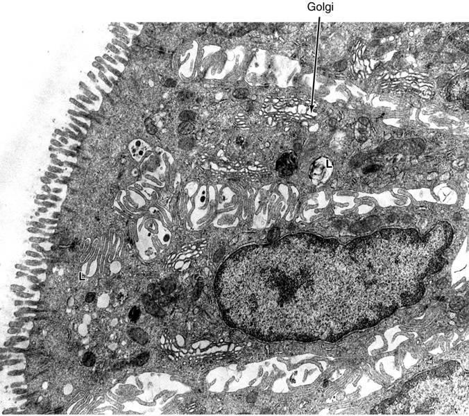
57 Continued Cells of Mucosa Absorptive cells: In both villi & crypts Brush border Glycocalyx Membranes of microvilli contains different enzymes: Disaccharidase Aminopeptidase Enterokinase in duodenum: Trypsinogen from pancreas to Trypsin Goblet cells: In both villi & crypts Increased toward end of intestine Paneth cells: At the base of gland Tall pyramidal Acidophilic granules Secretion of lysozyme & defensin

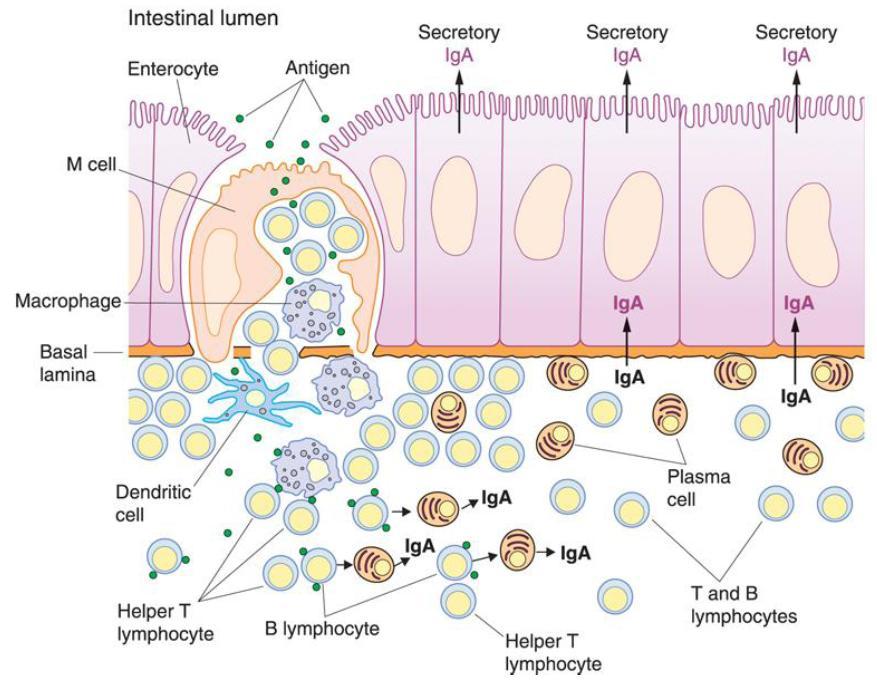
58 Continued Endocrine cells/apud: Base of the gland Secretions: Secretin Cholecystokinin Somatostatin Substance P Neurotensin Cells of Mucosa Undifferentiated cells M/Microfold cells: In Ep. Overlying lymphatic follicle Without microvilli Basolateral folds Antigen presenting cells
59
60

61 Leaf-like villi Brunner s glands: Mucous secretion ph: Duodenum

62 Long finger-like villi Jejunum

63 Peyer s patches M-cells in the EP. Covering Peyer s patches Ileum

64 Ileum

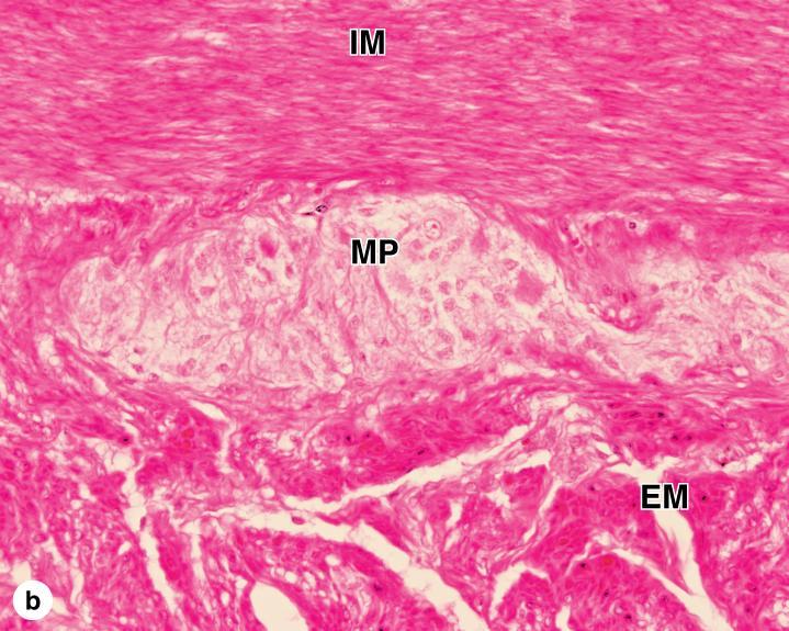
65 Muscularis Externa Myenteric Plexus
66
67

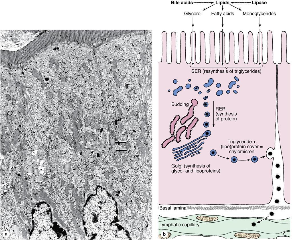
68 Fat Absorption
69
70
71 m Parts: Cecum Appendix Colon: Ascending Transverse Descending Sigmoid Rectum Large Intestine/Colon

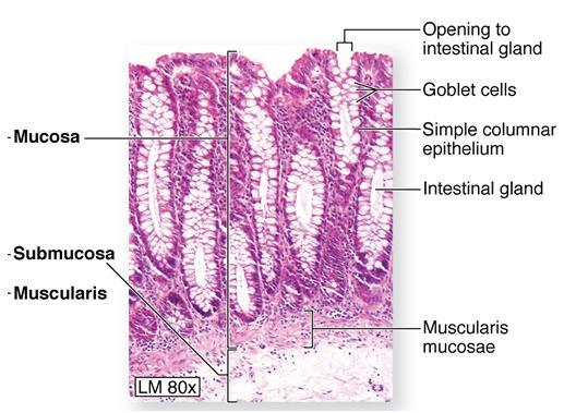
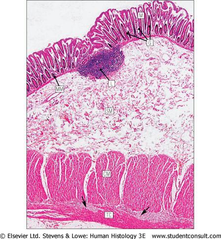
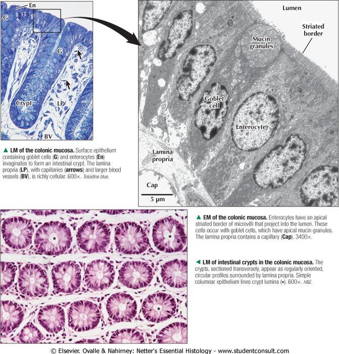
72 Continued Large Intestine/Colon Mucosa: Absence of pelica & villi Epithelium: Simple columnar Short & irregular microvilli Low ability of absorption Water absorption Cells of epithelium: Absorptive cells Goblet cells LP: High lymphatic follicles Muscularis mucosa Submucosa: High lymphatic follicles

73 Continued Large Intestine/Colon Muscularis externa: Inner circular Outer longitudinal: 3 muscular bands called Teniae coli In rectum: Similar to small intestine Serosa

74 Continued Large Intestine/Colon Cells of gland: Absorptive Goblet Undifferentiated APUD
75
76
77
78


79 Appendix 5-10 cm High lymphatic follicles in LP & submucosa 10-year-old child 36-year-old man
 Columns of morgagni/anal columns Anal sinuses Pectinate line Muscularis externa: Inner circular: Involuntary sphincter Outer striated: Voluntary")
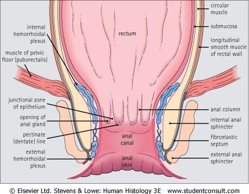
80 Anal Canal 2-3 cm Mucosa: Epithelium: Simple columnar Nonkeratinized stratified squamus Keratinized stratified squamus LP: Large venus network Hemorrhoid (enlarged veins) Columns of morgagni/anal columns Anal sinuses Pectinate line Muscularis externa: Inner circular: Involuntary sphincter Outer striated: Voluntary sphincter
81

82 Epithelium of Anal Canal
Alimentary Canal (I)
") Alimentary Canal (I) Esophagus and Stomach (Objectives) By the end of this lecture, the student should be able to discuss the microscopic structure in correlation with the function of the following organs:
Alimentary Canal (I) Esophagus and Stomach (Objectives) By the end of this lecture, the student should be able to discuss the microscopic structure in correlation with the function of the following organs:
HISTOLOGY VIRTUAL LABORATORY GASTROINTESTINAL SYSTEM
 HISTOLOGY VIRTUAL LABORATORY GASTROINTESTINAL SYSTEM LIP (Slides GI 1, 2) Identify the outer portion lined by stratified squamous (keratinized) epithelium. Note the hair follicles and sebaceous glands
Connective tissue The Digestive System
 Connective tissue The Digestive System Part 1 Structure of digestive system Functions Basic Structure of the Alimentary Canal Wall Tube is made up of four layers: 1. Mucosa 2. Submucosa 3. Muscularis externa
HISTOLOGY. GIT Block 432 Histology Team. Lecture 1: Alimentary Canal (1) (Esophagus & Stomach) Done by: Ethar Alqarni Reviewed by: Ibrahim Alfuraih
 (Esophagus & Stomach) Done by: Ethar Alqarni Reviewed by: Ibrahim Alfuraih") HISTOLOGY Lecture 1: Alimentary Canal (1) (Esophagus & Stomach) Done by: Ethar Alqarni Reviewed by: Ibrahim Alfuraih Color Guide: Black: Slides. Red: Important. Green: Doctor s notes. Blue: Explanation.
HISTOLOGY Lecture 1: Alimentary Canal (1) (Esophagus & Stomach) Done by: Ethar Alqarni Reviewed by: Ibrahim Alfuraih Color Guide: Black: Slides. Red: Important. Green: Doctor s notes. Blue: Explanation.
General Structure of Digestive Tract
 Dr. Nabil Khouri General Structure of Digestive Tract Common Characteristics: Hollow tube composed of a lumen whose diameter varies. Surrounded by a wall made up of 4 principal layers: Mucosa Epithelial
Digestive Anatomy Lab
 Digestive Anatomy Lab In-Lab Exercises I have included the word list in this document. Any descrepencies between this document and the wordlist, you should default to this document. There is a lot of repetition
The Digestive System. Chapter 25
 The Digestive System Chapter 25 Introduction Structure of the digestive system A tube that extends from mouth to anus Accessory organs are attached Functions include Ingestion Movement Digestion Absorption
Dana Alrafaiah. Dareen Abu Shalbak. Mohammad Almuhtaseb. 1 P a g e
 2 Dana Alrafaiah Dareen Abu Shalbak Mohammad Almuhtaseb 1 P a g e Esophagus: A muscular tube that is 25 cm long, but if measured from the incisors it would be 45cm long. Extends from C6 of cervical vertebra,
Dr Nadine Gravett School of Anatomical Sciences Room 2B10B
 Dr Nadine Gravett School of Anatomical Sciences Room 2B10B Nadine.Gravett@wits.ac.za Oral cavity Mechanical breakdown Formation of bolus Oesophagus Conduit from mouth to stomach Stomach Digestion Temporary
The Digestive System and Body Metabolism Premedical Biology
 The Digestive System and Body Metabolism Premedical Biology Copyright 2003 Pearson Education, Inc. publishing as Benjamin Cummings The Digestive System and Body Digestion Metabolism Breakdown of ingested
Small Intestine, Large Intestine and anal cannel
 Small Intestine, Large Intestine and anal cannel 32409 Small intestine Large intestine Small intestine General Structure of the Digestive Tract rat 32409 Epithelium with goblet cells and absorptive cells
Exercise. Digestive System. Digestive system function. 1. Define the following terms: a. Chemical digestionb. Mechanical digestionc.
 Exercise 7 The Digestive System NAME: DATE: INSTRUCTOR: SECTION: Digestive system function 1. Define the following terms: a. Chemical digestionb. Mechanical digestionc. Ingestiond. Digestione. Absorptionf.
DIGESTIVE TRACT ESOPHAGUS
 DIGESTIVE TRACT From the lower esophagus to the lower rectum four fundamental layers comprise the wall of the digestive tube: mucosa, submucosa, muscularis propria (externa), and adventitia or serosa (see
The doctor mentioned a few things about the esophagus from the previous lecture:
 السالم عليكم [HISOLOGY 2] April 27, 2014 The doctor mentioned a few things about the esophagus from the previous lecture: Esophagus - It is about 25 cm in length (from the incisor it is 45 cm) Histological
The Digestive System and Body Metabolism
 14 PART B The Digestive System and Body Metabolism PowerPoint Lecture Slide Presentation by Jerry L. Cook, Sam Houston University ESSENTIALS OF HUMAN ANATOMY & PHYSIOLOGY EIGHTH EDITION ELAINE N. MARIEB
Connective tissue The Digestive System
 Connective tissue The Digestive System Part 1 Structure of digestive system Functions Basic Structure of the Alimentary Canal Wall Tube is made up of four layers: 1. Mucosa 2. Submucosa 3. Muscularis externa
Bio 104 Digestive System
 13 Lecture Outline: Digestive System Hole s HAP [Chapters 17 & 18] General Characteristics of the Alimentary Canal A. Functions 1. Ingestion 2. Mechanical digestion 3. Chemical digestion 4. Propulsion
(b) Stomach s function 1. Dilution of food materials 2. Acidification of food (absorption of dietary Fe in small intestine) 3. Partial chemical digest
 Stomach s function 1. Dilution of food materials 2. Acidification of food (absorption of dietary Fe in small intestine) 3. Partial chemical digest") (1) General features a) Stomach is widened portion of gut-tube: between tubular and spherical; Note arranged of smooth muscle tissue in muscularis externa. 1 (b) Stomach s function 1. Dilution of food
(1) General features a) Stomach is widened portion of gut-tube: between tubular and spherical; Note arranged of smooth muscle tissue in muscularis externa. 1 (b) Stomach s function 1. Dilution of food
DIGESTIVE SYSTEM ALIMENTARY CANAL / GI TRACT & ACCESSORY ORGANS. Mar 16 10:34 PM
 DIGESTIVE SYSTEM ALIMENTARY CANAL / GI TRACT & ACCESSORY ORGANS Mar 16 10:34 PM 1 I. Digestive System Functions > Ingestion the taking in of food > Propulsion movement caused by force > Digestion breakdown
The Digestive System Laboratory
 The Digestive System Laboratory 1 The Digestive Tract The alimentary canal is a continuous tube stretching from the mouth to the anus. Liver Gallbladder Small intestine Anus Parotid, sublingual, and submaxillary
DIGESTIVE. CHAPTER 17 Lecture: Part 1 Part 2 BIO 212: ANATOMY & PHYSIOLOGY II
 BIO 212: ANATOMY & PHYSIOLOGY II 1 CHAPTER 17 Lecture: DIGESTIVE Part 1 Part 2 Dr. Lawrence G. Altman www.lawrencegaltman.com Some illustrations are courtesy of McGraw-Hill. Processes of DIGESTION Mechanical
Chapter 9. The digestive system. Glossary. Louise McErlean
 Chapter 9 The digestive system Louise McErlean Glossary Absorption Process whereby the products of digestion move into the blood or lymph fluid. Acini glands Produce pancreatic juice. Amylase Carbohydrate
Organs Histology D. Sahar AL-Sharqi. Digestive System
 Digestive System The digestive system consists of the digestive tract oral cavity, esophagus, stomach, small and large intestines, and anus and its associated glands salivary glands, liver, and pancreas.
Lab activity manual Histology of the digestive system
 Lab activity manual Histology of the digestive system Jeanne Adiwinata Pawitan Prerequisite: Histology of the 4 basic tissues In this module we learn about the histology of the digestive system, from the
Digestive system L 2. Lecturer Dr. Firdous M. Jaafar Department of Anatomy/Histology section
 Digestive system L 2 Lecturer Dr. Firdous M. Jaafar Department of Anatomy/Histology section objectives 1-Describe the general structure of digestive tract: a-mucosa. b-submucosa. c-muscularis externa d-adventitia
consists of: Muscular, hollow tube (= digestive tract ) + Various accessory organs
 + Various accessory organs") DIGESTIVE SYSTEM consists of: Muscular, hollow tube (= digestive tract ) + Various accessory organs FUNCTION Individual parts function in: ingestion mechanical digestion chemical and enzymatic digestion
DIGESTIVE SYSTEM consists of: Muscular, hollow tube (= digestive tract ) + Various accessory organs FUNCTION Individual parts function in: ingestion mechanical digestion chemical and enzymatic digestion
GI Histology Lab 1. Prepared by: Zeina Kalaji
 GI Histology Lab 1 Prepared by: Zeina Kalaji Lip ORAL MUCOSA -Arrow shows labial salivary glands in the submucosa. VERMILLION transitional zone. SKIN Stratified Squamous epithelium, keratinized -Arrow
The Digestive System. Chapter 16. Introduction. Overview of Digestive System. Histological Organization. Movement and Mixing of Digestive Materials
 The Digestive System Chapter 16 Introduction Structure of the digestive system A tube that extends from mouth to anus Accessory organs are attached Functions include Ingestion Movement Digestion Absorption
BIOL& 253 Lab Manual for Practical #2 Page 1 Rausch. For all slides, know a function for structures marked with a single asterisk (*).
.") BIOL& 253 Lab Manual for Practical #2 Page 1 Rausch Lab equipment: slides, models SLIDES For all slides, know a function for structures marked with a single asterisk (*). DIGESTIVE SYSTEM Layers of the
BIOL& 253 Lab Manual for Practical #2 Page 1 Rausch Lab equipment: slides, models SLIDES For all slides, know a function for structures marked with a single asterisk (*). DIGESTIVE SYSTEM Layers of the
Small intestine. Small intestine
 General features Tubular organ longest part; 5-6 m most of chemical digestion absorption of nutrients reabsorption of H2O occurs. Two structural features; maximize the lumenal surface area villi microvilli
Dorsum of the tongue. Oral Part exhibit lingual papillae of the 4 types. Oral Part of Tongue divided into Left & right halves by shallow median groove
 Histology of TONGUE Figure 22.13 Dorsum of the tongue Oral Part of Tongue divided into Left & right halves by shallow median groove Oral Part exhibit lingual papillae of the 4 types a. filiform papillae,
Anatomy & Histology of The Small intestine
 Anatomy & Histology of The Small intestine Prof. Abdulameer Al-Nuaimi E-mail: a.al-nuaimi@sheffield.ac.uk E. mail: abdulameerh@yahoo.com Jejunum Ileum Histology: Duodenum, jejunum, and ileum
Lab activity manual - Histology of the digestive system. Lab activity 1: esophagus stomach - small intestines
 Lab activity manual - Histology of the digestive system Jeanne Adiwinata Pawitan Prerequisite: Histology of the 4 basic tissues In this module we learn about the histology of the digestive system, from
An overview of the digestive system. mouth pharynx esophagus stomach small intestine large intestine rectum anus
 An overview of the digestive system mouth pharynx esophagus stomach small intestine large intestine rectum anus Why GIT? What are the main steps in the digestive process? Ingestion intake of food via the
I. The Alimentary Canal (GI track)
") A. About 9 meters long B. Passes through the ventral cavity. C.Movements of the Tube 1. Mixing movements- smooth muscles contract rhythmically. 2. Propelling movements- a wavelike motion called peristalsis.
A. About 9 meters long B. Passes through the ventral cavity. C.Movements of the Tube 1. Mixing movements- smooth muscles contract rhythmically. 2. Propelling movements- a wavelike motion called peristalsis.
Digestive System. In one end and out the other.
 Digestive System In one end and out the other. Overview Every cell in the body needs nourishment, yet most cells cannot leave their position in the body and travel to a food source, so the food must be
Digestive system. Dr. Sami Zaqout. IUG
 Digestive system Digestive system Digestive tract Associated glands Oral cavity Salivary glands Esophagus Liver Stomach Pancreas Small and large intestines Rectum and anus General Structure of the Digestive
The Digestive System
 The Digestive System Identify the Structure and Function. Mesentery of the Large Intestine The mesentery functions to connect the visceral organs to the abdominal wall. Identify the Structure. Nasal Cavity
MICROSTRUCTURES LIPS TOOTH TONGUE OESOPHAGUS STOMACH, CARDIAC, PYLORIC FUNDIC GLANDS
 MICROSTRUCTURES LIPS TOOTH TONGUE OESOPHAGUS STOMACH, CARDIAC, PYLORIC FUNDIC GLANDS HUMAN ANATOMY: MICROSTRUCTURES CLASSIFICATION: LOCATION AND BOUNDARIES, FORM, FUNCTION, MICROSCOPIC STRUCTURE: A hollow
Two main groups Alimentary canal continuous coiled hollow tube Accessory digestive organs
 Digestion Breakdown of ingested food Absorption of nutrients into the blood Metabolism Production of cellular energy (ATP) Constructive and degradative cellular activities Two main groups Alimentary canal
bolus. The bolus is passed to the pharynx which will convey it to the esophagus, the start of the digestive tube proper. This muscular tube will
 Chapter 13 Digestive System (Oral Cavity and the Alimentary Canal) 13.1. Basic Concepts The digestive system is involved with the intake, mechanical and chemical breakdown, and absorption of food. It also
Esophagus. Transport is achieved by peristaltic contractions and relaxation of the esophageal sphincters (upper and lower)
") GI Histology 2 Esophagus is a muscular tube whose function is to transport foodstuffs from the mouth to the stomach and to prevent the retrograde flow of gastric contents Transport is achieved by peristaltic
GI Histology 2 Esophagus is a muscular tube whose function is to transport foodstuffs from the mouth to the stomach and to prevent the retrograde flow of gastric contents Transport is achieved by peristaltic
DIGESTIVE SYSTEM CLASS NOTES. tube along with several
 DIGESTIVE SYSTEM CLASS NOTES Digestion Breakdown of food and the of nutrients in the bloodstream. Metabolism Production of for and cellular activities. The digestive system is composed of the canal which
ACTIVITY 11: RESPIRATORY AND DIGESTIVE SYSTEMS RESPIRATORY SYSTEM
 ACTIVITY 11: RESPIRATORY AND DIGESTIVE SYSTEMS OBJECTIVES: 1) How to get ready: Read Chapters 25 and 26, McKinley et al., Human Anatomy, 4e. All text references are for this textbook. 2) Identify structures
Lab 8: Digestive System
 BIOL 221 A&P II Lab 8: Digestive System Become familiar with the gross anatomy of the digestive system (Exercise 38) using the models, Fig. 38.1 (Activity 1), and the rat. Recognize and know the functions
RESPIRATORY SYSTEM. described: pp. 744,746 fig. 25.1, described: p. 746 fig described: p. 776 fig. 26.3
 ACTIVITY 11: RESPIRATORY AND DIGESTIVE SYSTEMS OBJECTIVES: 1) How to get ready: Read Chapters 25 and 26, McKinley et al., Human Anatomy, 5e. All text references are for this textbook. 2) Identify structures
Gastrointestinal System!
 Gastrointestinal System! Assoc. Prof. Prasit Suwannalert, Ph.D. (Email: prasit.suw@mahidol.ac.th)! Objectives: After learning, student should be able to describe and discuss in topics of! 1. Anatomical
Includes mouth, pharynx, esophagus, stomach, small intestine, large intestine, rectum, anus. Salivary glands, liver, gallbladder, pancreas
 Chapter 14 The Digestive System and Nutrition Digestive System Brings Nutrients Into the Body The digestive system includes Gastrointestinal (GI) tract (hollow tube) Lumen: space within this tube Includes
بسم هللا الرحمن الرحيم
 بسم هللا الرحمن الرحيم Today, we will leave all hormones and start with another topic which is GI system This lecture is talking about general histology of Gastrointestinal system.. The gastrointestinal
Digestive System 7/15/2015. Outline Digestive System. Digestive System
 Digestive System Biology 105 Lecture 18 Chapter 15 Outline Digestive System I. Functions II. Layers of the GI tract III. Major parts: mouth, pharynx, esophagus, stomach, small intestine, large intestine,
A deep groove encircles the body of the circumvallate papilla. Serous (von Ebner s) glands (serous) drain into the base of this groove.
 glands (serous) drain into the base of this groove.") By Dr. Raja Ali A deep groove encircles the body of the circumvallate papilla. Serous (von Ebner s) glands (serous) drain into the base of this groove. The flow of fluid from these glands serves to wash
By Dr. Raja Ali A deep groove encircles the body of the circumvallate papilla. Serous (von Ebner s) glands (serous) drain into the base of this groove. The flow of fluid from these glands serves to wash
الله الر ح م ن الر ح يم مسب
 بسم رلا هللارلا هللا This is the second histology lecture in the GI system. In this lecture, we will discuss the histology of the esophagus, stomach, and small intestine so prepare yourself.this sheet
Overview of the Digestive System Organs are divided into two groups
 C H A P T E R 23 The Digestive System Overview of the Digestive System Organs are divided into two groups The alimentary canal Mouth, pharynx, and esophagus Stomach, small intestine, and large intestine
Chapter 26 The Digestive System
 Chapter 26 The Digestive System Digestive System Gastroenterology is the study of the stomach and intestine. Digestion Catabolism Absorption Anabolism The actions of the digestive system are controlled
Objectives. Describe the cells of the GI tract and their function. Differentiate between different parts of the GI tract
 GI Histology 1 Objectives Describe the cells of the GI tract and their function Describe the histological features of each part of the GI tract. Differentiate between different parts of the GI tract Appreciate
Biology Human Anatomy Abdominal and Pelvic Cavities
 Biology 351 - Human Anatomy Abdominal and Pelvic Cavities Please place your name and I.D. number on the back of the last page of this exam. You must answer all questions on this exam. Because statistics
Oral cavity Lab exercises
 Oral cavity Lab exercises Slide #190 (GT-1-32). Oral cavity, goat. large conical buccal papillae stratified squamous epithelium keratinized or non-keratinized no muscularis mucosae connective tissue represents
Human Structure and Function GI Tract Exercises
 GI Tract Exercises Study Exercises. Review of the Elements of the Alimentary Tube. On the following two pages is a chart or matrix of blank spaces. Each space is the intersection of a horizontal row and
Respiratory & Digestive Organs of the Head and Neck, Human;
 Name Date Lab Exercise 5: Lab Exercise 6: Lab Exercise 7: Lab Exercise 8: Respiratory & Digestive Organs of the Head and Neck, Human; Histology of the Respiratory System Digestive System Models, Human
This lab activity is aligned with Visible Body s Human Anatomy Atlas app. Learn more at visiblebody.com/professors
 1 This lab activity is aligned with Visible Body s Human Anatomy Atlas app. Learn more at visiblebody.com/professors 2 A. Digestive System Overview To Start: Go to the Views menu and scroll down to the
Energy, Chemical Reactions and Enzymes
 Phosphorylation Hydrolysis Energy, Chemical Reactions and Enzymes Chapter 2 (selections) What is Energy? Energy is the capacity to do work Potential Energy Kinetic Energy Chemical Bond Energy Like a rechargeable
Slide 154: Pancreas, H&E
 Slide 154: Pancreas, H&E the pancreas, located adjacent to the duodenum, is a mixed exocrine and endocrine gland; it is usually readily identifiable by the presence of the interspersed endocrine pancreatic
Lungs a. d. b. c. e.
 Lungs d. e. Lungs Right superior lobe Right middle lobe Right inferior lobe d. Left superior lobe e. Left inferior lobe Sinuses d. Nasal Cavity & Sinuses g. g. i. Nasal Cavity & Sinuses g. h. d. f. e.
Histology Lab. looking at microscopic pictures of tissues, for more information use Junqueira book and you can use BlueHistolgy website
 Done By: Aseel Twaijer & Laith Sorour Histology Lab *These notes help in differentiating tissues and you must read them while looking at microscopic pictures of tissues, for more information use Junqueira
Digestion and Absorption. Food:
 Digestion and Absorption Food: Food is a basic requirement of all living beings. Food provides energy for different activities in the body. Food also provides organic materials for growth and repair. Carbohydrates,
The Digestive System 1
 The Digestive System 1 Digestion Processing of food Types Mechanical (physical) Chew Tear Grind Mash Mix Chemical Catabolic reactions Enzymatic hydrolysis Carbohydrate Protein Lipid 2 Digestion Phases
Nutrition. Autotrophs. plants, some protists & bacteria producers
 Nutrition Autotrophs plants, some protists & bacteria producers Nutrition Heterotrophs animals, fungi, some protists & bacteria consumers Animal Nutrition Most obtain food by ingestion take in their food
General functions of digestive system. Ch. 15 The Digestive System. General histology of the wall of the digestive tract. Overview of digestive organs
 Overall idea: obtain nutrients from food (for energy and raw materials for synthesis), and defecate the leftover waste 2 types of organs involved: 1. Parts of the digestive tract (= a long muscular tube
Lab 5 Digestion and Hormones of Digestion. 7/16/2015 MDufilho 1
 Lab 5 Digestion and Hormones of Digestion 1 Figure 23.1 Alimentary canal and related accessory digestive organs. Mouth (oral cavity) Tongue* Parotid gland Sublingual gland Submandibular gland Salivary
Al s 202 study guide answers Answers Respiratory System 1 External nares (nostrils) 33 Carina 2 Vestibule 34 Left primary bronchus 3 Nasal cavity 35
 33 Carina 2 Vestibule 34 Left primary bronchus 3 Nasal cavity 35") Trachea & Respiratory Histology 1 Epiglottis 26 Capillary 2 Larynx 27 Alveolar sac 3 Thyroid cartilage 28 Alveoli/Alveolus 4 Cricoid cartilage 29 Basement membrane 5 Vocal folds (True vocal cords) 30 Cilia
Trachea & Respiratory Histology 1 Epiglottis 26 Capillary 2 Larynx 27 Alveolar sac 3 Thyroid cartilage 28 Alveoli/Alveolus 4 Cricoid cartilage 29 Basement membrane 5 Vocal folds (True vocal cords) 30 Cilia
Gastrointestinal Anatomy and Physiology. Bio 219 Napa Valley College Dr. Adam Ross
 Gastrointestinal Anatomy and Physiology Bio 219 Napa Valley College Dr. Adam Ross Functions of digestive system Digestion Breakdown of food (chemically) using enzymes, acid, and water Absorption Nutrients,
DIGESTIVE SYSTEM. Chapter 25
 DIGESTIVE SYSTEM Chapter 25 DIGESTIVE SYSTEM Digestive Tract Mouth Pharynx Esophagus Stomach Small intestines Large intestines Anus Accessory Organs Teeth Tongue Salivary glands Pancreas Liver Gallbladder
Nutrition and the Digestive System
 C H A P T E R 16 Nutrition and the Digestive System OBJECTIVES After studying this chapter, you should be able to: 1. List and describe the five basic activities of the digestive process. 2. List the four
Tongue In the buccal cavity of the digestive system
 Tongue In the buccal cavity of the digestive system same layers as those of tubular organs Mucosa, submucosa, and muscularis muscularis = the muscularis externa no muscularis mucosa 1 Tongue ling = tongue
The stomach is formed of three parts: -
 The stomach is formed of three parts: - (a) CARDIAC STOMACH: - It receives the oesophagus through Cardiac aperture guarded by a cardiac sphincter which prevents regurgitation of food. (b) FUNDIC PART:
Anatomy and Physiology: The Unity of Form and Function
 Chapter 25 Anatomy and Physiology: The Unity of Form and Function Second Edition Kenneth S. Saladin (c) The McGraw-Hill Companies, Inc. Structures and Location A. Location from mouth through thoracic
Practical Histology o
 Practical Histology o 1.. Contents: Histology of the : Stomach Esophagus Small intestine Large intestine Liver Gallbladder Exocrine pancreas Spleen GNT Block Things you need to know before the exam : o
3/16/2016. Food--mixture of carbohydrates, proteins, and lipids
 Food--mixture of carbohydrates, proteins, and lipids Food being broken down into small molecules Takes place in the alimentary canal Complete digestive system 4 layers of tissue (in book) Lumen 1) MECHANICAL/PHYSICAL--
Bio 322 Human Anatomy Objectives for the laboratory exercise Digestive System
 Bio 322 Human Anatomy Objectives for the laboratory exercise Digestive System Required reading before beginning this lab: Saladin, KS: Human Anatomy 5 th ed (2017) Chapter 24 For this lab you will use
Soft palate elevates, closing off the nasopharynx. Hard palate Tongue Bolus Epiglottis. Glottis Larynx moves up and forward.
 The Cephalic Phase Chemical and mechanical digestion begins in the mouth Saliva is an exocrine secretion Salivary secretion is under autonomic control Softens and lubricates food Chemical digestion: salivary
1. Approximately 21 ft. long: duodenum (one ft.), jejunum (eight ft.), and ileum (twelve ft.)
, jejunum (eight ft.), and ileum (twelve ft.)") IV. Small Intestines A. General features and functions 1. Approximately 21 ft. long: duodenum (one ft.), jejunum (eight ft.), and ileum (twelve ft.) 2. Functions: move forward chyme, continue digestion,
IV. Small Intestines A. General features and functions 1. Approximately 21 ft. long: duodenum (one ft.), jejunum (eight ft.), and ileum (twelve ft.) 2. Functions: move forward chyme, continue digestion,
the serous membranes lining the peritoneal cavity continuously produce what?
 Basic A & P II Dr. L. Bacha Chapter Outline (Martini & Nath 2010) - two groups of organs form the digestive system (see Fig. 22-1): 1. digestive tract what is it also called? list the organs that make
Chapter 14: The Digestive System
 Chapter 14: The Digestive System Digestive system consists of Muscular tube (digestive tract) alimentary canal Accessory organs teeth, tongue, glandular organs 6 essential activities 1. 2. 3. 4. 5. 6.
Week 12 - Outline. Outline. Digestive System I Major Organs. Overview of Digestive System
 Outline Week 12 - Digestive System I Major Organs Copyright The McGraw-Hill Companies, Inc. Permission required for reproduction or display. Digestive Tract Function GI Tract Structure Regulation of the
Overview of the Digestive
 Overview of the Digestive System Bởi: OpenStaxCollege The function of the digestive system is to break down the foods you eat, release their nutrients, and absorb those nutrients into the body. Although
Chapter 23: The Digestive System
 Chapter 23: The Digestive System I. OVERVIEW Consists of alimentary canal and accessory organs. GIT (gastrointestinal tract) and teeth, tongue, gallbladder, salivary glands, liver and pancreas. (A) Digestive
The Digestive system
 The Digestive system The GI tract (gastrointestinal tract) Mouth Pharynx Esophagus Stomach Small intestine Large intestine Anus The accessory digestive organs Supply secretions contributing to the breakdown
-Ibrahim Al-Naser. -Dr Al- Muhtaseb. 1 P a g e
 -1 -Ibrahim Al-Naser - -Dr Al- Muhtaseb 1 P a g e The Digestive System The doctor started the lecture by talking about the class rules. The GI system is an organ system, it is divided into: The Alimentary
THE ORAL CAVITY
 THE ORAL CAVITY WALL OF ABDOMEN (ANTERIOR) The paraumbilical vein drains into the portal vein and then through the liver. This is an important clinical connection. THE ABDOMINAL VISCERA The small
Digestive System CHAPTER 14
 Digestive System CHAPTER 14 Digestive System http://media.pearsoncmg.com/bc/bc_marie b_ehap_8/activities/chapter14/act14a.ht ml Functions of Digestion Ingestion - intake of food Propulsion a. Peristalsis
Midterm 2 is Tuesday 5/28/13
 Business Reminder: No class Monday (Memorial Day) Midterm 2 is Tuesday 5/28/13 Optional review session tomorrow @ 5pm Homework due in Lab 1. PreLab 8 (1pt) 2. Replace a Missing Assignment (4 pts) Homework
- Digestion occurs during periods of low activity - Produces more energy than it uses. - Mucosa
 Introduction Digestive System Chapter 29 Provides processes to break down molecules into a state easily used by cells - A disassembly line: Starts at the mouth and ends at the anus Digestive functions
Digestive System II - Lower tract Revised
 ANAT D502 Basic Histology Digestive System II - Lower tract Revised 10.12.12 Outline: I. Small intestine II. Enterocyte digestion II. Hepatic portal system IV. Large intestine V. Enteric nervous system
Digestive System Lecture Notes Read Ch 14; review questions start on page 477 S/A # 4, 5, 7, 8, 10, 11, 12, 13, 16, 17, 19, 20, 21, 22, 26, 35
 The PRINCIPLE structure of the digestive system is the Alimentary Canal (Gastrointestinal Tract) = hollow tube much like a hallway in a home. I. Functions of the Digestive System a. Ingestion: Putting
Digestive Lecture Test Questions Set 4
 Digestive Lecture Test Questions Set 4 1. Which of the following is not associated directly with the small intestine: a. villi b. circular folds c. microvilli d. haustrae e. secretin 2. The largest (longest)
GASTROINTESTINAL SYSTEM
 GASTROINTESTINAL SYSTEM Topographic Anatomy of the Abdomen Surface Landmarks Xiphoid process T9/T10 Inferior costal margin L2/L3 Iliac Crest L4 level ASIS L5/S1 level Pubic symphysis level of greater trochanter
(A) Diarrhea. (B) Stomach cramps. (C) Dehydration due to excess fluid loss. (D) A, B, and C are correct. (E) Only answer B is correct.
 Diarrhea. (B) Stomach cramps. (C) Dehydration due to excess fluid loss. (D) A, B, and C are correct. (E) Only answer B is correct.") Human Anatomy - Problem Drill 21: The Digestive System Question No. 1 of 10 1. A 26-year-old male is treated in the emergency department for severe gastrointestinal disturbance. Which of the following
Human Anatomy - Problem Drill 21: The Digestive System Question No. 1 of 10 1. A 26-year-old male is treated in the emergency department for severe gastrointestinal disturbance. Which of the following
14 Cardiovascular System
 14 Cardiovascular System The goal of this topic is to examine and understand the structure of the heart, blood vessels and the lymphatic vessels. You should aim to understand how the structure of blood
Alimentary Canal (I) Salivatory Glands. (Esophagus and Stomach) Color index: Slides.. Important..Notes..Extra..
 Salivatory Glands. (Esophagus and Stomach) Color index: Slides.. Important..Notes..Extra..") Alimentary Canal (I) (Esophagus and Stomach) Salivatory Glands Color index: Slides.. Important..Notes..Extra.. Objectives: 1. By the end of this lecture, the student should be able to discuss the microscopic
Alimentary Canal (I) (Esophagus and Stomach) Salivatory Glands Color index: Slides.. Important..Notes..Extra.. Objectives: 1. By the end of this lecture, the student should be able to discuss the microscopic
Epithelium. Four primary tissue types:
 Epithelium Four primary tissue types: Epithelial (covering) Connective (support) Nervous (control) Muscular (movement) Smooth muscle Cardiac muscle Skeletal muscle 1 Epithelial Tissue Features Epithelial