Chapter 07. Lecture Outline. See separate PowerPoint slides for all figures and tables pre-inserted into PowerPoint without notes and animations.
|
|
|
- Ethel Murphy
- 5 years ago
- Views:
Transcription

1 Chapter 07 Lecture Outline See separate PowerPoint slides for all figures and tables pre-inserted into PowerPoint without notes and animations. 1-1

2 Chapter 7 Skeletal System Gross Anatomy
3 7.1 Skeletal System Overview Provides framework Provides levers upon which muscles act to move the body Protection of organs Mineral storage Hemopoiesis Energy storage Components Bones Cartilage Ligaments Tendons 7-3
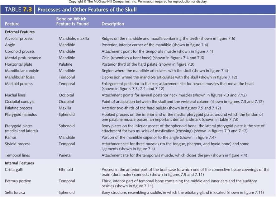
4 Skeletal System Overview Terms Body: main part Head: enlarged end Neck: constriction between head and body Margin or border: edge Angle: bend Ramus: branch off body Condyle: smooth rounded articular surface Facet: small flattened articular surface Projections Process: prominent projection Tubercle: small rounded bump Tuberosity: knob Trochanter: tuberosities on proximal femur Epicondyle: near or above condyle 7-4
5 Skeletal System Overview Ridges Line or linea: low ridge Crest or crista: prominent ridge Spine: very high ridge Openings Foramen: hole Canal or meatus: tunnel Fissure: cleft Sinus or labyrinth: cavity Depressions Fossa: general term for a depression Notch: depression in bone margin Fovea: little pit Groove or sulcus: deeper, narrow depression 7-5
6 TABLE 7.2 Term Body Head Neck Margin, border Angle Ramus Condyle Facet Ridges Line, linea Crest, crista Spine Projections Process Tubercle Tuberosity, tuber Trochanter Epicondyle Lingula Hamulus Cornu Openings Foramen Canal, meatus Fissure Sinus, labyrinth Depressions Fossa Notch Fovea Groove,ulcus Anatomical Terms for Bone Features Description Main part Enlarged, often rounded end Constriction between head and body Edge Bend Branch off the body beyond the angle Smooth, rounded articular surface Small, flattened aticular surface Low ridge Prominent ridge Very high ridge Prominent projection Small, rounded bump Knob; larger than a tubercle Tuberosity on the proximal femur Upon a condyle Flat, tongue-shaped Hook-shaped Horn-shaped Hole Tunnel Cleft Cavity General term for a depression Depression in the margin of a bone Little pit Deep, narrow depression 7-6

7 The Complete Skeleton Axial Skeleton Appendicular Skeleton Axial Skeleton Skull Skull Mandible Hyoid bone Mandible Clavicle Scapula Sternum Ribs Humerus Ribs Vertebral column Sacrum Ulna Radius Vertebral column Sacrum Carpal bones Metacarpal bones Phalanges Coxal bone Femur Coccyx Patella Tibia Fibula Tarsal bones Metatarsal bones Phalanges 7-7 Anterior view Posterior view
8 TABLE 7.1 Number of Named Bones Listed by Category Bones Number Bones Number AxialSkeleton Appendicular Skeleton Skull (Cranium) Braincase (neurocranium) Pectoral Girdle Scapula 2 Paired (left and right) Parietal 2 Clavicle 2 Temporal 2 Upper Limb Unpaired (single) Frontal 1 Humerus 2 Sphenoid Occipital Ethmoid Ulna Radius Carpal bones Face (viscerocranium) Metacarpal bones 10 Paired Maxilla Zygomatic 2 2 Phalanges Total girdle and upper limb bones Unpaired Palatine Lacrimal Nasal Inferior nasal concha Mandible Vomer Total skull bones Pelvic Girdle Coxal bone Lower Limb Femur Tibia Fibula Patella Bones Associated with the Skull Tarsal bones 14 Auditory ossicles Metatarsal bones 10 Malleus 2 Phalanges 28 Incus 2 Total girdle and lower limb bones 62 Stapes Hyoid Vertebral Column Cervical vertebrae Total associated bones Total appendicular skeleton bones Total axial skeleton bones Total appendicular skeleton bones Total bones Thoracic vertebrae 12 Lumbar vertebrae 5 Sacrum 1 Coccyx 1 Total vertebral column bones 26 Rib Cage (Thoracic Cage) Ribs 24 Sternum Total rib cage bones Total axial skeleton bones
9 Axial skeleton 7.2 Axial Skeleton Skull Hyoid bone Vertebral column Thoracic (rib) cage 7-9


10 Sagittal suture Superior View of the Skull Coronal suture Zygomatic arch Superior view Frontal bone Parietal bones Inferior temporal line Superior temporal line Parietal eminence Occipital bone Functions Protects brain Supports organs of special senses Provides foundation for structures that take air, food, and water into body Superior view of skull Parietal bones Frontal bone Sagittal suture Coronal suture 7-10
11 7-11


12 Posterior View of Skull Parietal bones Occipital bone Temporal bone Occipitomastoid suture Mastoid Zygom aticarch Posterior view Sagittal suture Lambdoid suture External occipital protuberance Superior nuchal line Inferior nuchal line Occipital condyle Parietal and occipital bones are major structures Lambdoid suture: between parietals and occipital Sutural bones may be present: variable External occipital protuberance Ligamentum nuchae: Helps keep head erect Nuchal lines: Neck muscle attachment points 7-12

13 Lateral View of Skull Parietal bones and squamous part of temporal bone form most of side of skull Squamous suture: joins the parietal and temporal bone Features of the temporal bone External auditory meatus Mastoid Temporal lines Coronal suture Superior temporal line Inferior temporal line Parietal bone Squamous suture Temporal bone Occipital bone Lambdoid suture Mandibular condyle External auditory canal Occipitomastoid suture Mastoid Zygomatic of the zygomatic arch Greater wing of the sphenoid bone anterior to the temporal bone Zygomatic bones with its temporal of the zygomatic arch Maxilla Mandible. Articulates with the temporal bone. Body, ramus, condyle, and coronoid Styloid Zygomatic arch Zygomatic of temporal bone Temporal of zygomatic bone Mandibular ramus Angle of mandible Body of mandible Lateral view Frontal bone Supraorbital foramen Supraorbital margin Sphenoid bone (greater wing) Nasal bone Lacrimal bone Nasolacrimal canal Infraorbital foramen Zygomatic bone Coronoid of mandible Maxilla Alveolar es Mental foramen Mandible Mental protuberance 7-13

14 Frontal bone Supraorbital margin Zygomatic arch Nasal bone Zygomatic bone Maxilla Mastoid Mental protuberance Mandible Angle of mandible McGraw-Hill Higher Education, Inc./Eric Wise, photographer 7-14

15 Anterior View of Skull Frontal bone Coronal suture Glabella Supraorbital margin Temporal bone Parietal bone Supraorbital foramen Optic canal Orbital plate of frontal bone Sphenoid bone (greater wing) Major structures are frontal bone, zygomatic bones, maxillae, and mandible Nasal septum Nasal bone Infraorbital margin Zygomatic bone Perpendicular plate of ethmoid bone Vomer Nasal cavity Maxilla Alveolar es Body of mandible Mental foramen Superior orbital fissure Lacrimal bone Infraorbital foramen Middle nasal concha Inferior nasal concha Anterior nasal spine Oblique line of mandible Mandible Mandibular symphysis Maxilla and mandible bear teeth Orbits. Cone-shaped fossae with their apices oriented posteriorly Anterior view Mental protuberance Nasolacrimal canal Optic foramen 7-15

16 Glabella Frontal bone Supraorbital margin Zygomatic bone Maxilla Mandible Mental protuberance McGraw-Hill Higher Education, Inc./Eric Wise, photographer 7-16


17 The Orbit Supraorbital foramen Frontal bone Superior orbital fissure Sphenoid bone Palatine bone Zygomatic bone Lesser wing Greater wing Optic canal Posterior and anterior ethmoidal for amina Ethmoid bone Lacrimal bone Opening to nasolacrimal canal Maxilla Inferior orbital fissure Infraorbital foramen Infraorbital groove Anterior view 7-17
 Medial view (b) Medial view Crista galli Horizontal plate of palatine bone Palatine of")
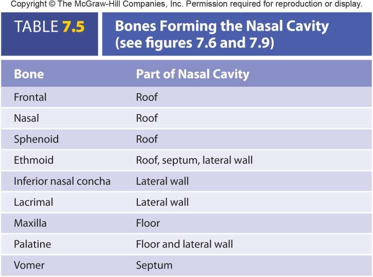
18 Bones of Nasal Cavity Nasal septum Frontal bone Frontal sinus Nasal bone Perpendicular plate of ethmoid bone Septal cartilage Vomer Greater alar cartilage Anterior nasal spine Central incisor Frontal bone Frontal sinus Nasal bone Maxilla bone Lateral nasal cartilage Greater alar cartilage Lateral incisor (a) Medial view (b) Medial view Crista galli Horizontal plate of palatine bone Palatine of maxilla Incisive canal Cribriform plate Olfactory foramina Sphenoidal sinus Sphenoid bone Hard palate Lacrimal bone Olfactory recess Superior nasal concha Middle nasal concha Sphenoidal sinus Sphenoid bone Inferior nasal concha Vertical plate Horizontal plate Medial pterygoid plate Part of ethmoid bone Palatine bone Palatine of maxilla Nasal cavity. Pear-shaped, open anteriorly Nasal septum divides nasal cavity into right and left halves Bony part is vomer and perpendicular plate of the ethmoid Hyaline cartilage anterior part Nasal conchae: form lateral walls Inferior: separate bones Middle and superior: projections of the ethmoid Increase surface of nasal cavity 7-18
19 7-19


 Frontal Sphenoidal")

20 Paranasal Sinuses Associated with the bones of the nasal cavity Functions Decrease skull weight Resonating chambers Named for bones in which they are found Frontal Maxillary Ethmoidal Sphenoidal Frontal sinus Sphenoidal sinus Ethmoidal labyrinth (sinuses) Maxillary sinus (b) Frontal sinus Ethmoidal labyrinth (sinuses) Sphenoidal sinus (c) Maxillary sinus (d) (a) c-d: Jupiter Media/Alamy 7-20
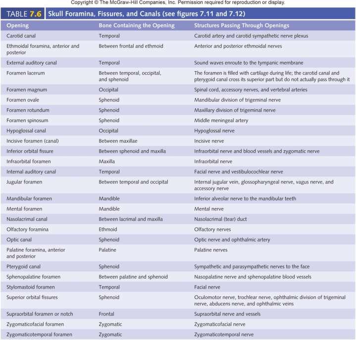
21 Interior of the Cranial Cavity Cranial cavity: occupied by the brain Floor divided into anterior, middle, and posterior fossae Crista galli: prominent ridge in center of anterior fossa. Point of attachment for the dura mater (one of the meninges) Olfactory fossae lateral to crista galli. Olfactory bulb within Cribriform plate of the ethmoid forms floor of olfactory fossae Olfactory nerves pass through the foramina of the cribriform plate Sella turcica: part of sphenoid bone that houses the pituitary gland Foramen magnum: opening where brain attaches to spinal cord 7-21
 Hypoglossal canal Foramen")
22 Interior of the Cranial Cavity Frontal sinuses Anterior cranial fossa Crista galli Olfactory foramina Cribriform plate Frontal bone Ethmoid bone Sphenoid bone Temporal bone Lesser wing Greater wing Middle cranial fossa Squamous portion Petrous portion Optic canal Sella turcica Foramen rotundum Foramen ovale Foramen spinosum Carotid canal (foramen lacerum is inferior) Hypoglossal canal Foramen magnum Parietal bone Occipital bone Internal auditory canal Jugular foramen Posterior cranial fossa Superior view 7-22
23 Inferior View of Skull Foramina Foramen magnum: spinal cord exits and vertebral arteries enter Carotid canals: internal carotid arteries Foramen lacerum: internal carotid Jugular foramen: internal jugular veins Specialized surfaces Occipital condyles: articulation between skull and vertebral column Styloid es: attachment site for muscles that move the tongue Mandibular fossa: site of articulation with mandibular condyles Medial and later pterygoid plates: parts of sphenoid bone that surround posterior opening of nasal cavities Vomer: posterior portion of nasal septum Hard palate: floor of the nasal cavity. With the soft palate, separates nasal from oral cavities 7-23

24 Inferior View of Skull Incisive fossa Maxilla Zygomatic bone Palatine of maxillary bone Horizontal plate of palatine bone Hard palate Sphenoid bone Anterior palatine foramen Posterior palatine foramen Inferior orbital fissure Lateral pterygoid plate Greater wing Medial pterygoid plate Foramen ovale Foramen spinosum External auditory canal Jugular foramen Occipital condyle Foramen magnum Pterygoid hamulus Temporal of zygomatic bone Zygomatic arch Zygomatic of temporal bone Vomer Foramen lacerum Styloid Mandibular fossa Carotid canal (posteroinferior opening) Stylomastoid foramen Mastoid Temporal bone Occipital bone Inferior nuchal line External occipital protuberance Superior nuchal line Inferior view 7-24
25 7-25
26 Bones of the Skull Twenty-two bones plus six auditory ossicles that function in hearing Of the twenty-two, two portions- Braincase (Neurocranium) Surrounds and protects brain Parietals, temporals, frontal, occipital, sphenoid, ethmoid Facial bones (Viscerocranium) Protect major sensory organs- eyes, nose, and tongue Provide attachment sites for muscles of mastication, facial expression, and eye movement Maxilla and mandible have alveolar es and sockets for tooth attachment Maxillae, zygomatics, palatines, lacrimals, nasals, inferior nasal conchae, mandible, vomer. Note: frontal and ethmoid contribute to the face and mandible is not part of the skull 7-26
 Parietal Bone (Right) Lateral View Landmark Parietal eminence Superior and inferior temporallines Special Feature Forms lateral wall of skull Description The")
27 TABLE 7.7 Skull Bones (a) Parietal Bone (Right) Lateral View Landmark Parietal eminence Superior and inferior temporallines Special Feature Forms lateral wall of skull Description The widest part of the head is from one parietal eminence to the other. Attachment point for temporalis muscle Parietal eminence Superior tem poralline Inferior temporal line 7-27


 Mandibular fossa Foramen through which the internal jugular vein exits the cranial cavity Articulation point between")

 Thick portion of the temporal bone Squamous portion (shown in figure 7.")

28 TABLE 7.7 Skull Bones (b) Temporal Bone (Right) Lateral and Medial Views Landmark Carotid canal (shownin figures 7.11 and 7.12 ) Description Canal through which the internal carotid artery enters the cranial cavity External auditory canal External canal of the ear; carries sound to the ear Squamous portion Internal auditory canal (shown in figure 7.11 ) Opening through which the facial (cranial nerve VII) and vestibulocochlear (cranial nerve VIII) nerves enter the petrous portion of the temporal bone Mastoid Zygomatic Mandibular fossa External auditory canal Forms one side of jugular for a men (shown in figures 7.11 and 7.12 ) Mandibular fossa Foramen through which the internal jugular vein exits the cranial cavity Articulation point between the mandible and skull Styloid Mastoid Attachment point for muscles moving the head and for a hyoid muscle Lateral view Middle cranial fossa (shown in figure 7.11) Depression in the floor of the cranial cavity formed by the temporall obesof the brain Petrous portion (shown in figure 7.11) Thick portion of the temporal bone Squamous portion (shown in figure 7.11 ) Flat, lateral portion of the temporal bone Mastoid Medial view Squamous portion Petrous portion Internal auditory canal Styloid Styloid Stylomastoid foramen (shown infigure 7.12 ) Zygomatic Attachment for muscles of the tongue, throat, and hyoid bone Foramen through which the facial nerve (cranial nerve VII ) exits the skull Helps form the bony bridge extending from the cheek to just anterior to the ear; attachment for a muscle that moves the mandible Special Features Contains the middle and inner ear and the mastoid air cells Place where the mandible articulates with the rest of the skull 7-28
 Frontal Bone Anterior View Landmark Glabella Nasal")


29 TABLE 7.7 Skull Bones Continued (c) Frontal Bone Anterior View Landmark Glabella Nasal spine Orbital plate Supraorbital foramenar Supraorbital margin Zygomatic Description Area between the supraorbital margins Superior part of the nasal bridge Roof of the orbit Opening through which nerves and vessels exit the skull to the skin of the forehead Ridge forming the anterior superior border of the orbit Connects to the zygomatic bone; helps form the lateral margin of the orbit Glabella Supraorbital foramen Orbital plate Supraorbital margin Zygomatic Nasal spine Special Features Forms the forehead and roof of the orbit Contains the frontal sinus 7-29
 Sphenoid Bone Superior and Posterior Views Landmark Description Body Thickest part of the bone; articulates with the occipital bone")
 exits the cranial cavity Lesser wing Greater wing Superior orbital fissure Sella turcica Foramen spinosum Greater")

")

30 TABLE 7.7 Skull Bones Continued (d) Sphenoid Bone Superior and Posterior Views Landmark Description Body Thickest part of the bone; articulates with the occipital bone Foramen ovale Opening through which a branch of the trigeminal nerve (cranial nerve V) exits the cranial cavity Optic canal Foramen rotundum Opening through which a branch of the trigeminal nerve (cranial nerve V) exits the cranial cavity Lesser wing Greater wing Superior orbital fissure Sella turcica Foramen spinosum Greater wing Opening through which a major artery to the meninges (membranes around the brain) enters the cranial cavity Forms the fl oor of the middle cranial fossa; several foramina pass through this wing Foramen rotundum Foramen ovale Foramen spinosum Superior view Groove of carotid canal Lateral pterygoid plate Attachment point for muscles of mastication (chewing) Lesser wing Superior border of the superior orbital fissure Medial pterygoid plate Posterolateral walls of the nasal cavity Lesser wing Greater wing Superior orbital fissure Body Optic canal Pterygoid canal Pterygoid hamulus Opening through which the optic nerve (cranial nerve II) passes from the orbit to the cranial cavity Opening through which nerves and vessels exit the cranial cavity Process around which the tendon passes from a muscle to the soft palate Sella turcica Fossa containing the pituitary gland Foramen rotundum Pterygoid canal Pterygoid hamulus Posterior view Lateral pterygoid plate Medial pterygoid plate Superior orbital fissure Special Feature Contains the sphenoidal sinus Opening through which nerves and vessels enter the orbit from the cranial cavity 7-30
 Occipital Bone Inferior View Landmark Condyle Description Articulation point between the skull and first vertebra External occipital protuberance Attachment point for a")
 Inferior nuchal line Posterior cranial fossa (shown in figure 7.")
31 TABLE 7.7 Skull Bones Continued (e) Occipital Bone Inferior View Landmark Condyle Description Articulation point between the skull and first vertebra External occipital protuberance Attachment point for a strong ligament (nuchal ligament) in the back of the neck Anterior Condyle Foramen magnum Foramen magnum Hypoglossal canal (shown in figure 7.11) Inferior nuchal line Posterior cranial fossa (shown in figure 7.11) Superior nuchal line Opening around the point where the brain and spinal cord connect Opening through which the hypoglossal nerve (cranial nerve XII) passes Attachment point for neck muscles Depression in the posterior of the cranial cavity formed by the cerebellum Attachment point for neck muscles Special Feature Inferior nuchalline Forms the base of the skull Superior nuchalline Posterior External occipital protuberance 7-31
 Zygomatic Bone (Right) Lateral View Frontal")


32 TABLE 7.7 Skull Bones Continued (f) Zygomatic Bone (Right) Lateral View Frontal Landmark Frontal Infraorbital margin Temporal Zygomaticofacial foramen Description Connection to the frontal bone; helps form the lateral margin of the orbit Ridge forming the inferior border of the orbit Helps form the bony bridge from the cheek to just anterior to the ear Opening through which a nerve and vessels exit the orbit to the face Temporal Zygomaticofacial foramen Infraorbital margin Maxillary Special Features Forms the prominence of the cheek Forms the anterolateral wall of the orbit 7-32
 Ethmoid Bone Superior, Lateral, and Anterior Views Land mark Description Cribriformplate Contains numerous olfactory")

 Middle nasal concha Ridge extending into the nasal cavity; increases surface area, helps warm and moisten air")


33 TABLE 7.7 Skull Bones Continued (g) Ethmoid Bone Superior, Lateral, and Anterior Views Land mark Description Cribriformplate Contains numerous olfactory foramina through which branches of the olfactory nerve (cranial nerve I) enter the cranial cavity from the nasal cavity Anterior Perpendicular plate Cristagalli Ethmoidalforamina (shown in figure7.8) Attachment for meninges (membranes around brain) Openings through which nerves and vessels pass from the orbit to the nasal cavity Crista galli Ethmoidal labyrinth (sinuses) Middle nasal concha Ridge extending into the nasal cavity; increases surface area, helps warm and moisten air in the cavity Cribriform plate Orbital plate Perpendicular plate Forms the medial wall of the orbit Forms the superior portion of the nasal septum Orbital plate Superior nasal concha Ridge extending into the nasal cavity; increases surface area, helps warm and moisten air in the cavity Posterior Special Features Forms part of the nasal septum and part of the lateral walls and roof of the nasal cavity Superior view Contains the ethmoidal labyrinth, or ethmoidal sinuses; the labyrinth is divided into anterior, middle, and posterior ethmoidal cells Crista galli Orbital plate Ethmoidal labyrinth (sinuses) Posterior Anterior Middle nasal concha Perpendicular plate Lateral view Crista galli Orbital plate Ethmoidal labyrinth (sinuses) Perpendicular plate Superior nasal concha Middle nasal concha Anterior view 7-33
 Maxilla (Right) Medial and Lateral Views Frontal Maxillary sinus Palatine Incisive canal Alveolar")


34 TABLE 7.7 Skull Bones Continued (h) Maxilla (Right) Medial and Lateral Views Frontal Maxillary sinus Palatine Incisive canal Alveolar Landmark Alveolar Anterior nasal spine Frontal Incisive canal Infraorbital foramen Orbital surface Palatine Maxillary Tuberosity Zygomatic Description Ridge containing the teeth Forms part of the nasal septum Forms the sides of the nasal bridge Opening through which a nerve exits the nasal cavity to the roof of the oral cavity Opening through which a nerve and vessels exit the orbit to the face Forms the fl oor of the orbit Forms the anterior two-thirds of the hard palate Lump posterior to the last maxillary molar tooth Connection to the zygomatic bone; helps form the interior margin of the orbit Special Features Contains the maxillary sinus and maxillary teeth Forms part of nasolacrimal canal Molars Premolars Canine Incisors Medial view Notch for lacrimal bone Frontal Orbital surface Zygomatic Tuberosity Infraorbital foramen Anterior nasal spine Alveolar Lateral view Incisors Canine Premolars Molars 7-34
 Palatine Bone (Right) Medial and")


35 TABLE 7.7 Skull Bones Continued (i) Palatine Bone (Right) Medial and Anterior Views Landmark Horizontal plate Vertical plate Description Forms the posterior one-third of the hard palate Forms part of the lateral nasal wall Special Feature Helps form part of the hard palate and a small part of the wall of the orbit Vertical plate Vertical plate Horizontal plate Horizontal plate Medial view Anterior view 7-35
 Lacrimal Bone (Right) Anterolateral View Special Features Forms a small")
36 TABLE 7.7 Skull Bones Continued (j) Lacrimal Bone (Right) Anterolateral View Special Features Forms a small portion of the orbital wall Forms part of the nasolacrimal canal Lacrimal bone Nasolacrimal canal 7-36
 Nasal")

37 TABLE 7.7 Skull Bones Continued (k) Nasal Bone (Right) Anterolateral View Special Feature Forms the bridge of the nose Nasal bone 7-37
 Mandible (Right Half) Medial and Lateral Views Landmark Alveolar Angle Description Ridge containing the teeth")
 Condylar Ramus Mandibular foramen Lingula Alveolar Mylohyoid line Angle Coronoid Molars")


38 TABLE 7.7 Skull Bones Continued (l) Mandible (Right Half) Medial and Lateral Views Landmark Alveolar Angle Description Ridge containing the teeth Corner between the body and ramus Molars Premolars Canine Incisors Body Mandibular notch Mandibular condyle Condylar Medial view Coronoid Mandibular notch Mandibular condyle (head) Condylar Ramus Mandibular foramen Lingula Alveolar Mylohyoid line Angle Coronoid Molars Premolars Canine Body Condylar Coronoid Mandibular condyle Mandibular foramen Mandibular notch Mental foramen Mylohyoid line Oblique line Ramus Major, horizontal portion of the bone Extension containing the mandibular condyle Attachment for a muscle of mastication Helps form the temporomandibular joint (the point of articulation between the mandible and the rest of the skull) Opening through which nerves and vessels to the mandibular teeth enter the bone Depression between the condylar and the coronoid Opening through which a nerve and vessels exit the mandible to the skin of the chin Attachment point of the mylohyoid muscle Ridge from the anterior edge of the ramus onto the body of the mandible Major, nearly vertical portion of the bone Oblique line Ramus Body Angle Lateral view Incisors Alveolar Mental foramen Special Features The only bone in the skull that is freely movable relative to the rest of the skull bones Holds the lower teeth 7-38
 Vomer Anterior and Lateral")


39 TABLE 7.7 Skull Bones Continued (m) Vomer Anterior and Lateral Views Alae Alae Landmark Alae Vertical plate Description Attachment points between the vomer and sphenoid Forms part of the nasal septum Special Feature Forms most of the posterior and inferior portions of the nasal septum Vertical plate Vertical plate Anterior view Lateral view 7-39

40 Hyoid Bone TABLE 7.8 Hyoid Bone Anterior and Lateral Views Greater cornu Landmark Body Greater cornu Lesser cornu Special Features Description Major portion of the bone Attachment point for muscles and ligaments Attachment point for muscles and ligaments One of the few bones of the body that does not articulate with another bone Attached to the skull by muscles and ligaments Lesser cornu Body Anterior view Lesser cornu Greater cornu Lateral view Body 7-40
41 Vertebral Column Functions Supports weight of head and trunk Protects the spinal cord Allows spinal nerves to exit the spinal cord Provides site for muscle attachment Permits movement of head and trunk Twenty-six bones in adult; in embryo 5 fuse to form sacrum 4 or 5 coccygeal fuse to form the coccyx Regions Cervical (7 vertebrae) Thoracic (12 vertebrae) Lumbar (5 vertebrae) Sacral bone (1) Coccygeal bone (1) 7-41
 Second cervical vertebra (axis) Coccyx Seventh cervical vertebra First thoracic vertebra Sacrum Intervertebral foramina Body Intervertebral disk Twelfth thoracic vertebra")
42 Vertebral Column Cervical region (convex anteriorly) Thoracic region (concave anteriorly) Lumbar region (convex anteriorly) Sacral and coccygeal regions (concave anteriorly) L a te r al view First cervical vertebra (atlas) Second cervical vertebra (axis) Coccyx Seventh cervical vertebra First thoracic vertebra Sacrum Intervertebral foramina Body Intervertebral disk Twelfth thoracic vertebra First lumbar vertebra Transverse Spinous Fifth lumbar vertebra Sacral promontory Four major curvatures in adults Cervical: anterior Thoracic: posterior Lumbar: anterior Sacral and coccygeal: posterior At birth, column is C shaped When head is raised, cervical curve appears When sitting and walking begin, lumbar curve develops Abnormal curvatures Lordosis. Exaggeration of lumbar Kyphosis. Exaggeration of thoracic Scoliosis. Lateral, often accompanied by kyphosis 7-42

43 General Features of the Vertebrae TABLE 7.9 Feature Body Vertebral foramen Vertebral arch Pedicle Lamina Transverse Spinous Articular es General Structure of a Vertebra Description Disk-shaped; usually the largest part with fl at surfaces directed superiorly and inferiorly; forms the anterior wall of the vertebral foramen; intervertebral disks are located between the bodies Hole in each vertebra through which the spinal cord passes; adjacent vertebral foramina form the vertebral canal Forms the lateral and posterior walls of the vertebral foramen; possesses several es and articular surfaces Foot of the arch with one on each side; forms the lateral walls of the vertebral foramen Posterior part of the arch; forms the posterior wall of the vertebral foramen Process projecting laterally from the junction of the lamina and pedicle; a site of muscle attachment Process projecting posteriorly at the point where the two laminae join; a site of muscle attachment; strengthens the vertebral column and allows for movement Superior and inferior projections containing articular facets where vertebrae articulate with each other; strengthen the vertebral column and allow for movement Intervertebral notches Form intervertebral foramina between two adjacent vertebrae through which spinal nerves exit the vertebral canal 7-43


 Lateral view, Sagittal section Anterior Posterior Superior")


44 General Features of the Vertebrae TABLE 7.9 General Structure of a Vertebra Transverse Spinous Lamina Body (cut) Part of vertebral canal Superior articular facet Superior articular Pedicle Vertebral arch Intervertebral disk Vertebral foramen Body Vertebral foramina Spinous (cut) ( a ) Superior view ( b ) Lateral view, Sagittal section Anterior Posterior Superior articular Anterior Posterior Transverse Pedicle Inferior articular of superior vertebra Superior articular of inferior vertebra Space for intervertebral disk Inferior intervertebral notch of superior vertebra Superior intervertebral notch of inferior vertebra Intervertebral foramen Body Spinous es ( c ) ( d ) 7-44

45 Spina Bifida Lamina of vertebrae: can be removed (laminectomy) when they inhibit a surgery such as a disc removal. Spina bifida: failure of the laminae to form or to fuse together during development. Can affect the spinal cord. Most often occurs in lumbar region. Posterior Dura mater Skin of back Enlarged fluid-filled space Spinal cord Cauda equina Body of first lumbar vertebra Back muscles Incomplete vertebral arch Superior view 7-45

46 Intervertebral Disks Intervertebral disk Vertebral body Annulus fibrosus Nucleus pulposus Intervertebral foramen Annulus fibrosus Nucleus pulposus (a) Lateral view (b) Superior view Located between adjacent vertebrae Functions Provide support Prevent vertebrae rubbing against each other Consist of Annulus fibrosus: external Nucleus pulposus: internal and gelatinous Becomes compressed with age and height decreases With age, more susceptible to herniation 7-46

47 Herniated or Ruptured Disk Part of the fibrosus has been removed to expose the pulposus. Breakage or ballooning of the annulus fibrosus with a partial or complete release of the nucleus pulposus. May push against spinal nerves impairing function and causing pain. Spinous Transverse Spinal cord in vertebral canal Compressed spinal nerve root in intervertebral foramen Herniated portion of disk Nucleus pulposus Annulus fibrosus Intervertebral disk Superior view 7-47


48 TABLE 7.10 Comparison of Vertebral Regions Feature Cervical Thoracic Lumbar Superior articular facet Transverse Body Body Body Transverse foramen Inferior articular facet Superior articular Facets for rib Transverse articulation Spinous Spinous Inferior articular facet Superior articular Transverse Spinous Body Absent in C1, small in others Medium-sized with articular facets for ribs Large Transverse Transverse foramen Articular facets for ribs, except T11 and T12 Square Spinous Absent in C1, bifid in others, except C7 Long, angled inferiorly Square Articular facets Face superior/inferior Face obliquely Face medial/lateral 7-48
49 Cervical Vertebrae Superior seven vertebrae Have very small bodies, tend to have bifid spinous es, and have transverse foramina Atlas: first cervical vertebra Articulates with skull and allows yes movement No body and no spinous Axis: second cervical vertebra Dens or odontoid extends superiorly into the vertebral foramen of the atlas Allows rotation of the atlas on the axis, the no movement Vertebral prominence: most prominent spinous in area. Usually 7 th cervical Superior articular facets face superiorly; inferior facets face inferiorly 7-49
 Atlas, lateral view Posterior arch Transverse Transverse foramen Dens Body Spinous (bifid) Posterior arch Vertebral foramen Superior")
50 Transverse Transverse foramen Posterior arch Vertebral foramen Superior articular facet (articulates with occipital condyle) Facet for dens Anterior arch (a) Atlas (first cervical vertebra), superior view Anterior arch Superior articular facet Transverse (b) Atlas, lateral view Posterior arch Transverse Transverse foramen Dens Body Spinous (bifid) Posterior arch Vertebral foramen Superior articular facet Dens Superior articular facet Body Spinous Transverse foramen (c) Axis (second cervical vertebra), superior view (d) Axis, lateral view Anterior arch Transverse ligament Dens of axis Atlas Posterior arch Axis Lamina Pedicle Transverse foramen Transverse Body Spinous (bifid) Vertebral foramen Superior articular facet (e) Atlas and axis articulated, superior view (f) Fifth cervical vertebra, superior view Vertebral body Superior articular Transverse Spinous Bifid tip of spinous Inferior articular Transverse foramen Inferior articular facet (g) Fifth cervical vertebra, lateral view (h) Trent Stephens Dens Body C1 C2 C3 C4 C5 C6 C7 (h) Anterolateral view Spinous es Transverse Transverse foramen 7-50
 Superior view (b) Lateral view Spinous Vertebral foramen Articular facet for tubercle of rib Superior articular facet Superior articular facet for rib head Superior")
51 Thoracic Vertebrae Anterior Lamina Transverse Body Superior articular Pedicle Superior intervertebral notch Superior articular facet for rib head Inferior articular facet for rib head Inferior intervertebral notch Body (a) Superior view (b) Lateral view Spinous Vertebral foramen Articular facet for tubercle of rib Superior articular facet Superior articular facet for rib head Superior articular Superior articular facet Pedicle Transverse Articular facet for tube rcle of rib Lamina Inferior articular Spinous Body Space for intervertebral disk T1 T2 T3 T4 T5 T6 T7 Posterior Articular facet for rib head Articular facet for tubercle of rib Transverse Spinous Intervertebral foramen Long, thin spinous es directed inferiorly Long transverse es Articular facets on transverse es for ribs (first 10 thoracic vertebrae) Facets on body for articulation with ribs Most ribs have heads that articulate with two sequential vertebrae (c) Posterolateral view (c) Trent Stephens 7-51
 Superior view Superior articular Transverse Spinous Adds strength Inferior articular Inferior articular facet Limits")
52 Lumbar Vertebrae Spinous Lamina Large thick bodies Transverse Pedicle Superior articular facet Vertebral foramen Heavy rectangular transverse and spinous es Body Superior articular facets face medially; inferior articular facets face laterally Vertebral body Pedicle (a) Superior view Superior articular Transverse Spinous Adds strength Inferior articular Inferior articular facet Limits rotation (b) Lateral view L1 Intervertebral foramen Body L2 Spinous Space for intervertebral disk L3 L4 Transverse L5 (c) Lateral view (c) Trent Stephens 7-52
 Median sacral crest Sacral hiatus Sacrum Alae: superior lateral parts of fused transverse es Auricular surface: articulates with")
53 Sacrum and Coccyx Ala Sacr al promontory Anterior sacral for amina Transverse lines Ala Sacral canal Auricular surface (articulates with coxal bone) Posterior sacral foramina (a) Anterior view Coccyx Superior articular facet (articulates with fifth lumbar vertebra) Median sacral crest Sacral hiatus Sacrum Alae: superior lateral parts of fused transverse es Auricular surface: articulates with pelvic bone Median sacral crest: partially fused spinous es Sacral hiatus: site of anesthesia injection Sacral foramina: intervertebral foramina Sacral promontory: anterior edge of body of first vertebra. Marks separation of abdominal and pelvic cavities Coccyx: tailbone Coccyx (b) Posterior view 7-53
 Anterior view Articular facet for transverse of vertebra (c) Lateral view (c) Trent Stephens Seventh cervical vertebra First thoracic")

 joined by common cartilage to sternum Floating or vertebral (2) do not")
54 Rib Cage Sternal end True ribs False ribs (8 12) Clavicle Sternal angle Costal cartilage Floating ribs Head Neck Tubercle Angle Body (b) Inferior view T12 12 L1 (a) Anterior view Articular facet for transverse of vertebra (c) Lateral view (c) Trent Stephens Seventh cervical vertebra First thoracic vertebra Jugular notch Manubrium Body Xiphoid Sternum Head of rib set against the inferior articular facet of the superior vertebra Head of rib set against the superior articular facet of the inferior vertebra Tubercle of rib set against the articular facet on the transverse of the inferior vertebra Angle of rib Body of rib Functions Protects vital organs Forms semi-rigid chamber for respiration Parts Thoracic vertebrae Ribs (12 pair) True or Vertebrosternal: superior seven. Attach directly to sternum via costal cartilages False: inferior five Vertebrochondral (3) joined by common cartilage to sternum Floating or vertebral (2) do not attach to sternum 7-54
55 Manubrium Sternum Articulates with first rib and clavicle Jugular notch superiorly Sternal angle: point where manubrium joins body. Second rib articulates here Body: third through seventh ribs articulate Also called gladiolus Xiphoid : inferior tip Clavicle 1 Seventh cervical vertebra First thoracic vertebra Jugular notch True ribs Sternal angle Costal cartilage Manubrium 6 7 Body Xiphoid Sternum False ribs (8 12) Floating ribs T12 12 L (a) Anterior view

56 7.3 Appendicular Skeleton Girdles Pectoral or shoulder Pelvic Upper Limbs Arm Forearm Wrist Hand Lower Limbs Thigh Leg Foot Ulna Radius Anterior view Carpal bones Metacarpal bones Phalanges Clavicle Scapula Humerus Pectoral girdle Upper limb Anterior view Sacrum Coxal bone Femur Patella Tibia Fibula Tarsal bones Metatarsal bones Phalanges Pelvic girdle Lower limb 7-56


 Superior view Superior angle Superior border Scapular notch Supraspinous")

 border (b) Posterior view")
57 Pectoral Girdle Scapula (2) Acromion Forms protective cover Attachment for clavicle Attachment for muscles Scapular spine: divides posterior surface into supraand infraspinous fossae Coracoid : attachment for muscles Glenoid cavity: articulates with humerus Clavicle (2): articulates with acromion and with manubrium of sternum Acromion Coracoid Supraglenoid tubercle Glenoid cavity Infraglenoid tubercle Lateral (axillary) border Inferior angle Acromial (lateral) end (a) Anterior view Body of clavicle (c) Superior view Superior angle Superior border Scapular notch Supraspinous fossa Scapular spine Subscapular fossa Medial (vertebral) border View in (d) Sternal (medial) end d: Trent Stephens Spine of scapula Supraspinous fossa of scapula Superior border of scapula Acromion of scapula Acromial end of clavicle Coracoid of scapula Body of clavicle Acromion Coracoid Glenoid cavity Infraglenoid tubercle Infraspinous fossa Lateral (axillary) border (b) Posterior view Posterior Anterior (d) Superior view 7-57

58 Arm (Humerus) Greater tubercle Lesser tubercle Intertubercular groove Deltoid tuberosity Lateral supracondylar ridge Radial fossa Lateral epicondyle Capitulum Head Anatomical neck Surgical neck Medial supracondylar ridge Coronoid fossa Medial epicondyle Radial groove Olecranon fossa Lateral epicondyle Head Neck: anatomic and surgical Tubercles: greater and lesser Intertubercular groove Deltoid tuberosity Capitulum: rounded, articulates with radius Trochlea: spool-shaped, articulates with ulna Epicondyles Trochlea Trochlea (a) Anterior view (b) Posterior view 7-58
59 Forearm: Radius Radial notch of ulna Olecranon View in (a) Head of radius Radial notch of ulna Head Neck Radial tuberosity Interosseous ridges Radius (a) Proximal view Trochlear notch Coronoid Trochlear notch Coronoid Radial notch of ulna Ulnar tuberosity Ulna Olecranon Medial: thumb side Proximal end Head rotates in radial notch of ulna. Radial tuberosity: site of biceps brachii insertion Distal end Articulates with carpals and ulna Styloid Head Styloid (b) Anterior view Styloid Ulnar notch of radius (c) Medial view of ulna 7-59
60 Forearm: Ulna Lateral: little finger side Proximal end View in (a) Radial notch of ulna Head of radius (a) Proximal view Olecranon Trochlear notch Coronoid Trochlear notch: fits over trochlea of humerus Olecranon : point of elbow Radial notch of ulna Head Neck Radial tuberosity Trochlear notch Coronoid Radial notch of ulna Ulnar tuberosity Olecranon Coronoid Distal end Interosseous ridges Ulna Head articulates with radius and with carpals Radius Styloid Head Styloid (b) Anterior view Styloid Ulnar notch of radius (c) Medial view of ulna 7-60

61 Heads of metacarpal bones (knuckles) Head of ulna Acromion Lateral epicondyle Olecranon Medial border of scapula Olecranon Medial epicondyle The McGraw-Hill Companies, Inc./Eric Wise, photographer 7-61
62 Wrist and Hand Wrist: eight carpal bones In order from lateral to medial for proximal row and medial to lateral for distal row: So Long Top Part, Here Comes The Thumb Scaphoid, Lunate, Triquetrum, Pisiform, Hamate, Capitate, Trapezoid, Trapezium As a unit are convex posteriorly and concave anteriorly Carpal tunnel: on anterior surface. Ligament from tubercle of trapezium to hook of hamate Hand: five metacarpals (palm of hand); five digits with their phalanges 7-62
 Metacarpal bones 5 4 3 2 1 1 2 3 4 5 Proximal phalanx of thumb Distal phalanx of thumb Digits Proximal phalanx of")
63 Wrist and Hand Radius Ulna Carpal bones (distal row) Carpal bones (proximal row) Scaphoid bone Lunate bone Triquetrum bone Pisiform bone Hamate bone Capitate bone Trapezoid bone Trapezium bone Scaphoid bone Lunate bone Triquetrum bone Pisiform bone Carpal bones (proximal row) Metacarpal bones Proximal phalanx of thumb Distal phalanx of thumb Digits Proximal phalanx of finger Middle phalanx of finger Distal phalanx of finger (a) Posterior view (b) Anterior view 7-63

64 Pelvic Girdle Sacrum Sacroiliac joint Anterior superior iliac spine Acetabulum Symphysis pubis Obturator foramen Subpubic angle Anterosuperior view Sacral promontory Ilium Pubis Ischium Coxal bone Coxal bones and sacrum form ring Pelvis: pelvic girdle and coccyx Coxal bones: Right and Left Ilium Ischium Pubis Acetabulum: articulates with head of femur Obturator foramen Sacrum 7-64

65 Sacrum Coxal bone Pelvic girdle Femur Patella Tibia Lower limb Fibula Anterior view Tarsal bones Metatarsal bones Phalanges 7-65
66 Coxal Bones Formed as fusion of embryonic ilium, ischium, pubis. All three contribute to acetabulum Ilium: iliac crest, anterior and posterior superior iliac spines, greater sciatic notch, auricular surface, sacroiliac joint, iliac fossa Ischium: ischial tuberosity Pubis: pubic crest, symphysis pubis (pubic symphysis) Pelvic brim False (greater pelvis) pelvis superior to brim True pelvis inferior to brim Pelvic inlet Pelvic outlet 7-66

67 Coxal Bones Ilium Acetabulum Cartilage in young pelvis Pubis Ischium Obturator foramen (a) Lateral view Tubercle of iliac crest Posterior superior Iliac spine Posterior inferior iliac spine Greater sciatic notch Ischial spine Lesser sciatic notch Ischial tuberosity Lunate surface Acetabulum Iliac crest Ilium Iliac fossa Anterior superior iliac spine Anterior inferior iliac spine Linea terminalis Superior pubic ramus Pubis Acetabular notch Pubic tubercle Pubic crest Inferior pubic ramus Obturator foramen Arcuate line Pectineal line Auricular surface (articulates with sacrum) Posterior superior iliac spine Posterior inferior iliac spine Greater sciatic notch Ischium Ischial spine Lesser sciatic notch Ischial ramus 7-67 (b) Lateral view Ischial ramus (c) Medial view
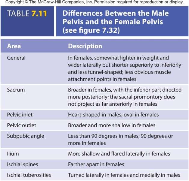
 Male (a) Anterosuperior view Female (b) Anterosuperior view Pelvic brim Sacral promontory Pelvic inlet (c) Medial view Coccyx Pelvic outlet")
68 Comparison of the Male and Female Pelvis Pelvic Inlet (red dashed line) Sacral promontory Pelvic brim Symphysis pubis Subpubic angle Ischial spine Coccyx Symphysis pubis Pelvic outlet (blue dashed line) Male (a) Anterosuperior view Female (b) Anterosuperior view Pelvic brim Sacral promontory Pelvic inlet (c) Medial view Coccyx Pelvic outlet 7-68
69 7-69
70 Thigh Femur Head: articulates with acetabulum Neck Trochanters: attachment for muscles that fasten lower extremity to hip Greater and lesser Distal condyles: articulate with tibia Medial and lateral Epicondyles: ligament attachment Medial and lateral Patella or kneecap: sesamoid In tendon of quadriceps femoris Changes force relationship between femur and tibia 7-70
 of femur Adductor tubercle Medial epicondyle Lateral epicondyle Lateral epicondyle Intercondylar fossa Patellar groove Medial condyle Lateral condyle 7-71 (a) Anterior view (b) Posterior")
71 Thigh Head Greater trochanter Neck Intertrochanteric line Fovea capitis Lesser trochanter Head Greater trochanter Neck Intertrochanteric crest Pectineal line Gluteal tuberosity Linea aspera Body (shaft) of femur Adductor tubercle Medial epicondyle Lateral epicondyle Lateral epicondyle Intercondylar fossa Patellar groove Medial condyle Lateral condyle 7-71 (a) Anterior view (b) Posterior view
 Posterior")
72 Anterior surface (a) Anterior view Posterior surface Medial facet Lateral facet (b) Posterior view 7-72

73 Leg Intercondylar eminence Lateral condyle Apex Head Proximal articulation of tibia and fibula Distal articulation of tibia and fibula Lateral malleolus Fibula Anterior view Medial condyle Tibial tuberosity Anterior crest Tibia Medial malleolus Tibia Larger and supports most of weight Tibial tuberosity: attachment of quadriceps femoris Anterior crest: shin Condyles: medial and lateral; articulate with condyles of femur Intercondylar eminence Medial malleolus: medial side of ankle Fibula Articulates with tibia not femur Lateral malleolus: lateral wall of ankle 7-73
 Superior view Calcaneus Talus Cuboid Navicular")
 Proximal row: No Thanks Cow = Navicular, Talus, Calcaneus Distal row: MILC =")
 Medial inferior view")
74 Foot: Tarsals, Metatarsals, Phalanges Digits Metatarsal bones Proximal phalanx Middle phalanx Distal phalanx Talus Navicular Intermediate cuneiform Medial cuneiform (a) Superior view Calcaneus Talus Cuboid Navicular Medial cuneiform Intermediate cuneiform Lateral cuneiform Proximal phalanx of great toe Distal phalanx of great toe Tarsal bones Fibula Tibia Tarsals (7) Proximal row: No Thanks Cow = Navicular, Talus, Calcaneus Distal row: MILC = Medial, Intermediate and Lateral Cuneiforms Metatarsals (5): foot Phalanges: toes Medial longitudinal arch Talus Lateral longitudinal arch Transverse arch Cuboid Calcaneus Phalanges Metatarsal bones (b) Medial inferior view Tarsal bones 7-74
75 Function Arches of the Foot Distribute weight of body between heel and ball of foot: weight transferred from the tibia and fibula to the talus. From there, the weight is distributed first to the calcaneus then through the arch system along the lateral side of the foot to the ball (head of the metatarsals). Footprint in wet sand: only heel, lateral margin, ball, and toes of foot imprinted. Three major arches Transverse arch Longitudinal arches: Medial and lateral 7-75
Chapter 7: Skeletal System: Gross Anatomy
 Chapter 7: Skeletal System: Gross Anatomy I. General Considerations A. How many bones in an average adult skeleton? B. Anatomic features of bones are based on II. Axial Skeleton A. Skull 1. Functionally
Axial skeleton bones and markings
 Axial skeleton bones and markings Skull Cranial bones Frontal x 1 Supraorbital foramen Occipital x 1 Foramen magnum Occipital condyles Superior nuchal line Inferior nuchal line Anterior cranial fossa External
Bone Flashcards for 10a
 Bone Flashcards for 0a CLAVICLE (collar bone). Sternal extremity (end) flat end. Acromial extremity (end) rounded end. SCAPULA (shoulder blade). Right or left scapula?. Superior border (superior margin).
Biology 2401 The Skeletal System
 Biology 2401 The Skeletal System Purpose: The lab will describe the microscopic and gross anatomy of bone, identify bones of the body, and identify important bone markings. I. Overview of the Skeleton
TEST YOURSELF- Chapter 7
 TEST YOURSELF- Chapter 7 Cranial Bones 1. Give the name of the bone for each of the following markings. Some of the markings are found on more than one bone. List all that apply. Cranium a. Frontal squama:
the Skeletal System provided by Academic Web Services Grand Canyon University
 Anatomy Resource Center Study Guides the Skeletal System HEAD & NECK REGIONAL VIEW SKULL BONES CRANIUM FACE SKULL LANDMARKS ANTERIOR SIDE SUPERIOR/INFERIOR VERTEBRAL COLUMN VERTEBRAL REGIONS CERVICAL C1
Bone List Anatomy
 1 Frontal Bone Skull 2 Parietal Bone Skull 3 Occipital Bone Skull 4 Temporal Bone Skull 5 Coronal Suture Skull 6 Sagittal Suture Skull 7 Squamous suture Skull 8 Lambdoid Suture Skull 9 Surpaorbital Ridge
Spring Written By: J. E. Sutton. Contents: I. Overview of the Skeleton: II. Appendicular Skeleton III. Axial Skeleton IV.
 Spring 2012 Written By: J. E. Sutton Contents: I. Overview of the Skeleton: II. Appendicular Skeleton III. Axial Skeleton IV. Articulations Overview of the Skeleton: I. Orientation to Human Skeleton: a.
Perpendicular Plate Zygomatic Bone. Mental Foramen Mandible
 Glabella Frontal Middle Nasal Concha Nasal Lacrimal Perpendicular Plate Zygomatic Inferior Nasal Concha Maxilla Mental Mandible Skull (anterior view) Squamosal Suture Coronal Suture Frontal Parietal Nasal
bio4165 lab quiz 1 Posterior View Anterior View Lateral View Anterior View bio fall.quarter lab.quiz.1...page.1 of 6
 B A Posterior View D C E Lateral View bio.4165...fall.quarter.2005...lab.quiz.1...page.1 of 6 F I G 35 Posterior View H bio.4165...fall.quarter.2005...lab.quiz.1...page.2 of 6 J Posterior View L K Inferior
Riverside Community College Anatomy & Physiology 2B SPRING 2012 EXAM #1-ABC (Nervous System)
") Riverside Community College Anatomy & Physiology 2B SPRING 2012 EXAM #1-ABC (Nervous System) Name: 1) This vertebra is an example of a(n). 1) A) thoracic B) axis C) atlas D) lumbar E) sacral 1 2) W hich
Riverside Community College Anatomy & Physiology 2B SPRING 2012 EXAM #1-ABC (Nervous System) Name: 1) This vertebra is an example of a(n). 1) A) thoracic B) axis C) atlas D) lumbar E) sacral 1 2) W hich
Chapter 8. The Appendicular Skeleton. Lecture Presentation by Lee Ann Frederick University of Texas at Arlington Pearson Education, Inc.
 Chapter 8 The Appendicular Skeleton Lecture Presentation by Lee Ann Frederick University of Texas at Arlington An Introduction to the Appendicular Skeleton The Appendicular Skeleton 126 bones Allows us
Lab Unit One Flashcards
 CLAVICLE (collar bone). Sternal extremity (end) flat end. Acromial extremity (end) rounded end.. Conoid tubercle near round end SCAPULA (shoulder blade). Right or left scapula?. Superior border (superior
The Appendicular Skeleton
 8 The Appendicular Skeleton PowerPoint Lecture Presentations prepared by Jason LaPres Lone Star College North Harris 8-1 The Pectoral Girdle The Pectoral Girdle Also called shoulder girdle Connects the
BIOLOGY 113 LABORATORY Skeletal System
 BIOLOGY 113 LABORATORY Skeletal System Objectives Distinguish between the axial and appendicular skeleton. Distinguish between the cranium and facial skeleton. Locate and name the bones of the skull and
Biology 218 Human Anatomy
 Chapter 8 Adapted from Tortora 10 th ed. LECTURE OUTLINE A. Introduction (p. 203) 1. The appendicular skeleton contains 126 bones that form: i. two pectoral (shoulder) girdles two upper limbs i one pelvic
Biology 218 Human Anatomy. Adapted from Martini Human Anatomy 7th ed. Chapter 6 The Skeletal System: Axial Division
 Adapted from Martini Human Anatomy 7th ed. Chapter 6 The Skeletal System: Axial Division Introduction The axial skeleton: Composed of bones along the central axis of the body Divided into three regions:
Biology 218 Human Anatomy. Adapted from Martini Human Anatomy 7th ed. Chapter 7 The Skeletal System Appendicular Division
 Adapted from Martini Human Anatomy 7th ed. Chapter 7 The Skeletal System Appendicular Division Introduction The appendicular skeleton includes: Pectoral girdle Shoulder bones Upper limbs Pelvic girdle
CHAPTER 7, PART II (BONES)
") Anatomy Name: CHAPTER 7, PART II (BONES) Entry #: INSTRUCTIONS: 1) READ Chapter 7, pg. 140-161. 2) Using the outline, make a note card for each underlined bone name or phrase. 3) On each note card, put
Anatomy Name: CHAPTER 7, PART II (BONES) Entry #: INSTRUCTIONS: 1) READ Chapter 7, pg. 140-161. 2) Using the outline, make a note card for each underlined bone name or phrase. 3) On each note card, put
External Acoustic Meatus. Mastoid Process. Zygomatic Process. Temporal Bone
 Bone lab review 1. Frontal Bone 2. Supra-Orbital Foramen 3. Orbit (Orbital Cavity) 4. Superior Orbital Fissure 5. Inferior Orbital Fissure 6. Zygomatic Bone 7. Infra-Orbital Foramen 8. Maxilla 9. Mandible
APPENDICULAR SKELETON 126 AXIAL SKELETON SKELETAL SYSTEM. Cranium. Skull. Face. Skull and associated bones. Auditory ossicles. Associated bones.
 SKELETAL SYSTEM 206 AXIAL SKELETON 80 APPENDICULAR SKELETON 26 Skull Skull and associated s 29 Cranium Face Auditory ossicles 8 4 6 Associated s Hyoid Thoracic cage 25 Sternum Ribs 24 Vertebrae 24 column
Skeletal System - Prelab 1
 Skeletal System - Prelab 1 1. Which bones contain the paranasal sinuses? What function do the sinuses serve? 2. What two areas are separated from each other by the hard palate? Name the two bones that
Pectoral (Shoulder) Girdle
 Girdle") Chapter 8 Skeletal System: Appendicular Skeleton Pectoral girdle Pelvic girdle Upper limbs Lower limbs 8-1 Pectoral (Shoulder) Girdle Consists of scapula and clavicle Clavicle articulates with sternum
Chapter 8 Skeletal System: Appendicular Skeleton Pectoral girdle Pelvic girdle Upper limbs Lower limbs 8-1 Pectoral (Shoulder) Girdle Consists of scapula and clavicle Clavicle articulates with sternum
Exercise Science Section 2: The Skeletal System
 Exercise Science Section 2: The Skeletal System An Introduction to Health and Physical Education Ted Temertzoglou Paul Challen ISBN 1-55077-132-9 Role of the Skeleton Protection Framework Attachments for
Lab 6, 7, 8: Skeletal System
 107 Lab 6, 7, 8: Skeletal System Adult Skull Bony orbit (FLEZMS) Frontal bone supraorbital foramen frontal sinus Lacrimal bone Ethmoid bone perpendicular plate of ethmoid middle nasal conchae cribriform
BONE CHALLENGE DANIL HAMMOUDI.MD
 BONE CHALLENGE DANIL HAMMOUDI.MD Bone Basic functions? A. support B. protection C. movement assistance in D. RBC formation-hemopoiesis E. mineral homeostasis +importance of calcium F. energy supply -yellow
SKELETON FUNCTIONS OF BONE:
 SKELETON FUNCTIONS OF BONE: SKELETON: 1. Performs a mechanical function in forming the skeletal support of the body and in forming a leverage system whereby work and movement are possible. 2. Serves as
11/25/2012. Chapter 7 Part 2: Bones! Skeletal Organization. The Skull. Skull Bones to Know Cranium
 Chapter 7 Part 2: Bones! 5) Distinguish between the axial and appendicular skeletons and name the major parts of each 6) Locate and identify the bones and the major features of the bones that compose the
Chapter 7. Skeletal System
 Chapter 7 Skeletal System 1 Skull A. The skull is made up of 22 bones: 8 cranial bones, 13 facial bones, and the mandible. B. The Cranium encloses and protects the brain, provides attachments for muscles,
THE SKELETAL SYSTEM. Focus on the Skull
 THE SKELETAL SYSTEM Focus on the Skull Review Anatomical Terms Anterior/Posterior Dorsal/Ventral Medial/Lateral Superior/Inferior Bone Markings - Review Projections for attachment of muscles, ligaments
Cranium Facial bones. Sternum Rib
 Figure 7.1 The human skeleton. Skull Thoracic cage (ribs and sternum) Cranium Facial bones Sternum Rib Bones of pectoral girdle Vertebral column Sacrum Vertebra Bones of pelvic girdle (a) Anterior view
Important Parts of Bones
 Important Parts of Bones For 2015 Know: Humerus (posterior) Clavical Femur (Anterior) Foot Hand Mandible Os Coxa Scapula Skull (Anterior, Inferior, Lateral) Sternum Humerus (posterior) A. olecranon fossa
Chapter 8B. The Skeletal System: Appendicular Skeleton. The Appendicular Skeleton. Clavicle. Pectoral (Shoulder) Girdle
 Girdle") The Appendicular Skeleton Chapter 8B The Skeletal System: Appendicular Skeleton 126 bones Pectoral (shoulder) girdle Pelvic (hip) girdle Upper limbs Lower limbs Functions primarily to facilitate movement
The Appendicular Skeleton Chapter 8B The Skeletal System: Appendicular Skeleton 126 bones Pectoral (shoulder) girdle Pelvic (hip) girdle Upper limbs Lower limbs Functions primarily to facilitate movement
Anatomy Lab: The skeletal system. Part I: Vertebrae and Thoracic cage
 ANA Lab: Bone 1 Anatomy Lab: The skeletal system Part I: Vertebrae and Thoracic cage Spine (Vertebrae) Body Vertebral arch Vertebral canal Pedicle Lamina Spinous process Transverse process Sup. articular
The Appendicular Skeleton
 8 The Appendicular Skeleton PowerPoint Lecture Presentations prepared by Jason LaPres Lone Star College North Harris An Introduction to the Appendicular Skeleton Learning Outcomes 8-1 Identify the bones
SKELETAL SYSTEM 206. AXIAL SKELETON 80 APPENDICULAR SKELETON 126 (see Figure 6.1) Clavicle. Clavicle. Pectoral girdles. Scapula. Scapula.
 Clavicle. Clavicle. Pectoral girdles. Scapula. Scapula.") SKELETAL SYSTEM 206 AXIAL SKELETON 80 APPENDICULAR SKELETON 126 (see Figure 6.1) Pectoral girdles 4 Clavicle Scapula 2 2 Clavicle Scapula Humerus 2 Humerus Upper limbs 60 Radius 2 Ulna Carpal bones Metacarpal
SKELETAL SYSTEM 206 AXIAL SKELETON 80 APPENDICULAR SKELETON 126 (see Figure 6.1) Pectoral girdles 4 Clavicle Scapula 2 2 Clavicle Scapula Humerus 2 Humerus Upper limbs 60 Radius 2 Ulna Carpal bones Metacarpal
Copyright 2010 Pearson Education, Inc.
 E. VERTEBRAL COLUMN 1. The vertebral column extends from the skull to the pelvis and forms the vertical axis of the skeleton. 2. The vertebral column is composed of vertebrae that are separated by intervertebral
Axial Skeleton BONE TERMINOLOGY FEATURES
 Axial Skeleton BONE TERMINOLOGY FEATURES Tuberosity Rounded area on bone often roughened for muscle attachment. Tubercle Rounded projection on bone. This is called a tuberosity on the femur. Crest Ridgeline
Hole s Human Anatomy and Physiology
 Hole s Human Anatomy and Physiology 1 Chapter 7 Skeletal System Bone Classification Long Bones Short Bones Flat Bones Irregular Bones Sesamoid (Round) Bones 2 Parts of a Long Bone epiphysis distal proximal
PRE-LAB EXERCISES. Before we get started, look up the definitions of these common bone marking terms: Canal: Condyle: Facet: Fissure:
 1 PRE-LAB EXERCISES When studying the skeletal system, the bones are often sorted into two broad categories: the axial skeleton and the appendicular skeleton. This lab focuses on the appendicular skeleton,
Chapter 8A. The Skeletal System: The Axial Skeleton. The Skeletal System: The Axial Skeleton. Types of Bones. Types of Bones
 Chapter 8A The Skeletal System: The Axial Skeleton The Skeletal System: The Axial Skeleton 206 named bones Axial Skeleton 80 bones lie along longitudinal axis skull, hyoid, vertebrae, ribs, sternum, ear
Labs 6, 7, 8: Skeletal System
 153 Labs 6, 7, 8: Skeletal System Unit 6: Skeletal System: Bone tissue, Bones and Joints (p. 105-152) Ex. 6-1: Histology of Osseous Tissue, p. 113 Model: Osteon Tiss Lamella Osteocyte Lacunae Canaliculi
Lab Activity 9. Appendicular Skeleton Martini Chapter 8. Portland Community College BI 231
 Lab Activity 9 Appendicular Skeleton Martini Chapter 8 Portland Community College BI 231 Appendicular Skeleton Upper & Lower extremities Shoulder Girdle Pelvic Girdle 2 Humerus 3 Humerus: Proximal End
10/12/2010. Upper Extremity. Pectoral (Shoulder) Girdle. Clavicle (collarbone) Skeletal System: Appendicular Skeleton
 Girdle. Clavicle (collarbone) Skeletal System: Appendicular Skeleton") Skeletal System: Appendicular Skeleton Pectoral girdle Pelvic girdle Upper limbs Lower limbs 8-1 Pectoral (Shoulder) Girdle Consists of scapula and clavicle Clavicle articulates with sternum (Sternoclavicular
Skeletal System: Appendicular Skeleton Pectoral girdle Pelvic girdle Upper limbs Lower limbs 8-1 Pectoral (Shoulder) Girdle Consists of scapula and clavicle Clavicle articulates with sternum (Sternoclavicular
Skeletal system. Prof. Abdulameer Al-Nuaimi. E. mail:
 Skeletal system Prof. Abdulameer Al-Nuaimi E-mail: a.al-nuaimi@sheffield.ac.uk E. mail: abdulameerh@yahoo.com Functions of Bone and The Skeletal System Support: The skeleton serves as the structural framework
Chapter 7 Part A The Skeleton
 Chapter 7 Part A The Skeleton Why This Matters Understanding the anatomy of the skeleton enables you to anticipate problems such as pelvic dimensions that may affect labor and delivery The Skeleton The
Nervous & Skeletal Systems. Virtual Science University
 Nervous & Skeletal Systems Virtual Science University 1 Nervous & Skeletal Systems Texas TEK B.10(A) The student will interpret the function of systems in organisms (humans) including the nervous and skeletal
Copyright 2003 Pearson Education, Inc. publishing as Benjamin Cummings. Dr. Nabil khouri
 Dr. Nabil khouri Appendicular Skeleton The appendicular skeleton is made up of the bones of the upper and lower limbs and their girdles Two girdles: Pectoral girdles attach the upper limbs to the body
Anatomy images for MSS practical exam- 2019
 Anatomy images for MSS practical exam- 2019 Ilium Ischium Pubis Acetabulaum Iliac crest Iliac tubercle ASIS (muscle and ligament attached) AIIS (muscle attached) PSIS PIIS Ischial spine Ischial tuberosity
Bones of the skull & face
 Bones of the skull & face Cranium= brain case or helmet Copyright The McGraw-Hill Companies, Inc. Permission required for reproduction or display. The cranium is composed of eight bones : frontal Occipital
YOU MUST BRING YOUR OWN GLOVES FOR THIS ACTIVITY.
 ACTIVITY 3: AXIAL SKELETON AND LONG BONE DISSECTION Objectives: 1) How to get ready: Read Chapter 7, McKinley et al., Human Anatomy, 5e. All text references are for this textbook. Learning the meanings
Hole s Human Anatomy and Physiology Tenth Edition. Chapter 7
 PowerPoint Lecture Outlines to accompany Hole s Human Anatomy and Physiology Tenth Edition Shier Butler Lewis Chapter 7 Copyright The McGraw-Hill Companies, Inc. Permission required for reproduction or
Amy Warenda Czura, Ph.D. 1 SCCC BIO130 Lab 7 Appendicular Skeleton & Articulations
 The Skeletal System II: Appendicular Skeleton and Articulations Exercises 11, 13 (begins: page 145 in 9 th and 10 th editions) Exercises 10, 11 (begins: page 147 in 11 th edition, page 149 in 12 th edition)
BIO 137 AXIAL SKELETON BONE STUDY THE HUMAN SKELETON
 BIO 137 THE AXIAL SKELETON MARY CATHERINE FLATH, Ph.D. THE HUMAN SKELETON AXIAL SKULL HYOID THORACIC CAGE VERTEBRAL COLUMN APPENDICULAR PECTORAL GIRDLE UPPER LIMBS PELVIC GIRDLE LOWER LIMBS AXIAL SKELETON
Bio 5/6 5 The Skeletal System Study Guide
 Name: THE SKELETAL SYSTEM: 5 The Skeletal System Study Guide Period: The skeleton is constructed of two of the most supportive tissues found in the human body - cartilage and bone. Besides supporting and
ACTIVITY 3: AXIAL SKELETON AND LONG BONE DISSECTION COW BONE DISSECTION
 ACTIVITY 3: AXIAL SKELETON AND LONG BONE DISSECTION Objectives: 1) How to get ready: Read Chapter 7, McKinley et al., Human Anatomy, 4e. All text references are for this textbook. Learning the meanings
Biology 152 Appendicular Skeleton Anatomy Objectives
 Biology 152 Appendicular Skeleton Anatomy Objectives We will learn proper bone names, left/right/medial, and the parts of bones in this exercise. Start by learning the names of the bones. As you gain comfort
Anatomy and Physiology. Bones, Sutures, Teeth, Processes and Foramina of the Human Skull
 Anatomy and Physiology Chapter 6 DRO Bones, Sutures, Teeth, Processes and Foramina of the Human Skull Name: Period: Bones of the Human Skull Bones of the Cranium: Frontal bone: forms the forehead and the
Frontal Anterior cranium Supraorbital margins. Glabella Frontal sinus Coronal suture Parietal Superior/lateral cranium Sagittal suture
 The Skeleton Outline PART 1: THE AXIAL SKELETON I. The skull consists of 8 cranial bones and 14 facial bones (pp. 200 218; Figs. 7.1 7.18; Table 7.1). A. The cranial and facial bones form the framework
Overview of the Skeleton: Bone Markings
 Name Overview of the Skeleton: Bone Markings Match the terms in column B with the appropriate description in column A. Column A 1. sharp, slender process* 2. small rounded projection* 3. narrow ridge of
Lab Exercise #04 The Skeletal System Student Performance Objectives
 Lab Exercise #04 The Skeletal System Student Performance Objectives The material that you are required to learn in this exercise can be found in either the lecture text or the supplemental materials provided
Anatomy & Physiology Skeletal System Worksheet
 1. Name the five functions of the skeleton. c) d) e) Anatomy & Physiology Skeletal System Worksheet 2. The term for the shaft of a bone is:. 3. The bony struts found in spongy bone are called. 4. In ossification,
The Skeletal System: Axial Skeleton
 The Skeletal System: Axial Skeleton The Big Idea The Axial Skeleton & Homeostasis The bones of the axial skeleton contribute to homeostasis by protecting many of the body s organs such as the brain, spinal
Principles of Anatomy and Physiology
 Principles of Anatomy and Physiology 14 th Edition CHAPTER 8 The Skeletal System: The Appendicular Skeleton The Appendicular Skeleton The 126 bones of the appendicular skeleton are primarily concerned
Chapter 8 The Skeletal System: The Appendicular Skeleton. Copyright 2009 John Wiley & Sons, Inc.
 Chapter 8 The Skeletal System: The Appendicular Skeleton Appendicular Skeleton The primary function is movement It includes bones of the upper and lower limbs Girdles attach the limbs to the axial skeleton
ANATOMY & PHYSIOLOGY I Laboratory Version B Name Section. REVIEW SHEET Exercise 10 Axial Skeleton
 ANATOMY & PHYSIOLOGY I Laboratory Version B Name Section REVIEW SHEET Exercise 10 Axial Skeleton 1 POINT EACH. THE SKULL MULTIPLE CHOICE 1. The major components of the axial skeleton include the 7. The
The Skeletal System. overview of the skeleton. the skull. the vertebral column and. thoracic cage. the pectoral girdle and upper limb
 The Skeletal System Parietal bone Frontal bone overview of the skeleton Skull the vertebral column and Mandible Mandible Pectoral girdle the skull Maxilla Clavicle Scapula Sternum Thoracic cage Humerus
Dr.Israa H. Mohsen. Lecture 5. The vertebral column
 Anatomy Lecture 5 Dr.Israa H. Mohsen The vertebral column The vertebral column a flexible structure consisting of 33 vertebrae holds the head and torso upright, serves as an attachment point for the legs,
Copyright 2010 Pearson Education, Inc. Copyright 2010 Pearson Education, Inc. Figure Sectioned spinous process. Interspinous.
 PowerPoint Lecture Slides prepared by Janice Meeking, Mount Royal College C H A P T E R 7 The Skeleton: Part B Vertebral Column Transmits weight of trunk to lower limbs Surrounds and protects spinal cord
Chapter 8 The Skeletal System: The Appendicular Skeleton. Copyright 2009 John Wiley & Sons, Inc.
 Chapter 8 The Skeletal System: The Appendicular Skeleton Appendicular Skeleton It includes bones of the upper and lower limbs Girdles attach the limbs to the axial skeleton The pectoral girdle consists
Chapter 7 Part C The Skeleton
 Chapter 7 Part C The Skeleton Part 2 The Appendicular Skeleton Consists of bones of the limbs and their girdles Pectoral girdle Attaches upper limbs to body trunk Pelvic girdle Attaches lower limbs to
Ch. 5 - Skeletal System
 Ch. 5 - Skeletal System Bones are living, ever-changing structures. This allows them grow and adapt to new situations that the body encounters. The functions of the skeletal system: 1) support bones are
The Axial Skeleton. C h a p t e r. PowerPoint Lecture Slides prepared by Jason LaPres Lone Star College - North Harris
 C h a p t e r 7 The Axial Skeleton PowerPoint Lecture Slides prepared by Jason LaPres Lone Star College - North Harris Copyright 2009 Pearson Education, Inc., publishing as Pearson Benjamin Cummings An
The Axial Skeleton Hyoid Bone. Lecture Overview. Marieb s Human Anatomy and Physiology. Chapter 7 The Axial and Appendicular Skeleton Lecture 14
 Marieb s Human Anatomy and Physiology Marieb Hoehn Chapter 7 The Axial and Appendicular Skeleton Lecture 14 1 Axial Skeleton Hyoid bone Bones of the orbit Paranasal sinuses Infantile skull Vertebral column
Chapter 7 The Skeletal System:The Axial Skeleton
 Chapter 7 The Skeletal System:The Axial Skeleton Axial Skeleton 80 bones lie along longitudinal axis skull, hyoid, vertebrae, ribs, sternum, ear ossicles Appendicular Skeleton 126 bones upper & lower limbs
Introduction. Skeletal Nomenclature. Lesson One: Bones of the Upper Extremity. Shoulder Girdle: Clavicle. Scapula: Bony Markings
 Chapter 14 Skeletal Nomenclature Susan G. Salvo Bones help locate muscles Can also be areas to avoid Chapter divided into 5 lessons covering bones, bony markings, and joints Introduction 1 2 Lesson One:
Chapter 8 Outline. Pectoral Girdle Upper Limb Pelvic Girdle Lower Limb Aging of the Appendicular Skeleton Development of the Appendicular Skeleton
 Chapter 8 Outline Pectoral Girdle Upper Limb Pelvic Girdle Lower Limb Aging of the Appendicular Skeleton Development of the Appendicular Skeleton Figure 8.1 Appendicular Skeleton Pectoral Girdle Clavicle
VERTEBRAL COLUMN REFERENCES: TEST: CHAPTER 7 / LAB MANUAL 1: SK1-12
 VERTEBRAL COLUMN REFERENCES: TEST: CHAPTER 7 / LAB MANUAL 1: SK1-12 1. A vertebra consists of two main masses of bone: an anterior vertebral _?_ and a posterior vertebral or neural _?_. 2. In the articulated
Figure 1: Bones of the upper limb
 BONES OF THE APPENDICULAR SKELETON The appendicular skeleton is composed of the 126 bones of the appendages and the pectoral and pelvic girdles, which attach the limbs to the axial skeleton. Although the
Bones of the wrist and ankle Bones that form within tendons (e.g., patella)
") Skeletal System Review Surface Anatomy Dr. Gary Mumaugh Function of Bones Support form the framework that supports the body and cradles soft organs Protection provide a protective case for the brain, spinal
Skeletal System Review Surface Anatomy Dr. Gary Mumaugh Function of Bones Support form the framework that supports the body and cradles soft organs Protection provide a protective case for the brain, spinal
Chapter 7: Skeletal System
 Chapter 7: Skeletal System The Skeletal System Introduction P. 182 Bone is an organ made up of tissues: It is made up of the following components. Cartilage Blood Nerves Bone Connective Bone Classification
Skeletal System -Axial System. Chapter 7 Part A
 Skeletal System -Axial System Chapter 7 Part A Skeleton Learn: Names of the s. Identify specific landmarks that allow: Bones to fit into each other, Organs to fit into the cavities, Muscles to attach,
Appendicular Skeleton. Prof. Abdulameer Al-Nuaimi
 Appendicular Skeleton Prof. Abdulameer Al-Nuaimi a.alnuaimi@sheffield.ac.uk abdulameerh@yahoo.com Hi Prof, It is great to hear from you, I really enjoyed your teaching last year. You taught me the hardest
Biology 210 Chapter 8: Skeletal Tissues Supplement 1
 Biology 210 Chapter 8: Skeletal Tissues Supplement 1 By John McGill Material contributed by Beth Wyatt & Jack Bagwell DIVISIONS OF THE SKELETAL SYSTEM AXIAL SKELETON (80 BONES) Bones of the Head, Neck,
Chapter 8. The Pectoral Girdle & Upper Limb
 Chapter 8 The Pectoral Girdle & Upper Limb Pectoral Girdle pectoral girdle (shoulder girdle) supports the arm consists of two on each side of the body // clavicle (collarbone) and scapula (shoulder blade)
Structure Location Function
 Frontal Bone Cranium forms the forehead and roof of the orbits Occipital Bone Cranium forms posterior and inferior portions of the cranium Temporal Bone Cranium inferior to the parietal bone forms the
Crafton Hills College Human Anatomy & Physiology Axial Skeleton
 A. Major Divisions Crafton Hills College Human Anatomy & Physiology Axial keleton 1. Axial: Part of skeleton lies along long axis of body 2. Appendicular: Bones & features of the appendages B. AXIAL KELETON
Axial Skeleton: Vertebrae and Thorax
 Axial Skeleton: Vertebrae and Thorax Function of the vertebral column (spine or backbone): 1) 2) 3) Composition of Vertebral column The vertebral column is formed by 33 individual vertebrae (some of which
Human Anatomy and Physiology - Problem Drill 07: The Skeletal System Axial Skeleton
 Human Anatomy and Physiology - Problem Drill 07: The Skeletal System Axial Skeleton Question No. 1 of 10 Which of the following statements about the axial skeleton is correct? Question #01 A. The axial
Nervous/Vascular/Skeletal Systems
 L 2 A B O R A T O R Y Nervous/Vascular/Skeletal Systems NERVOUS SYSTEM Identify the structures associated with the central nervous system and the peripheral nervous system. Identify the location of gray
Chapter 08 Lecture Outline
 Chapter 08 Lecture Outline See separate PowerPoint slides for all figures and tables preinserted into PowerPoint without notes. Copyright McGraw-Hill Education. Permission required for reproduction or
Human Anatomy & Physiology I Dr. Sullivan Unit VIIIa The Axial Skeleton Chapter 8 (Sections )
") Human Anatomy & Physiology I Dr. Sullivan Unit VIIIa The Axial Skeleton Chapter 8 (Sections 8.1-8.3) I. Divisions of the skeletal system a) An adult human skeleton has 206 named bones b) Most are paired
Human Anatomy & Physiology I Dr. Sullivan Unit VIIIa The Axial Skeleton Chapter 8 (Sections 8.1-8.3) I. Divisions of the skeletal system a) An adult human skeleton has 206 named bones b) Most are paired
The Skeleton. Bone Markings. The Axial Skeleton
 The Skeleton Consists of bones, cartilage, joints, and ligaments Composed of 206 named bones grouped into two divisions Axial skeleton (80 bones) Appendicular skeleton (126 bones) Bone Markings Bone markings
1. In regards to the bones of the face, which of the following is NOT TRUE?
 Anatomy and Physiology Fall Exam II: Form B Name: 1. In regards to the bones of the face, which of the following is NOT TRUE? A. The vomer bone articulates with the dorsal surface of the palatine process
SHORT ANSWER. Write the word or phrase that best completes each statement or answers the question.
 Exam Name SHORT ANSWER. Write the word or phrase that best completes each statement or answers the question. Figure 7.1 Using Figure 7.1, match the following: 1) Articulates with hip bones of the pelvis.
Copyright 2003 Pearson Education, Inc. publishing as Benjamin Cummings. Dr. Nabil Khouri MD, MSc, Ph.D
 Dr. Nabil Khouri MD, MSc, Ph.D Pelvic Girdle (Hip) Organization of the Lower Limb It is divided into: The Gluteal region The thigh The knee The leg The ankle The foot The thigh and the leg have compartments
Overview of the Skeleton
 Overview of the Skeleton Skull Pectoral girdle Thoracic cage Pelvis Copyright The McGraw-Hill Companies, Inc. Permission required for reproduction or display. Frontal bone Maxilla Mandible Clavicle Scapula
Skeletal system overview. Classification of Bones
 Skeletal system overview BIOL241 Lab #9 Classification of Bones Bone are identified by: shape internal tissues bone markings 1 1. Flat bones 2. Long bones 3. Short bones 4. Irregular bones 5. Sutural bones
Labs 9 and 10. Classification of Bones. Bone Shapes 1/05/13. Skeletal system overview. Bone are identified by:
 Labs 9 and 10 Skeletal system overview Classification of Bones Bone are identified by: shape internal tissues bone markings 1. Flat bones 2. Long bones 3. Short bones 4. Irregular bones 5. Sutural bones
I. Organization of the Skeletal System A. Types of Bone Tissue - Architectural Arrangement 1. Compact (Dense) Bone 2. Spongy (Cancellous) Bone B.
 Bone 2. Spongy (Cancellous) Bone B.") I. Organization of the Skeletal System A. Types of Bone Tissue - Architectural Arrangement 1. Compact (Dense) Bone 2. Spongy (Cancellous) Bone B. Bone Structure 1. Periosteum a. Osteoblasts b. Osteocytes
I. Organization of the Skeletal System A. Types of Bone Tissue - Architectural Arrangement 1. Compact (Dense) Bone 2. Spongy (Cancellous) Bone B. Bone Structure 1. Periosteum a. Osteoblasts b. Osteocytes