Bone Densitometry Radiation dose: what you need to know
|
|
|
- Deirdre Stephens
- 6 years ago
- Views:
Transcription


1 Bone Densitometry Radiation dose: what you need to know John Damilakis, PhD Associate Professor and Chairman University of Crete, Iraklion, Crete, GREECE
2 Estimation of bone status using X-rays Assessment of low energy fractures Dual-energy X-ray absorptiometry (DXA) Quantitative Computed Tomography (QCT) High Resolution CT imaging Peripheral QCT (pqct)
3 Spinal Radiography Spinal radiography is used for identification of vertebral fractures The dose from a lateral radiograph of the thoracic and lumbar spine is about 600 µsv Vokes T et al (2006) J Clin Densitom 9:37-46
 1.15 2.")
4 Vertebral Fracture Assessment VFA is a low dose technique with doses reported to be from 1 to about 50 µsv Effective Dose (µsv) PAVA LVA Ferrar L et al (2005) Osteoporos Int 16:717-28

5 Multi-Detector CT Fracture assessment of the spine is possible without any additional radiation burden by routinely performing sagittal reformations in 3D CT of the thorax and abdomen which have been performed for other clinical indications. Damilakis J et al (2007) Eur Radiol 17: Bauer JS et al (2006) Osteoporos Int 17:608-15
6 Estimation of bone status using X-rays Assessment of low energy fractures Dual-energy X-ray absorptiometry (DXA) Quantitative Computed Tomography (QCT) High Resolution CT imaging Peripheral QCT (pqct)


 Eur Radiol 20: 2707-14")
7 Dual energy X-ray absorptiometry For the first generation pencil-beam devices the patient effective dose was negligible i.e. up to 1 µsv for for a spine and femur DXA. For fan-beam DXA devices, patient effective doses vary between systems of different models and manufacturers depending on a number of variables Damilakis J, Adams J, Guglielmi G, Link T (2010) Eur Radiol 20:
8 Effective dose from spine DXA Hologic DXA scanners (scan lengths adjusted to the child s body size) Array Effective dose (µsv) µsv Fast Express 4-13 µsv Blake G et al, Bone 38:935-42, Age at time of DXA (yr)
9 Effective dose from hip DXA Hologic DXA scanners (scan lengths adjusted to the child s body size) Array Effective dose (µsv) µsv Fast Express 3-9 µsv Blake G et al, Bone 38:935-42, Age at time of DXA (yr)

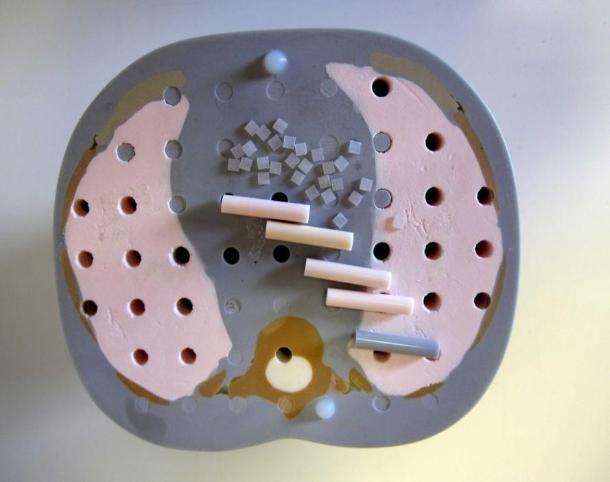
10 Physical phantoms and TLDs
11 Radiation dose from DXA GE-Lunar makes use of a fan-beam with a angle orientated parallel to the longitudinal body axis. A filter splits the X-ray spectrum into high- and low-energy components and provides energies of 38 kev and 70 kev. Mazess et al (2011) Osteoporos Int 11:

12 Radiation dose from DXA Hologic uses a linear fan-beam with a detector array sufficiently wide to image an entire vertebral column in a single sweep. Hologic uses rapid switching between high and low tube potentials e.g. from 70 to 140 kvp
13 Effective dose from DXA: Comparison with other examinations 30 Chest radiography Dental panoramic radiography Spine + hip DXA (Hologic) ED (µsv) 18 6 Spine + hip DXA (GE) Intra-oral radiography
 Source :")
14 Risk coefficients for cancer Risk per Unit Dose (% per Sv) % per Sv Age at acute exposure (yr) Source : BEIR VII
15 Risks for cancer 50-year-old female patient Dose per screening: 20 µsv (Hologic) Risk = x 20 x 10-6 = 1.48x cases of cancer / 1 million patients (Hologic) Effective doses from DXA and potential risks are negligible compared with the benefits
16 Exposure during flights 20 µsv µsv The worldwide average effective dose from natural Bg radiation is 2.4 msv/y

17 Conceptus dose from DXA Conceptus Dose (µgy) Spine Hip Photo by L. Nilsson Fetus at 13 weeks: Arm and leg bones begin to calcify. 1 First Trimester Second Trimester Third Trimester J. Damilakis et al, Osteoporos Int (2002) 913:716-22
18 DXA: How to reduce patient dose Patient preparation is important for reducing the radiation dose Avoid patient movement during imaging Select the most appropriate acquisition protocol Use dose reduction tools such as SmartScan The length of the DXA should take into account patient s body size
19 Estimation of bone status using X-rays Assessment of low energy fractures Dual-energy X-ray absorptiometry (DXA) Quantitative Computed Tomography (QCT) High Resolution CT imaging Peripheral QCT (pqct)

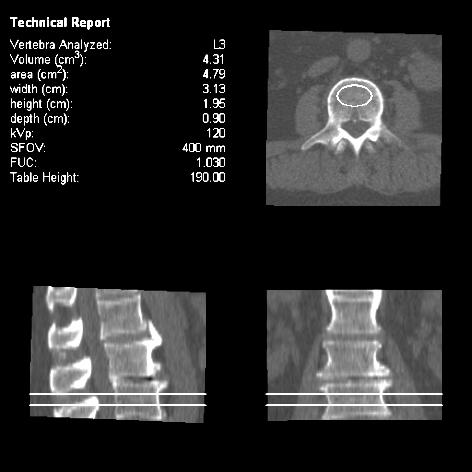
20 QCT using body CT

21 Radiation dose from QCT Effective Dose (msv) D QCT Spine 3D MDQCT Spine 3D MDQCT Hip 3D MDQCT Radius 0.06 <0.01 Huda W et al, BJR (1996) 69: Adams J, EJR (2009) 71:415-24
22 Estimation of bone status using X-rays Assessment of low energy fractures Dual-energy X-ray absorptiometry (DXA) Quantitative Computed Tomography (QCT) High Resolution CT imaging Peripheral QCT (pqct)

23 High-Resolution CT imaging Important information can be obtained from structure analysis of high-resolution image data. Effective dose of about 3 msv Ito M et al, (2005) J Bone Miner Res 20:
24 Parameters that affect CT dose Beam shaping filter Collimation kv, mas Filtration Detection system efficiency Scanning length, scanner geometry, beam collimation, rsw, pitch, algorithms, dose reduction tools
25 Normalized Conceptus Dose Distance (cm)
26

27 The body is not irradiated uniformly


28 Patient-specific MC Simulation Allows machine-specific and patient-specific calculations of the dose distributions J. Damilakis et al, Radiology 257:483-9, 2010 J. Damilakis et al, Medical Physics 37: , 2010
29 Estimation of bone status using X-rays Assessment of low energy fractures Dual-energy X-ray absorptiometry (DXA) Quantitative Computed Tomography (QCT) High Resolution CT imaging Peripheral QCT (pqct)

 Osteoporos Int")
30 Peripheral QCT Isotropic voxel size in the order of 80 µm which allows direct or indirect evaluation of cortical and trabecular bone architecture Effective dose lower than 3 µsv Link T et al, (2010) Skeletal Radiol 39: Burrows M et al, (2010) Osteoporos Int 21:515-20


31 Occupational doses and shielding Examine isodose curves provided by the manufacturer! Dose rates at 1m from the central axis of the imaging table range from about 0.01 µsv/h to about 5 µsv/h, depending on the model.
32 Messages to take home VFA is associated with considerably l ower exposure than lateral spine radiography (1-50 µsv vs. 600 µsv) Radiation doses associated with DXA are very low Pencil beam : less than 1 µsv/scan Fan beam : up to 25 µsv/scan Emphasis must be on dose optimization, especially for paediatric examinations

33 Messages to take home 2D QCT of the lumbar spine is a low-dose technique with doses reported to be from 60 to about 90 µsv Protocols used to examine vertebral microstructure using HR MDCT provide an effective dose of 3 msv The effective dose from pqct is negligible i.e. from 3 to 10 µsv Examine isodose curves provided by the manufacturer
34 Thank you! Ευχαριστώ!
Radiation exposure in X-ray-based imaging techniques used in osteoporosis
 Eur Radiol (2010) 20: 2707 2714 DOI 10.1007/s00330-010-1845-0 MUSCULOSKELETAL John Damilakis Judith E. Adams Giuseppe Guglielmi Thomas M. Link Radiation exposure in X-ray-based imaging techniques used
The Bone Densitometry Examination
 The Bone Densitometry Examination The purpose of The American Registry of Radiologic Technologist (ARRT ) Bone Densitometry Examination is to assess the knowledge and cognitive skills underlying the intelligent
Prevalence of Osteoporosis p. 262 Consequences of Osteoporosis p. 263 Risk Factors for Osteoporosis p. 264 Attainment of Peak Bone Density p.
 Dedication Preface Acknowledgments Continuing Education An Introduction to Conventions in Densitometry p. 1 Densitometry as a Quantitative Measurement Technique p. 2 Accuracy and Precision p. 2 The Skeleton
EXAMINATION CONTENT SPECIFICATIONS ARRT BOARD APPROVED: JANUARY 2017 IMPLEMENTATION DATE: JULY 1, 2017
 EXAMINATION CONTENT SPECIFICATIONS Bone Densitometry The purpose of the bone densitometry examination is to assess the knowledge and cognitive skills underlying the intelligent performance of the tasks
STRUCTURED EDUCATION REQUIREMENTS IMPLEMENTATION DATE: JULY 1, 2017
 STRUCTURED EDUCATION REQUIREMENTS Bone Densitometry The purpose of structured education is to provide the opportunity for individuals to develop mastery of discipline-specific knowledge that, when coupled
Bone Densitometry. Total 30 Maximum CE 14. DXA Scanning (10) 7
 7") STRUCTURED SELF ASSESSMENT CONTENT SPECIFICATIONS SSA LAUNCH DATE: JANUARY 1, 2018 Bone Densitometry The purpose of continuing qualifications requirements (CQR) is to assist registered technologists in
STRUCTURED SELF ASSESSMENT CONTENT SPECIFICATIONS SSA LAUNCH DATE: JANUARY 1, 2018 Bone Densitometry The purpose of continuing qualifications requirements (CQR) is to assist registered technologists in
Accuracy of DEXA scanning & other methods for determining BMD.
 BMD- Measurement Site Accuracy of DEXA scanning & other methods for determining BMD. Ann Larkin In general, densitometry techniques can be performed in either the axial or the appendicular skeleton, depending
CT Imaging of skeleton in small animals. Massimo Marenzana
 CT Imaging of skeleton in small animals Massimo Marenzana Introduction Osteoporosis is a disease in which bones become fragile and more likely to break. It can be defined as a systemic skeletal disease
Estimating the Absorbed Dose to Critical Organs During Dual X-ray Absorptiometry
 Estimating the Absorbed Dose to Critical Organs During Dual X-ray Absorptiometry M Mokhtari-Dizaji, PhD 1 A A Sharafi, PhD 2 B Larijani, MD 3 N Mokhlesian, MSc 4 H Hasanzadeh, MS 1 Index terms: Dual X-ray
Risk of secondary cancers after radiotherapy. for benign diseases
 Risk of secondary cancers after radiotherapy for benign diseases John Damilakis, PhD Professor and Chairman University of Crete, Iraklion, Crete, Greece john.damilakis@med.uoc.gr Radiogenic cancer risk:
2013 ISCD Official Positions Adult
 2013 ISCD Official Positions Adult These are the Official Positions of the ISCD as updated in 2013. The Official Positions that are new or revised since 2007 are in bold type. Indications for Bone Mineral
Introducing the future of DXA. Powerful images. Clear answers. Horizon DXA System
 Introducing the future of DXA Powerful images. Clear answers. Horizon DXA System Hologic turns ideas into innovation. Again. Hologic cares about you and your patients about keeping their bones healthy,
Prodigy. from GE Healthcare. Most trusted, reliable and best-selling DXA system with one of the largest installed base in the world. gehealthcare.
 Prodigy from GE Healthcare Most trusted, reliable and best-selling DXA system with one of the largest installed base in the world gehealthcare.com Prodigy High performance, efficient and reliable DXA system
Lunar idxa. The intelligent DXA. gehealthcare.com
 Lunar idxa The intelligent DXA gehealthcare.com The best of DXA technology for bone and metabolic health assessment With a state-of-the-art design, Lunar idxa offers research-grade image resolution and
Bone Densitometry Equipment Operator
 Bone Densitometry Equipment Operator The purpose of the Bone Densitometry Equipment Operator Examination, which is made available to state licensing agencies, is to assess the knowledge and cognitive skills
Bone Mineral Densitometry with Dual Energy X-Ray Absorptiometry
 Bone Mineral Densitometry with Dual Energy X-Ray Absorptiometry R Gilles, Laurentius Ziekenhuis Roermond 1. Introduction Osteoporosis is characterised by low bone mass, disruption of the micro-architecture
CURRENT CT DOSE METRICS: MAKING CTDI SIZE-SPECIFIC
 CURRENT CT DOSE METRICS: MAKING CTDI SIZE-SPECIFIC Keith Strauss, MSc, FAAPM, FACR Cincinnati Children s Hospital University of Cincinnati College of Medicine Acknowledgments John Boone, PhD Michael McNitt-Grey,
X-Ray & CT Physics / Clinical CT
 Computed Tomography-Basic Principles and Good Practice X-Ray & CT Physics / Clinical CT INSTRUCTORS: Dane Franklin, MBA, RT (R) (CT) Office hours will be Tuesdays from 5pm to 6pm CLASSROOM: TIME: REQUIRED
Thickness Computation Under In-Vivo Trabecular Bone CT Imaging
 Thickness Computation Under In-Vivo Trabecular Bone CT Imaging Gokul. S PG Scholar,M.E - II year Department of Computer Science and Engineering Jansons Institute of Technology Coimbatore, Tamilnadu Aarthi.K
Quantitative Computed Tomography 4 Introduction
 Quantitative Computed Tomography 4 Introduction Quantitative Computed Tomography (QCT) is a well recognised technique for the measurement of bone mineral density (BMD) in the lumbar spine1 and forearm2.
Managing Radiation Risk in Pediatric CT Imaging
 Managing Radiation Risk in Pediatric CT Imaging Mahadevappa Mahesh, MS, PhD, FAAPM, FACR, FACMP, FSCCT. Professor of Radiology and Cardiology Johns Hopkins University School of Medicine Chief Physicist
Dual-Energy CT: The Technological Approaches
 Dual-Energy CT: The Technological Approaches Dushyant Sahani, M.D Director of CT Associate Professor of Radiology Massachusetts General Hospital Harvard Medical School Email-dsahani@partners.org Disclosure
Measurement of organ dose in abdomen-pelvis CT exam as a function of ma, KV and scanner type by Monte Carlo method
 Iran. J. Radiat. Res., 2004; 1(4): 187-194 Measurement of organ dose in abdomen-pelvis CT exam as a function of ma, KV and scanner type by Monte Carlo method M.R. Ay 1, M. Shahriari 2, S. Sarkar 3, P.
Densitometry Techniques
 2 Densitometry Techniques CONTENTS PLAIN RADIOGRAPHY IN THE ASSESSMENT OF BONE DENSITY QUALITATIVE MORPHOMETRY QUALITATIVE SPINAL MORPHOMETRY THE SINGH INDEX QUANTITATIVE MORPHOMETRIC TECHNIQUES CALCAR
8/18/2011. Acknowledgements. Managing Pediatric CT Patient Doses INTRODUCTION
 Managing Pediatric CT Patient Doses Keith J. Strauss, MSc, FAAPM, FACR President X-Ray Computations, Inc. Boston, Massachusetts Acknowledgements Marilyn Goske, MD John Boone, PhD Cynthia McCollough, PhD
New Dual-energy X-ray Absorptiometry Machines (idxa) and Vertebral Fracture Assessment
 and Vertebral Fracture Assessment") Case 1 New Dual-energy X-ray Absorptiometry Machines (idxa) and Vertebral Fracture Assessment (VFA) History and Examination Your wealthy friend who is a banker brings his 62-year-old mother to your office
Case 1 New Dual-energy X-ray Absorptiometry Machines (idxa) and Vertebral Fracture Assessment (VFA) History and Examination Your wealthy friend who is a banker brings his 62-year-old mother to your office
PhenX Measure: Body Composition (#020300) PhenX Protocol: Body Composition - Body Composition by Dual-Energy X-Ray Absorptiometry (#020302)
 PhenX Protocol: Body Composition - Body Composition by Dual-Energy X-Ray Absorptiometry (#020302)") PhenX Measure: Body Composition (#020300) PhenX Protocol: Body Composition - Body Composition by Dual-Energy X-Ray Absorptiometry (#020302) Date of Interview/Examination (MM/DD/YYYY): A downloadable PDF
PhenX Measure: Body Composition (#020300) PhenX Protocol: Body Composition - Body Composition by Dual-Energy X-Ray Absorptiometry (#020302) Date of Interview/Examination (MM/DD/YYYY): A downloadable PDF
Bone Densitometry. What is a Bone Density Scan (DXA)? What are some common uses of the procedure?
? What are some common uses of the procedure?") Scan for mobile link. Bone Densitometry What is a Bone Density Scan (DXA)? Bone density scanning, also called dual-energy x-ray absorptiometry (DXA) or bone densitometry, is an enhanced form of x-ray technology
Scan for mobile link. Bone Densitometry What is a Bone Density Scan (DXA)? Bone density scanning, also called dual-energy x-ray absorptiometry (DXA) or bone densitometry, is an enhanced form of x-ray technology
Managing Patient Dose in Computed Tomography (CT) INTERNATIONAL COMMISSION ON RADIOLOGICAL PROTECTION
 INTERNATIONAL COMMISSION ON RADIOLOGICAL PROTECTION") Managing Patient Dose in Computed Tomography (CT) International Commission on Radiological Protection Information abstracted from ICRP Publication 87 Available at www.icrp.org Task Group: M.M. Rehani,
Managing Patient Dose in Computed Tomography (CT) International Commission on Radiological Protection Information abstracted from ICRP Publication 87 Available at www.icrp.org Task Group: M.M. Rehani,
Doses from pediatric CT examinations in Norway Are pediatric scan protocols developed and in daily use?
 Doses from pediatric CT examinations in Norway Are pediatric scan protocols developed and in daily use? Eva Godske Friberg * Norwegian Radiation Protection Authority, P.O. Box, Østerås, Norway Abstract.
THE DIAGNOSIS OF OSTEOPOROSIS BY MEASURING LUMBAR VERTEBRAE DENSITY WITH MDCT: A COMPARATIVE STUDY WITH QUANTITATIVE COMPUTERIZED TOMOGRAPHY (QCT)
") Acta Medica Mediterranea, 2013, 29: 775 THE DIAGNOSIS OF OSTEOPOROSIS BY MEASURING LUMBAR VERTEBRAE DENSITY WITH MDCT: A COMPARATIVE STUDY WITH QUANTITATIVE COMPUTERIZED TOMOGRAPHY (QCT) KEMAL KARA 1,
Acta Medica Mediterranea, 2013, 29: 775 THE DIAGNOSIS OF OSTEOPOROSIS BY MEASURING LUMBAR VERTEBRAE DENSITY WITH MDCT: A COMPARATIVE STUDY WITH QUANTITATIVE COMPUTERIZED TOMOGRAPHY (QCT) KEMAL KARA 1,
Utility of Dual-Energy CT to Evaluate Patients with Hip and Pelvis Pain in the ER Setting
 Utility of Dual-Energy CT to Evaluate Patients with Hip and Pelvis Pain in the ER Setting Johnson, T., Moran, E., Glazebrook, K., Leng, S., Fletcher, J., and McCollough, C. An educational review ER011
pqct Measurement of Bone Parameters in Young Children
 Journal of Clinical Densitometry, vol. 3, no. 1, 9 14, Spring 2000 Copyright 2000 by Humana Press Inc. All rights of any nature whatsoever reserved. 0169-4194/00/3:9 14/$11.50 Original Article pqct Measurement
DXA Best Practices. What is the problem? 9/29/2017. BMD Predicts Fracture Risk. Dual-energy X-ray Absorptiometry: DXA
 BMD Predicts Fracture Risk Ten Year Fracture Probability (%) 50 40 30 20 10 Age 80 70 60 50 E. Michael Lewiecki, MD Director, New Mexico Clinical Research & Osteoporosis Center Director, Bone TeleHealth
To Shield or Not to Shield? Lincoln L. Berland, M.D.
 To Shield or Not to Shield? Lincoln L. Berland, M.D. Disclosures Consultant to: Nuance, Inc. Page 2 Breast Radiation on CT Use of chest CT has increased in women vulnerable to cancer induction by radiation.
Shielding Calculation: Radiographic Room. Jerry Williams
 Shielding Calculation: Radiographic Room Jerry Williams What you need to know Room use and layout DAP workload DAP averaged kv Distance to barrier Construction details Walls Ceilings/ floors Surrounding
9 Quality Assurance in Bone Densitometry section
 9 Quality Assurance in Bone Densitometry section Introduction Bone densitometry is frequently used to determine an individual's fracture risk at a particular point in time but may also be used to assess
Radiation exposure of the Yazd population from medical conventional X-ray examinations
 Iran. J. Radiat. Res., 2007; 4 (4): 195-200 Radiation exposure of the Yazd population from medical conventional X-ray examinations F. Bouzarjomehri 1*, M.H. Dashti 2, M.H. Zare 1 1 Department of Medical
Norland Densitometry A Tradition of Excellence
 Norland Densitometry A Tradition of Excellence Norland DXA Bone Density Measurement Osteoporosis is a disease marked by reduced bone strength leading to an increased risk of fractures. About 54 million
Cross-reference: MP Whole Body Dual X-Ray Absorptiometry (DEXA) to Determine Body Composition MP Bone Mineral Density
 to Determine Body Composition MP Bone Mineral Density") Original Issue Date (Created): April 26, 2011 Most Recent Review Date (Revised): September 24, 2013 Effective Date: November 1, 2013 I. POLICY Screening for vertebral fractures using dual x-ray absorptiometry
Original Issue Date (Created): April 26, 2011 Most Recent Review Date (Revised): September 24, 2013 Effective Date: November 1, 2013 I. POLICY Screening for vertebral fractures using dual x-ray absorptiometry
Radiation Physics Principles of DXA Basic Statistics for DXA Essential Anatomy for DXA
 Strong Bone Asia 2013 Osteoporosis in ASEAN Radiation Physics Principles of DXA Basic Statistics for DXA Essential Anatomy for DXA Chris Schultz Scientist-in-Charge (Bone Densitometry) Royal Adelaide Hospital,
QCT and CT applications in Osteoporosis Imaging
 Q appli in Osteoporosis Imaging Thomas M. Link, MD, PhD Department of Radiology Biomedical Imaging University of California, San Francisco Goals 1. To identify advantages disadvantages of Q compared to
2013 ISCD Combined Official Positions
 2013 ISCD Combined Oicial Positions Oicial Positions of the International Society for Clinical Densitometry The International Society for Clinical Densitometry (ISCD) is a not-for-profit multidisciplinary
Interpreting DEXA Scan and. the New Fracture Risk. Assessment. Algorithm
 Interpreting DEXA Scan and the New Fracture Risk Assessment Algorithm Prof. Samir Elbadawy *Osteoporosis affect 30%-40% of women in western countries and almost 15% of men after the age of 50 years. Osteoporosis
Does standardized BMD still remove differences between Hologic and GE-Lunar state-of-the-art DXA systems?
 Osteoporos Int (2010) 21:1227 1236 DOI 10.1007/s00198-009-1062-3 ORIGINAL ARTICLE Does standardized BMD still remove differences between Hologic and GE-Lunar state-of-the-art DXA systems? B. Fan & Y. Lu
PACS: ERGONOMIC CONSIDERATIONS 1
 RADIOLOGY RESEARCH Radiographic Tomosynthesis: Acquisition Parameters Michael J. Flynn, PhD Henry Ford Health System Detroit, MI Learning Objectives Learn.. 1. Appreciate the importance of scan direction,
A basic study for automatic recognition of osteoporosis using abdominal X-ray CT images
 A basic study for automatic recognition of osteoporosis using abdominal X-ray CT images Sadamitsu Nishihara* a, Hiroshi Fujita b, Tadayuki Iida a, Atsushi Takigawa a, Takeshi Hara b and Xiangrong Zhou
Dual-energy Vertebral Assessment
 Dual-energy Vertebral Assessment gehealthcare.com Dual-energy Vertebral Assessment More than 40% of women with normal or osteopenic BMD had a moderate or severe vertebral deformation seen with DVA. Patrick
Thoracic examinations with 16, 64, 128 and 256 slices CT: comparison of exposure doses measured with an anthropomorphic phantom and TLD dosimeters
 Thoracic examinations with 16, 64, 128 and 256 slices CT: comparison of exposure doses measured with an anthropomorphic phantom and TLD dosimeters Poster No.: C-2584 Congress: ECR 2015 Type: Scientific
Mineral Density Of Subchondral Bone May Be Quantitatively Evaluated Using A Clinical Cone Beam Computed Tomography Scanner
 Mineral Density Of Subchondral Bone May Be Quantitatively Evaluated Using A Clinical Cone Beam Computed Tomography Scanner Mikael J. Turunen, PhD 1, Juha Töyräs, PhD 1, Harri Kokkonen, PhD 2, Jukka S.
Standard Operating Procedure TCRC Dual-Energy X-ray Absorptiometry (DXA)
") 1. RELEVANCE a. This SOP outlines the instructions to completing Duel Energy X-Ray Absorptiometry (DXA) including: scanning, analysis, review and filing. 2. SCOPE a. This SOP applies to all TCRC RDs. 3.
1. RELEVANCE a. This SOP outlines the instructions to completing Duel Energy X-Ray Absorptiometry (DXA) including: scanning, analysis, review and filing. 2. SCOPE a. This SOP applies to all TCRC RDs. 3.
DXA When to order? How to interpret? Dr Nikhil Tandon Department of Endocrinology and Metabolism All India Institute of Medical Sciences New Delhi
 DXA When to order? How to interpret? Dr Nikhil Tandon Department of Endocrinology and Metabolism All India Institute of Medical Sciences New Delhi Clinical Utility of Bone Densitometry Diagnosis (DXA)
Clinical Study Comparison of QCT and DXA: Osteoporosis Detection Rates in Postmenopausal Women
 International Endocrinology Volume 3, Article ID 895474, 5 pages http://dx.doi.org/.55/3/895474 Clinical Study Comparison of QCT and DXA: Osteoporosis Detection Rates in Postmenopausal Women Na Li, Xin-min
Cone Beam CT Protocol Optimisation for Prostate Imaging with the Varian Radiotherapy OBI imaging system. Dr Craig Moore & Dr Tim Wood
 Cone Beam CT Protocol Optimisation for Prostate Imaging with the Varian Radiotherapy OBI imaging system Dr Craig Moore & Dr Tim Wood Background With the increasing use of CBCT imaging alongside complex
Low Dose Era in Cardiac CT
 Low Dose Era in Cardiac CT DIANA E. LITMANOVICH, MD Department of Radiology Beth Israel Deaconess Medical Center Harvard Medical School Disclosures Neither I nor my immediate family members have a financial
Annotations Part III Vertebral Fracture Initiative. International Osteoporosis Foundation March 2011
 Annotations Part III Vertebral Fracture Initiative International Osteoporosis Foundation March 2011 Slide 1-3 Topics to be covered: What is vertebral fracture assessment? How does VFA compare to standard
CT Radiation Risks and Dose Reduction
 CT Radiation Risks and Dose Reduction Walter L. Robinson, M.S. D.A.B.S.N.M., D.A.B.M.P., D.A.B.R. Consultant Certified Medical Radiation Health & Diagnostic Imaging Physicist Medical Radiation and Children
Why is CT Dose of Interest?
 Why is CT Dose of Interest? CT usage has increased rapidly in the past decade Compared to other medical imaging CT produces a larger radiation dose. There is direct epidemiological evidence for a an increase
Managing Cone Beam CT Dose in Paediatric Dental Imaging
 Ask EuroSafe Imaging Tips & Tricks Paediatric Imaging Working Group Managing Cone Beam CT Dose in Paediatric Dental Imaging Raija Seuri (HUS Medical Imaging Center, FI) Cristina Almeida (Centro Hospitalar
Douglas J. Simpkin, Ph.D. Aurora St. Luke s Medical Center Milwaukee, Wisconsin. www.
 PET/CT Shielding Design Examples Douglas J. Simpkin, Ph.D. Aurora St. Luke s Medical Center Milwaukee, Wisconsin dsimpkin@wi.rr.com www. http://geocities.com/djsimpkin 1 Sources of Exposure: F-18 in Patients
Preliminary technical data
 Preliminary technical data 2D-FanBeam Whole Body Densitometer PROGRESS THROUGH INNOVATION 2D-Fan Beam Whole Body Densitometer Acquisition chain parameters Dual Emission X-ray Absorptiometry (DEXA) 2D-FanBeam
Accounting for Imaging Dose
 Accounting for Imaging Dose High Profile Over-exposures Lead to Growing Concern FDA issues warning in October 2009-209 patients exposed to 8 times typical dose for CT brain perfusion scan (3-4 Gy) - Some
IMAGE GENTLY HOW CAN YOU HELP?
 IMAGE GENTLY HOW CAN YOU HELP? Keith J. Strauss, MSc, FAAPM, FACR Director, Radiology Physics & Engineering Children s s Hospital Boston Harvard Medical School Acknowledgment Marilyn J. Goske,, MD Robert
Clinical Densitometry
 Volume 8 Number 3 Fall 2005 ISSN: 1094 6950 Journal of Clinical Densitometry The Official Journal of The International Society for Clinical Densitometry Editor-in-Chief Paul D. Miller, MD HumanaJournals.com
Diagnosis of Vertebral Fractures by Vertebral Fracture Assessment
 Journal of Clinical Densitometry, vol. 9, no. 1, 66 71, 2006 Ó Copyright 2006 by The International Society for Clinical Densitometry 1094-6950/06/9:66 71/$32.00 DOI: 10.1016/j.jocd.2005.11.002 Original
Medical Diagnostic Imaging
 Medical Diagnostic Imaging Laboratories Medical Diagnostic Imaging Lab Name Location Person in Charge Programs Served Courses Served Patient Care and Management (2) Introduction to MDI Radiographic Technique
ReviewArticle. Frequently Asked Questions About Bone Mineral Density Test
 ReviewArticle Frequently Asked Questions About Bone Mineral Density Test Jiraporn Sriprapaporn, M.D.*,** *Division of Nuclear Medicine, Department of Radiology, Faculty of Medicine Siriraj Hospital, Mahidol
X-Ray Board Reviewing Rules
 Volume 24, Issue 3 July-Sept 2018 Tennessee X-Ray X-Ray Board Reviewing Rules The Tennessee Board of Radiologic Imaging and Radiation Therapy had their first meeting on January 25, 2018. The minutes from
Chapter 16 Worksheet Code It
 Name: Class: Date: ID: A Chapter 16 Worksheet 3 2 1 Code It True/False Indicate whether the statement is true or false. 1. CT scans generate three-dimensional images. 2. An ultrasound produces images of
2
 1 2 3 4 5 6 7 8 9 10 11 12 13 Cine loop of tomosynthesis slice images through the chest. 14 Standard PA chest radiograph (left) and single slice from the tomosynthesis image dataset (right) of a patient
Trabecular bone analysis with tomosynthesis in diabetic patients: comparison with CT-based finite-element method
 Trabecular bone analysis with tomosynthesis in diabetic patients: comparison with CT-based finite-element method Poster No.: C-1789 Congress: ECR 2015 Type: Scientific Exhibit Authors: M. Fujii, T. Aoki,
Bureau of Laboratory Quality Standards
 1. Condom -Lubricant ISO 4074:2002/Cor.1:2003 Annex C -Thickness ISO 4074:2002/Cor.1:2003 Annex F - Length ISO 4074:2002 / Cor1 : 2003 - Width ISO 4074 : 2002/Cor 1 : 2003 - Bursting pressure and Volume
Automated measurement method of bone mineral density of vertebral bones and spinal curvature using sagittal plane in X-ray torso CT images
 501-1194 1-1 501-1194 1-1 501-1194 1-1 501-1194 1-1 501-1194 1-1 2009 2 9 2009 6 4 Automated measurement method of bone mineral density of vertebral bones and spinal curvature using sagittal plane in X-ray
Regional diagnostic reference levels and collective effective doses from CT scanners in India
 Regional diagnostic reference levels and collective effective doses from CT scanners in India Roshan S Livingstone and Paul M Dinakaran Department of Radiology Christian Medical College, Vellore, S India
Survey of patients CT radiation dose in Jiangsu Province
 Original Article Page 1 of 6 Survey of patients CT radiation dose in Jiangsu Province Yuanyuan Zhou 1, Chunyong Yang 1, Xingjiang Cao 1, Xiang Du 1, Ningle Yu 1, Xianfeng Zhou 2, Baoli Zhu 1, Jin Wang
Prof. Dr. Doğan BOR Ankara University Institute of Nuclear Science
 PATIENT DOSIMETRY IN DIAGNOSTIC RADIOLOGY MODALITIES Prof. Dr. Doğan BOR Ankara University Institute of Nuclear Science Ankara University Institute of Nuclear Science USE OF RADIATION! INCREASING? Natural
3/5/2015. Don t Electrocute Me!: Common Misconceptions in Imaging and Radiation Safety (and What to Do About Them)
") Don t Electrocute Me!: Common Misconceptions in Imaging and Radiation Safety (and What to Do About Them) Rebecca Milman Marsh, Ph.D. University of Colorado Department of Radiology Who in the Facility Works
Don t Electrocute Me!: Common Misconceptions in Imaging and Radiation Safety (and What to Do About Them) Rebecca Milman Marsh, Ph.D. University of Colorado Department of Radiology Who in the Facility Works
Opportunistic screening for osteoporosis on routine computed tomography? An external validation study
 Eur Radiol (2015) 25:2074 2079 DOI 10.1007/s00330-014-3584-0 COMPUTED TOMOGRAPHY Opportunistic screening for osteoporosis on routine computed tomography? An external validation study Constantinus F. Buckens
THE TUFFEST STUFF CT REGISTRY REVIEW Live Lecture Seminar SATURDAY CURRICULUM
 1. The CT Imaging Chain-10 major components & their functions a. The x-ray tube b. Generator c. Filter d. Pre-patient collimator e. Pre-detector collimator f. Detector system g. Analog to digital converter
Clinical Application of Computed Radiography in Orthopedic Surgery
 Clinical Application of Computed Radiography in Orthopedic Surgery Satoru Fujita, Masamichi Tanaka, Sigeaki Hirota, and Takeshi Fuji Since 1988, Fuji Computed Radiography (FCR) system (Fuji Medical Systems,
LUMBAR IS IT IMPORTANT? S. Tantawy,, M.D.
 بسم االله الرحمن الرحيم DEXA LATERAL LUMBAR IS IT IMPORTANT? By S. Tantawy,, M.D. Osteopenia,, bone mineral deficiency in the absence of fracture, is an indicator of the bone structural integrity and compared
The Fluoroscopic Technique for Monitoring Distraction of a Non-Invasive Lengthening Device in Early Onset Scoliosis
 Send Orders for Reprints to reprints@benthamscience.net Open Medicine Journal, 2014, 1, 23-28 23 Open Access The Fluoroscopic Technique for Monitoring Distraction of a Non-Invasive Lengthening Device in
Lateral Vertebral Analysis DXA Body Composition Quality Assurance in DXA
 Strong Bone Asia 2013 Osteoporosis in ASEAN Lateral Vertebral Analysis DXA Body Composition Quality Assurance in DXA Chris Schultz Scientist-in-Charge (Bone Densitometry) Royal Adelaide Hospital, Adelaide,
Translating Protocols Across Patient Size: Babies to Bariatric
 Translating Protocols Across Patient Size: Babies to Bariatric Cynthia H. McCollough, PhD, FACR, FAAPM Professor of Radiologic Physics Director, CT Clinical Innovation Center Department of Radiology Mayo
Debra Pennington, MD Director of Imaging Dell Children s Medical Center
 Debra Pennington, MD Director of Imaging Dell Children s Medical Center 1 Gray (Gy) is 1 J of radiation energy/ 1 kg matter (physical quantity absorbed dose) Diagnostic imaging doses in mgy (.001 Gy)
Acknowledgments. A Specific Diagnostic Task: Lung Nodule Detection. A Specific Diagnostic Task: Chest CT Protocols. Chest CT Protocols
 Personalization of Pediatric Imaging in Terms of Needed Indication-Based Quality Per Dose Acknowledgments Duke University Medical Center Ehsan Samei, PhD Donald Frush, MD Xiang Li PhD DABR Cleveland Clinic
Selecting regions of interest on intra oral radiographs. for the prediction of bone mineral density
 Selecting regions of interest on intra oral radiographs for the prediction of bone mineral density W.G.M. Geraets a, J.G.C. Verheij a, P.F. van der Stelt a, K. Horner b, C. Lindh c, K. Nicopoulou-Karayianni
Powerful images. Clear answers.
 SKELETAL HEALTH Introducing the future of DXA Powerful images. Clear answers. Horizon DXA System Every Life is Extraordinary Keeping life in motion. 1 in 2 women over 50 are at risk of a vertebral fracture
chapter Bone Density (Densitometry) RADIOPHARMACY INDICATIONS Radionuclide Localization Quality Control Adult Dose Range Method of Administration
 RADIOPHARMACY INDICATIONS Radionuclide Localization Quality Control Adult Dose Range Method of Administration") 10766-04_CH04_redo.qxd 12/3/07 3:47 PM Page 17 chapter 4 Bone Density (Densitometry) RADIOPHARMACY Radionuclide Single radionuclide: 125 I t 1/2 : 60.1 days Energies: 23 31 kev Type: EC, x, γ, accelerator
10766-04_CH04_redo.qxd 12/3/07 3:47 PM Page 17 chapter 4 Bone Density (Densitometry) RADIOPHARMACY Radionuclide Single radionuclide: 125 I t 1/2 : 60.1 days Energies: 23 31 kev Type: EC, x, γ, accelerator
Ask EuroSafe Imaging. Tips & Tricks. CT Working Group. Optimization of scan length to reduce CT radiation dose
 Ask EuroSafe Imaging Tips & Tricks CT Working Group Optimization of scan length to reduce CT radiation dose Alban Gervaise (Centre Hospitalier Universitaire Nancy, FR) Mika Kortesniemi (HUS Medical Imaging
Introduction Pediatric malignancies Changing trends & Radiation burden Radiation exposure from PET/CT Image gently PET & CT modification - PET/CT
 Introduction Pediatric malignancies Changing trends & Radiation burden Radiation exposure from PET/CT Image gently PET & CT modification - PET/CT protocols Tips Leukaemia / lymphoma: ~ 35% acute lymphoblastic
Certified Clinical Densitometrist (CCD ) DXA Resource Materials
 DXA Resource Materials") Certified Clinical Densitometrist (CCD ) DXA Resource Materials International Society for Densitometry DXA Resource Materials Editors Steven Petak, MD, JD, CCD Neil Binkley, MD, CCD Sue Broy, MD, CCD
Certified Clinical Densitometrist (CCD ) DXA Resource Materials International Society for Densitometry DXA Resource Materials Editors Steven Petak, MD, JD, CCD Neil Binkley, MD, CCD Sue Broy, MD, CCD
Managing the imaging dose during Image-guided Radiotherapy. Martin J Murphy PhD Department of Radiation Oncology Virginia Commonwealth University
 Managing the imaging dose during Image-guided Radiotherapy Martin J Murphy PhD Department of Radiation Oncology Virginia Commonwealth University Radiographic image guidance has emerged as the new paradigm
S. N. Khan 1, R. M. Warkhedkar 2, A. Shyam 3, B. R. Patil 4
 IOSR Journal of Mechanical and Civil Engineering (IOSR-JMCE) ISSN(e) : 2278-1684, ISSN(p) : 2320 334X, PP : 40-44 www.iosrjournals.org Correlation between BMD and Radio intensity of Human Bones for Strength
ROMANIA MEDICAL EXPOSURE IN 2016 IN ROMANIAN RADIOLOGICAL DEPARTMENTS. Olga GIRJOABA and Alexandra CUCU
 ROMANIA MEDICAL EXPOSURE IN 2016 IN ROMANIAN RADIOLOGICAL DEPARTMENTS Olga GIRJOABA and Alexandra CUCU National Institute of Public Health, Bucharest, Romania olga.garjoaba@insp.gov.ro PURPOSE Improvement
COMPUTED TOMOGRAPHY COURSE
 CT Radiography RAD 421 4 th year semester 2 Course Lecture Tutorial Practical Credit hours CT Radiography 2-1 2 Course Description The course explores the basic physical and technical principles of CT
Journal of Dental Research, Dental Clinics, Dental Prospects. Original Article
 Journal of Dental Research, Dental Clinics, Dental Prospects Original Article Efficacy of radiographic density values of the first and second cervical vertebrae recorded by CBCT technique to identify patients
Estimating Patient Radiation Dose from Computed Tomography
 Estimating Patient Radiation Dose from Computed Tomography C. Cagnon, J. DeMarco, E. Angel, M. McNitt-Gray UCLA David Geffen School of Medicine 1 Patient Dose from CT Advances in Technology... Helical
Radiation Dose Reduction Strategies in Coronary CT Angiography
 Radiation Dose Reduction Strategies in Coronary CT Angiography Noor Diyana Osman, PhD noordiyana@usm.my Contents: Introduction Radiation dosimetry in CT Radiation risk associated with coronary CT angiography
X-ray Safety Discussion and Tour for Health Physicists
 X-ray Safety Discussion and Tour for Health Physicists April 1, 2016 Health Physics Society Spring Meeting Jerry Bai Environmental Administrator, Field Operations Bureau of Radiation Control Florida Department