The Lower Extremity. OPSO Summer Primary Care CME Conference August 15, 2014 K. Turner Slicho, DO
|
|
|
- Scott Douglas
- 6 years ago
- Views:
Transcription

1 The Lower Extremity OPSO Summer Primary Care CME Conference August 15, 2014 K. Turner Slicho, DO
2 Case Presentation 53-year-old Caucasian female complains of left sided SI pain described as an ache w/o radiation Initial onset of pain was 25 years ago, when her back seized up bending over to put her toddler to bed. Infrequent issues over the next years, then began to be more problematic Now, constant pain over the last 2 years Numbness in a small area of left shin. Denies weakness, tingling
3 Case Presentation Unable to sit for long periods, bend over, cross legs comfortably Does not lift her grandson Does not brush teeth bending over without holding the sink No longer exercising due to pain 15 chiropractic session with two different providers over 2 years with absolutely zero effect on her pain 12 physical therapy sessions with marginal effect
4 Case Presentation PMHx: Unremarkable** FHx: Cancer, DM, Food Allergies, Meniere s Disease, Restless leg syndrome, Ulcerative colitis PSHx: C-section 1990, Appendectomy 2009, Hernia repair 2005 Meds: Diclofenac 100mg BID
5 Case Presentation X-Ray: L4-L5 degenerative changes. Normal pelvis and SI joints bilaterally MRI: mild degenerative changes L3-S1 without significant neuroforaminal or spinal stenosis Neurosurgical evaluation : non-surgical back
6 Case Presentation Gen: NAD, A&O x 3 Neuro: 2+/4 DTR s, Strength 5/5, no motor deficits, mild sensory loss in left superio-lateral shin Extremities: no edema, ecchymosis, peripheral pulses normal
7 Case Presentation: Somatic Dysfunction findings Cervical: Left posterior OA facet restriction Lumbar: L5 ERSl, left iliolumbar ligament hypertonicity, mild left psoas spasm and left erector spinae m. spasm Sacrum: L on LOA torsion Pelvis: Left innominate anteriorly, bilateral gluteus medius m. hypertonicity, right TFL hypertonicity LE: Left talus plantarflexed, left fibular head posterior, bil. interosseus membrane restriction
8 Case presentation Patient treated with MFR, BLT, FPR, ME techniques Instructed to follow-up in 1 week, and continue diclofenac as prescribed Patient to avoid heavy lifting until F/U Patient returned 1 week later, reporting she was symptom free since her last treatment

9 Case Presentation: Somatic Dysfunction findings Cervical: Left posterior OA facet restriction Lumbar: L5 ERSl, left iliolumbar ligament hypertonicity, mild left psoas spasm and left erector spinae m. spasm Sacrum: L on LOA torsion Pelvis: Left innominate anteriorly, bilateral gluteus medius m. hypertonicity, right TFL hypertonicity LE: Left talus plantarflexed, left fibular head posterior, bil. interosseus membrane restriftion
10 The Lower leg
11 Why did the ankle become such a pain in the back?
12 Gait cycle Left foot steps forward Heel strike :Left innominate posteriorly rotated, right anterior right foot plantar flexed During this motion, the sacrum rotates right Toward mid-stance, the left leg is straight and the innominate is rotated anteriorly Sacrum rotates left, lumbar spine side-bends left, rotates right Left leg swings through, foot into plantar flexion Right leg then steps forward, repeating the cycle on the other side

13 Gait cycle image
14 Where did the numbness in the shin come from?
15


16 `
17 Fibular Head Reciprocal motion Plantar flexion of the ankle causes posterio-medial Posterior S/D can cause common peroneal nerve injury Fibular head is most common site of injury Foot drop Dorsiflexion of the ankle causes anterio-lateral movement of fibular head Take Home: The ANKLE FUNCTIONALLY STOPS AT THE KNEE!
18 Interosseous Membrane Extends between the interosseus crests of the tibia and fibula Helps to stabilize the Tib-Fib relationship Separates the muscles of the front and the back of the leg

19 Interosseous membrane
20
21 Ankle Sprains Inversion Most common Anterior/Posterior Talofibular ligaments Fibulocalcaneal ligament Eversion Deltoid ligament High Ankle Sprain

22
23
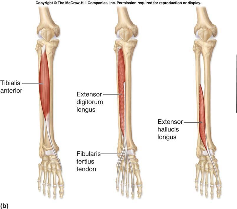
24 Extensor digitorum longus Origin: Lateral condyle of the tibia Upper ¾ of the anterior surface of the body of the fibula Upper part of the interosseus membrane Deep surface of the fascia Intermuscular septa between it and the tibialis anterior Extends toes and dorsiflexes the foot Opposite motion of inversion ankle sprain
25 High Ankle Sprain Sprain of the syndesmotic ligaments that connect the tibia and fibula on the lower leg Described as high because they are located above the ankle Sudden forceful outward twisting of the foot Common in contact and cutting sports

26
27 OMT and Acute Ankle Sprain N = 55 (28 in treatment group, 27 control) Patients 18+ y.o. with unilateral ankle sprain Both groups received current standard of care for ankle sprains Treatment group had one session of OMT Both groups returned for F/U one week later
28 OMT and Acute Ankle Sprain Results OMT group had statistically significant improvement in edema and pain and a trend towards increased ROM immediately following OMT At F/U, both groups were significantly improved Patients in OMT group had statistically significant improvement in ROM compared to the control JAOA, Vol 103, No. 9, Sept. 2003, Eisenhart et. al. Osteopathic Manipulative Treatment in the Emergency Department for Patients with Acute Ankle Injuries
29 Acute Ankle Sprain OMT HVLA likely not tolerated Make sure to address lymphatic restrictions Popliteal fascia Pelvic/abdominal diaphragm Thoracic outlet on left Effleurage Counterstrain/MFR to gastrocnemius/soleus m.
30 Ankle Injury Recurrence Epidemiologic study conducted among 3 categories of Hong Kong Chinese athletes National teams Competitive athletes Recreational athletes Questionnaire sent to athletes having a history of ankle sprain
31 Ankle Injury Recurrence Only athletes involved in sports on a regular basis chosen All must have sprained ankle(s) at least once, with detectable swelling and pain around the injured ankle Athletes with acute ankle sprain within a 3-month period excluded 400 questionnaires distributed and collected, 20 incomplete 380 for data analysis
32 Ankle Injury Recurrence 73% of all athletes had recurrent ankle sprain 59% of these athletes had significant disability and residual symptoms which led to impairment of their athletic performance Residual problems included: Pain, instability, crepitus, weakness, stiffness, swelling British Journal of Sports Medicine 1994; 28(2). MPhil et. al. An epidemiological survey on ankle sprain
33 Fibular head posterior

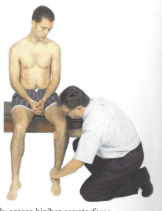
34 Seated modification Can be modified for anterior or posterior fibular head MFR of IOM can be done from this position as well

35 Muscle Energy

36 Technique 3
37

38 IT Band
39 Functional Anatomy of Lower Extremity IT band doesn t contract! Transfers contractile forces TFL Superior Gluteal Nerve L5/S1 Courses between glut med and min Stabilizes hip in extension Glut Maximus Glut Medius

40 Fascia Lata deep fascia of the thigh
41 Fascia Lata Fibrous sheath that encircles the thight like a subcutaneous stocking and tightly binds its muscle Lateral Surface: combines with tendons of glut max and TFL to form IT band IT band extends from iliac crest to lateral condyle of the tibia In the erect posture, acting from below, it will serve to steady the pelvis upon the had of the femur, and by means of the IT band, it steadies the condyles of the femur on the articular surfaces of the tibia, and assists glut max in supporting the knee in a position of extension

42 TFL Basic functional movement is walking TFL is a hip abductor Works in synergy with glut med/min to abduct the thigh and internally rotate the thigh
43 TFL Injury? Stand on one leg with other leg raised If pain at the hip and knee and difficulty maintaining balance, can indicate TFL injury
44 The Glutes: Beyond the Piriformis School OMT courses tend to focus on piriformis m. Gluteus maximus muscle is the LARGEST muscle in the human body Antagonist to iliopsoas muscle LBP implications Dysfunction of all the gluteal muscles can easily be assessed and treated with minimal patient assistance
45 Treatment of Glutes Patient in lateral recumbent position Doctor stands behind the patient Gluteal region TPs assessed as well as myofascial tension Can move from one TP to the next assessing, and stopping to treat if warranted
46 Treatment of Glutes Myofascial release Vector of force generally aimed towards the pubic symphysis 3D vector aimed at epicenter of tension Goal is to match your force vector to the tension of the tissue NOT ischemic compression/inhibition Good visual: Melt a chocolate chip to the tip on a hot dash board


47 Gluteus medius Gluteus minimus

48 Tensor Fascia Lata Piriformis All TP images from triggerpoints.net

49 Deeper muscles of the pelvis
50 Questions? Please feel free to leave topics of interest for future talks in the course comments section of the evaluation Welcome to me as well: - personal - practice
Muscles of the Hip 1. Tensor Fasciae Latae O: iliac crest I: lateral femoral condyle Action: abducts the thigh Nerve: gluteal nerve
 Muscles of the Hip 1. Tensor Fasciae Latae O: iliac crest I: lateral femoral condyle Action: abducts the thigh Nerve: gluteal nerve 2. Gluteus Maximus O: ilium I: femur Action: abduct the thigh Nerve:
RN(EC) ENC(C) GNC(C) MN ACNP *** MECHANISM OF INJURY.. MOST IMPORTANT ***
 ENC(C) GNC(C) MN ACNP *** MECHANISM OF INJURY.. MOST IMPORTANT ***") HISTORY *** MECHANISM OF INJURY.. MOST IMPORTANT *** Age of patient - Certain conditions are more prevalent in particular age groups (Hip pain in children may refer to the knee from Legg-Calve-Perthes
HISTORY *** MECHANISM OF INJURY.. MOST IMPORTANT *** Age of patient - Certain conditions are more prevalent in particular age groups (Hip pain in children may refer to the knee from Legg-Calve-Perthes
MUSCLES OF THE LOWER LIMBS
 MUSCLES OF THE LOWER LIMBS Naming, location and general function Dr. Nabil khouri ROLES THAT SHOULD NOT BE FORGOTTEN Most anterior compartment muscles of the hip and thigh Flexor of the femur at the hip
Human Anatomy Biology 351
 Human Anatomy Biology 351 Lower Limb Please place your name on the back of the last page of this exam. You must answer all questions on this exam. Because statistics demonstrate that, on average, between
Muscles of the lower extremities. Dr. Nabil khouri MD, MSc, Ph.D
 Muscles of the lower extremities Dr. Nabil khouri MD, MSc, Ph.D Posterior leg Popliteal fossa Boundaries Biceps femoris (superior-lateral) Semitendinosis and semimembranosis (superior-medial) Gastrocnemius
Human Anatomy Biology 351
 Human Anatomy Biology 351 Lower Limb Please place your name on the back of the last page of this exam. You must answer all questions on this exam. Because statistics demonstrate that, on average, between
Human Anatomy Biology 255
 Human Anatomy Biology 255 Exam #4 Please place your name and I.D. number on the back of the last page of this exam. You must answer all questions on this exam. Because statistics demonstrate that, on average,
The Muscular System. Chapter 10 Part D. PowerPoint Lecture Slides prepared by Karen Dunbar Kareiva Ivy Tech Community College
 Chapter 10 Part D The Muscular System Annie Leibovitz/Contact Press Images PowerPoint Lecture Slides prepared by Karen Dunbar Kareiva Ivy Tech Community College Table 10.14: Muscles Crossing the Hip and
Where should you palpate the pulse of different arteries in the lower limb?
 Where should you palpate the pulse of different arteries in the lower limb? The femoral artery In the femoral triangle, its pulse is easily felt just inferior to the inguinal ligament midway between the
What This Is! What This Isn t! Insights Into Functional Training 5/27/15. #ideaworld. Chuck Wolf, MS, FAFS Thank you for coming!!!
 Insights Into Functional Training Insights Into Functional Training 2015 IDEA Health & Fitness Association. All Rights Reserved. www.ideafit.com/world P R E S E N T E D B Y Chuck Wolf, MS, FAFS Human Motion
From Childhood to Adulthood OMT for LOWER EXTREMITY Hip, Knee, Ankle, Foot. Objectives
 From Childhood to Adulthood OMT for LOWER EXTREMITY Hip, Knee, Ankle, Foot Jan Hendryx, DO, FAAO Peek n Peak CME March 1, 2019 Objectives 1. Demonstrate knowledge of the anatomy of the lower extremity-
OTM Lecture Gait and Somatic Dysfunction of the Lower Extremity
 OTM Lecture Gait and Somatic Dysfunction of the Lower Extremity Somatic Dysfunction Tenderness Asymmetry Range of Motion Tissue Texture Changes Any one of which must be present to diagnosis somatic dysfunction.
Lower Limb Dr. Robin Paudel
 Lower Limb n What is a limb? n Skeleton n Joints n Pelvis or limb girdle n Hip/Hip Muscles n Lumber and sacral plexus getting spinal nerves out onto limb n Muscles anterior and posterior compartments n
Myoskeletal Alignment for Low Back, Hip, and Leg Pain DVDs
 Myoskeletal Alignment for Low Back, Hip, and Leg Pain DVDs Use these handy time markers to locate the specific treatment techniques on the Level 4 Dynamic Body 6 DVD set as demonstrated by Erik Dalton
musculoskeletal system anatomy nerves of the lower limb 2 done by: Dina sawadha & mohammad abukabeer
 musculoskeletal system anatomy nerves of the lower limb 2 done by: Dina sawadha & mohammad abukabeer #Sacral plexus : emerges from the ventral rami of the spinal segments L4 - S4 and provides motor and
Lumbar Plexus. Ventral rami L1 L4 Supplies: Major nerves.. Abdominal wall External genitalia Anteromedial thigh
 Lower Limb Nerves Lectures Objectives Describe the structure and relationships of the plexuses of the lower limb. Describe the course, relationships and structures supplied for the major nerves of the
Lower Limb Nerves. Clinical Anatomy
 Lower Limb Nerves Clinical Anatomy Lumbar Plexus Ventral rami L1 L4 Supplies: Abdominal wall External genitalia Anteromedial thigh Major nerves.. Lumbar Plexus Nerves relation to psoas m. : Obturator n.
MUSCULOSKELETAL LOWER LIMB
 MUSCULOSKELETAL LOWER LIMB Spinal Cord Lumbar and Sacral Regions Spinal cord Dorsal root ganglion Conus medullaris Cauda equina Dorsal root ganglion of the fifth lumbar nerve End of subarachnoid space
Muscles of Lesson Five. Muscular Nomenclature and Kinesiology - Two. Muscles of Lesson Five, cont. Chapter 16
 Chapter 16 Muscular Nomenclature and Kinesiology - Two Lessons 5-6 Muscles of Lesson Five Iliopsoas (psoas major, iliacus) Hip outward rotators (piriformis, gemellus superior, gemellus inferior, obturator
lesser trochanter of femur lesser trochanter of femur iliotibial tract (connective tissue) medial surface of proximal tibia
 medial surface of proximal tibia") LOWER LIMB MUSCLES OF THE APPENDICULAR SKELETON The muscles that act on the lower limb fall into three groups: those that move the thigh, those that move the lower leg, and those that move the ankle, foot,
LOWER LIMB MUSCLES OF THE APPENDICULAR SKELETON The muscles that act on the lower limb fall into three groups: those that move the thigh, those that move the lower leg, and those that move the ankle, foot,
ANKLE PLANTAR FLEXION
 ANKLE PLANTAR FLEXION Evaluation and Measurements By Isabelle Devreux 1 Ankle Plantar Flexion: Gastrocnemius and Soleus ROM: 0 to 40-45 A. Soleus: Origin: Posterior of head of fibula and proximal1/3 of
Anatomy & Physiology. Muscles of the Lower Limbs.
 Anatomy & Physiology Muscles of the Lower Limbs http://www.ishapeup.com/musclecharts.html Muscles of the Lower Limbs Among the strongest muscles in the body. Because pelvic girdle is composed of heavy,
The Lower Limb. Anatomy RHS 241 Lecture 2 Dr. Einas Al-Eisa
 The Lower Limb Anatomy RHS 241 Lecture 2 Dr. Einas Al-Eisa The bony pelvis Protective osseofibrous ring for the pelvic viscera Transfer of forces to: acetabulum & head of femur (when standing) ischial
Balanced Body Movement Principles
 Balanced Body Movement Principles How the Body Works and How to Train it. Module 3: Lower Body Strength and Power Developing Strength, Endurance and Power The lower body is our primary source of strength,
The Hip (Iliofemoral) Joint. Presented by: Rob, Rachel, Alina and Lisa
 Joint. Presented by: Rob, Rachel, Alina and Lisa") The Hip (Iliofemoral) Joint Presented by: Rob, Rachel, Alina and Lisa Surface Anatomy: Posterior Surface Anatomy: Anterior Bones: Os Coxae Consists of 3 Portions: Ilium Ischium Pubis Bones: Pubis Portion
The Hip (Iliofemoral) Joint Presented by: Rob, Rachel, Alina and Lisa Surface Anatomy: Posterior Surface Anatomy: Anterior Bones: Os Coxae Consists of 3 Portions: Ilium Ischium Pubis Bones: Pubis Portion
OMT for Chronic Low Back Pain: OSTEOPATHIC Trial Protocol. Learning Objectives. Chronic Low Back Pain 8/5/2016
 OMT for Chronic Low Back Pain: OSTEOPATHIC Trial Protocol David C. Mason, DO, MBA, FACOFP Chair Family Medicine and Osteopathic Manipulative Medicine Texas College of Osteopathic Medicine Learning Objectives
The Leg. Prof. Oluwadiya KS
 The Leg Prof. Oluwadiya KS www.oluwadiya.sitesled.com Compartments of the leg 4 Four Compartments: 1. Anterior compartment Deep fibular nerve Dorsiflexes the foot and toes 2. Lateral Compartment Superficial
Lower limb summary. Anterior compartment of the thigh. Done By: Laith Qashou. Doctor_2016
 Lower limb summary Done By: Laith Qashou Doctor_2016 Anterior compartment of the thigh Sartorius Anterior superior iliac spine Upper medial surface of shaft of tibia 1. Flexes, abducts, laterally rotates
The psoas minor is medial to the psoas major. The iliacus is a fan-shaped muscle that when contracted helps bring the swinging leg forward in walking
 1 p.177 2 3 The psoas minor is medial to the psoas major. The iliacus is a fan-shaped muscle that when contracted helps bring the swinging leg forward in walking and running. The iliopsoas and adductor
LUMBAR SPINE CASE 3. Property of VOMPTI, LLC. For Use of Participants Only. No Use or Reproduction Without Consent 1. L4-5, 5-S1 disc, facet (somatic)
") LUMBAR SPINE CASE 3 A.J. Lievre, PT, DPT, OCS, CMPT Aaron Hartstein, PT, DPT, OCS, FAAOMPT Orthopaedic Manual Physical Therapy Series Richmond 2018-2019 L4-5, 5-S1 disc, facet (somatic) L5/S1 Radiculopathy
LUMBAR SPINE CASE 3 A.J. Lievre, PT, DPT, OCS, CMPT Aaron Hartstein, PT, DPT, OCS, FAAOMPT Orthopaedic Manual Physical Therapy Series Richmond 2018-2019 L4-5, 5-S1 disc, facet (somatic) L5/S1 Radiculopathy
Main Menu. Joint and Pelvic Girdle click here. The Power is in Your Hands
 1 Hip Joint and Pelvic Girdle click here Main Menu K.6 http://www.handsonlineeducation.com/classes//k6entry.htm[3/23/18, 2:01:12 PM] Hip Joint (acetabular femoral) Relatively stable due to : Bony architecture
A.J. Lievre, PT, DPT, OCS, CMPT Aaron Hartstein, PT, DPT, OCS, FAAOMPT
 LUMBAR SPINE CASE #3 A.J. Lievre, PT, DPT, OCS, CMPT Aaron Hartstein, PT, DPT, OCS, FAAOMPT Orthopaedic Manual Physical Therapy Series Charlottesville 2017-2018 L4-5, 5-S1 disc, facet (somatic) L5/S1 Radiculopathy
THE LOWER EXTREMITY EXAM FOR THE FAMILY PRACTITIONER
 THE LOWER EXTREMITY EXAM FOR THE FAMILY PRACTITIONER Melinda A. Scott, D.O. Orthopedic Associates of Dayton Board Certified in Primary Care Sports Medicine GOALS Identify landmarks necessary for exam of
Leg. Dr. Heba Kalbouneh Associate Professor of Anatomy and Histology
 Leg Dr. Heba Kalbouneh Associate Professor of Anatomy and Histology Skin of the Leg Cutaneous Nerves Medially: The saphenous nerve, a branch of the femoral nerve supplies the skin on the medial surface
Understanding Leg Anatomy and Function THE UPPER LEG
 Understanding Leg Anatomy and Function THE UPPER LEG The long thigh bone is the femur. It connects to the pelvis to form the hip joint and then extends down to meet the tibia (shin bone) at the knee joint.
The University Of Jordan Faculty Of Medicine THE LOWER LIMB. Dr.Ahmed Salman Assistant Prof. of Anatomy. The University Of Jordan
 The University Of Jordan Faculty Of Medicine THE LOWER LIMB Dr.Ahmed Salman Assistant Prof. of Anatomy. The University Of Jordan Gluteal Region Cutaneous nerve supply of (Gluteal region) 1. Lateral cutaneous
Sky Ridge Medical Center, Aspen Building Ridgegate Pkwy., Suite 309 Lone Tree, Colorado Office: Fax:
 ANKLE SPRAIN What is the ATFL? The ankle joint is made up of the tibia, fibula (bones in the lower leg) and the talus (bone below the tibia and fibula). Ligaments in the ankle connect bone to bone and
Year 2004 Paper one: Questions supplied by Megan
 QUESTION 47 A 58yo man is noted to have a right foot drop three days following a right total hip replacement. On examination there is weakness of right ankle dorsiflexion and toe extension (grade 4/5).
9 PROGRESSED YOGA HIP
 MY ACEACCOUNT ACE Professional ResourcesExpert Articles9 Progressed Yoga Hip Opener Postures 9 PROGRESSED YOGA HIP OPENER POSTURES /2/2015 Yoga is an ideal form of exercise to open tight and stiff hips.
The Lower Limb VI: The Leg. Anatomy RHS 241 Lecture 6 Dr. Einas Al-Eisa
 The Lower Limb VI: The Leg Anatomy RHS 241 Lecture 6 Dr. Einas Al-Eisa Muscles of the leg Posterior compartment (superficial & deep): primary plantar flexors of the foot flexors of the toes Anterior compartment:
Figure 1 - Hip and Pelvis
 Hip Figure 1 - Hip and Pelvis The terms hip and pelvis are frequently used interchangeably, but strictly speaking, the pelvis is a girdle of bones and the hip is a joint. The pelvis consists of The sacrum
In-Depth Foundations: Anatomy Terms to Know
 Be familiar with / able to identify and define all the following parts. The Spine Cranium Vertebrae Cervical, Thoracic, Lumbar Sacrum Coccyx Bones of Upper Body Cranium Mastoid process; Occipital condyle,
Swedish Technique Class
 Swedish Technique Class Massage of the Back Establish contact at the sacrum and occiput, and relax. Effleurage the whole back to apply oil, warm, and soften (3-6 times). Each effleurage of the whole back
Feet First. Michael K. Cooper, DO FACOFP Family Practice/OMM St John Clinic - Claremore OOA 2018 Annual Convention
 Feet First Michael K. Cooper, DO FACOFP Family Practice/OMM St John Clinic - Claremore OOA 2018 Annual Convention Disclaimer I have no conflict of interest. I am not on any pharmaceutical company payroll
BLUE SKY SCHOOL OF PROFESSIONAL MASSAGE AND THERAPEUTIC BODYWORK Musculoskeletal Anatomy & Kinesiology KNEE & ANKLE MUSCLES
 BLUE SKY SCHOOL OF PROFESSIONAL MASSAGE AND THERAPEUTIC BODYWORK Musculoskeletal Anatomy & Kinesiology KNEE & ANKLE MUSCLES MSAK201-I Session 3 1) REVIEW a) THIGH, LEG, ANKLE & FOOT i) Tibia Medial Malleolus
Muscles of the Gluteal Region
 Muscles of the Gluteal Region 1 Some of the most powerful in the body Extend the thigh during forceful extension Stabilize the iliotibial band and thoracolumbar fascia Related to shoulders and arms because
5 Testing the Muscles of the Lower Extremity
 C H A P T E R 5 Testing the Muscles of the Lower Extremity Hip Flexion Hip Flexion, Abduction, and External Rotation with Knee Flexion Hip Extension Hip Abduction Hip Abduction from Flexed Position Hip
Main Menu. Ankle and Foot Joints click here. The Power is in Your Hands
 1 The Ankle and Foot Joints click here Main Menu Copyright HandsOn Therapy Schools 2009 K.8 http://www.handsonlineeducation.com/classes/k8/k8entry.htm[3/27/18, 1:40:03 PM] Ankle and Foot Joint 26 bones
Information within the handout. Brief Introduction Anatomy & Biomechanics Assessment & Diagnosis Treatment through Muscle Energy
 Manual Medicine Diagnosis and Treatment for Somatic Dysfunction of the Pelvis Through Muscle Energy Greenman s Priciples of Manual Medicine (5 th Ed.)- Lisa DeStefano,DO Speaker disclosure I declare I
DISTANCE RUNNER MECHANICS AMY BEGLEY
 DISTANCE RUNNER MECHANICS AMY BEGLEY FORM Forward motion is thought to be automatic and hard to change. Changing one thing can cause a chain reaction. Can improve: Balance Strength Flexibility Alignment
Lectures of Human Anatomy
 Lectures of Human Anatomy Lower Limb Gluteal Region and Hip Joint By DR. ABDEL-MONEM AWAD HEGAZY M.B. with honor 1983, Dipl."Gynecology and Obstetrics "1989, Master "Anatomy and Embryology" 1994, M.D.
The thigh. Prof. Oluwadiya KS
 The thigh Prof. Oluwadiya KS www.oluwadiya.com The Thigh: Boundaries The thigh is the region of the lower limb that is approximately between the hip and knee joints Anteriorly, it is separated from the
DISSECTION SCHEDULE. Session I - Hip (Front) & Thigh (Superficial)
 & Thigh (Superficial)") DISSECTION SCHEDULE Session I - Hip (Front) & Thigh (Superficial) Surface anatomy Inguinal region Gluteal region Thigh Leg Foot bones Hip bone Femur Superficial fascia Great saphenous vein Superficial
DISSECTION SCHEDULE Session I - Hip (Front) & Thigh (Superficial) Surface anatomy Inguinal region Gluteal region Thigh Leg Foot bones Hip bone Femur Superficial fascia Great saphenous vein Superficial
Scapula Spine Lateral edge of clavicle. Medial border Scapula. Medial border of Scapula, between superior angle and root of spine. Scapula.
 Muscle attachments and actions answer sheet Muscle Origins insertions Movements Joints crossed Trapezius Base of skull Spinous process of C7 Thoracic Spine Lateral edge of clavicle Elevation Retraction
Active-Assisted Stretches
 1 Active-Assisted Stretches Adequate flexibility is fundamental to a functional musculoskeletal system which represents the foundation of movement efficiency. Therefore a commitment toward appropriate
9/4/10. James J. Lehman, DC, MBA, DABCO. Why is posture important to you, the chiropractic physician?
 James J. Lehman, DC, MBA, DABCO The posture of homo sapiens is a complex biomechanical continuum, which involves the function of muscles, ligaments, fascia, nerves, osseous structures, neuromuscular control,
Role Of The Fitness Professional. Causes of Fitness Related Injuries. The Assessments. Screening & Assessing: A Holistic Approach 2/9/2016
 Screening & Assessing: A Holistic Approach Role Of The Fitness Professional Fitness professionals must assess clientele, but need to understand the difference between medical diagnosis vs fitness limitations.
Posture. Kinesiology RHS 341 Lecture 10 Dr. Einas Al-Eisa
 Posture Kinesiology RHS 341 Lecture 10 Dr. Einas Al-Eisa Posture = body alignment = the relative arrangement of parts of the body Changes with the positions and movements of the body throughout the day
Topic 7: Hip and pelvis. Parts of the hip. Parts of the femur
 Topic 7: Hip and pelvis Parts of the hip Parts of the femur Classifying the hip joint Ball and socket Synovial Multiaxial Movements of the hip: Abduction/adduction Flexion/extension Medial/lateral rotation
Everything. You Should Know. About Your Ankles
 Everything You Should Know About Your Ankles How Your Ankle Works The ankle joint is a hinge type joint that participates in movement and is involved in lower limb stability. There are 2 types of motions
OMT for the ACOFP Boards: A Review of Clinical and Basic Information
 OMT for the ACOFP Boards: A Review of Clinical and Basic Information Kevin D. Treffer, D.O., FACOFP Associate Professor, Department of OMM and Primary Care Interim Chair, Department of OMM Kansas City
Copyright 2004, Yoshiyuki Shiratori. All right reserved.
 Ankle and Leg Evaluation 1. History Chief Complaint: A. What happened? B. Is it a sharp or dull pain? C. How long have you had the pain? D. Can you pinpoint the pain? E. Do you have any numbness or tingling?
CHAPTER 8: THE BIOMECHANICS OF THE HUMAN LOWER EXTREMITY
 CHAPTER 8: THE BIOMECHANICS OF THE HUMAN LOWER EXTREMITY _ 1. The hip joint is the articulation between the and the. A. femur, acetabulum B. femur, spine C. femur, tibia _ 2. Which of the following is
Sports Medicine 15. Unit I: Anatomy. The knee, Thigh, Hip and Groin. Part 4 Anatomies of the Lower Limbs
 Sports Medicine 15 Unit I: Anatomy Part 4 Anatomies of the Lower Limbs The knee, Thigh, Hip and Groin Anatomy of the lower limbs In Part 3 of this section we focused upon 11 of the 12 extrinsic muscles
Scar Engorged veins. Size of the foot [In clubfoot, small foot]
![Scar Engorged veins. Size of the foot [In clubfoot, small foot]](/thumbs/78/77722241.jpg "Scar Engorged veins. Size of the foot [In clubfoot, small foot]") 6. FOOT HISTORY Pain: Walking, Running Foot wear problem Swelling; tingly feeling Deformity Stiffness Disability: At work; recreation; night; walk; ADL, Sports Previous Rx Comorbidities Smoke, Sugar, Steroid
6. FOOT HISTORY Pain: Walking, Running Foot wear problem Swelling; tingly feeling Deformity Stiffness Disability: At work; recreation; night; walk; ADL, Sports Previous Rx Comorbidities Smoke, Sugar, Steroid
Walking Theory DR.MURALI KRISHNA P.V M.S.(ORTHO)
") Walking Theory DR.MURALI KRISHNA P.V M.S.(ORTHO) G E N E R A L H O S P I T A L A N E K A L, D E P A R T M E N T O F O R T H O P E D I C S, B A N G A L O R E G O V E R N M E N T O F K A R N A T A K A Walking
Walking Theory DR.MURALI KRISHNA P.V M.S.(ORTHO) G E N E R A L H O S P I T A L A N E K A L, D E P A R T M E N T O F O R T H O P E D I C S, B A N G A L O R E G O V E R N M E N T O F K A R N A T A K A Walking
Deep Massage - Pelvis and Legs
 Deep Massage - Pelvis and Legs GLUTEUS MAXIMUS Place your forearm (near the elbow) onto the apex of gluteus maximus Take out the looseness (pause) Take up the slack, with a satisfying level of pressure
PART ONE. Belly Dance Fitness Technique
 PART ONE Belly Dance Fitness Technique OVERVIEW Understanding belly dance movement The gentle, symmetrical, rhythmic undulations that we practice in Belly dance can help to revitalize almost every part
Prater Chiropractic Wellness Center 903 W. South St. Kalamazoo, MI PH: (269)
") Purpose of Program After an injury or surgery, an exercise conditioning program will help you return to daily activities and enjoy a more active, healthy lifestyle. Following a well-structured conditioning
Purpose of Program After an injury or surgery, an exercise conditioning program will help you return to daily activities and enjoy a more active, healthy lifestyle. Following a well-structured conditioning
ANKLE JOINT ANATOMY 3. TALRSALS = (FOOT BONES) Fibula. Frances Daly MSc 1 CALCANEUS 2. TALUS 3. NAVICULAR 4. CUBOID 5.
 Fibula. Frances Daly MSc 1 CALCANEUS 2. TALUS 3. NAVICULAR 4. CUBOID 5.") ANKLE JOINT ANATOMY The ankle joint is a synovial joint of the hinge type. The joint is formed by the distal end of the tibia and medial malleolus, the fibula and lateral malleolus and talus bone. It is
ANKLE JOINT ANATOMY The ankle joint is a synovial joint of the hinge type. The joint is formed by the distal end of the tibia and medial malleolus, the fibula and lateral malleolus and talus bone. It is
Prime movers provide the major force for producing a specific movement Antagonists oppose or reverse a particular movement Synergists
 Dr. Gary Mumaugh Prime movers provide the major force for producing a specific movement Antagonists oppose or reverse a particular movement Synergists Add force to a movement Reduce undesirable or unnecessary
EXERCISE PHOTOS, TIPS AND INSTRUCTIONS
 Page 1 of 21 EXERCISE PHOTOS, TIPS AND INSTRUCTIONS Page 2. Squat Page 12. Crab Walks Page 3. Single Leg Squat Page 13. Bench Press Page 4. Split Squat Page 14. Bench Pull Page 5. Deadlift Page 15. Shoulder
Evaluating the Athlete Questionnaire
 Evaluating the Athlete Questionnaire Prior to developing the strength and conditioning training plan the coach should first evaluate factors from the athlete s questionnaire that may impact the strength
Section Three: The Leg, Ankle, and Foot Lecture: Review of Clinical Anatomy, Patterns of Dysfunction and Injury, and
 Section Three: The Leg, Ankle, and Foot Lecture: Review of Clinical Anatomy, Patterns of Dysfunction and Injury, and Treatment Implications for the Leg, Ankle, and Foot Levels I and II Demonstration and
Primary Movements. Which one? Rational - OHS. Assessment. Rational - OHS 1/1/2013. Two Primary Movement Assessment: Dynamic Assessment (other)
") Primary Movements Practical Application for Athletic Trainers Two Primary Movement Assessment: NASM-CES Overhead Squat Single-leg Squat Dynamic Assessment (other) Single-leg Step Off Functional Movement
Primary Movements Practical Application for Athletic Trainers Two Primary Movement Assessment: NASM-CES Overhead Squat Single-leg Squat Dynamic Assessment (other) Single-leg Step Off Functional Movement
ANATYOMY OF The thigh
 ANATYOMY OF The thigh 1- Lateral cutaneous nerve of the thigh Ι) Skin of the thigh Anterior view 2- Femoral branch of the genitofemoral nerve 5- Intermediate cutaneous nerve of the thigh 1, 2 and 3 are
Hip Conditioning Program
 Prepared for: Prepared by: Purpose of Program After an injury or surgery, an exercise conditioning program will help you return to daily activities and enjoy a more active, healthy lifestyle. Following
Certified Personal Trainer Re-Certification Manual
 Certified Personal Trainer Re-Certification Manual Section II 1 Anatomy & Physiology Terms Anatomy and physiology are closely related fields of study: anatomy is the study of form, and physiology is the
ANATYOMY OF The thigh
 ANATYOMY OF The thigh 1- Lateral cutaneous nerve of the thigh Ι) Skin of the thigh Anterior view 2- Femoral branch of the genitofemoral nerve 1, 2 and 3 are From the lumber plexus 5- Intermediate cutaneous
Compiled and Designed by: Sport Dimensions - 2 -
 SOCCER TRAINING While all reasonable care has been taken during the preparation of this edition, neither the publisher, nor the authors can accept responsibility for any consequences arising from the use
MUSCLES OF THE LOWER EXTREMITY
 MUSCLES OF THE LOWER EXTREMITY Muscles of the lower extremity are divisible into groups, corresponding with the different regions of the limb. I. Muscles of the Iliac Region II. Muscles of the Thigh III.
The Muscular System PART C. PowerPoint Lecture Slide Presentation by Patty Bostwick-Taylor, Florence-Darlington Technical College
 PowerPoint Lecture Slide Presentation by Patty Bostwick-Taylor, Florence-Darlington Technical College The Muscular System 6 PART C Five Golden Rules of Skeletal Muscle Activity Table 6.2 Muscles and Body
Muscle Anatomy Review Chart
 Muscle Anatomy Review Chart BACK Superficial (5) Trapezius Transverse cervical a. Latissimus dorsi Thoracodorsal a. Rhomboideus major Dorsal scapular a. Rhomboideus minor Levator scapulae Intermediate
Leo Kormanik DC, MS, CCSP Ohio Sports Chiropractic
 Leo Kormanik DC, MS, CCSP Ohio Sports Chiropractic ! Been running at a high level for 15 years.! 2012 Olympics Trials qualifier in the marathon and 6-time All-American in college! Owner of Ohio Sports
Chapter Four. The Lower Extremities
 Chapter Four The Lower Extremities 40 Ligamentous Articular Strain The lower extremities are among the most important structures of the body and yet are often overlooked. Each lower extremity contains
ANATOMY TEAM GLUTEAL REGION & BACK OF THIGH
 ANATOMY TEAM GLUTEAL REGION & BACK OF THIGH OBJECTIVES By the end of this lecture, the student should be able to identify and discuss: Contents of gluteal region: Groups of Glutei muscles and small muscles
The Hip Joint. Shenequia Howard David Rivera
 The Hip Joint Shenequia Howard David Rivera Topics Of Discussion Movement Bony Anatomy Ligamentous Anatomy Muscular Anatomy Origin/Insertion/Action/Innervation Common Injuries MOVEMENT Flexion Extension
In which arm muscle are intramuscular injections most often given? (not in text)
") AP1 Lab 9 - Muscles of the Arms and Legs Locate the following muscles on the models and on yourself. Recall anatomical position. Directional terms such as anterior, posterior, lateral, etc. all assume
AP1 Lab 9 - Muscles of the Arms and Legs Locate the following muscles on the models and on yourself. Recall anatomical position. Directional terms such as anterior, posterior, lateral, etc. all assume
Trigger Point Management
 Trigger Point Management What is a Trigger Point (TrP)? Ø A trigger point is a hyperirritable spot located in a taut band of skeletal muscle. They may form following a sudden trauma or may develop on a
HOME EXERCISE PROGRAM FOR HIP CONDITIONING
 Exercise Program for: Prepared by: Seasons Family Medicine 37 South 2nd East Rexburg ID, 83440 (208) 356-9231 HOME EXERCISE PROGRAM FOR HIP CONDITIONING The stretching exercises below may be done in addition
Muscle Release Techniques for. Low Back Pain and Hip Pain
 Muscle Release Techniques for Low Back Pain and Hip Pain The movement of the lower back is very closely correlated to the upper back, pelvis and hips. When the deep muscles of the hip are tight and contracted
Ankle Tendons in Athletes. Laura W. Bancroft, M.D.
 Ankle Tendons in Athletes Laura W. Bancroft, M.D. Outline Protocols Normal Anatomy Tendinopathy, partial and complete tears Posterior tibial, Flexor Hallucis Longus, Achilles, Peroneal and Anterior Tibial
بسم هللا الرحمن الرحيم
 بسم هللا الرحمن الرحيم Laboratory RHS 221 Manual Muscle Testing Theory 1 hour practical 2 hours Dr. Ali Aldali, MS, PT Department of Physical Therapy King Saud University Talocrural and Subtalar Joint
Mohammad Ashraf. Abdulrahman Al-Hanbali. Ahmad Salman. 1 P a g e
 - 7 Mohammad Ashraf Abdulrahman Al-Hanbali Ahmad Salman 1 P a g e Structures under the cover of Gluteus Maximus: 1-Bones: Ileum, Femur (Head, greater trochanter and gluteal tuberosity), Ischium (ischial
Muscles of Gluteal Region
 1 The Gluteal Region In the gluteal region the skin is tough with many layers underneath. Directly under it is the superficial fascia followed by the deep fascia then the muscles and the bones of the thigh.
2. Iliotibial Band syndrome
 2. Iliotibial Band syndrome Iliotibial band (ITB) syndrome (so called runners knee although often seen in other sports e.g. cyclists and hill walkers). It is usually an overuse injury with pain felt on
OMT for the Pregnant Patient
 OMT for the Pregnant Patient Presented by: Kristie Petree, DO Assistant Professor of Neuromusculoskeletal Medicine and Osteopathic Manipulative Medicine Georgia Campus Philadelphia College of Osteopathic
REVIEW OF LOWER EXTREMITY
 REVIEW OF LOWER EXTREMITY I. OVERVIEW - UPPER AND LOWER EXTREMITY ROTATION, DERMATOME MAP, REFLEXES II. REGIONS - HIP, KNEE, ANKLE, FOOT DEVELOPMENT OF EXTREMITIES: ROTATION CLAPPING BABY'S HANDS AND FEET
THE INNATE PHYSICAL FITNESS PROGRAM ENERGY EXPENDITURE AND DAILY ACTIVITY PATTERN PROFILES
 Phase 2 - Stretches THE INNATE PHYSICAL FITNESS PROGRAM ENERGY EXPENDITURE AND DAILY ACTIVITY PATTERN PROFILES Activities to Avoid or Minimize 1. Sitting 2. Standing with weight on one foot 3. Reading