MUSCLES OF THE LOWER EXTREMITY
|
|
|
- Darrell Mathews
- 6 years ago
- Views:
Transcription


 the leg forward, flexes the thigh Psoas minor: Weak trunk flexor; (only present")
 Anterior Femoral Muscles: Sartorius(tailor's muscle): longest muscle in")

1 MUSCLES OF THE LOWER EXTREMITY Muscles of the lower extremity are divisible into groups, corresponding with the different regions of the limb. I. Muscles of the Iliac Region II. Muscles of the Thigh III. Muscles of the Leg IV. Muscles of the Foot I. MUSCLES OF THE ILIAC REGION Iliopsoas Muscle Group Psoas major : contributes to flexion and external rotation in the hip joint. Iliacus: lifting (flexing) the leg forward, flexes the thigh Psoas minor: Weak trunk flexor; (only present in about 50 % of population) o II. MUSCLES OF THE THIGH (1) Anterior Femoral Muscles: Sartorius(tailor's muscle): longest muscle in body,narrow, ribbon-like; rotation of hip, flexion of knee Quadriceps femoris (Quadriceps extensor): Great extensor muscle of leg, forming a large fleshy mass which covers the front and sides of the femur. Includes 4 remaining muscles on the front of the thigh- Rectus femoris, Vastus lateralis, Vastus medialis, Vastus intermedius Articularis genu (Subcrureus):Just above the knee ;Prevents friction of the synovial membrane between the patella and the femur. 1

 Muscles of the Gluteal Region Gluteus maximus: most superficial muscle in gluteal region; it is with the power he can maintain the trunk in")

: small muscular fasciculi, accessories to the tendon of")
2 II. MUSCLES OF THE THIGH (2) Medial Femoral Muscles: Gracilis: most superficial muscle on the medial side of the thigh. Pectineus: quadrangular muscle, situated t at medial aspect of thigh. h Adductor longus (most superficial),adductor magnus,adductor brevis: adducts, flexes, and medially rotates the femur; adduct the thigh powerfully; used in horse exercise (3) Muscles of the Gluteal Region Gluteus maximus: most superficial muscle in gluteal region; it is with the power he can maintain the trunk in the erect posture. Gluteus medius: thick, radiating muscle, situated on the outer surface of the pelvis. Gluteus minimus: smallest of three Glutei, fan-shaped Tensor fasciae latae/femoris: oblique direction of its fibers enables it to stabilizes knee in extension (helps Gluteus Maximus during knee extension). Obturator internus: situated partly within lesser pelvis Gemelli (Gemellus superior and inferior): small muscular fasciculi, accessories to the tendon of Obturator internus Quadratus femoris: laterally rotates the thigh Piriformis: laterally rotates and abducts thigh Obturator externus: flat, triangular muscle, which covers outer surface of the anterior wall of pelvis; rotates the femur outward; extends the femur, supports knee in the extended position 2
-- extends the thigh; Hamstrings also bend the knees (knee flexion)--flexes the leg Biceps femoris")

 Anterior Crural Muscles Tibialis anterior (anticus) :lateral side of tibia; Extensor digitorum longus:extends")
:everts the foot 2.")
.")
3 II. MUSCLES OF THE THIGH (4) Posterior Femoral Muscles (Hamstring Muscles) -Function by pulling the leg backward and propelling body forward while walking or running (hip extension)-- extends the thigh; Hamstrings also bend the knees (knee flexion)--flexes the leg Biceps femoris (Biceps):situated on the posterior and lateral aspect of the thigh. Semitendinosus: remarkable for the great length of its tendon of insertion Semimembranosus: membranous tendon of origin;situated at the back and medial side of thigh III. MUSCLES OF THE LEG (1) Anterior Crural Muscles Tibialis anterior (anticus) :lateral side of tibia; Extensor digitorum longus:extends metatarsophalangeal, proximal interphalangeal and distal interphalangeal joints of the lateral 4 toes Extensor hallucis longus:extends the metatarsophalangeal interphalangeal joints of the great toe Peronus tertius (fibularis tertius):everts the foot 2. Posterior Crural Muscles (I) Superficial Group Gastrocnemius (most superficial muscle). Soleus (Tendo calcaneus- common tendon of Soleus and gastrocnemius, thickest and strongest in the body). Plantaris (between Gastrocnemius and Soleus): Constitute a powerful muscular mass, forming calf of leg. Constantly called into use in standing, walking, dancing, leaping. Muscles of calf are chief extensors of foot at the ankle-joint. In walking, these muscles raise the heel from the ground Plantaris is accessory to Gastrocnemius, extending or bending ankle. 3


4 III. MUSCLES OF THE LEG (II) Deep Group Popliteus: assists in flexing the leg upon the thigh; Flexor digitorum longus (on tibial side of leg) and Flexor hallucis longus (on fibular side of the leg): direct flexors of phalanges; extend the foot;assist Gastrocnemius and Soleus in extending foot, as in walking, standing on tiptoe. Tibialis posterior: most deeply seated of muscles on back of leg; direct extensor of the foot at the ankle-joint 3. Lateral Crural Muscles Peroneus longus: more superficial Peroneus brevis: a shorter and smaller muscle Peronei serve to steady the leg upon the foot; extend the foot upon the leg, in conjunction with Tibialis posterior, antagonizing the Tibialis anterior and Peroneus tertius, which are flexors of the foot. Peroneus longus also everts the sole of the foot. 4


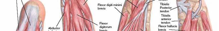
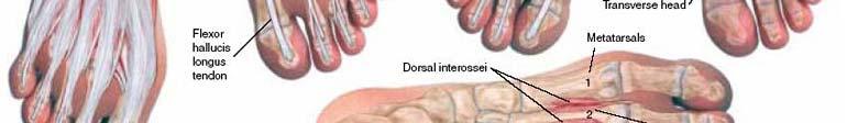
5 IV. MUSCLES OF THE FOOT (1) Dorsal Muscle of the Foot Extensor digitorum brevis: extends phalanges of the four toes into which it is inserted, but in the great toe acts only on the first phalanx. Extensor hallucis brevis: part of extensor digitorum brevis that goes to the great toe 2. Plantar Muscles of Foot First Layer: Abductor hallucis, Flexor digitorum brevis, Abductor digiti quinti Second Layer: Quadratus plantae (Flexor accessorius), Lumbricales Third Layer: Flexor hallucis brevis, Adductor hallucis, Flexor digiti quinti brevis Fourth Layer : Interossei dorsales Abductors Interossei dorsales, Abductor hallucis, and Abductor digiti quinti. Adductors Interossei plantares, Adductor hallucis. Flexors Flexor digitorum brevis, Quadratus plantæ, Flexor hallucis brevis, Flexor digiti quinti brevis, and Lumbricales. 5

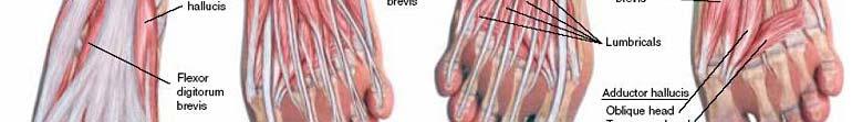
6 Plantar Muscles of Foot 6


 foot does not have a normal arch when standing Fibromyalgia :(algia = pain) a mysterious chronic-pain syndrome; affects mostly women; affecting tendons,")
7 Muscular Dystrophy: Progressive degeneration and atrophy of skeletal muscles. Duchenne muscular dystrophy lack dystrophin, most common & serious form, inherited as a sex-linked recessive disease Myotonic dystrophy: slow-progressing disease, can be present anytime between birth and age 60 Spasm: Sudden, involuntary contraction of muscle lasting a short time. Prolonged spasm is called cramp, when muscle becomes taut & painful; common in calf, thigh & hip muscle, usually occurs at night or after exercise. Cramps have been attributed to a lack of salt or other minerals (like calcium, potassium), muscle fatigue, or dehydration. Pes planus ( flatfoot or fallen arches ) foot does not have a normal arch when standing Fibromyalgia :(algia = pain) a mysterious chronic-pain syndrome; affects mostly women; affecting tendons, ligaments, fibrous tissues. symptoms: fatigue, sleep abnormalities, severe musculoskeletal pain, and headache. Rhabdomyolysis: (Rhabdomyo- skeletal muscle) destruction of muscle tissue, resulting in the release of muscle fiber content into the bloodstream. The fibers release myoglobin, which blocks the kidney structures and can lead to kidney failure. A common cause is a crushing muscle injury. Also a side effect of statin cholesterol medications. 7
8 Myasthenia- muscular weakness Myodynia- pain of the muscle Myomalacia- abnormal muscle softening Myositis- inflammation of muscle Rigor Mortis Stiffening of the body beginning 3 to 4 hours after death; deteriorating sarcoplasmic reticulum releases calcium. Calcium activates myosin-actin cross-bridging and muscle contracts, but can not relax.muscle relaxation requires ATP and ATP production is no longer produced after death.fibers remain contracted until myofilaments decay. Facial muscles are affected first. Joints are stiff for 1-3 days, but after this time general tissue decay and leaking of lysosomal intracellular digestive enzymes will cause the muscles to relax.
The Muscular System. Chapter 10 Part D. PowerPoint Lecture Slides prepared by Karen Dunbar Kareiva Ivy Tech Community College
 Chapter 10 Part D The Muscular System Annie Leibovitz/Contact Press Images PowerPoint Lecture Slides prepared by Karen Dunbar Kareiva Ivy Tech Community College Table 10.14: Muscles Crossing the Hip and
Muscles of the Gluteal Region
 Muscles of the Gluteal Region 1 Some of the most powerful in the body Extend the thigh during forceful extension Stabilize the iliotibial band and thoracolumbar fascia Related to shoulders and arms because
lesser trochanter of femur lesser trochanter of femur iliotibial tract (connective tissue) medial surface of proximal tibia
 medial surface of proximal tibia") LOWER LIMB MUSCLES OF THE APPENDICULAR SKELETON The muscles that act on the lower limb fall into three groups: those that move the thigh, those that move the lower leg, and those that move the ankle, foot,
LOWER LIMB MUSCLES OF THE APPENDICULAR SKELETON The muscles that act on the lower limb fall into three groups: those that move the thigh, those that move the lower leg, and those that move the ankle, foot,
Muscles of the Hip 1. Tensor Fasciae Latae O: iliac crest I: lateral femoral condyle Action: abducts the thigh Nerve: gluteal nerve
 Muscles of the Hip 1. Tensor Fasciae Latae O: iliac crest I: lateral femoral condyle Action: abducts the thigh Nerve: gluteal nerve 2. Gluteus Maximus O: ilium I: femur Action: abduct the thigh Nerve:
Muscles of Lesson Five. Muscular Nomenclature and Kinesiology - Two. Muscles of Lesson Five, cont. Chapter 16
 Chapter 16 Muscular Nomenclature and Kinesiology - Two Lessons 5-6 Muscles of Lesson Five Iliopsoas (psoas major, iliacus) Hip outward rotators (piriformis, gemellus superior, gemellus inferior, obturator
Muscles of the lower extremities. Dr. Nabil khouri MD, MSc, Ph.D
 Muscles of the lower extremities Dr. Nabil khouri MD, MSc, Ph.D Posterior leg Popliteal fossa Boundaries Biceps femoris (superior-lateral) Semitendinosis and semimembranosis (superior-medial) Gastrocnemius
Anatomy & Physiology. Muscles of the Lower Limbs.
 Anatomy & Physiology Muscles of the Lower Limbs http://www.ishapeup.com/musclecharts.html Muscles of the Lower Limbs Among the strongest muscles in the body. Because pelvic girdle is composed of heavy,
Lower limb summary. Anterior compartment of the thigh. Done By: Laith Qashou. Doctor_2016
 Lower limb summary Done By: Laith Qashou Doctor_2016 Anterior compartment of the thigh Sartorius Anterior superior iliac spine Upper medial surface of shaft of tibia 1. Flexes, abducts, laterally rotates
Muscles to know. Lab 21. Muscles of the Pelvis and Lower Limbs. Muscles that Position the Lower Limbs. Generally. Muscles that Move the Thigh
 Muscles to know Lab 21 Muscles of the Pelvis, Leg and Foot psoas major iliacus gluteus maximus gluteus medius sartorius quadriceps femoris (4) gracilus adductor longus biceps femoris semitendinosis semimembranosus
Human Anatomy Biology 351
 Human Anatomy Biology 351 Lower Limb Please place your name on the back of the last page of this exam. You must answer all questions on this exam. Because statistics demonstrate that, on average, between
MUSCLES OF THE LOWER LIMBS
 MUSCLES OF THE LOWER LIMBS Naming, location and general function Dr. Nabil khouri ROLES THAT SHOULD NOT BE FORGOTTEN Most anterior compartment muscles of the hip and thigh Flexor of the femur at the hip
DISSECTION SCHEDULE. Session I - Hip (Front) & Thigh (Superficial)
 & Thigh (Superficial)") DISSECTION SCHEDULE Session I - Hip (Front) & Thigh (Superficial) Surface anatomy Inguinal region Gluteal region Thigh Leg Foot bones Hip bone Femur Superficial fascia Great saphenous vein Superficial
DISSECTION SCHEDULE Session I - Hip (Front) & Thigh (Superficial) Surface anatomy Inguinal region Gluteal region Thigh Leg Foot bones Hip bone Femur Superficial fascia Great saphenous vein Superficial
Human Anatomy Biology 351
 Human Anatomy Biology 351 Lower Limb Please place your name on the back of the last page of this exam. You must answer all questions on this exam. Because statistics demonstrate that, on average, between
Peripheral Nervous System: Lower Body
 Peripheral Nervous System: Lower Body MSTN121 - Neurophysiology Session 11 Department of Myotherapy Lumbar Plexus Iliohypogastric nerve (T12-L1) Motor: Transverse abdominis and internal obliques Sensory:
HUMAN BODY COURSE LOWER LIMB NERVES AND VESSELS
 HUMAN BODY COURSE LOWER LIMB NERVES AND VESSELS October 22, 2010 D. LOWER LIMB MUSCLES 2. Lower limb compartments ANTERIOR THIGH COMPARTMENT General lfunction: Hip flexion, knee extension, other motions
Muscle Anatomy Review Chart
 Muscle Anatomy Review Chart BACK Superficial (5) Trapezius Transverse cervical a. Latissimus dorsi Thoracodorsal a. Rhomboideus major Dorsal scapular a. Rhomboideus minor Levator scapulae Intermediate
Lowe w r e r B ody Resistance Training
 Lower Body Resistance Training Tibialis Anterior Extensor Hallucis Longus Extensor Digitorum Longus Proneus Tertius AROM 25-40 degrees Extensor active-insufficiency Flexion & Eversion (Pronation) Flexion
Human Anatomy Biology 255
 Human Anatomy Biology 255 Exam #4 Please place your name and I.D. number on the back of the last page of this exam. You must answer all questions on this exam. Because statistics demonstrate that, on average,
CHAPTER 8: THE BIOMECHANICS OF THE HUMAN LOWER EXTREMITY
 CHAPTER 8: THE BIOMECHANICS OF THE HUMAN LOWER EXTREMITY _ 1. The hip joint is the articulation between the and the. A. femur, acetabulum B. femur, spine C. femur, tibia _ 2. Which of the following is
Muscles of the Thigh. 6.1 Identify, describe the attachments of and deduce the actions of the muscles of the thigh: Anterior group
 Muscles of the Thigh 6.1 Identify, describe the attachments of and deduce the actions of the muscles of the thigh: Anterior group Sartorius: This is a long strap like muscle with flattened tendons at each
Topic 7: Hip and pelvis. Parts of the hip. Parts of the femur
 Topic 7: Hip and pelvis Parts of the hip Parts of the femur Classifying the hip joint Ball and socket Synovial Multiaxial Movements of the hip: Abduction/adduction Flexion/extension Medial/lateral rotation
Head & Neck The muscle names are followed by the chapter number
 Head & Neck The muscle names are followed by the chapter number. Splenius capitis (9) 2. Occipitalis (2) Temporalis () 3. Temporalis () 4. Semispinalis capitis (9) Facial / Scalp (2) 5. Temporalis () Facial
Organization of the Lower Limb
 Organization of the Lower Limb Limb Development Lower limb develops in an aterolateral position at the level of the L2 to S3 trunk segments Great toe positioned cephalic direction with the soles of the
Walking Theory DR.MURALI KRISHNA P.V M.S.(ORTHO)
") Walking Theory DR.MURALI KRISHNA P.V M.S.(ORTHO) G E N E R A L H O S P I T A L A N E K A L, D E P A R T M E N T O F O R T H O P E D I C S, B A N G A L O R E G O V E R N M E N T O F K A R N A T A K A Walking
Walking Theory DR.MURALI KRISHNA P.V M.S.(ORTHO) G E N E R A L H O S P I T A L A N E K A L, D E P A R T M E N T O F O R T H O P E D I C S, B A N G A L O R E G O V E R N M E N T O F K A R N A T A K A Walking
The Hay is in the Barn
 Anatomy 1 Practical 1 Review Made by Forrest Allen (nerd) Edited by TJ Williamson (not nerd) The Hay is in the Barn 2019 Thunderbringers Too much to handle https://www.youtube.com/watch?v=glii-kaza d8
musculoskeletal system anatomy nerves of the lower limb 2 done by: Dina sawadha & mohammad abukabeer
 musculoskeletal system anatomy nerves of the lower limb 2 done by: Dina sawadha & mohammad abukabeer #Sacral plexus : emerges from the ventral rami of the spinal segments L4 - S4 and provides motor and
The Foot. Dr. Wegdan Moh.Mustafa Medicine Faculty Assistant Professor Mob:
 The Foot Dr. Wegdan Moh.Mustafa Medicine Faculty Assistant Professor Mob: 0127155717 The skeleton of the foot Cutaneous innervations Sole of foot layers of muscles First layer -Abductor hallucis -Flexor
The psoas minor is medial to the psoas major. The iliacus is a fan-shaped muscle that when contracted helps bring the swinging leg forward in walking
 1 p.177 2 3 The psoas minor is medial to the psoas major. The iliacus is a fan-shaped muscle that when contracted helps bring the swinging leg forward in walking and running. The iliopsoas and adductor
The Leg. Prof. Oluwadiya KS
 The Leg Prof. Oluwadiya KS www.oluwadiya.sitesled.com Compartments of the leg 4 Four Compartments: 1. Anterior compartment Deep fibular nerve Dorsiflexes the foot and toes 2. Lateral Compartment Superficial
Main Menu. Joint and Pelvic Girdle click here. The Power is in Your Hands
 1 Hip Joint and Pelvic Girdle click here Main Menu K.6 http://www.handsonlineeducation.com/classes//k6entry.htm[3/23/18, 2:01:12 PM] Hip Joint (acetabular femoral) Relatively stable due to : Bony architecture
BLUE SKY SCHOOL OF PROFESSIONAL MASSAGE AND THERAPEUTIC BODYWORK Musculoskeletal Anatomy & Kinesiology KNEE & ANKLE MUSCLES
 BLUE SKY SCHOOL OF PROFESSIONAL MASSAGE AND THERAPEUTIC BODYWORK Musculoskeletal Anatomy & Kinesiology KNEE & ANKLE MUSCLES MSAK201-I Session 3 1) REVIEW a) THIGH, LEG, ANKLE & FOOT i) Tibia Medial Malleolus
Organization of the Lower Limb
 Organization of the Lower Limb Most illustrations from: Thieme Atlas of Anatomy: Musculoskeletal System. M Schuenke, et al, 2006. Anatomy: A Regional Atlas of the Human Body. Carmine Clemente, 4th edition.
Lumbar Plexus. Ventral rami L1 L4 Supplies: Major nerves.. Abdominal wall External genitalia Anteromedial thigh
 Lower Limb Nerves Lectures Objectives Describe the structure and relationships of the plexuses of the lower limb. Describe the course, relationships and structures supplied for the major nerves of the
Organization of the Lower Limb Audrone Biknevicius, Ph.D. Dept. Biomedical Sciences, OU HCOM at Dublin Clinical Anatomy Immersion 2014
 Organization of the Lower Limb Audrone Biknevicius, Ph.D. Dept. Biomedical Sciences, OU HCOM at Dublin Clinical Anatomy Immersion 2014 www.thestudio1.co.za LIMB FUNCTION choco-locate.com blog.coolibar.com
Main Menu. Ankle and Foot Joints click here. The Power is in Your Hands
 1 The Ankle and Foot Joints click here Main Menu Copyright HandsOn Therapy Schools 2009 K.8 http://www.handsonlineeducation.com/classes/k8/k8entry.htm[3/27/18, 1:40:03 PM] Ankle and Foot Joint 26 bones
Lectures of Human Anatomy
 Lectures of Human Anatomy Lower Limb Gluteal Region and Hip Joint By DR. ABDEL-MONEM AWAD HEGAZY M.B. with honor 1983, Dipl."Gynecology and Obstetrics "1989, Master "Anatomy and Embryology" 1994, M.D.
Practical 1 Worksheet
 Practical 1 Worksheet ANATOMICAL TERMS 1. Use the word bank to fill in the missing words. reference side stand body arms palms anatomical forward All anatomical terms have a(n) point which is called the
5/21/2013. Muscle Anatomy. Thursday January, 24 th, Skeletal Muscle. Smooth Muscle. Cardiac Muscle
 Muscle Anatomy Thursday January, 24 th, 2013 Skeletal Muscle Cardiac Muscle Smooth Muscle 1 Smooth Muscle 1. Found in the walls of the digestive system, bladder, uterus and blood vessels 2. Involuntary
Location Terms. Anterior and posterior. Proximal and Distal The term proximal (Latin proximus; nearest) describes where the appendage joins the body.
 describes where the appendage joins the body.") HUMAN ANAT OMY Location Terms Anterior and posterior In human anatomical usage, anterior refers to the front of the individual. Similarly, posterior refers to the back of the subject. In standard anatomical
HUMAN ANAT OMY Location Terms Anterior and posterior In human anatomical usage, anterior refers to the front of the individual. Similarly, posterior refers to the back of the subject. In standard anatomical
Anatomage Table Instructors Guide- Lower Limb
 The Lower Limb Anatomage Table Instructors Guide- Lower Limb Table of Contents Lower Limb 1- The Skeletal System...3 1: Hip Bone...3 2: Hip Joint and Femur...4 3: Patella and Knee Joint...7 4: Tibia, Fibula,
Temporalis Elevates & retracts mandible. Masseter Elevates mandible. Sternocleidomastoid Neck flexion. Trapezius Elevates & depresses shoulders
 Anterior Posterior Temporalis Elevates & retracts mandible Masseter Elevates mandible Sternocleidomastoid Neck flexion Trapezius Elevates & depresses shoulders Masseter Elevates mandible Temporalis Elevates
The Hip (Iliofemoral) Joint. Presented by: Rob, Rachel, Alina and Lisa
 Joint. Presented by: Rob, Rachel, Alina and Lisa") The Hip (Iliofemoral) Joint Presented by: Rob, Rachel, Alina and Lisa Surface Anatomy: Posterior Surface Anatomy: Anterior Bones: Os Coxae Consists of 3 Portions: Ilium Ischium Pubis Bones: Pubis Portion
The Hip (Iliofemoral) Joint Presented by: Rob, Rachel, Alina and Lisa Surface Anatomy: Posterior Surface Anatomy: Anterior Bones: Os Coxae Consists of 3 Portions: Ilium Ischium Pubis Bones: Pubis Portion
Chapter 9. The Muscular System
 1 Chapter 9 The Muscular System 2 Introduction Skeletal muscles: movement in environment Smooth muscles: intestines, ureters, veins and arteries Cardiac muscle: pumps blood through heart and blood vessels
Understanding Leg Anatomy and Function THE UPPER LEG
 Understanding Leg Anatomy and Function THE UPPER LEG The long thigh bone is the femur. It connects to the pelvis to form the hip joint and then extends down to meet the tibia (shin bone) at the knee joint.
Anatomy MCQs Week 13
 Anatomy MCQs Week 13 1. Posterior to the medial malleolus of the ankle: The neurovascular bundle lies between Tibialis Posterior and Flexor Digitorum Longus The tendon of Tibialis Posterior inserts into
this makes sense, however this is lower order thinking and does not solve the lower leg
 Functional Knee Valgus in a Barbell Squat 1 One of the most common lower leg dysfunction we see in athletes, particularly general population is functional knee valgus, or better referred to as the knees
Hip joint and pelvic girdle. Lower Extremity. Pelvic Girdle 6/5/2017
 Hip joint and pelvic girdle Lower Extremity The relationship between the pelvic girdle and hip is similar to that between the shoulder girdle and shoulder joint. The lower limbs are attached to the axial
Gluteal region DR. GITANJALI KHORWAL
 Gluteal region DR. GITANJALI KHORWAL Gluteal region The transitional area between the trunk and the lower extremity. The gluteal region includes the rounded, posterior buttocks and the laterally placed
Where should you palpate the pulse of different arteries in the lower limb?
 Where should you palpate the pulse of different arteries in the lower limb? The femoral artery In the femoral triangle, its pulse is easily felt just inferior to the inguinal ligament midway between the
Lower Limb Nerves. Clinical Anatomy
 Lower Limb Nerves Clinical Anatomy Lumbar Plexus Ventral rami L1 L4 Supplies: Abdominal wall External genitalia Anteromedial thigh Major nerves.. Lumbar Plexus Nerves relation to psoas m. : Obturator n.
5 Testing the Muscles of the Lower Extremity
 C H A P T E R 5 Testing the Muscles of the Lower Extremity Hip Flexion Hip Flexion, Abduction, and External Rotation with Knee Flexion Hip Extension Hip Abduction Hip Abduction from Flexed Position Hip
Due in Lab weeks because of Thanksgiving Prelab #10. Homework #8. Both sides! Both sides!
 Lab 8 MUSCLES Due in Lab 10 2 weeks because of Thanksgiving Prelab #10 Both sides! Homework #8 Both sides! Refer to Muscles 22-23 Naming of muscles Origin Site of muscle attachment that doesn t move during
5.1 Identify, describe the attachments of and deduce the actions of the muscles of the thigh:
 5.1 Identify, describe the attachments of and deduce the actions of the muscles of the thigh: Anterior group Proximal attachment Distal attachment Sartorius ASIS» Upper part of shaft tibia (middle surface)»
~, /' ~::'~ EXTENSOR HALLUCIS LONGUS. Leg-anterolateral :.:~ / ~\,
 TIBIALIS ANTERIOR Lateral condyle of tibia, upper half of lateral surface of tibia, interosseous membrane Medial side and plantar surface of medial cuneiform bone, and base of first metatarsal bone Dorsiflexes
The thigh. Prof. Oluwadiya KS
 The thigh Prof. Oluwadiya KS www.oluwadiya.com The Thigh: Boundaries The thigh is the region of the lower limb that is approximately between the hip and knee joints Anteriorly, it is separated from the
11/15/2018. Temporalis Elevates & retracts mandible. Masseter = Prime mover of jaw closure. Levator scapulae Supraspinatus Clavicle.
 Due in Lab 10 Lab 8 MUSCLES 2 weeks because of Thanksgiving Prelab #10 Both sides! Homework #8 Both sides! Refer to Muscles 22-23 Examples of Origin & Insertion Naming of muscles Origin Site of muscle
Synergist Muscles. Shoulder (glenohumeral joint) Flexion Deltoid (anterior fibers) Pectoralis major (upper fibers) Biceps Brachii Coracobrachialis
 Flexion Deltoid (anterior fibers) Pectoralis major (upper fibers) Biceps Brachii Coracobrachialis") Synergist Muscles Dr Gene Desepoli DrGeneLMT@gmail.com Shoulder (glenohumeral joint) Deltoid (anterior fibers) Pectoralis major (upper fibers) Biceps Brachii Coracobrachialis Deltoid (posterior fibers)
Synergist Muscles Dr Gene Desepoli DrGeneLMT@gmail.com Shoulder (glenohumeral joint) Deltoid (anterior fibers) Pectoralis major (upper fibers) Biceps Brachii Coracobrachialis Deltoid (posterior fibers)
First & second layers of muscles of the sole
 The FOOT First & second layers of muscles of the sole introduction The muscles acting on the foot can be divided into two distinct groups; extrinsic and intrinsic muscles. The extrinsic muscles arise from
MUSCULOSKELETAL LOWER LIMB
 MUSCULOSKELETAL LOWER LIMB Spinal Cord Lumbar and Sacral Regions Spinal cord Dorsal root ganglion Conus medullaris Cauda equina Dorsal root ganglion of the fifth lumbar nerve End of subarachnoid space
Biology 2401 Muscles List for CPC models
 Biology 2401 List for CPC models Italicized muscles are dissect and similar in the cat = Dissect and note the differences in human and cat Major of the Human Head Facial Expression Epicranius frontalis
Human Anatomy and Physiology I Laboratory
 Human Anatomy and Physiology I Laboratory Gross Anatomy of the Muscular System (Two weeks) 1 This lab involves study of the laboratory exercise Gross Anatomy of the Muscular System. Complete the Review
Human Anatomy Lab #7: Muscles of the Cadaver
 Human Anatomy Lab #7: Muscles of the Cadaver Table of Contents: Expected Learning Outcomes.... 1 Introduction...... 1 Identifying Muscles on Yourself.... 2 Muscles of the Anterior Trunk and Arm.. 2 Muscles
The Human Muscular System Required reading before beginning this lab: Saladin, KS: Human Anatomy 5th ed (2017) Chapters 10, 11, 12 INTRODUCTION
 Chapters 10, 11, 12 INTRODUCTION") Biology 322: Human Anatomy The Human Muscular System Required reading before beginning this lab: Saladin, KS: Human Anatomy 5 th ed (2017) Chapters 10, 11, 12 INTRODUCTION We will use a number of lab periods
Biology 322: Human Anatomy The Human Muscular System Required reading before beginning this lab: Saladin, KS: Human Anatomy 5 th ed (2017) Chapters 10, 11, 12 INTRODUCTION We will use a number of lab periods
Prime movers provide the major force for producing a specific movement Antagonists oppose or reverse a particular movement Synergists
 Dr. Gary Mumaugh Prime movers provide the major force for producing a specific movement Antagonists oppose or reverse a particular movement Synergists Add force to a movement Reduce undesirable or unnecessary
phag/o-, psych/i-, spir/o-, Quick Reference pharyng/o-, pulmon/o, stat/i-, pyel/o-, CORE PRINCIPLES in A&P phleb/o-, systol/i-, phot/o-, py/o-,
 phag/o-, eat: autophagy = self eating; condition of a cell digesting itself pharyng/o-, throat: glossopharyngeal = pertaining to the tongue and throat phleb/o-, vein: phlebotomy = incision into a vein
Lower Limb Dr. Robin Paudel
 Lower Limb n What is a limb? n Skeleton n Joints n Pelvis or limb girdle n Hip/Hip Muscles n Lumber and sacral plexus getting spinal nerves out onto limb n Muscles anterior and posterior compartments n
rotation of the hip Flexion of the knee Iliac fossa of iliac Lesser trochanter Femoral nerve Flexion of the thigh at the hip shaft of tibia
 Anatomy of the lower limb Anterior & medial compartments of the thigh Dr. Hayder The fascia lata encloses the entire thigh like a sleeve/stocking. Three intramuscular fascial septa (lateral, medial, and
Sports Medicine 15. Unit I: Anatomy. The knee, Thigh, Hip and Groin. Part 4 Anatomies of the Lower Limbs
 Sports Medicine 15 Unit I: Anatomy Part 4 Anatomies of the Lower Limbs The knee, Thigh, Hip and Groin Anatomy of the lower limbs In Part 3 of this section we focused upon 11 of the 12 extrinsic muscles
Leg. Dr. Heba Kalbouneh Associate Professor of Anatomy and Histology
 Leg Dr. Heba Kalbouneh Associate Professor of Anatomy and Histology Skin of the Leg Cutaneous Nerves Medially: The saphenous nerve, a branch of the femoral nerve supplies the skin on the medial surface
The Muscular System PART C. PowerPoint Lecture Slide Presentation by Patty Bostwick-Taylor, Florence-Darlington Technical College
 PowerPoint Lecture Slide Presentation by Patty Bostwick-Taylor, Florence-Darlington Technical College The Muscular System 6 PART C Five Golden Rules of Skeletal Muscle Activity Table 6.2 Muscles and Body
Muscle fiber (cell) Blood vessel. Perimysium. Epimysium. Fascicle (wrapped by perimysium) Endomysium (between fibers) Tendon. Bone
 Blood vessel. Perimysium. Epimysium. Fascicle (wrapped by perimysium) Endomysium (between fibers) Tendon. Bone") Figure 6.1 Connective tissue wrappings of skeletal muscle. Blood vessel Muscle fiber (cell) Perimysium Epimysium Fascicle (wrapped by perimysium) Tendon Endomysium (between fibers) Bone Figure 6.15 Superficial
Figure 6.1 Connective tissue wrappings of skeletal muscle. Blood vessel Muscle fiber (cell) Perimysium Epimysium Fascicle (wrapped by perimysium) Tendon Endomysium (between fibers) Bone Figure 6.15 Superficial
LOWER LIMB. As we know the bony part of the body is divided into Axial and Appendicular (upper and lower Limbs)
") LOWER LIMB As we know the bony part of the body is divided into Axial and Appendicular (upper and lower Limbs) Bones of the Lower limb: 1-Pelvic Girdle: composed of: 1. Right hip bone : is formed by 3
LOWER LIMB As we know the bony part of the body is divided into Axial and Appendicular (upper and lower Limbs) Bones of the Lower limb: 1-Pelvic Girdle: composed of: 1. Right hip bone : is formed by 3
Human Muscles (Anterior View) Model 3-44
 Model 3-44") Human Muscles (Anterior View) Model 3-44 Temporalis Frontalis Orbicularis Occuli Orbicularis Oris Masseter Sternocleidomastoid Orbicularis Occuli Human Muscles (Anterior View) Model 3-65 Temporalis Masseter
Human Muscles (Anterior View) Model 3-44 Temporalis Frontalis Orbicularis Occuli Orbicularis Oris Masseter Sternocleidomastoid Orbicularis Occuli Human Muscles (Anterior View) Model 3-65 Temporalis Masseter
In which arm muscle are intramuscular injections most often given? (not in text)
") AP1 Lab 9 - Muscles of the Arms and Legs Locate the following muscles on the models and on yourself. Recall anatomical position. Directional terms such as anterior, posterior, lateral, etc. all assume
AP1 Lab 9 - Muscles of the Arms and Legs Locate the following muscles on the models and on yourself. Recall anatomical position. Directional terms such as anterior, posterior, lateral, etc. all assume
Anterior and Medial compartments of the thigh. Dr. Heba Kalbouneh Associate Professor of Anatomy and Histology
 Anterior and Medial compartments of the thigh Dr. Heba Kalbouneh Associate Professor of Anatomy and Histology Terms Related to Movements Movement Flexion Extension Abduction Adduction Medial (internal)
Clarification of Terms
 Clarification of Terms The plantar aspect of the foot refers to the role or its bottom The dorsal aspect refers to the top or its superior portion The ankle and foot perform three main functions: 1. shock
Match the types of muscle tissues with the words and phrases. 1) Skeletal 2) Smooth 3) Cardiac 2 Walls of blood vessels. 2 Walls of digestive tract
 Skeletal 2) Smooth 3) Cardiac 2 Walls of blood vessels. 2 Walls of digestive tract") S T U D Y G U I D E. Types of Muscle Tissues Match the types of muscle tissues with the words and phrases. ) Skeletal ) Smooth ) Cardiac, Striated Walls of blood vessels, Single nucleus Heart muscle, Involuntary
S T U D Y G U I D E. Types of Muscle Tissues Match the types of muscle tissues with the words and phrases. ) Skeletal ) Smooth ) Cardiac, Striated Walls of blood vessels, Single nucleus Heart muscle, Involuntary
Acland's DVD Atlas of Human Anatomy. Transcript for Volume Robert D Acland
 Acland's DVD Atlas of Human Anatomy Transcript for Volume 2 2007 Robert D Acland This free downloadable pdf file is to be used for individual study only. It is not to be reproduced in any form without
Figure 1 - Hip and Pelvis
 Hip Figure 1 - Hip and Pelvis The terms hip and pelvis are frequently used interchangeably, but strictly speaking, the pelvis is a girdle of bones and the hip is a joint. The pelvis consists of The sacrum
The Muscular System Lab Power Point
 The Muscular System Lab Power Point Myoneural Junction Sarcoplasm Nucleus Myofibrils Sarcomere (black line to black line) Sarcolemma Myoneural space Nucleus Endomysium Motor Neuron Muscles of Facial Expression
TABLE OF MUSCLES OF LOWER EXTREMITY 2018zillmusom ANTERIOR THIGH
 TABLE OF MUSCLES OF LOWER EXTREMITY 2018zillmusom ANTERIOR THIGH MUSCLE ORIGIN INSERTION ACTION NERVE Iliopsoas Ilium, vertebra Femur Flex hip joint Femoral nerve (T12-L5) Pectineus Pubis Femur Flex hip
In-Depth Foundations: Anatomy Terms to Know
 Be familiar with / able to identify and define all the following parts. The Spine Cranium Vertebrae Cervical, Thoracic, Lumbar Sacrum Coccyx Bones of Upper Body Cranium Mastoid process; Occipital condyle,
The University Of Jordan Faculty Of Medicine THE LOWER LIMB. Dr.Ahmed Salman Assistant Prof. of Anatomy. The University Of Jordan
 The University Of Jordan Faculty Of Medicine THE LOWER LIMB Dr.Ahmed Salman Assistant Prof. of Anatomy. The University Of Jordan Gluteal Region Cutaneous nerve supply of (Gluteal region) 1. Lateral cutaneous
Identify the muscles associated with the medial compartment of the thigh. Identify the attachment points of the medial thigh muscles.
 L 8 A B O R A T O R Y Thigh MEDIAL THIGH Identify the muscles associated with the medial compartment of the thigh. Identify the attachment points of the medial thigh muscles. Identify the actions of these
Cadaver Muscular System Practice Practical
 Cadaver Muscular System Practice Practical Station 1 Station 1 1. Specific structure 1. Rectus sheath 2. Red line 2. Linea alba Station 2 Station 2 3. Red muscle 1. Rectus abdominis 4. Red muscle actions
Mohammad Ashraf. Abdulrahman Al-Hanbali. Ahmad Salman. 1 P a g e
 - 7 Mohammad Ashraf Abdulrahman Al-Hanbali Ahmad Salman 1 P a g e Structures under the cover of Gluteus Maximus: 1-Bones: Ileum, Femur (Head, greater trochanter and gluteal tuberosity), Ischium (ischial
Muscular System. IB Sports, exercise and health science 1.2
 Muscular System IB Sports, exercise and health science 1.2 Characteristics Common to Contractility-ability to shorten the muscles length Extensibility-ability to lengthen the muscles length Elasticity-muscle
Anatomy of the lower limb
 Anatomy of the lower limb 1. Bones of the lower limb Pelvis Hip bone/coxal bone Acetabulum o Acetabular margin o Acetabular fossa o Acetabular notch o Lunate surface Ischiopubic ramus Obturator foramen
Contents. Preface xv. SECTION 1: Introduction to the Bodynamic System 1. SECTION 2: The Bodynamic Psycho-Motor Anatomy 29
 Contents Preface xv SECTION 1: Introduction to the Bodynamic System 1 Definitions in the Bodynamic System 3 Ego Formation through the Coding Elements 9 Examples of Formation of Coding 17 Using This Book
Baraa Ayed حسام أبو عوض. Ahmad Salman. 1 P a g e
 4 Baraa Ayed حسام أبو عوض Ahmad Salman 1 P a g e Today we are going to cover these concepts: Iliotibial tract Anterior compartment of the thigh and the hip Medial compartment of the thigh Femoral triangle
musculoskeletal system anatomy muscles of foot sheet done by: dina sawadha & mohammad abukabeer
 musculoskeletal system anatomy muscles of foot sheet done by: dina sawadha & mohammad abukabeer Extensor retinaculum : A- superior extensor retinaculum (SER) : originates from the distal ends of the tibia
BIOL 4260 Human Evolu3onary Anatomy Lecture 12: Limb Development. Lecture 2: Fossil Record
 BIOL 4260 Human Evolu3onary Anatomy Lecture 12: Limb Development Lecture 2: Fossil Record Outline Limb Evolution Limb Development Limb Function A Few Definitions Appendicular skeleton girdles & limbs Pectoral
Scapula Spine Lateral edge of clavicle. Medial border Scapula. Medial border of Scapula, between superior angle and root of spine. Scapula.
 Muscle attachments and actions answer sheet Muscle Origins insertions Movements Joints crossed Trapezius Base of skull Spinous process of C7 Thoracic Spine Lateral edge of clavicle Elevation Retraction
The Hip Joint. Shenequia Howard David Rivera
 The Hip Joint Shenequia Howard David Rivera Topics Of Discussion Movement Bony Anatomy Ligamentous Anatomy Muscular Anatomy Origin/Insertion/Action/Innervation Common Injuries MOVEMENT Flexion Extension
Stretching the Major Muscle Groups of the Lower Limb
 Preface In 1964, at the beginning of my PhD studies at Southern Illinois University, I was first introduced to the theory and practice of proprioceptive neuromuscular facilitation (PNF). One specific application,
Muscles of the Cat. N Deltoid MUSCLES OF THE CHEST. Pectoralis major. (This muscle is superior to Pectoralis minor) MUSCLES OF THE CHEST
 MUSCLES OF THE CHEST") MUSCLES OF THE CHEST Pectoralis major (This muscle is superior to Pectoralis minor) 1. MUSCLES OF THE CHEST Pectoralis minor (This muscle is inferior to Pectoralis major) 2. MUSCLES OF THE ARM Deltoid
MUSCLES OF THE CHEST Pectoralis major (This muscle is superior to Pectoralis minor) 1. MUSCLES OF THE CHEST Pectoralis minor (This muscle is inferior to Pectoralis major) 2. MUSCLES OF THE ARM Deltoid
Lesson 24. A & P Hip
 Lesson 24 A & P Hip 1 Aims of the Session This session will allow candidates to have an understanding of the bony prominences and soft tissues of the hip 2 Learning Outcomes By the end of the lesson the
ANATYOMY OF The thigh
 ANATYOMY OF The thigh 1- Lateral cutaneous nerve of the thigh Ι) Skin of the thigh Anterior view 2- Femoral branch of the genitofemoral nerve 5- Intermediate cutaneous nerve of the thigh 1, 2 and 3 are
Epicranius (frontal belly) Zygomaticus minor. Zygomaticus major Buccinator
 Zygomaticus minor. Zygomaticus major Buccinator") Epicranius (frontal belly) Zygomaticus minor Zygomaticus major Buccinator Masseter Digastric (posterior belly) Stylohyoid Sternocleidomastoid Trapezius Scalenus Omohyoid (inferior belly) Orbicularis oris
Epicranius (frontal belly) Zygomaticus minor Zygomaticus major Buccinator Masseter Digastric (posterior belly) Stylohyoid Sternocleidomastoid Trapezius Scalenus Omohyoid (inferior belly) Orbicularis oris
ANATOMY TEAM GLUTEAL REGION & BACK OF THIGH
 ANATOMY TEAM GLUTEAL REGION & BACK OF THIGH OBJECTIVES By the end of this lecture, the student should be able to identify and discuss: Contents of gluteal region: Groups of Glutei muscles and small muscles