Case Example. Nerve Entrapments in the Lower limb
|
|
|
- Rudolf Simon
- 6 years ago
- Views:
Transcription

1 Nerve Entrapments in the Lower limb February, 2013 William S. Pease, M.D. Ernest W. Johnson Professor of PM&R Case Example CC: Right ankle dorsiflexion weakness with minimal paresthesias HPI: 87 year-old physician with chronic lumbar pain and h/o prostate cancer, had recent weight loss. He had no history of diabetes or of neuropathy. Prostate had been excised. PE: Ankle dorsiflexion and eversion of affected right side was 2/5 with normal inversion and knee flexion, and no other significant weakness noted. Minimal sensory loss in the dorsal foot. 1
2 Case Example-Motor NCS Nerves Latency Amplitude Distance Velocity Ms mv Cm m/s R Fibular (EDB) Ankle Fib Head Knee No response 0 R Fibular (Ant Tib) Fib Head Knee First testing-6 weeks after onset Ankle stim-edb Fibula-EDB Politeal-EDB (NR) Fib-Tibialis Ant Pop-Tibialis Ant Strength ADF 2/5 2
3 Peroneal (Fibular) Motor Study All three possible changes agree: Focal slowing of conduction Conduction block (neurapraxia) Change is shape of response (wider) Temporal dispersion =Nerve entrapment (As good as it gets!) Case Example-Motor NCS 3 months later Nerves Latency Amplitude Distance Velocity Ms mv Cm m/s R Fibular (EDB) Ankle Fib Head Knee R Fibular (Ant Tib) Fib Head Knee

4 Follow-up testing- 3 months later Strength ADF 4+/5 R Comm Fibular - Default 2 Fib-Tibialis Ant Fib hd TA 1 30ms 5mV 2 Pop-Tibialis Ant 1 3 Site ms 5mV Improved amplitude with proximal stimulation = improved strength! Fibular Nerve Compression at the Knee-Etiology Crossing Legs 87 yo m Electrodiagnostician Acute fibular neuropathy Ernest W. Johnson, M.D. 4

5 Fibular (Peroneal) Nerve Compression Drop foot Weakness of dorsiflexion and eversion Paresthesia/sensory loss dorsal foot Exclusions Not CNS, reflexes WNL, Babinski - Not cauda equina-opposite side normal No incontinence Common Fibular Nerve 28 yo woman 1 st pregnancy Rt dorsiflexion weakness Onset s/p deliveryvaginal Excessive knee flexion while pushing at end of labor 5

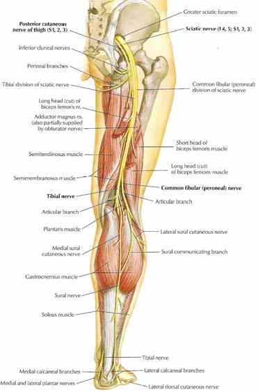
6 Fibular (Peroneal) Nerve Compression Entrapped between popliteal space and fibular head Passes between tendons in area Contributing factors Rapid weight loss Crossing legs Squatting- strawberry picker s palsy Tx: Eliminate cause, heals well May need temporary orthosis 6


7 Fibular Nerve Below knee Tendons Biceps femoris Popliteus Lateral Gastrocnemius Common fibular Both divisions affected typically 7

8 What is it you want to know? Is this nerve working? Amid pain, edema, restricted motion, paresthesia, weakness Is it a peripheral nerve problem? Where is the problem? Is anything else going on? How severe is the problem? What is the prognosis? 8


9 Nerve Entrapment Always stimulate proximal and distal to the point of suspected entrapment Note amplitude, duration and latency Best measure of prognosis is most distal stimulation Best measure of strength is response to proximal stimulation 9

10 10
11 11

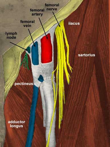
12 Stimulate Femoral N Above inguinal ligament Stimulate Femoral N Below inguinal ligament Iliacus Muscle Needle Insertion 12

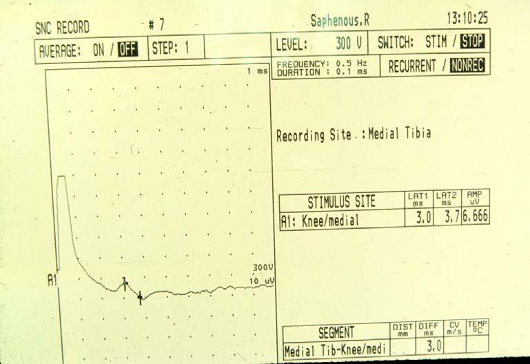
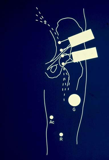
13 Saphenous Nerve 13


14 Meralgia Paresthetica Lateral Cutaneous Nerve Burning pain in lateral thigh to just above the knee No weakness Risk factors Weight gain Diabetes Mellitus Tight clothing/tool belt Meralgia Paresthetica Benign sensory-only nerve palsy Rule outs are important L4 root injury Lumbar plexus Femoral Nerve Tx: relieve pressure, Symptomatic meds Eg, gabapentin Lateral Cutaneus Nv. 14


 Stimulate with monopolar needle 1 cm medial to ASIS")
15 Lateral Cutaneous Nerve of the Thigh Stim 1 cm medial to ASIS Record 12 cm along line to lateral patella Onset Lat < 2.9 ms At 14 cm Amplitude > 3 µv MERALGIA PARESTHETICA FREQUENT IN DIABETICS; overweight persons with tight belts Place recording electrodes 2/3 down on anterior-lateral thigh (use disposable sensory recording strips) Stimulate with monopolar needle 1 cm medial to ASIS Compare with contralateral 15


16 MERALGIA PARESTHETICA Differential Diagnosis Radiculopathy L3 or L4 Lumbar plexopathy Femoral Neuropathy Polyneuropathy 16


17 MERALGIA PARESTHETICA EMG Evaluation Needle EMG to evaluate plexus and roots Iliospsoas, quadriceps, adductors, tibialis anterior and gastrocnemius Sural and tibial NCS with F wave Lateral femoral nerve study Piriformis Syndrome 17
18 Jesse Owens Memorial Plaza The Ohio State University 1935 Big Ten Championships Jesse Owens set Four World Records in One hour during the finals Then went on to Berlin Jesse Owens Memorial Plaza The Ohio State University 18

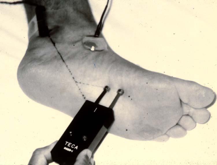
19 Tarsal Tunnel Syndrome Tibial nerve compression at medial malleolus is relatively rare NOT analogous to CTS Anatomy of tendons and ligaments is very different Associated with altered anatomy of foot, usually following trauma Tarsal Tunnel Syndrome EDX Motor latency medial plantar n < 6 ms; lateral plantar n - < 6.5 ms If Medial Plantar comes within 0.5 ms of lateral plantar latency suspect medial plantar entrapment 19


20 Lateral plantar nerve entrapment More frequently seen in diabetic peripheral neuropathy CNAP will be reduced or absent with stimulation at foot sole (Lat Pl N) Needle EMG abnormalities in abd dig V ped and lateral interosseus muscles Tibial nerve Motor study Abd Hall Medial Plantar Abd Dig Min Lateral Plantar 20


21 Trans-Tarsal technique Medial plantar nerve Amplitudes :10-30 uv Latency: 3.2 msec Lateral plantar nerve Amplitudes 8-20 uv Latency: 3.2 msec NB. This is a MIXED nerve action potential 21
22 22

23 Go Bucks! 23
Year 2004 Paper one: Questions supplied by Megan
 QUESTION 47 A 58yo man is noted to have a right foot drop three days following a right total hip replacement. On examination there is weakness of right ankle dorsiflexion and toe extension (grade 4/5).
A/Professor Arun Aggarwal Balmain Hospital
 A/Professor Arun Aggarwal Balmain Hospital Nerve Conduction Studies Test to evaluate the function of motor / sensory nerves Evaluate Paraesthesia (numbness, tingling, burning) Weakness of arms and legs
The Lower Limb II. Anatomy RHS 241 Lecture 3 Dr. Einas Al-Eisa
 The Lower Limb II Anatomy RHS 241 Lecture 3 Dr. Einas Al-Eisa Tibia The larger & medial bone of the leg Functions: Attachment of muscles Transfer of weight from femur to skeleton of the foot Articulations
Electrodiagnostics for Back & Neck Pain. Steven Andersen, MD Providence Physiatry Clinic
 Electrodiagnostics for Back & Neck Pain Steven Andersen, MD Providence Physiatry Clinic Electrodiagnostics Electromyography (EMG) Needle EMG exam (NEE) Nerve conduction studies (NCS) Motor Sensory Late
Leg. Dr. Heba Kalbouneh Associate Professor of Anatomy and Histology
 Leg Dr. Heba Kalbouneh Associate Professor of Anatomy and Histology Skin of the Leg Cutaneous Nerves Medially: The saphenous nerve, a branch of the femoral nerve supplies the skin on the medial surface
The Lower Limb VI: The Leg. Anatomy RHS 241 Lecture 6 Dr. Einas Al-Eisa
 The Lower Limb VI: The Leg Anatomy RHS 241 Lecture 6 Dr. Einas Al-Eisa Muscles of the leg Posterior compartment (superficial & deep): primary plantar flexors of the foot flexors of the toes Anterior compartment:
Quillen College of Medicine
 Ea s t T e n n e s s e e St a t e Un i v e r s i t y Quillen College of Medicine Failing to prepare is preparing to fail. John Wooden, UCL A Dr. Tom Kwasigroch Associate Dean Director, Medical Human Gross
Regional Anaesthesia
 Regional Anaesthesia Lower limb anatomy and blocks Hip and Knee Joint Hip Joint: Nerve supply Lumbar plexus Femoral nerve through the nerve to the Rectus Femoris Ant division of the Obturator nerve The
Lower Limb Nerves. Clinical Anatomy
 Lower Limb Nerves Clinical Anatomy Lumbar Plexus Ventral rami L1 L4 Supplies: Abdominal wall External genitalia Anteromedial thigh Major nerves.. Lumbar Plexus Nerves relation to psoas m. : Obturator n.
Borderline Moderately out of normal range Severely out of normal range Technical
 Urgent Care Specialists 2200 Universal Drive, Boston, MA Phone: 761-621-5216 Fax: 761-564-0336 Report: EMG-1133-28616 Physician: Dr. John Smith Age Range: 40-59 Ref Phys: Dr. Joe Ranier Height Range cm:
HUMAN BODY COURSE LOWER LIMB NERVES AND VESSELS
 HUMAN BODY COURSE LOWER LIMB NERVES AND VESSELS October 22, 2010 D. LOWER LIMB MUSCLES 2. Lower limb compartments ANTERIOR THIGH COMPARTMENT General lfunction: Hip flexion, knee extension, other motions
Where should you palpate the pulse of different arteries in the lower limb?
 Where should you palpate the pulse of different arteries in the lower limb? The femoral artery In the femoral triangle, its pulse is easily felt just inferior to the inguinal ligament midway between the
BLUE SKY SCHOOL OF PROFESSIONAL MASSAGE AND THERAPEUTIC BODYWORK Musculoskeletal Anatomy & Kinesiology KNEE & ANKLE MUSCLES
 BLUE SKY SCHOOL OF PROFESSIONAL MASSAGE AND THERAPEUTIC BODYWORK Musculoskeletal Anatomy & Kinesiology KNEE & ANKLE MUSCLES MSAK201-I Session 3 1) REVIEW a) THIGH, LEG, ANKLE & FOOT i) Tibia Medial Malleolus
MUSCULOSKELETAL LOWER LIMB
 MUSCULOSKELETAL LOWER LIMB Spinal Cord Lumbar and Sacral Regions Spinal cord Dorsal root ganglion Conus medullaris Cauda equina Dorsal root ganglion of the fifth lumbar nerve End of subarachnoid space
CNS & PNS Entrapment. Disclosure - Nothing
 Peripheral Nerve Entrapments That Mimic Spinal Pathology: Evaluation And Treatment Both Medical And Surgical Michel Kliot MD Clinical Professor UCSF Department of NeuroSurgery Director Center For Evaluation
Compound Nerve Action Potential of Common Peroneal Nerve and Sural Nerve Action Potential in Common Peroneal Neuropathy
 J Korean Med Sci 2008; 23: 117-21 ISSN 1011-8934 DOI: 10.3346/jkms.2008.23.1.117 Copyright The Korean Academy of Medical Sciences Compound Nerve Action Potential of Common Peroneal Nerve and Sural Nerve
Organization of the Lower Limb Audrone Biknevicius, Ph.D. Dept. Biomedical Sciences, OU HCOM at Dublin Clinical Anatomy Immersion 2014
 Organization of the Lower Limb Audrone Biknevicius, Ph.D. Dept. Biomedical Sciences, OU HCOM at Dublin Clinical Anatomy Immersion 2014 www.thestudio1.co.za LIMB FUNCTION choco-locate.com blog.coolibar.com
Lumbar Plexus. Ventral rami L1 L4 Supplies: Major nerves.. Abdominal wall External genitalia Anteromedial thigh
 Lower Limb Nerves Lectures Objectives Describe the structure and relationships of the plexuses of the lower limb. Describe the course, relationships and structures supplied for the major nerves of the
Index. Note: Page numbers of article titles are in boldface type.
 Neurol Clin N Am 20 (2002) 605 617 Index Note: Page numbers of article titles are in boldface type. A ALS. See Amyotrophic lateral sclerosis (ALS) Amyotrophic lateral sclerosis (ALS) active denervation
VENOUS DRAINAGE OF THE LOWER LIMB
 Anatomy of the lower limb Superficial veins & nerve injuries Dr. Hayder VENOUS DRAINAGE OF THE LOWER LIMB The venous drainage of the lower limb is of huge clinical & surgical importance. Since the venous
Disclosure. Entrapment Neuropathies - Overview. Common mononeuropathy sites. Definitions. Common mononeuropathy sites. Common mononeuropathy sites
 Disclosure Entrapment Neuropathies - Overview I receive compensation from Wiley- Blackwell publishers for my work as Editor-in-Chief of Muscle & Nerve Lawrence H. Phillips, II, MD Definitions Mononeuropathy:
Guide to the use of nerve conduction studies (NCS) & electromyography (EMG) for non-neurologists
 & electromyography (EMG) for non-neurologists") Guide to the use of nerve conduction studies (NCS) & electromyography (EMG) for non-neurologists What is NCS/EMG? NCS examines the conduction properties of sensory and motor peripheral nerves. For both
Guide to the use of nerve conduction studies (NCS) & electromyography (EMG) for non-neurologists What is NCS/EMG? NCS examines the conduction properties of sensory and motor peripheral nerves. For both
Section Three: The Leg, Ankle, and Foot Lecture: Review of Clinical Anatomy, Patterns of Dysfunction and Injury, and
 Section Three: The Leg, Ankle, and Foot Lecture: Review of Clinical Anatomy, Patterns of Dysfunction and Injury, and Treatment Implications for the Leg, Ankle, and Foot Levels I and II Demonstration and
Making sense of Nerve conduction & EMG
 Making sense of Nerve conduction & EMG Drs R Arunachalam Consultant Clinical Neurophysiologist Wessex Neurological Centre Southampton University Hospital EMG/NCS EMG machine For the assessment of patients
Muscles of the lower extremities. Dr. Nabil khouri MD, MSc, Ph.D
 Muscles of the lower extremities Dr. Nabil khouri MD, MSc, Ph.D Posterior leg Popliteal fossa Boundaries Biceps femoris (superior-lateral) Semitendinosis and semimembranosis (superior-medial) Gastrocnemius
musculoskeletal system anatomy nerves of the lower limb 2 done by: Dina sawadha & mohammad abukabeer
 musculoskeletal system anatomy nerves of the lower limb 2 done by: Dina sawadha & mohammad abukabeer #Sacral plexus : emerges from the ventral rami of the spinal segments L4 - S4 and provides motor and
Lecture 09. Popliteal Fossa. BY Dr Farooq Khan Aurakzai
 Lecture 09 Popliteal Fossa BY Dr Farooq Khan Aurakzai Dated: 14.02.2018 What is popliteus? Introduction Anything relating to, or near the part of the leg behind the knee. From New Latin popliteus the muscle
For convenience values outside the normal range are bolded. Normal values for the specified patient are stated below the tables.
 Case tudy 8 or convenience values outside the normal range are bolded. Normal values for the specified patient are stated below the tables. History: 60 year-ol man with a history of left hand weakness
Anatomy MCQs Week 13
 Anatomy MCQs Week 13 1. Posterior to the medial malleolus of the ankle: The neurovascular bundle lies between Tibialis Posterior and Flexor Digitorum Longus The tendon of Tibialis Posterior inserts into
Neurologic complications - whom to blame? Benno Rehberg Médecin adjoint agrégé Unité d anesthésiologie gynéco-obstétricale, HUG
 Neurologic complications - whom to blame? Benno Rehberg Médecin adjoint agrégé Unité d anesthésiologie gynéco-obstétricale, HUG SAOA spring meeting 2015 The simple surgical answer: outline Epidemiology
Lower Limb Dr. Robin Paudel
 Lower Limb n What is a limb? n Skeleton n Joints n Pelvis or limb girdle n Hip/Hip Muscles n Lumber and sacral plexus getting spinal nerves out onto limb n Muscles anterior and posterior compartments n
The Muscular System. Chapter 10 Part D. PowerPoint Lecture Slides prepared by Karen Dunbar Kareiva Ivy Tech Community College
 Chapter 10 Part D The Muscular System Annie Leibovitz/Contact Press Images PowerPoint Lecture Slides prepared by Karen Dunbar Kareiva Ivy Tech Community College Table 10.14: Muscles Crossing the Hip and
1-Muscles: 2-Blood supply: Branches of the profunda femoris artery. 3-Nerve supply: Sciatic nerve
 1-Muscles: B i c e p s f e m o r i s S e m i t e n d i n o s u s S e m i m e m b r a n o s u s a small part of the adductor magnus (h a m s t r i n g p a r t o r i s c h i a l p a r t ) 2-Blood supply:
Contents of the Posterior Fascial Compartment of the Thigh
 Contents of the Posterior Fascial Compartment of the Thigh 1-Muscles: B i c e p s f e m o r i s S e m i t e n d i n o s u s S e m i m e m b r a n o s u s a small part of the adductor magnus (h a m s t
Nerve Site Latency ms Amplitude mv Distance mm Conduction Velocity m/s
 Clinical Electrophysiology Instructions and Sample Reports NCS/EMG REPORT Date: August 2007 Reason for Electrophysiologic Referral: Bilateral upper extremity pain and numbness. History: 59 year-old female
DISSECTION SCHEDULE. Session I - Hip (Front) & Thigh (Superficial)
 & Thigh (Superficial)") DISSECTION SCHEDULE Session I - Hip (Front) & Thigh (Superficial) Surface anatomy Inguinal region Gluteal region Thigh Leg Foot bones Hip bone Femur Superficial fascia Great saphenous vein Superficial
DISSECTION SCHEDULE Session I - Hip (Front) & Thigh (Superficial) Surface anatomy Inguinal region Gluteal region Thigh Leg Foot bones Hip bone Femur Superficial fascia Great saphenous vein Superficial
The Leg. Prof. Oluwadiya KS
 The Leg Prof. Oluwadiya KS www.oluwadiya.sitesled.com Compartments of the leg 4 Four Compartments: 1. Anterior compartment Deep fibular nerve Dorsiflexes the foot and toes 2. Lateral Compartment Superficial
The Hip (Iliofemoral) Joint. Presented by: Rob, Rachel, Alina and Lisa
 Joint. Presented by: Rob, Rachel, Alina and Lisa") The Hip (Iliofemoral) Joint Presented by: Rob, Rachel, Alina and Lisa Surface Anatomy: Posterior Surface Anatomy: Anterior Bones: Os Coxae Consists of 3 Portions: Ilium Ischium Pubis Bones: Pubis Portion
The Hip (Iliofemoral) Joint Presented by: Rob, Rachel, Alina and Lisa Surface Anatomy: Posterior Surface Anatomy: Anterior Bones: Os Coxae Consists of 3 Portions: Ilium Ischium Pubis Bones: Pubis Portion
Ultrasound Guided Lower Extremity Blocks
 Ultrasound Guided Lower Extremity Blocks CONTENTS: 1. Femoral Nerve Block 2. Popliteal Nerve Block Updated December 2017 1 1. Femoral Nerve Block Indications Surgery involving the knee, anterior thigh,
Mohammad Ashraf. Abdulrahman Al-Hanbali. Ahmad Salman. 1 P a g e
 - 7 Mohammad Ashraf Abdulrahman Al-Hanbali Ahmad Salman 1 P a g e Structures under the cover of Gluteus Maximus: 1-Bones: Ileum, Femur (Head, greater trochanter and gluteal tuberosity), Ischium (ischial
Femoral Artery. Its entrance to the thigh Position Midway between ASIS and pubic symphysis
 Lower Limb Vessels Lecture Objectives Describe the major arteries of the lower limb. Describe the deep and superficial veins of the lower limb. Describe the topographical relationships of the arteries
Jose Santiago Campos, MD and Eric L. Altschuler, MD, PhD
 Numb Toes Jose Santiago Campos, MD and Eric L. Altschuler, MD, PhD No one involved in the planning of this CME activity have anyy relevant financial relationships to disclose. Authors/faculty have nothingg
Anatomy & Physiology. Muscles of the Lower Limbs.
 Anatomy & Physiology Muscles of the Lower Limbs http://www.ishapeup.com/musclecharts.html Muscles of the Lower Limbs Among the strongest muscles in the body. Because pelvic girdle is composed of heavy,
MUSCLES OF THE LOWER LIMBS
 MUSCLES OF THE LOWER LIMBS Naming, location and general function Dr. Nabil khouri ROLES THAT SHOULD NOT BE FORGOTTEN Most anterior compartment muscles of the hip and thigh Flexor of the femur at the hip
Leo Kormanik DC, MS, CCSP Ohio Sports Chiropractic
 Leo Kormanik DC, MS, CCSP Ohio Sports Chiropractic ! Been running at a high level for 15 years.! 2012 Olympics Trials qualifier in the marathon and 6-time All-American in college! Owner of Ohio Sports
Copyright 2004, Yoshiyuki Shiratori. All right reserved.
 Ankle and Leg Evaluation 1. History Chief Complaint: A. What happened? B. Is it a sharp or dull pain? C. How long have you had the pain? D. Can you pinpoint the pain? E. Do you have any numbness or tingling?
Muscles of the Hip 1. Tensor Fasciae Latae O: iliac crest I: lateral femoral condyle Action: abducts the thigh Nerve: gluteal nerve
 Muscles of the Hip 1. Tensor Fasciae Latae O: iliac crest I: lateral femoral condyle Action: abducts the thigh Nerve: gluteal nerve 2. Gluteus Maximus O: ilium I: femur Action: abduct the thigh Nerve:
Yoshihisa Masakado, Michiyuki Kawakami, Kanjiro Suzuki, Leon Abe, Tetsuo Ota, and Akio Kimura
 REVIEW Clinical Neurophysiology in the Diagnosis of Peroneal Nerve Palsy Yoshihisa Masakado, Michiyuki Kawakami, Kanjiro Suzuki, Leon Abe, Tetsuo Ota, and Akio Kimura Keio University Tsukigase Rehabilitation
Gross Anatomy Coloring Book Series. Lower Extremity Arteries
 Gross Anatomy Coloring Book Series Lower Extremity Arteries 1 Femoral Artery and Associated Branches For the life of the flesh is in the blood. Leviticus 17:11 Femoral Artery and Associated Branches After
Lumbar and Sacral Plexuses. Dr. Heba Kalbouneh Associate Professor of Anatomy and Histology
 Lumbar and Sacral Plexuses Dr. Heba Kalbouneh Associate Professor of Anatomy and Histology Structure of Spinal Nerves: Somatic Pathways dorsal root CNS interneuron spinal nerve dorsal ramus somatic sensory
ANKLE PLANTAR FLEXION
 ANKLE PLANTAR FLEXION Evaluation and Measurements By Isabelle Devreux 1 Ankle Plantar Flexion: Gastrocnemius and Soleus ROM: 0 to 40-45 A. Soleus: Origin: Posterior of head of fibula and proximal1/3 of
The near-nerve sensory nerve conduction in tarsal tunnel syndrome
 Journal of Neurology, Neurosurgery, and Psychiatry 1985;48: 999-1003 The near-nerve sensory nerve conduction in tarsal tunnel syndrome SHN J OH, HYUN S KM, BASHRUDDN K AHMAD From the Department ofneurology,
1. A worker falls from a height and lands on his feet. Radiographs reveal a fracture of the sustentaculum tali. The muscle passing immediately
 1. A worker falls from a height and lands on his feet. Radiographs reveal a fracture of the sustentaculum tali. The muscle passing immediately beneath it that would be adversely affected is the: fibularis
Sensory nerve conduction studies
 Genomläst 2007-01-31/meg Sensory nerve conduction studies Department of clinical neurophysiology University hospital Uppsala, Sweden 1997-12-16 Björn Falck, Erik Stålberg and Lena Eriksson Department of
Nerve Conduction Studies and EMG
 Nerve Conduction Studies and EMG Limitations of other methods of investigations of the neuromuscular system - Dr Rob Henderson, Neurologist Assessment of Weakness Thanks Peter Silburn PERIPHERAL NEUROPATHY
The thigh. Prof. Oluwadiya KS
 The thigh Prof. Oluwadiya KS www.oluwadiya.com The Thigh: Boundaries The thigh is the region of the lower limb that is approximately between the hip and knee joints Anteriorly, it is separated from the
موسى صالح عبد الرحمن الحنبلي أحمد سلمان
 8 موسى صالح عبد الرحمن الحنبلي أحمد سلمان 1 P a g e Today we will talk about a new region, which is the leg. And as always, we will start with studying the sensory innervation of the leg. What is the importance
UltraEMG Course Schedule 2015
 Ultra EMG February 20-25 Manchester Grand Hyatt San Diego, California Thursday February 19 Travel Date 5:00-6:00 Registration UltraEMG Course Schedule 2015 UltraEMG-MSK (Musculoskeletal Emphasis) Friday,
Located Deep to Flexor Retinaculum on medial aspect of ankle. Posterior to Posterior Tibial Artery. Tom, Dick, and Very Nervous Harry
 ANKLE BLOCK ANESTHESIA GREGORY CLARK D.P.M. HEAD, SECTION OF PODIATRY SCRIPPS CLINIC LA JOLLA, CALIFORNIA A METHOD BY WHICH ONE MAY PROVIDE AN ANESTHETIC BLOCK TO THE FOOT OR ANKLE WITH A MINIMUM OF PATIENT
Ankle and Foot Orthopaedic Tests Orthopedics and Neurology DX 612
 Ankle and Foot Orthopaedic Tests Orthopedics and Neurology DX 612 James J. Lehman, DC, MBA, DABCO University of Bridgeport College of Chiropractic Ankle & Foot Anatomy Stability of the ankle is dependent
5.1 Identify, describe the attachments of and deduce the actions of the muscles of the thigh:
 5.1 Identify, describe the attachments of and deduce the actions of the muscles of the thigh: Anterior group Proximal attachment Distal attachment Sartorius ASIS» Upper part of shaft tibia (middle surface)»
EDL EHL. Extensor Hallucis Longus L5 Extensor Digitorum longus L5,1 Peroneus Tertius L5 1 Extensor Digitorum Brevis S1,2 [like intrinsic muscle]
![EDL EHL. Extensor Hallucis Longus L5 Extensor Digitorum longus L5,1 Peroneus Tertius L5 1 Extensor Digitorum Brevis S1,2 [like intrinsic muscle]](/thumbs/78/77875930.jpg "EDL EHL. Extensor Hallucis Longus L5 Extensor Digitorum longus L5,1 Peroneus Tertius L5 1 Extensor Digitorum Brevis S1,2 [like intrinsic muscle]") ANATOMY OF ANKLE AND FOOT Lateral aspect: [Dorsal medial to lateral= dorsal under extensor retinaculum] Tibialis Anterior EHL Artery [Dorsal pedal A] and Anterior tibial N EDL Peroneus Tertius Behind the
ANATOMY OF ANKLE AND FOOT Lateral aspect: [Dorsal medial to lateral= dorsal under extensor retinaculum] Tibialis Anterior EHL Artery [Dorsal pedal A] and Anterior tibial N EDL Peroneus Tertius Behind the
Peripheral Nervous System: Lower Body
 Peripheral Nervous System: Lower Body MSTN121 - Neurophysiology Session 11 Department of Myotherapy Lumbar Plexus Iliohypogastric nerve (T12-L1) Motor: Transverse abdominis and internal obliques Sensory:
Joints of the Lower Limb II
 Joints of the Lower Limb II Lecture Objectives Describe the components of the knee and ankle joint. List the ligaments associated with these joints and their attachments. List the muscles acting on these
Understanding Leg Anatomy and Function THE UPPER LEG
 Understanding Leg Anatomy and Function THE UPPER LEG The long thigh bone is the femur. It connects to the pelvis to form the hip joint and then extends down to meet the tibia (shin bone) at the knee joint.
stimulation with a magnetic coil. This approach has the great advantage of being relatively painless. We
 Journal ofneurology, Neurosurgery, and Psychiatry 1989;52:767-772 Magnetic stimulation in the diagnosis of lumbosacral radiculopathy S CHOKROVERTY, R SACHDEO, J DILULLO, R C DUVOISIN From the Department
OTM Lecture Gait and Somatic Dysfunction of the Lower Extremity
 OTM Lecture Gait and Somatic Dysfunction of the Lower Extremity Somatic Dysfunction Tenderness Asymmetry Range of Motion Tissue Texture Changes Any one of which must be present to diagnosis somatic dysfunction.
Topic 7: Hip and pelvis. Parts of the hip. Parts of the femur
 Topic 7: Hip and pelvis Parts of the hip Parts of the femur Classifying the hip joint Ball and socket Synovial Multiaxial Movements of the hip: Abduction/adduction Flexion/extension Medial/lateral rotation
Mononeuropathies of the Upper and Lower Extremity
 15 Mononeuropathies of the Upper and Lower Extremity Kevin R. Scott and Milind J. Kothari Summary Nerves of both the upper and lower extremities are frequently injured for a variety of reasons. In the
Back Pain. John W. Engstrom, MD December 16, Disclosures. A Clinical Approach to the Evaluation of Back Pain and Lumbar Radiculopathy
 Disclosures Nothing to declare --- or --- Significant ownership interests Speaker bureaus, honorarium, grants A Clinical Approach to the Evaluation of and Lumbar Radiculopathy John Engstrom, MD Acute Low
Electrophysiology of Brachial and Lumbosacral Plexopathies
 18 Electrophysiology of Brachial and Lumbosacral Plexopathies Juan A. Acosta and Elizabeth M. Raynor Summary Brachial and lumbosacral plexopathies represent a heterogeneous group of disorders including
Introduction to Anatomy. Dr. Maher Hadidi. Laith Al-Hawajreh. Mar/25 th /2013
 Introduction to Anatomy Dr. Maher Hadidi Laith Al-Hawajreh 22 Mar/25 th /2013 Lower limb - The leg The skeleton of the leg is formed by two bones: 1) Medial: Tibia 2) Lateral: Fibula The two bones are
Department of Rehabilitation Medicine, Michuhol Rehabilitation Center, Incheon; 2
 Case Report Ann Rehabil Med 2013;37(6):886-890 pissn: 2234-0645 eissn: 2234-0653 http://dx.doi.org/10.5535/arm.2013.37.6.886 Annals of Rehabilitation Medicine Sciatic Nerve Injury Caused by a Stretching
Human Anatomy Biology 351
 Human Anatomy Biology 351 Lower Limb Please place your name on the back of the last page of this exam. You must answer all questions on this exam. Because statistics demonstrate that, on average, between
Recognizing common injuries to the lower extremity
 Recognizing common injuries to the lower extremity Bones Femur Patella Tibia Tibial Tuberosity Medial Malleolus Fibula Lateral Malleolus Bones Tarsals Talus Calcaneus Metatarsals Phalanges Joints - Knee
بسم هللا الرحمن الرحيم
 بسم هللا الرحمن الرحيم Laboratory RHS 221 Manual Muscle Testing Theory 1 hour practical 2 hours Dr. Ali Aldali, MS, PT Department of Physical Therapy King Saud University Talocrural and Subtalar Joint
lesser trochanter of femur lesser trochanter of femur iliotibial tract (connective tissue) medial surface of proximal tibia
 medial surface of proximal tibia") LOWER LIMB MUSCLES OF THE APPENDICULAR SKELETON The muscles that act on the lower limb fall into three groups: those that move the thigh, those that move the lower leg, and those that move the ankle, foot,
LOWER LIMB MUSCLES OF THE APPENDICULAR SKELETON The muscles that act on the lower limb fall into three groups: those that move the thigh, those that move the lower leg, and those that move the ankle, foot,
Nerve Conduction Studies NCS
 Nerve Conduction Studies NCS Nerve conduction studies are an essential part of an EMG examination. The clinical usefulness of NCS in the diagnosis of diffuse and local neuropathies has been thoroughly
Lower limb summary. Anterior compartment of the thigh. Done By: Laith Qashou. Doctor_2016
 Lower limb summary Done By: Laith Qashou Doctor_2016 Anterior compartment of the thigh Sartorius Anterior superior iliac spine Upper medial surface of shaft of tibia 1. Flexes, abducts, laterally rotates
Muscles of the Gluteal Region
 Muscles of the Gluteal Region 1 Some of the most powerful in the body Extend the thigh during forceful extension Stabilize the iliotibial band and thoracolumbar fascia Related to shoulders and arms because
Nerve Conduction Studies NCS
 Nerve Conduction Studies NCS Nerve conduction studies are an essential part of an EMG examination. The clinical usefulness of NCS in the diagnosis of diffuse and local neuropathies has been thoroughly
Mononeuropathies: A Practical Approach to Diagnosis and Treatment. Dr. Simran Singh Basi MD, FRCPC, CSCN Diplomate (EMG) February 28, 2018
 February 28, 2018") Mononeuropathies: A Practical Approach to Diagnosis and Treatment Dr. Simran Singh Basi MD, FRCPC, CSCN Diplomate (EMG) February 28, 2018 Faculty/Presenter Disclosure Faculty: Dr. Simran Singh Basi Relationships
Mononeuropathies: A Practical Approach to Diagnosis and Treatment Dr. Simran Singh Basi MD, FRCPC, CSCN Diplomate (EMG) February 28, 2018 Faculty/Presenter Disclosure Faculty: Dr. Simran Singh Basi Relationships
PRIMARY CARE EXAMINATION OF KEY JOINTS. Thomas M. Howard, MD, FACSM FFPC Sports Medicine
 PRIMARY CARE EXAMINATION OF KEY JOINTS Thomas M. Howard, MD, FACSM FFPC Sports Medicine General exam principles: Expose entire joint and opposite limb for comparison Have a Differential Diagnosis Exam
Neuro Exam Workshop. AAO Convocation, 2018 Drew Lewis, DO, FAAO, FAOCPMR Associate Professor, OMM Department Des Moines University
 Neuro Exam Workshop AAO Convocation, 2018 Drew Lewis, DO, FAAO, FAOCPMR Associate Professor, OMM Department Des Moines University Table of Contents I. Neuro Exam Screen... 2 A. Inspection... 2 B. Reflexes...
The five P signs of acute arterial occlusion are pain, pallor, paresthesia, paralysis, and pulselessness.
 The five P signs of acute arterial occlusion are pain, pallor, paresthesia, paralysis, and pulselessness. Where should you palpate the pulse of different arteries in the lower limb? بطال Dr.Shataratد.
This presentation is the intellectual property of the author. Contact them for permission to reprint and/or distribute.
 Introduction Compartment Syndromes of the Leg Related to Athletic Activity Mark M. Casillas, M.D. Consequences of a misdiagnosis persistence of a performance limitation loss of function/compartment loss
The Hay is in the Barn
 Anatomy 1 Practical 1 Review Made by Forrest Allen (nerd) Edited by TJ Williamson (not nerd) The Hay is in the Barn 2019 Thunderbringers Too much to handle https://www.youtube.com/watch?v=glii-kaza d8
Practical 1 Worksheet
 Practical 1 Worksheet ANATOMICAL TERMS 1. Use the word bank to fill in the missing words. reference side stand body arms palms anatomical forward All anatomical terms have a(n) point which is called the
Zahid H. Bajwa, MD. Boston Headache Institute Boston PainCare HMS 2018
 Zahid H. Bajwa, MD Boston Headache Institute Boston PainCare HMS 2018 Zahid H. Bajwa, M.D. Director, Boston Headache Institute Director, Clinical Research, Boston PainCare ZBAJWA@BOSTONPAINCARE.COM BOARD
Anatomage Table Instructors Guide- Lower Limb
 The Lower Limb Anatomage Table Instructors Guide- Lower Limb Table of Contents Lower Limb 1- The Skeletal System...3 1: Hip Bone...3 2: Hip Joint and Femur...4 3: Patella and Knee Joint...7 4: Tibia, Fibula,
The Knee. Tibio-Femoral
 The Knee Tibio-Femoral Osteology Distal Femur with Proximal Tibia Largest Joint Cavity in the Body A modified hinge joint with significant passive rotation Technically, one degree of freedom (Flexion/Extension)
perivascular, sensory and motor effects, 62, side effects, 64 and subfascial hematoma, 221 axillary plexus, 6 7 axonotmesis, 221, 222
 249 Index A abscess, and continuous peripheral nerve block, 244 Achilles tendon rupture, 173 and saphenous nerve block, 208 and sural nerve block, 209 and tibial nerve block, 203, 204, 210, 211 acromion,
Motor and sensory nerve conduction studies
 3 rd Congress of the European Academy of Neurology Amsterdam, The Netherlands, June 24 27, 2017 Hands-on Course 2 Assessment of peripheral nerves function and structure in suspected peripheral neuropathies
~, /' ~::'~ EXTENSOR HALLUCIS LONGUS. Leg-anterolateral :.:~ / ~\,
 TIBIALIS ANTERIOR Lateral condyle of tibia, upper half of lateral surface of tibia, interosseous membrane Medial side and plantar surface of medial cuneiform bone, and base of first metatarsal bone Dorsiflexes
fig fig For the following diagrams
 fig. 1271 For the following diagrams Please draw small circles at the following points (pts in bold are main syllabus pts): Liver-1 Liver-2 Liver-3 Liver-4 Spleen-4 Spleen-5 Stomach-41 Stomach-42 Stomach-43
Anatomy of Foot and Ankle
 Anatomy of Foot and Ankle Surface anatomy of the ankle & foot Surface anatomy of the ankle & foot Medial orientation point medial malleous sustentaculum tali tuberosity of navicular TA muscle TP muscle
Posterior compartment of the thigh. Dr. Heba Kalbouneh Associate Professor of Anatomy and Histology
 Posterior compartment of the thigh Dr. Heba Kalbouneh Associate Professor of Anatomy and Histology Posterior compartment of the thigh 1-Muscles: Biceps femoris Semitendinosus Semimembranosus Adductor magnus
Anterior and Medial compartments of the thigh. Dr. Heba Kalbouneh Associate Professor of Anatomy and Histology
 Anterior and Medial compartments of the thigh Dr. Heba Kalbouneh Associate Professor of Anatomy and Histology Terms Related to Movements Movement Flexion Extension Abduction Adduction Medial (internal)
حسام أبو عوض. - Ahmad. 1 P a g e
 - 9 حسام أبو عوض - - Ahmad 1 P a g e In the last lecture, we finished discussing the superficial part of the posterior compartment and the popliteus muscle of the deep layer[reminder: The entire posterior
REVIEW OF LOWER EXTREMITY
 REVIEW OF LOWER EXTREMITY I. OVERVIEW - UPPER AND LOWER EXTREMITY ROTATION, DERMATOME MAP, REFLEXES II. REGIONS - HIP, KNEE, ANKLE, FOOT DEVELOPMENT OF EXTREMITIES: ROTATION CLAPPING BABY'S HANDS AND FEET