Regional Anaesthesia
|
|
|
- Herbert McCormick
- 5 years ago
- Views:
Transcription
1 Regional Anaesthesia
2 Lower limb anatomy and blocks
3 Hip and Knee Joint

4 Hip Joint: Nerve supply Lumbar plexus Femoral nerve through the nerve to the Rectus Femoris Ant division of the Obturator nerve The Accessory Obturator nerve Sacral plexus The nerve to the quadratus femoris The Sup Gluteal nerve

5 Knee Joint Femoral nerve, through its branches to the VasD esp VasD Medialis Obturator through its post division Scia=c nerve through genicular branches of the Tibial and Common Peroneal


6 Ankle Joint Scia=c nerve Tibial n Superficial Peroneal n Deep Peroneal n
7 Lumbo- Sacral Plexus FormaDon and Branches
8 Lumbo- sacral plexus Lumbar plexus Ant rami L 1-4, Frq include branches from T 12 and occ from L 5 Sacral plexus S 1,2,&3 Sacral nerves Plus branches from the ant rami of L 4,5
9 Dorsal division Upper and lower divisions L 1 Ventral division Iliohypogastric nerve Lat cut n. of thigh L 2 L 3 Genitofemoral nerve Ilioinguinal nerve L 4 Femoral nerve Branch to lumbosacral trunk Obturator nerve
10 L 4 L 5 Sup gluteal nerve S 1 Inf gluteal nerve S 2 Sciatic nerve S 3 Post cut n. of thigh S 4 Pudendal nerve
11 Motor responses if using PNS + US Ant Femoral Sartorius Post Femoral Quads ( Dancing of Patella) Common Peroneal n sdmuladon Dorsiflexion Eversion of the foot Tibial nerve sdmuladon Plantar flexion Inversion of the foot
12 Genito-femoral nerve Lateral cutaneous nerve of thigh Obturator nerve Femoral nerve Sciatic nerve
13 Obturator nerve Lateral cutaneous nerve of thigh Femoral nerve Sciatic nerve
14 Femoral Nerve
15 PELVIS ILIACUS L 2 L 3 L 4 Femoral nerve RECTUS FEMORIS HIP JOINT Lat circumflex femoral artery PECTINEUS SARTORIUS VASTUS LATERALIS INTERMEDIUS MEDIALIS MED FEMORAL CUT NERVE INTERMEDIATE FEMORAL CUT NERVE KNEE JOINT FEMORAL ARTERY INFRAPATELLAR BRANCH DEEP FASCIA BELOW KNEE SAPHENOUS NERVE


16 Femoral Nerve above the Inguinal Ligament Femoral Nerve below the Inguinal Ligament Iliac Vessels Femoral vessels Sartorius

17 Fascia Lata Fatty tissue between the two fascia often mistaken for the nerve Femoral Nerve Facia Iliaca


18 Fascia Lata Fatty tissue between the two fascia often mistaken for the nerve Femoral Nerve Femoral Artery Facia Iliaca
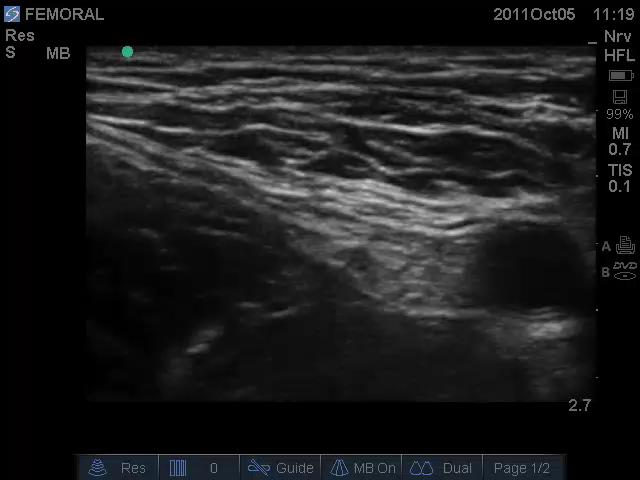
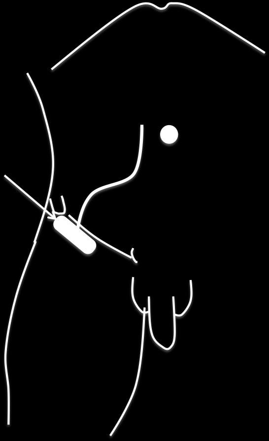
19 Ultrasound Guided
20 Probe Gel Iliopsoas
21
22 Saphenous nerve

23 Sub- sartorial

24 Not a single nerve but Nerve to Vastus Medialis and Saphenous nerve lie together and a muscular response can be elicited by using PNS Femoral artery

25 Nerve to Vastus Medialis Saphenous Nerve
26 LCFN

27 Lat cut nerve of the thigh
28 Internal oblique muscle Iliacus Fascia External oblique Aponeurosis ASIS Single pop through fascia lata, deposit LA above and below the fascia to catch the main and the cut branches 1 cm Iliacus Muscle Inguinal ligament Fascia Lata
29
30

31 Fascia Lata Fascia Iliaca ASIS
32 Obturator Nerve

33 AL OBTURATOR NERVE
34 OBTURATOR NERVE L 2 L 3 PELVIS OBTURATOR EXTERNUS L 4 OBTURATOR FORAMEN ADDUCTOR BREVIS THIGH ADDUCTOR PORTION OF ADDUCTOR MAGNUS HIP JOINT ADDUCTOR BREVIS ADDUCTOR LONGUS Ant Div Post Div KNEE JOINT TWIG TO PECTINIUS GRACILIS MEDIAL THIGH SKIN OVER ADDUCTORS
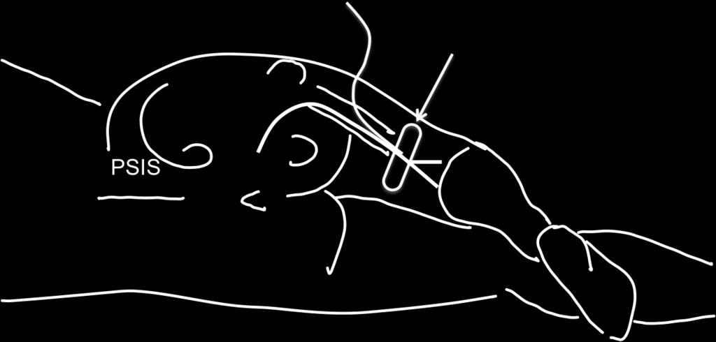
35 FEMORAL VESSELS OBTURATOR NERVE FEMORAL NERVE PT 2Cms 2Cms


36 AL Adductor Longus AB Adductor Brevis AM Adductor Magnus
37 3 in 1 Block!!!
38 Lat Cut Nerve of Thigh Psoas Major Single injection large volume misses out Obturator as it lies on the medial side ( sometimes within the substance) of the Psoas muscle Iliacus Femoral Nerve Iliopsoas Inguinal ligament Obturator Nerve
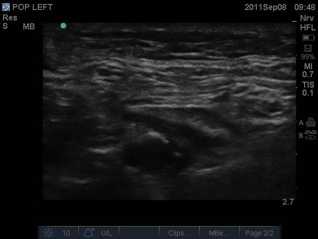
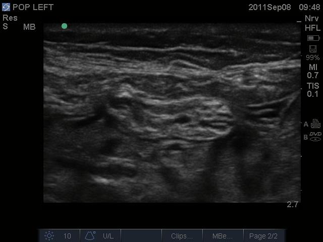
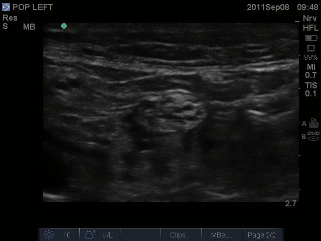
39 SciaDc Nerve
40 Sciatic nerve Popliteal fossa Tibial nerve Common peroneal Med & lat plantar nerves Sural nerve Superficial Peroneal Deep Peroneal

41 SciaDc nerve
42 Approaches to SciaDc Nerve in the Gluteal Region
43 GT IT PSIS Sacral Hiatus
44


45 Gluteus Maximus Sciatic Nerve
46 Popliteal Fossa
47 Artery Vein Nerve Medial Lateral Tibial Sural Common Peroneal
48 Tibial n Semimembranous Biceps Femoris Common Peroneal n Sural communicating branch Medial and lateral heads of Gastronemius Sural n

49
50

51

52 LA around the nerve

53 Distal spread of LA around the main baranches
54 ANKLE
55 Superficial Peroneal nerves Great Saphenous Vein and Saphenous Nerve Medial Malleolus Dorsal Paedis Artery Deep Peroneal Nerve Lateral Malleolus Post Tibial Artery Tibial Nerve Achilles Tendon Sural Nerve Small Saphenous Vein
56 Saphenous Sural Superficial Peroneal Deep Peroneal Medial plantar Lateral plantar

57 Post Tibial

58 Sural nerve at level of Lat Malleolus Short Saphenous vein


59 Deep Peroneal at the ankle level Ant Tibial Artery
60 Cases Anaesthesia and Blocks


61 82 yrs old padent listed for Dynamic Hip Screw ( DHS)
62

63 78 yrs old padent for hemi- arthroplasty
64

65 28yr old padent for ORIF of Tibia and Fibula
66
67 55 yrs old diabedc padent for amputadon of the big toe of Rt foot
68

69
Lower Limb Nerves. Clinical Anatomy
 Lower Limb Nerves Clinical Anatomy Lumbar Plexus Ventral rami L1 L4 Supplies: Abdominal wall External genitalia Anteromedial thigh Major nerves.. Lumbar Plexus Nerves relation to psoas m. : Obturator n.
The thigh. Prof. Oluwadiya KS
 The thigh Prof. Oluwadiya KS www.oluwadiya.com The Thigh: Boundaries The thigh is the region of the lower limb that is approximately between the hip and knee joints Anteriorly, it is separated from the
Lumbar Plexus. Ventral rami L1 L4 Supplies: Major nerves.. Abdominal wall External genitalia Anteromedial thigh
 Lower Limb Nerves Lectures Objectives Describe the structure and relationships of the plexuses of the lower limb. Describe the course, relationships and structures supplied for the major nerves of the
Muscles of the lower extremities. Dr. Nabil khouri MD, MSc, Ph.D
 Muscles of the lower extremities Dr. Nabil khouri MD, MSc, Ph.D Posterior leg Popliteal fossa Boundaries Biceps femoris (superior-lateral) Semitendinosis and semimembranosis (superior-medial) Gastrocnemius
DISSECTION SCHEDULE. Session I - Hip (Front) & Thigh (Superficial)
 & Thigh (Superficial)") DISSECTION SCHEDULE Session I - Hip (Front) & Thigh (Superficial) Surface anatomy Inguinal region Gluteal region Thigh Leg Foot bones Hip bone Femur Superficial fascia Great saphenous vein Superficial
DISSECTION SCHEDULE Session I - Hip (Front) & Thigh (Superficial) Surface anatomy Inguinal region Gluteal region Thigh Leg Foot bones Hip bone Femur Superficial fascia Great saphenous vein Superficial
Lower Limb Dr. Robin Paudel
 Lower Limb n What is a limb? n Skeleton n Joints n Pelvis or limb girdle n Hip/Hip Muscles n Lumber and sacral plexus getting spinal nerves out onto limb n Muscles anterior and posterior compartments n
rotation of the hip Flexion of the knee Iliac fossa of iliac Lesser trochanter Femoral nerve Flexion of the thigh at the hip shaft of tibia
 Anatomy of the lower limb Anterior & medial compartments of the thigh Dr. Hayder The fascia lata encloses the entire thigh like a sleeve/stocking. Three intramuscular fascial septa (lateral, medial, and
musculoskeletal system anatomy nerves of the lower limb 1 done by: dina sawadha & mohammad abukabeer
 musculoskeletal system anatomy nerves of the lower limb 1 done by: dina sawadha & mohammad abukabeer What is the importance of plexuses? plexuses provides us the advantage of a phenomenon called convergence
lower limb Anterior Compartment: lecture 3 The deep fascia ( fascia lata) divides the thigh into 3 compartments:
 divides the thigh into 3 compartments:") lower limb lecture 3 The deep fascia ( fascia lata) divides the thigh into 3 compartments: 1. Anterior Extensor compartment 2. Medial Adductor compartment 3. Posterior Flexor compartment Anterior Compartment:
lower limb lecture 3 The deep fascia ( fascia lata) divides the thigh into 3 compartments: 1. Anterior Extensor compartment 2. Medial Adductor compartment 3. Posterior Flexor compartment Anterior Compartment:
HUMAN BODY COURSE LOWER LIMB NERVES AND VESSELS
 HUMAN BODY COURSE LOWER LIMB NERVES AND VESSELS October 22, 2010 D. LOWER LIMB MUSCLES 2. Lower limb compartments ANTERIOR THIGH COMPARTMENT General lfunction: Hip flexion, knee extension, other motions
Anterior and Medial compartments of the thigh. Dr. Heba Kalbouneh Associate Professor of Anatomy and Histology
 Anterior and Medial compartments of the thigh Dr. Heba Kalbouneh Associate Professor of Anatomy and Histology Terms Related to Movements Movement Flexion Extension Abduction Adduction Medial (internal)
ANATYOMY OF The thigh
 ANATYOMY OF The thigh 1- Lateral cutaneous nerve of the thigh Ι) Skin of the thigh Anterior view 2- Femoral branch of the genitofemoral nerve 5- Intermediate cutaneous nerve of the thigh 1, 2 and 3 are
Muscles of the Hip 1. Tensor Fasciae Latae O: iliac crest I: lateral femoral condyle Action: abducts the thigh Nerve: gluteal nerve
 Muscles of the Hip 1. Tensor Fasciae Latae O: iliac crest I: lateral femoral condyle Action: abducts the thigh Nerve: gluteal nerve 2. Gluteus Maximus O: ilium I: femur Action: abduct the thigh Nerve:
The Muscular System. Chapter 10 Part D. PowerPoint Lecture Slides prepared by Karen Dunbar Kareiva Ivy Tech Community College
 Chapter 10 Part D The Muscular System Annie Leibovitz/Contact Press Images PowerPoint Lecture Slides prepared by Karen Dunbar Kareiva Ivy Tech Community College Table 10.14: Muscles Crossing the Hip and
The Hay is in the Barn
 Anatomy 1 Practical 1 Review Made by Forrest Allen (nerd) Edited by TJ Williamson (not nerd) The Hay is in the Barn 2019 Thunderbringers Too much to handle https://www.youtube.com/watch?v=glii-kaza d8
MUSCULOSKELETAL LOWER LIMB
 MUSCULOSKELETAL LOWER LIMB Spinal Cord Lumbar and Sacral Regions Spinal cord Dorsal root ganglion Conus medullaris Cauda equina Dorsal root ganglion of the fifth lumbar nerve End of subarachnoid space
ANATYOMY OF The thigh
 ANATYOMY OF The thigh 1- Lateral cutaneous nerve of the thigh Ι) Skin of the thigh Anterior view 2- Femoral branch of the genitofemoral nerve 5- Intermediate cutaneous nerve of the thigh 1, 2 and 3 are
Peripheral Nervous System: Lower Body
 Peripheral Nervous System: Lower Body MSTN121 - Neurophysiology Session 11 Department of Myotherapy Lumbar Plexus Iliohypogastric nerve (T12-L1) Motor: Transverse abdominis and internal obliques Sensory:
ANATYOMY OF The thigh
 ANATYOMY OF The thigh 1- Lateral cutaneous nerve of the thigh Ι) Skin of the thigh Anterior view 2- Femoral branch of the genitofemoral nerve 1, 2 and 3 are From the lumber plexus 5- Intermediate cutaneous
Lecture 08 THIGH MUSCLES ANTERIOR COMPARTMENT. Dr Farooq Khan Aurakzai. Dated:
 Lecture 08 THIGH MUSCLES ANTERIOR COMPARTMENT BY Dr Farooq Khan Aurakzai Dated: 11.02.2017 INTRODUCTION to the thigh Muscles. The musculature of the thigh can be split into three sections by intermuscular
musculoskeletal system anatomy nerves of the lower limb 2 done by: Dina sawadha & mohammad abukabeer
 musculoskeletal system anatomy nerves of the lower limb 2 done by: Dina sawadha & mohammad abukabeer #Sacral plexus : emerges from the ventral rami of the spinal segments L4 - S4 and provides motor and
The Hip (Iliofemoral) Joint. Presented by: Rob, Rachel, Alina and Lisa
 Joint. Presented by: Rob, Rachel, Alina and Lisa") The Hip (Iliofemoral) Joint Presented by: Rob, Rachel, Alina and Lisa Surface Anatomy: Posterior Surface Anatomy: Anterior Bones: Os Coxae Consists of 3 Portions: Ilium Ischium Pubis Bones: Pubis Portion
The Hip (Iliofemoral) Joint Presented by: Rob, Rachel, Alina and Lisa Surface Anatomy: Posterior Surface Anatomy: Anterior Bones: Os Coxae Consists of 3 Portions: Ilium Ischium Pubis Bones: Pubis Portion
Lower Extremity Ultrasound-Guided Regional Anesthesia. Stephanie Duffy, CRNA Regional Anesthesia Faculty Acute Pain Service NMCSD
 Lower Extremity Ultrasound-Guided Regional Anesthesia Stephanie Duffy, CRNA Regional Anesthesia Faculty Acute Pain Service NMCSD Objectives Review anatomy of lumbosacral plexus Lumbar plexus blocks Psoas
Baraa Ayed حسام أبو عوض. Ahmad Salman. 1 P a g e
 4 Baraa Ayed حسام أبو عوض Ahmad Salman 1 P a g e Today we are going to cover these concepts: Iliotibial tract Anterior compartment of the thigh and the hip Medial compartment of the thigh Femoral triangle
Identify the muscles associated with the medial compartment of the thigh. Identify the attachment points of the medial thigh muscles.
 L 8 A B O R A T O R Y Thigh MEDIAL THIGH Identify the muscles associated with the medial compartment of the thigh. Identify the attachment points of the medial thigh muscles. Identify the actions of these
The Lower Limb II. Anatomy RHS 241 Lecture 3 Dr. Einas Al-Eisa
 The Lower Limb II Anatomy RHS 241 Lecture 3 Dr. Einas Al-Eisa Tibia The larger & medial bone of the leg Functions: Attachment of muscles Transfer of weight from femur to skeleton of the foot Articulations
Lower limb summary. Anterior compartment of the thigh. Done By: Laith Qashou. Doctor_2016
 Lower limb summary Done By: Laith Qashou Doctor_2016 Anterior compartment of the thigh Sartorius Anterior superior iliac spine Upper medial surface of shaft of tibia 1. Flexes, abducts, laterally rotates
Femoral Artery. Its entrance to the thigh Position Midway between ASIS and pubic symphysis
 Lower Limb Vessels Lecture Objectives Describe the major arteries of the lower limb. Describe the deep and superficial veins of the lower limb. Describe the topographical relationships of the arteries
Human Anatomy Biology 351
 Human Anatomy Biology 351 Lower Limb Please place your name on the back of the last page of this exam. You must answer all questions on this exam. Because statistics demonstrate that, on average, between
Muscles of Lesson Five. Muscular Nomenclature and Kinesiology - Two. Muscles of Lesson Five, cont. Chapter 16
 Chapter 16 Muscular Nomenclature and Kinesiology - Two Lessons 5-6 Muscles of Lesson Five Iliopsoas (psoas major, iliacus) Hip outward rotators (piriformis, gemellus superior, gemellus inferior, obturator
Where should you palpate the pulse of different arteries in the lower limb?
 Where should you palpate the pulse of different arteries in the lower limb? The femoral artery In the femoral triangle, its pulse is easily felt just inferior to the inguinal ligament midway between the
Human Anatomy Biology 255
 Human Anatomy Biology 255 Exam #4 Please place your name and I.D. number on the back of the last page of this exam. You must answer all questions on this exam. Because statistics demonstrate that, on average,
Gluteal region DR. GITANJALI KHORWAL
 Gluteal region DR. GITANJALI KHORWAL Gluteal region The transitional area between the trunk and the lower extremity. The gluteal region includes the rounded, posterior buttocks and the laterally placed
Ali Yaghi. Omar Eyad. Ahmad Salman. 1 P a g e
 5 Ali Yaghi Omar Eyad Ahmad Salman 1 P a g e **There are two types of groin hernia; the femoral hernia and the inguinal hernia. But how can we differentiate between the inguinal hernia and the femoral
Mohammad Ashraf. Abdulrahman Al-Hanbali. Ahmad Salman. 1 P a g e
 - 7 Mohammad Ashraf Abdulrahman Al-Hanbali Ahmad Salman 1 P a g e Structures under the cover of Gluteus Maximus: 1-Bones: Ileum, Femur (Head, greater trochanter and gluteal tuberosity), Ischium (ischial
Anatomage Table Instructors Guide- Lower Limb
 The Lower Limb Anatomage Table Instructors Guide- Lower Limb Table of Contents Lower Limb 1- The Skeletal System...3 1: Hip Bone...3 2: Hip Joint and Femur...4 3: Patella and Knee Joint...7 4: Tibia, Fibula,
Organization of the Lower Limb Audrone Biknevicius, Ph.D. Dept. Biomedical Sciences, OU HCOM at Dublin Clinical Anatomy Immersion 2014
 Organization of the Lower Limb Audrone Biknevicius, Ph.D. Dept. Biomedical Sciences, OU HCOM at Dublin Clinical Anatomy Immersion 2014 www.thestudio1.co.za LIMB FUNCTION choco-locate.com blog.coolibar.com
Anatomy of the lower limb
 Anatomy of the lower limb 1. Bones of the lower limb Pelvis Hip bone/coxal bone Acetabulum o Acetabular margin o Acetabular fossa o Acetabular notch o Lunate surface Ischiopubic ramus Obturator foramen
Abdominal muscles. Subinguinal hiatus and ingiunal canal. Femoral and adductor canals. Neurovascular system of the lower limb. Sándor Katz M.D.,Ph.D.
 Abdominal muscles. Subinguinal hiatus and ingiunal canal. Femoral and adductor canals. Neurovascular system of the lower limb. Sándor Katz M.D.,Ph.D. External oblique muscle Origin: outer surface of the
LOWER LIMB. As we know the bony part of the body is divided into Axial and Appendicular (upper and lower Limbs)
") LOWER LIMB As we know the bony part of the body is divided into Axial and Appendicular (upper and lower Limbs) Bones of the Lower limb: 1-Pelvic Girdle: composed of: 1. Right hip bone : is formed by 3
LOWER LIMB As we know the bony part of the body is divided into Axial and Appendicular (upper and lower Limbs) Bones of the Lower limb: 1-Pelvic Girdle: composed of: 1. Right hip bone : is formed by 3
1-Muscles: 2-Blood supply: Branches of the profunda femoris artery. 3-Nerve supply: Sciatic nerve
 1-Muscles: B i c e p s f e m o r i s S e m i t e n d i n o s u s S e m i m e m b r a n o s u s a small part of the adductor magnus (h a m s t r i n g p a r t o r i s c h i a l p a r t ) 2-Blood supply:
Quillen College of Medicine
 Ea s t T e n n e s s e e St a t e Un i v e r s i t y Quillen College of Medicine Failing to prepare is preparing to fail. John Wooden, UCL A Dr. Tom Kwasigroch Associate Dean Director, Medical Human Gross
inerve Guide to Nerves 2009
 inerve Guide to Nerves 2009 A guide to self learning and self assessment Context: The following guide is intended to help interpret the sono-anatomy and follow a systematic stepwise approach to the practice
Lumbar and Sacral Plexuses. Dr. Heba Kalbouneh Associate Professor of Anatomy and Histology
 Lumbar and Sacral Plexuses Dr. Heba Kalbouneh Associate Professor of Anatomy and Histology Structure of Spinal Nerves: Somatic Pathways dorsal root CNS interneuron spinal nerve dorsal ramus somatic sensory
lesser trochanter of femur lesser trochanter of femur iliotibial tract (connective tissue) medial surface of proximal tibia
 medial surface of proximal tibia") LOWER LIMB MUSCLES OF THE APPENDICULAR SKELETON The muscles that act on the lower limb fall into three groups: those that move the thigh, those that move the lower leg, and those that move the ankle, foot,
LOWER LIMB MUSCLES OF THE APPENDICULAR SKELETON The muscles that act on the lower limb fall into three groups: those that move the thigh, those that move the lower leg, and those that move the ankle, foot,
Anatomy & Physiology. Muscles of the Lower Limbs.
 Anatomy & Physiology Muscles of the Lower Limbs http://www.ishapeup.com/musclecharts.html Muscles of the Lower Limbs Among the strongest muscles in the body. Because pelvic girdle is composed of heavy,
Topic 7: Hip and pelvis. Parts of the hip. Parts of the femur
 Topic 7: Hip and pelvis Parts of the hip Parts of the femur Classifying the hip joint Ball and socket Synovial Multiaxial Movements of the hip: Abduction/adduction Flexion/extension Medial/lateral rotation
Muscles of the Thigh. 6.1 Identify, describe the attachments of and deduce the actions of the muscles of the thigh: Anterior group
 Muscles of the Thigh 6.1 Identify, describe the attachments of and deduce the actions of the muscles of the thigh: Anterior group Sartorius: This is a long strap like muscle with flattened tendons at each
Muscles of the Gluteal Region
 Muscles of the Gluteal Region 1 Some of the most powerful in the body Extend the thigh during forceful extension Stabilize the iliotibial band and thoracolumbar fascia Related to shoulders and arms because
Lowe w r e r B ody Resistance Training
 Lower Body Resistance Training Tibialis Anterior Extensor Hallucis Longus Extensor Digitorum Longus Proneus Tertius AROM 25-40 degrees Extensor active-insufficiency Flexion & Eversion (Pronation) Flexion
THE LOWER LIMB NERVES VESSELS
 THE LOWER LIMB NERVES VESSELS LOWER LIMB: FEMORAL TRIANGLE FEMORAL TRIANGLE LOWER LIMB: FEMORAL TRIANGLE FEMORAL TRIANGLE is a triangular landmark useful in dissection and in understanding relationships
ANATOMY TEAM GLUTEAL REGION & BACK OF THIGH
 ANATOMY TEAM GLUTEAL REGION & BACK OF THIGH OBJECTIVES By the end of this lecture, the student should be able to identify and discuss: Contents of gluteal region: Groups of Glutei muscles and small muscles
The University Of Jordan Faculty Of Medicine THE LOWER LIMB. Dr.Ahmed Salman Assistant Prof. of Anatomy. The University Of Jordan
 The University Of Jordan Faculty Of Medicine THE LOWER LIMB Dr.Ahmed Salman Assistant Prof. of Anatomy. The University Of Jordan Gluteal Region Cutaneous nerve supply of (Gluteal region) 1. Lateral cutaneous
Year 2004 Paper one: Questions supplied by Megan
 QUESTION 47 A 58yo man is noted to have a right foot drop three days following a right total hip replacement. On examination there is weakness of right ankle dorsiflexion and toe extension (grade 4/5).
MUSCLES OF THE LOWER LIMBS
 MUSCLES OF THE LOWER LIMBS Naming, location and general function Dr. Nabil khouri ROLES THAT SHOULD NOT BE FORGOTTEN Most anterior compartment muscles of the hip and thigh Flexor of the femur at the hip
Adductor canal (Subsartorial) or Hunter s canal
 or Hunter s canal") Adductor canal (Subsartorial) or Hunter s canal John Hunter described the exposure and ligation of the femoral artery in this canal for aneurysm of the popliteal artery; this method has the advantage that
Adductor canal (Subsartorial) or Hunter s canal John Hunter described the exposure and ligation of the femoral artery in this canal for aneurysm of the popliteal artery; this method has the advantage that
Ultrasound Guided Lower Extremity Blocks
 Ultrasound Guided Lower Extremity Blocks CONTENTS: 1. Femoral Nerve Block 2. Popliteal Nerve Block Updated December 2017 1 1. Femoral Nerve Block Indications Surgery involving the knee, anterior thigh,
Lectures of Human Anatomy
 Lectures of Human Anatomy Lower Limb Gluteal Region and Hip Joint By DR. ABDEL-MONEM AWAD HEGAZY M.B. with honor 1983, Dipl."Gynecology and Obstetrics "1989, Master "Anatomy and Embryology" 1994, M.D.
Acland's DVD Atlas of Human Anatomy. Transcript for Volume Robert D Acland
 Acland's DVD Atlas of Human Anatomy Transcript for Volume 2 2007 Robert D Acland This free downloadable pdf file is to be used for individual study only. It is not to be reproduced in any form without
Muscles to know. Lab 21. Muscles of the Pelvis and Lower Limbs. Muscles that Position the Lower Limbs. Generally. Muscles that Move the Thigh
 Muscles to know Lab 21 Muscles of the Pelvis, Leg and Foot psoas major iliacus gluteus maximus gluteus medius sartorius quadriceps femoris (4) gracilus adductor longus biceps femoris semitendinosis semimembranosus
Muscle Anatomy Review Chart
 Muscle Anatomy Review Chart BACK Superficial (5) Trapezius Transverse cervical a. Latissimus dorsi Thoracodorsal a. Rhomboideus major Dorsal scapular a. Rhomboideus minor Levator scapulae Intermediate
Lesson 24. A & P Hip
 Lesson 24 A & P Hip 1 Aims of the Session This session will allow candidates to have an understanding of the bony prominences and soft tissues of the hip 2 Learning Outcomes By the end of the lesson the
Femoral Triangle and Adductor Canal. Dr. Heba Kalbouneh Associate Professor of Anatomy and Histology
 Femoral Triangle and Adductor Canal Dr. Heba Kalbouneh Associate Professor of Anatomy and Histology Femoral Triangle and Adductor Canal Femoral triangle Is a triangular depressed area located in the upper
Lecture 09. Popliteal Fossa. BY Dr Farooq Khan Aurakzai
 Lecture 09 Popliteal Fossa BY Dr Farooq Khan Aurakzai Dated: 14.02.2018 What is popliteus? Introduction Anything relating to, or near the part of the leg behind the knee. From New Latin popliteus the muscle
Regional Blocks a practical guide:
 Regional Blocks a practical guide: Author Fleur Roberts email: fleur_roberts@hotmail.com Please use this guide in conjunction with the previous tutorial (16/01/06) which describes the rules for performing
Contents of the Posterior Fascial Compartment of the Thigh
 Contents of the Posterior Fascial Compartment of the Thigh 1-Muscles: B i c e p s f e m o r i s S e m i t e n d i n o s u s S e m i m e m b r a n o s u s a small part of the adductor magnus (h a m s t
VENOUS DRAINAGE OF THE LOWER LIMB
 Anatomy of the lower limb Superficial veins & nerve injuries Dr. Hayder VENOUS DRAINAGE OF THE LOWER LIMB The venous drainage of the lower limb is of huge clinical & surgical importance. Since the venous
THIEME. Anterior and Medial Compartments of the Thigh
 CHAPTER Anterior and Medial Compartments of the Thigh Learning Objectives 2 At the end of the dissection, you should be able to identify the following: Cutaneous nerves innervating the skin of the anterior
Leg. Dr. Heba Kalbouneh Associate Professor of Anatomy and Histology
 Leg Dr. Heba Kalbouneh Associate Professor of Anatomy and Histology Skin of the Leg Cutaneous Nerves Medially: The saphenous nerve, a branch of the femoral nerve supplies the skin on the medial surface
5.1 Identify, describe the attachments of and deduce the actions of the muscles of the thigh:
 5.1 Identify, describe the attachments of and deduce the actions of the muscles of the thigh: Anterior group Proximal attachment Distal attachment Sartorius ASIS» Upper part of shaft tibia (middle surface)»
Bony Anatomy. Femur. Femoral Head Femoral Neck Greater Trochanter Lesser Trochanter Intertrochanteric Crest Intertrochanteric Line Gluteal Tuberosity
 Hip Anatomy Bony Anatomy Femur Femoral Head Femoral Neck Greater Trochanter Lesser Trochanter Intertrochanteric Crest Intertrochanteric Line Gluteal Tuberosity Bony Anatomy Pelvic Girdle Acetabulum 3 bones
Anatomy MCQs Week 13
 Anatomy MCQs Week 13 1. Posterior to the medial malleolus of the ankle: The neurovascular bundle lies between Tibialis Posterior and Flexor Digitorum Longus The tendon of Tibialis Posterior inserts into
Organization of the Lower Limb
 Organization of the Lower Limb Limb Development Lower limb develops in an aterolateral position at the level of the L2 to S3 trunk segments Great toe positioned cephalic direction with the soles of the
Human Anatomy Biology 351
 Human Anatomy Biology 351 Lower Limb Please place your name on the back of the last page of this exam. You must answer all questions on this exam. Because statistics demonstrate that, on average, between
Skeleton. The left forearm is in the position of supination, the right in pronation.
 S ystemic review A Skeleton A from the front B B from behind The left forearm is in the position of supination, the right in pronation. Skull Mandible Hyoid bone Cervical vertebrae Clavicle Sternum Costal
USRA OF THE LOWER EXTREMITY
 USRA OF THE LOWER EXTREMITY Christian R. Falyar, CRNA, DNAP Department of Nurse Anesthesia Virginia Commonwealth University Disclosure Statement of Financial Interest I, Christian Falyar, DO NOT have a
CHAPTER 8: THE BIOMECHANICS OF THE HUMAN LOWER EXTREMITY
 CHAPTER 8: THE BIOMECHANICS OF THE HUMAN LOWER EXTREMITY _ 1. The hip joint is the articulation between the and the. A. femur, acetabulum B. femur, spine C. femur, tibia _ 2. Which of the following is
Organization of the Lower Limb
 Organization of the Lower Limb Most illustrations from: Thieme Atlas of Anatomy: Musculoskeletal System. M Schuenke, et al, 2006. Anatomy: A Regional Atlas of the Human Body. Carmine Clemente, 4th edition.
Anatomy images for MSS practical exam- 2019
 Anatomy images for MSS practical exam- 2019 Ilium Ischium Pubis Acetabulaum Iliac crest Iliac tubercle ASIS (muscle and ligament attached) AIIS (muscle attached) PSIS PIIS Ischial spine Ischial tuberosity
The Hip Joint. Shenequia Howard David Rivera
 The Hip Joint Shenequia Howard David Rivera Topics Of Discussion Movement Bony Anatomy Ligamentous Anatomy Muscular Anatomy Origin/Insertion/Action/Innervation Common Injuries MOVEMENT Flexion Extension
Located Deep to Flexor Retinaculum on medial aspect of ankle. Posterior to Posterior Tibial Artery. Tom, Dick, and Very Nervous Harry
 ANKLE BLOCK ANESTHESIA GREGORY CLARK D.P.M. HEAD, SECTION OF PODIATRY SCRIPPS CLINIC LA JOLLA, CALIFORNIA A METHOD BY WHICH ONE MAY PROVIDE AN ANESTHETIC BLOCK TO THE FOOT OR ANKLE WITH A MINIMUM OF PATIENT
To describe he knee joint, ligaments, structure & To list the main features of other lower limb joints
 To describe he knee joint, ligaments, structure & neurovascular supply To demonstrate the ankle joint anatomy To list the main features of other lower limb joints To list main groups of lymph nodes in
Posterior compartment of the thigh. Dr. Heba Kalbouneh Associate Professor of Anatomy and Histology
 Posterior compartment of the thigh Dr. Heba Kalbouneh Associate Professor of Anatomy and Histology Posterior compartment of the thigh 1-Muscles: Biceps femoris Semitendinosus Semimembranosus Adductor magnus
Muscle fiber (cell) Blood vessel. Perimysium. Epimysium. Fascicle (wrapped by perimysium) Endomysium (between fibers) Tendon. Bone
 Blood vessel. Perimysium. Epimysium. Fascicle (wrapped by perimysium) Endomysium (between fibers) Tendon. Bone") Figure 6.1 Connective tissue wrappings of skeletal muscle. Blood vessel Muscle fiber (cell) Perimysium Epimysium Fascicle (wrapped by perimysium) Tendon Endomysium (between fibers) Bone Figure 6.15 Superficial
Figure 6.1 Connective tissue wrappings of skeletal muscle. Blood vessel Muscle fiber (cell) Perimysium Epimysium Fascicle (wrapped by perimysium) Tendon Endomysium (between fibers) Bone Figure 6.15 Superficial
BOGOMOLETS NATIONAL MEDICAL UNIVERSITY. Department of Human Anatomy GUIDELINES. The theme of the lesson The vessels of the lower limb.
 BOGOMOLETS NATIONAL MEDICAL UNIVERSITY Department of Human Anatomy GUIDELINES Academic discipline HUMAN ANATOMY Module 2 The theme of the lesson The vessels of the lower limb. Course Faculties І Medical
Figure 1 - Hip and Pelvis
 Hip Figure 1 - Hip and Pelvis The terms hip and pelvis are frequently used interchangeably, but strictly speaking, the pelvis is a girdle of bones and the hip is a joint. The pelvis consists of The sacrum
Misc Anatomy. Upper Limb! 2. Lower Limb! 5. Venous Drainage! Head & neck! 8
 Misc Anatomy Upper Limb! 2 Arteries!... 2 Veins!... 2 Spaces!... 4 Lower Limb! 5 Arteries!... 5 Venous Drainage!... 6 Spaces!... 7 Head & neck! 8 Artery!... 8 Ultrasound View for IJ CVL!... 8 Arteries
In-Depth Foundations: Anatomy Terms to Know
 Be familiar with / able to identify and define all the following parts. The Spine Cranium Vertebrae Cervical, Thoracic, Lumbar Sacrum Coccyx Bones of Upper Body Cranium Mastoid process; Occipital condyle,
Hip joint and pelvic girdle. Lower Extremity. Pelvic Girdle 6/5/2017
 Hip joint and pelvic girdle Lower Extremity The relationship between the pelvic girdle and hip is similar to that between the shoulder girdle and shoulder joint. The lower limbs are attached to the axial
The Leg. Prof. Oluwadiya KS
 The Leg Prof. Oluwadiya KS www.oluwadiya.sitesled.com Compartments of the leg 4 Four Compartments: 1. Anterior compartment Deep fibular nerve Dorsiflexes the foot and toes 2. Lateral Compartment Superficial
Due in Lab weeks because of Thanksgiving Prelab #10. Homework #8. Both sides! Both sides!
 Lab 8 MUSCLES Due in Lab 10 2 weeks because of Thanksgiving Prelab #10 Both sides! Homework #8 Both sides! Refer to Muscles 22-23 Naming of muscles Origin Site of muscle attachment that doesn t move during
Gross Anatomy Coloring Book Series. Lower Extremity Arteries
 Gross Anatomy Coloring Book Series Lower Extremity Arteries 1 Femoral Artery and Associated Branches For the life of the flesh is in the blood. Leviticus 17:11 Femoral Artery and Associated Branches After
musculoskeletal system <lower limb vessle> <1> done by:renad abu ruman &rama alawamleh
 musculoskeletal system done by:renad abu ruman &rama alawamleh The entrance to the anterior compartment of the leg is through lingual lig. & superior ramus, & to the posterior compartment
The psoas minor is medial to the psoas major. The iliacus is a fan-shaped muscle that when contracted helps bring the swinging leg forward in walking
 1 p.177 2 3 The psoas minor is medial to the psoas major. The iliacus is a fan-shaped muscle that when contracted helps bring the swinging leg forward in walking and running. The iliopsoas and adductor
TABLE OF MUSCLES OF LOWER EXTREMITY 2018zillmusom ANTERIOR THIGH
 TABLE OF MUSCLES OF LOWER EXTREMITY 2018zillmusom ANTERIOR THIGH MUSCLE ORIGIN INSERTION ACTION NERVE Iliopsoas Ilium, vertebra Femur Flex hip joint Femoral nerve (T12-L5) Pectineus Pubis Femur Flex hip
Scapula Spine Lateral edge of clavicle. Medial border Scapula. Medial border of Scapula, between superior angle and root of spine. Scapula.
 Muscle attachments and actions answer sheet Muscle Origins insertions Movements Joints crossed Trapezius Base of skull Spinous process of C7 Thoracic Spine Lateral edge of clavicle Elevation Retraction
TRAINING TOOLS THE MOST ADVANCED. ULTRASOUND
 THE MOST ADVANCED ULTRASOUND TRAINING TOOLS Introducing NYSORA ULTRASOUND SIMULATORS Developed by opinion leaders and educators, NYSORA ULTRASOUND SIMULATORS are the most anatomically accurate, tissue-like
Ultrasound-guided Sciatic Nerve Blocks: Higher and Popliteal Approaches
 10.5005/jp-journals-10027-1026 K Kondov, S Fransis REVIEW ARTICLE Ultrasound-guided Sciatic Nerve Blocks: Higher and Popliteal Approaches K Kondov, S Fransis ABSTRACT Background and objective: In modern
BONES JOINTS MUSCLES OF THE LOWER LIMB
 BONES JOINTS MUSCLES OF THE LOWER LIMB LOWER LIMB: BONES LOWER LIMB GLUTEAL REGION consists of 6 major segments: FEMORAL REGION (THIGH) KNEE REGION LEG REGION TALOCRURAL REGION (ANKLE) FOOT REGION LOWER