We will review how to:
|
|
|
- Hugo Leonard
- 5 years ago
- Views:
Transcription
1 Lesson 2
2 We will review how to: Define the key terms of this chapter and state the meanings of the word origins of this chapter. Describe the relationship between cranial suture joints and childbirth. List the major muscles of mastication and describe their role in mastication. Explain the possible relationship between TMJ dysfunction and the muscular system. Describe the structure and function of the spine. Define the curves of the spine and describe their development. 2
3 1. Suture joints 2. Temporomandibular joints (TMJs) 3. Atlanto-occipital and atlantoaxial joints 4. Cervical spinal joints 5. Thoracic spinal joints a. Rib joints 6. Lumbar spinal joints 3

4 Structure Classification: Fibrous joint Suture joint Function Classification: Synarthrotic Major Motions Allowed: Nonaxial 4

5 Structure Classification: Synovial joint Modified hinge Function Classification: Diarthrotic Uniaxial Major Motions Allowed: Elevation and depression Protraction and retraction Left and right lateral deviation 5


6 1 2 depression elevation protraction retraction 3 Left lateral deviation Right lateral deviation 6


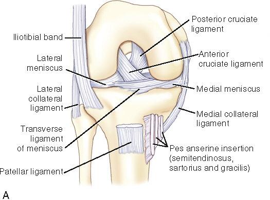
7 Major Ligaments of the TMJ: Fibrous joint capsule Temporomandibular ligament Stylomandibular ligament Sphenomandibular ligament 7


8 Major Muscles of the TMJ: Lateral pterygoid Medial pterygoid Temporalis Masseter Causes of TMJ Dysfunction: Tightness/imbalance of muscles that cross the TMJ Forward-head posture 8
9 Elements of the Spine: Cervical spine Thoracic spine Lumbar spine Sacrococcygeal spine Shape of the Adult Spine Primary spinal curves Thoracic curve Sacrococcygeal curve Secondary spinal curves Cervical curve Lumbar curve 9

10 Development of the Spinal Curves: - Born with one kyphotic curve - Develops a cervical lordosis - Develops a lumbar lordosis 10
11 Functions of the Spine: Provides structural support Allows movement Protects the spinal cord Provides shock absorption 11
12 1. Suture joints 2. Temporomandibular joints (TMJs) 3. Atlanto-occipital and atlantoaxial joints 4. Cervical spinal joints 5. Thoracic spinal joints Rib joints 6. Lumbar spinal joints 12
 Vertebral facet joints (facet joints)")
13 Segmental Structure: One median joint Two lateral joints Types of Spinal Joints: Intervertebral disc joints (disc joints) Vertebral facet joints (facet joints) 13

14 Structure Classification: Cartilaginous joint Symphysis Function Classification: Amphiarthrotic Functions of a Disc Joint: Determines amount of movement Absorbs shock Bears the weight of the body Maintains opening for spinal nerves 14


15 B Structure Classification: Synovial joint Plane Function Classification: Diarthrotic Function of a Facet Joint: Guides movement Motion Freely Allowed by the Facet Joints: Cervical facets - Right and left rotation in transverse plane - Right and left lateral flexion in frontal plane 15


16 B Motion Freely Allowed by the Facet Joints Thoracic facets Right and left lateral flexion in frontal plane C Motion Freely Allowed by the Facet Joints Lumbar facets Flexion and extension in sagittal plane 16


17 1. Flexion and Extension: Sagittal plane Mediolateral axis Right and Left Lateral Flexion: Frontal plane Anteroposterior axis Right and Left Rotation: Transverse plane Vertical axis 17

18 Gliding Translational Movements: Right-side and left-side translation Anterior and posterior translation Superior and inferior translation 18
19 19
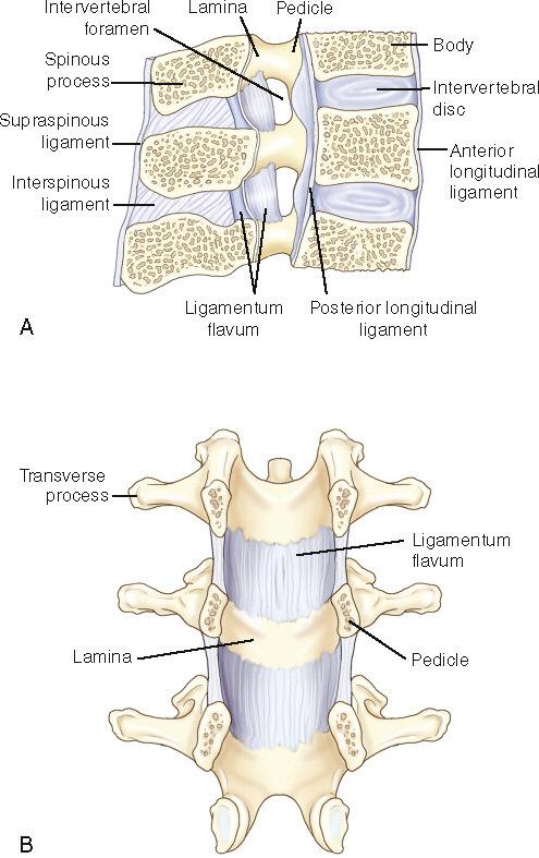
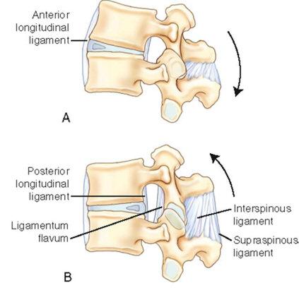
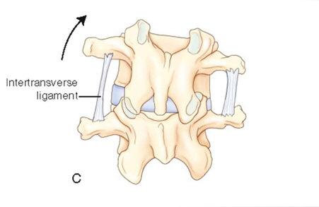
20 Major Ligaments of the Spinal Joints: Fibrous capsules of the facet joints Annulus fibrosus of the disc joints Anterior longitudinal ligament Posterior longitudinal ligament Ligamentum flava Interspinous ligaments Supraspinous ligament Intertransverse ligaments Nuchal ligament 20
21 21
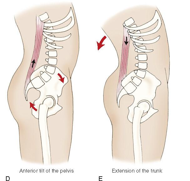
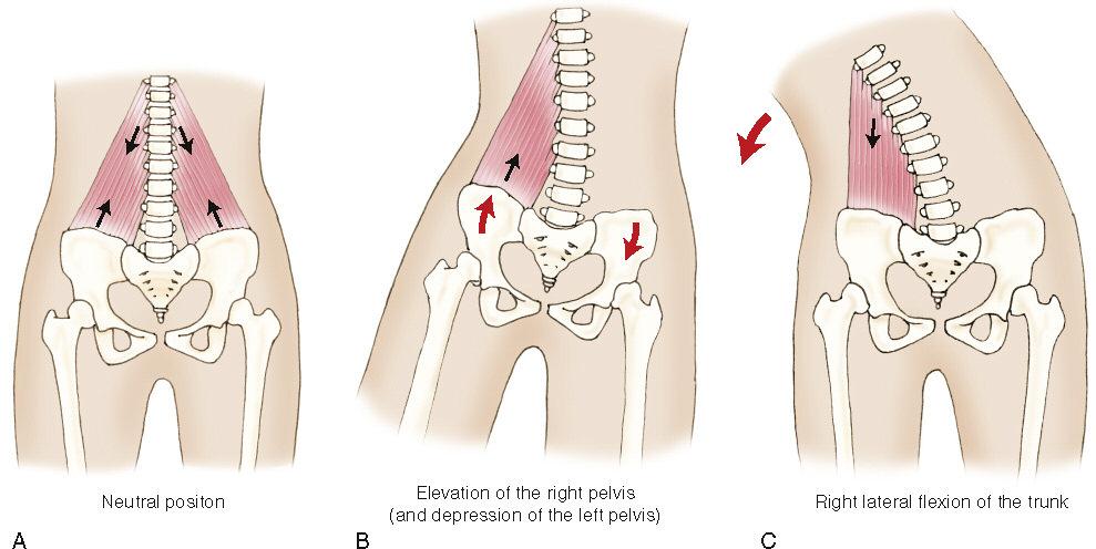
22 Major Muscles of the Spinal Joints: Spinal extensors What are some examples of muscles Spinal flexors that belong to these groups of muscles? Lateral flexors The erector spinae group, the Rotators transversospinalis group, and other muscles of the posterior neck and trunk are examples of spinal extensor muscles. Muscles of the anterior abdominal wall and muscles in the anterior neck are examples of spinal flexors. Most flexors and extensors are also lateral flexors. All lateral flexors are ipsilateral lateral flexors. Prominent rotators of the trunk include the external and internal abdominal obliques and the transversospinalis group muscles. 22

23 Structure Classification: Synovial joint Condyloid Function Classification: Diarthrotic Triaxial Major Motions Allowed: Flexion and extension Right and left lateral flexion Right and left lateral rotation 23


24 Flexion/extension Right lateral flexion and left lateral flexion Right rotation and left rotation 24
 is a cervical joint that is located between the atlas (C1) and the axis (C2).")
25 This bump is the odontoid process, often called the dens. The atlantoaxial joint (AAJ) is a cervical joint that is located between the atlas (C1) and the axis (C2). The AAJ allows the atlas to move on the axis. 25
26 Structure Classification: Synovial joints Atlanto-odontoid joint: Pivot joint Lateral facet joints: Plane joints Function Classification: Diarthrotic Biaxial Major Motions Allowed: Right and left rotation Flexion and extension Right and left lateral flexion Major Muscles of the Occipito- Atlantoaxial Region: Suboccipital group Rectus capitis anterior Rectus capitis lateralis 26

27 Special Vertebrae of the Cervical Spine: C1 atlas C2 axis C7 vertebral prominens 27
28 Special Characteristics of the Cervical Vertebrae: Transverse foramina Bifid spinous processes Bifid transverse processes Uncinate Processes Uncovertebral joint 28


29 Functions of the Cervical Spine: Bears the weight of the head Allows movement in all three planes Major Motions Allowed: Flexion and extension Right and left lateral flexion Right and left rotation Gliding translational movements in all three directions 29
30 List the joints at which rib motion occurs; explain how the movement of a bucket handle is used to describe how rib motion occurs. Describe the roles of the muscles of respiration. Explain the mechanism of thoracic breathing versus abdominal breathing. Describe the general structure and function of the cervical spine, thoracic spine, and lumbar spine. Describe the structure and function of the thoracolumbar fascia and abdominal aponeurosis. 30
31 Suture joints Temporomandibular joints (TMJs) Atlanto-occipital and atlantoaxial joints Cervical spinal joints Thoracic spinal joints Rib joints Lumbar spinal joints 31

32 Costospinal Joints of the Thoracic Spine: Costospinal articulations Costovertebral joint Costotransverse joint Structure classification Synovial joints 32
33 Costospinal Joints of the Thoracic Spine: Function classification Nonaxial Functions Stabilize ribs Allow ribs to move relative to the spine Spinal Joints of the Thoracic Spine: Major motions allowed Flexion and extension Right and left lateral flexion Right and left rotation Gliding translational movements 33
34 Costospinal Joints Sternocostal Joints Intrasternal Joints 34

35 Types of Costospinal Joints: Costovertebral joint Costotransverse joint 35
36 Structure of the Thoracic Spine: Two costal hemifacets per vertebra Intervertebral disc Ligaments of a Costovertebral Joint: Fibrous joint capsule Radiate ligament Structure of the Thoracic Spine: One full costal facet per vertebra Ligaments of a Costotransverse Joint: Fibrous joint capsule Costotransverse ligament Lateral costotransverse ligament Superior costotransverse ligament 36

37 Types of Ribs: True ribs False ribs Floating ribs Structure Classification: Cartilaginous joint Synchondrosis Function Classification: Amphiarthrotic Gliding 37

38 Types of Intrasternal Joints: Manubriosternal joint Sternoxiphoid joint Ligaments of the Intrasternal Joints: Manubriosternal ligament Sternoxiphoid ligament Both of the intrasternal joints are fibrocartilaginous amphiarthrotic joints. 38

39 Inspiration and Expiration Roles of Muscles of Respiration: Inspiration Elevate the ribs Expand the thoracic cavity downward Expiration Depress the ribs Expand the abdominal cavity upward 39
40 Abdominal Breathing: Contraction of the diaphragm Expanding thoracic cavity downward Thoracic Breathing: Further contraction of the diaphragm Elevating the lower ribs Roles of Muscles of Respiration: Inspiration Elevate the ribs Expand the thoracic cavity downward Expiration Depress the ribs Expand the abdominal cavity upward 40

41 Functions of the Lumbar Spine: Bears the weight of the body Allows movement Major Motions Allowed: Flexion and extension Right and left lateral flexion Right and left rotation Gliding translational movements in all three directions 41

42 Special Characteristics: a. Lumbosacral joint Allows the pelvis to move relative to the trunk b. Sacral base angle Determines the curvature of the spine 42


43 Major Motions Allowed: Flexion and extension Right and left lateral flexion Right and left rotation Flexion/extension Right/left lateral flexion Right/left rotation 43

44 Structure: Layer of fascia located in thoracic and lumbar regions Located posteriorly in the trunk Divided into three layers Functions: Provides attachment sites for muscles Adds to the stability of the trunk 44

45 Abdominal Aponeurosis Structure: Layers of fibrous connective tissue located in the abdominal region Located anteriorly in the trunk Left and right aponeuroses Functions: Provides attachment sites for muscles Adds to the stability of the trunk 45
46 Define the key terms of this chapter and state the meanings of the word origins of this chapter. Describe the structure of the pelvis and explain the difference between intrapelvic motion and motion of the pelvis relative to an adjacent body part. Describe the sacral movements of nutation and counternutation. Describe and compare movements of the pelvis at the lumbosacral and hip joints. Explain the reverse action relationships between pelvic movements and movements of the trunk and thighs. 46
47 ~Symphysis pubis joint ~Sacroiliac joints ~Lumbosacral joint ~Hip joints ~Knee joint complex ~Tibiofibular joints ~Talocrural (ankle) joint ~Tarsal joints ~Tarsometatarsal joints ~Intermetatarsal joints ~Metatarsophalangeal (MTP) joints ~Interphalangeal (IP) joints 47

48 Joints Located within the Pelvis: Symphysis pubis joint Sacroiliac joints Pelvic Motion: Intrapelvic motion Motion of pelvis as a unit relative to an adjacent body part 48

49 Symphysis Pubis Joint: Structure: Cartilaginous joint Symphysis joint Function: Amphiarthrotic Major motions: Nonaxial gliding Major ligaments: Arcuate pubic ligament Sacroiliac Joints: Structure: Mixed synovial/fibrous joint Plane joint Function: Mixed diarthrotic/amphiarthrotic Major motions allowed: Nonaxial gliding Nutation and counternutation 49

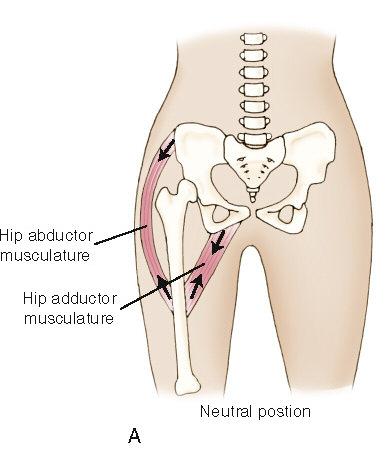
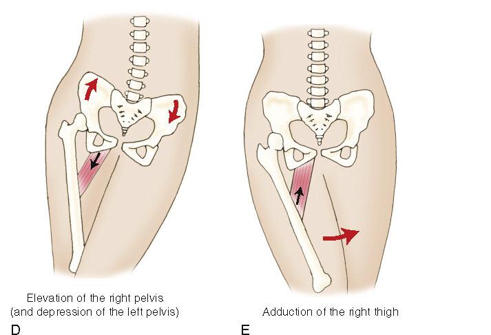
50 Major ligaments of the SI joint: Sacroiliac ligaments Sacrotuberous ligament Sacrospinous ligament Iliolumbar ligament 50 Intrapelvic Motion


51 Posterior/anterior tilt Right rotation pelvis/left rotation pelvis Elevation right pelvis Elevation left pelvis 51


52 Posterior tilt anterior tilt Depression right pelvis depression left pelvis Right rotation pelvis left rotation pelvis 52

53 When the pelvis moves at both the lumbosacral joint and the hip joints, it changes in position relative to both the spine and the femurs. When the pelvis approaches its maximum range of movement at the lumbosacral joint, motion also occurs at the lumbar spinal joints, changing the lumbar spinal curve. 53


54 Sagittal plane movements Frontal plane movements Transverse plane movements 54
55 55
 and right-sided contralateral rotators of the trunk (the right transversospinalis group and right external abdominal")
56 Muscles involved include both left-sided ipsilateral rotators of the trunk (the erector spinae group and left internal abdominal oblique) and right-sided contralateral rotators of the trunk (the right transversospinalis group and right external abdominal oblique). 56


57 Sagittal plane movements Frontal plane movements Transverse plane movements 57

58 58


59 Pelvic/thigh movement in the transverse plane results from the action of posterior and anterior groups of musculature. The posterior group is often called the lateral rotators of the hip joint. The anterior group is often called the medial rotators of the hip joint. 59

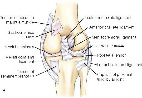
60 Sacral base angle Lumbopelvic rhythm Righting reflex 60
61 Explain the relationship between pelvic posture (and, specifically, the sacral base angle) and spinal posture. Discuss the meaning of open-chain and closed-chain activities and give examples of each. Explain the concepts of the femoral angle of inclination and femoral torsion angle, and explain the possible consequences of these femoral angulations. Describe and give an example of the concept of femoropelvic rhythm. 61
62 Symphysis pubis joint Sacroiliac joints Lumbosacral joint Hip joints Knee joint complex Tibiofibular joints Talocrural (ankle) joint Tarsal joints Tarsometatarsal joints Intermetatarsal joints Metatarsophalangeal (MTP) joints Interphalangeal (IP) joints 62

63 Structure Classification: Synovial joint Ball-and-socket Function Classification: Diarthrotic Triaxial Major Motions Allowed: Flexion and extension of thigh Abduction and adduction of thigh Medial rotation and lateral rotation of thigh Reverse Actions: Movement of pelvis 63

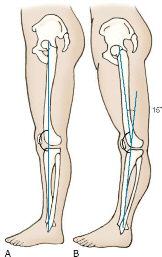
.")

64 Open-chain activity Distal bone of joint free to move Closed-chain activity Distal bone of joint fixed Chain activities involve linked kinematic elements (such as bones). An open chain allows movement of the distal element. A closed chain does not, requiring that the proximal element move instead. 64
65 Major Ligaments of the Hip Joint: Fibrous joint capsule Iliofemoral ligament Pubofemoral ligament Ischiofemoral ligament Ligamentum teres Closed-Packed Position: Full extension Major Muscles of the Hip Joint: Anterior muscles Posterior muscles Medial muscles Lateral muscles 65

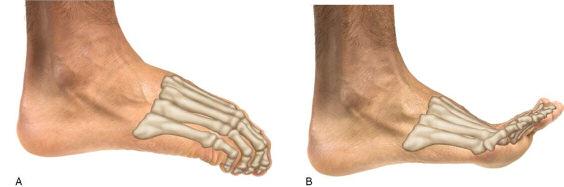
66 Femoral Angles of Inclination: The head, neck and shaft that compose the femur do not lie in a straight line. 66

67 There tends to be a rhythm to how the femur of the thigh and the pelvis move. When the actions of the thigh and the pelvis are coupled to allow a greater elevation of the foot in the air, this coordination of movement is known as femoropelvic rhythm. Femoropelvic motion couples right thigh extension at the hip joint with an anterior tilt of the pelvis at the left (contralateral) hip joint. 67
68 Symphysis pubis joint Sacroiliac joints Lumbosacral joint Hip joints Knee joint complex Tibiofibular joints Talocrural (ankle) joint Tarsal joints Tarsometatarsal joints Intermetatarsal joints Metatarsophalangeal (MTP) joints Interphalangeal (IP) joints 68

69 Tibiofemoral joint Patellofemoral joint Structure Classification: Synovial joint Modified hinge joint Function Classification: Diarthrotic Biaxial Major Motions Allowed: Flexion and extension of leg Medial rotation and lateral rotation of leg Reverse Actions: Flexion and extension of thigh Lateral rotation and medial rotation of thigh 69


70 Major Ligaments of the Knee Joint: Fibrous joint capsule Medial collateral ligament Lateral collateral ligament Anterior cruciate ligament Posterior cruciate ligament 70
71 71

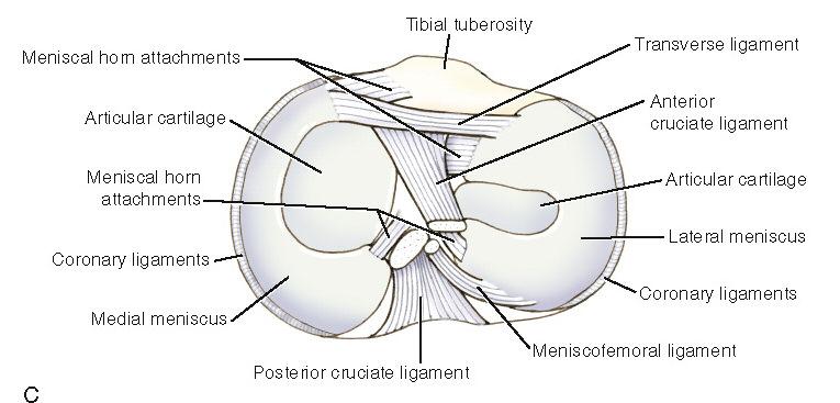
72 Closed-Packed Position: Full extension Major Muscles of the Knee Joint: Anterior muscles Posterior muscles Medial muscles Lateral muscles Menisci Medial meniscus Lateral meniscus Major bursae Sub patellar Screw-home mechanism The patellofemoral joint is formed by the articulation between the patella and the femur and located within the capsule of the knee joint along with the tibiofemoral joint. However, the tibia is not directly involved in the movement of the patella. The intercondylar groove shown in the figure indicates the path along which the patella tracks the femur during its nonaxial gliding movement. P a t e ll o f e m o r a l j o i n t 72
73 Structure of the Patella: Medial facet Lateral facet Articular cartilage Major Motions Allowed: Superior and inferior gliding (nonaxial) movements Closed-Packed Position: Flexion (of the knee joint) Functions of the Patella: Acts as an anatomic pulley Reduces friction Protects the femoral condyles 73
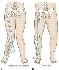
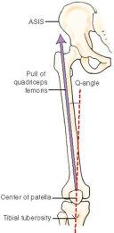
74 Angulations of the Knee Joint Knock-Knees Bowleg Q-Angle: 74

75 Tibiofibular Joints: Proximal tibiofibular joint Middle tibiofibular joint Distal tibiofibular joint Interosseus membrane Two purposes Tibial torsion Lateral twist of shaft of tibia The interosseus membrane that unites the shafts of the tibia and fibula allows the joined bones to grip the talus of the ankle joint between them, making the middle tibiofibular joint vital to the stability of the ankle joint. It also transfers the force of muscles pulling on the fibula to the tibia, moving the leg at the knee joint. 75
76 Symphysis pubis joint Sacroiliac joints Lumbosacral joint Hip joints Knee joint complex Tibiofibular joints 76
 Pes planus (decreased arch) The joints of the lower extremity include the talocrural (ankle) joint, the tarsal joints, the tarsometatarsal joints, the intermetatarsal joints,")
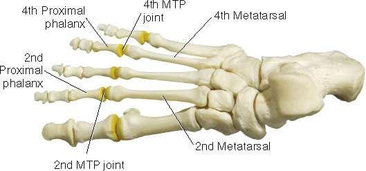
77 Functions of the Foot: Provides stability Bears weight of body Absorbs shock from motion Propels body through space Provides flexibility Adapts to uneven ground Evaluating the Arches of the Foot: Pes cavus (excessive arch) Pes planus (decreased arch) The joints of the lower extremity include the talocrural (ankle) joint, the tarsal joints, the tarsometatarsal joints, the intermetatarsal joints, the metatarsophalangeal joints, and the interphalangeal joints. How are the names for the metatarsophalangeal and interphalangeal joints often abbreviated? The metatarsophalangeal joints are often referred to as the MTP joints, and the interphalangeal joints are often referred to as the IP joints. 77
78 Plantar Fascia: Superficial layer Deep layer The foot has a thick layer of dense fibrous tissue on the plantar side known as the plantar fascia, whose purpose is to maintain and stabilize the longitudinal arches of the foot. There are two layers of plantar fascia: The superficial layer, located in the dermis of the skin of the foot, and the deep layer, which attaches to the calcaneal tuberosity posteriorly and the plantar plates of the MTP joints and adjacent flexor tendons of the toes anteriorly. Plantar fascitis is an inflammation that can tighten the muscles attaching to the fascia, placing tension on the fascia s calcaneal attachment, which can cause a heel spur. The condition often responds well to soft-tissue work. 78

79 Structure Classification: Synovial joint Hinge joint Function Classification: Diarthrotic Uniaxial Major Motions Allowed: Dorsiflexion and plantarflexion of foot Reverse Actions: Dorsiflexion and plantarflexion of leg 79

80 The terms dorsiflexion and plantarflexion are used to avoid confusion regarding which ankle joint motion is flexion and which is extension. Technically, flexion is plantarflexion because that is the direction of flexion from the knee joint and further distal. What movement is possible at the ankle joint if the foot is fixed in place? Moving the ankle joint with the foot fixed produces a reverse action: in this case, dorsiflexion and plantarflexion of the leg in the sagittal plane at the talocrural joint. 80

81 Figure above shows most of the bursae, retinacula, and tendon sheaths of the ankle joint region. The retinacula hold down the tendons that cross the ankle joint, preventing the tendon action called bowstringing. Why are tendon sheaths important to the talocrural joint? Tendon sheaths, found around most tendons that cross the ankle joint, minimize friction between the tendons and the underlying bony structures. 81
 Structure Classification: Synovial joint(s) Function Classification: Diarthrotic Uniaxial Major Motions Allowed: Pronation and supination of foot Reverse Actions: Medial rotation and lateral")
82 Subtalar Tarsal Joint (cont d.) Structure Classification: Synovial joint(s) Function Classification: Diarthrotic Uniaxial Major Motions Allowed: Pronation and supination of foot Reverse Actions: Medial rotation and lateral rotation of leg Closed-Packed Position: Supination Three separate talocalcaneal articulations compose the subtalar joint. Each is a facet articulation either slightly concave/convex or flat in shape. The dashed lines in the Figure indicate how the facets line up with each other. Where is the largest of these articulations? The largest articulation is between the posterior facets of the talus and calcaneus. 82


83 Component Cardinal Plane Actions: Pronation Eversion Dorsiflexion Abduction Supination Inversion Plantarflexion Adduction 83
84 84


85 The interphalangeal joints pedis are located between the head of the more proximal phalanx and the base of the more distal phalanx of the toes. The big toe has only one IP joint, but toes #2-5 each have two IP joints (a proximal interphalangeal [PIP] joint and a distal interphalangeal [DIP] joint). 85

86 86
Biology 325 Fall 2003
 Name: pre-lab exercise due at beginning of your lab session Matching a. fibrous joints b. cartilaginous joints c. synovial joints 1. exhibit a joint cavity 2. types are sutures and syndesmoses 3. bones
Exercise 13. Articulations and Body Movements
 Exercise 13 Articulations and Body Movements Articulations Articulations, or joints, are points where a bone is connected to one or more other bones. Articulations hold the skeleton together. Articulations
Pelvic Girdle
 ARTICULATIONS OF LOWER EXTREMITY Pages 429-437 Pelvic Girdle formed by connection of the hip bones and the sacrum Sacroiliac Joints compound joints synovial joint - anterior, between the auricular surfaces
Arthrology joint, articulation or union between two or more bones Classification by degree of movement or tissue that bind the bones together
 ARTICULATIONS OF THE SPINE AND THORAX Pages 8-12, 42 and 57 Arthrology joint, articulation or union between two or more bones Classification by degree of movement or tissue that bind the bones together
The Dance Hall by Vincent van Gogh,1888
 The Dance Hall by Vincent van Gogh,1888 Articulations of the pelvic girdle Lumbosacral joints, sacroiliac joints & pubic symphysis The remaining joints of the lower limb Hip joint Knee joint Tibiofibular
CHAPTER 8: THE BIOMECHANICS OF THE HUMAN LOWER EXTREMITY
 CHAPTER 8: THE BIOMECHANICS OF THE HUMAN LOWER EXTREMITY _ 1. The hip joint is the articulation between the and the. A. femur, acetabulum B. femur, spine C. femur, tibia _ 2. Which of the following is
Articulations. Articulation. Joint between bones. Does not mean movement! Some joints are immovable; sutures.
 Articulations Joint between bones Articulation Does not mean movement Some joints are immovable; sutures. Classification of joints Two questions about joints: 1- How does it move? - functional 2- How is
Anatomy. Anatomy deals with the structure of the human body, and includes a precise language on body positions and relationships between body parts.
 Anatomy deals with the structure of the human body, and includes a precise language on body positions and relationships between body parts. Proper instruction on safe and efficient exercise technique requires
Answers to Pre-Lab Quiz (p. 171) Answers to Activity Questions
 Answers to Activity Questions") Answers to Pre-Lab Quiz (p. 171) 1. Holds bones together; allows the rigid skeleton some flexibility so that gross body movements can occur 2. c, amount of movement allowed by the joint 3. synovial 4.
Answers to Pre-Lab Quiz (p. 171) 1. Holds bones together; allows the rigid skeleton some flexibility so that gross body movements can occur 2. c, amount of movement allowed by the joint 3. synovial 4.
Clarification of Terms
 Clarification of Terms The plantar aspect of the foot refers to the role or its bottom The dorsal aspect refers to the top or its superior portion The ankle and foot perform three main functions: 1. shock
Clarification of Terms
 Clarification of Terms The Spine, Spinal Column, and Vertebral Column are synonymous terms referring to the bony components housing the spinal cord Spinal Cord = made of nervous tissue Facet = a small,
Clarification of Terms
 Clarification of Terms The Spine, Spinal Column, and Vertebral Column are synonymous terms referring to the bony components housing the spinal cord Spinal Cord = made of nervous tissue Facet = a small,
CLASSIFICATION OF JOINTS STRUCTURAL VS FUNCTIONAL
 CHAPTER 8 JOINTS CLASSIFICATION OF JOINTS STRUCTURAL VS FUNCTIONAL The most moveable type of joint is a 1) Synarthrosis 2) Amphiarthrosis 3) Diarthrosis FIBROUS JOINTS Figure 8.1 Fibrous joints. (a) Suture
Joints Dr. Ali Ebneshahidi
 Joints Dr. Ali Ebneshahidi Function of Joints 1. Serve as functional junctions between bones. 2. Bind bones, strokes, and other related tissues together. 3. Allow bone growth to occur. 4. Permit certain
Clarification of Terms
 Clarification of Terms The Spine, Spinal Column, and Vertebral Column are synonymous terms referring to the bony components housing the spinal cord Spinal Cord = made of nervous tissue Facet = a small,
UNIT 2 - CHAPTER 8: JOINTS OF THE SKELETAL SYSTEM LEARNING OUTCOMES:
 LEARNING OUTCOMES: 8.1 Introduction 1. List the functions of joints. 2. Explain how joints can be classified according to the type of tissue that binds the bones together and the degree of movement possible
Introduction. Fibrous Joints. 8.1: Types of Joints. Cartilaginous Joints. Fibrous Joints 12/14/2016. Chapter 08 Lecture Outline
 Introduction Chapter 08 Lecture Outline See separate PowerPoint slides for all figures and tables preinserted into PowerPoint without notes. Joints (Articulations): Functional junctions between bones Bind
Chapter 9 Articulations Articulations joints where two bones interconnect. Two classification methods are used to categorize joints:
 Chapter 9 Articulations Articulations joints where two bones interconnect Two classification methods are used to categorize joints: Functional classification Structural classification Functional classification
Balanced Body Movement Principles
 Balanced Body Movement Principles How the Body Works and How to Train it. Module 3: Lower Body Strength and Power Developing Strength, Endurance and Power The lower body is our primary source of strength,
Structure and Function of the Vertebral Column
 Structure and Function of the Vertebral Column Posture Vertebral Alignment Does it really matter? Yes it does! Postural Curves The vertebral column has a series of counterbalancing curves posterior anterior
I. Introduction. Unit Two. of the Skeletal System. II. Classification of Joints. URLs for this chapter:
 8 URLs for this chapter: http://www.vh.org/adult/provider/radiology/joint Fluoro/JointFluoroHP.html of the Skeletal System Karen Webb Smith Unit Two http://www.science.ubc.ca/~biomania/tutorial/bonejt/
Main Menu. Trunk and Spinal Column click here. The Power is in Your Hands
 1 The Trunk and Spinal Column click here Main Menu K.9 http://www.handsonlineeducation.com/classes/k9/k9entry.htm[3/27/18, 2:00:55 PM] The Trunk and Spinal Column Vertebral column complex 24 intricate
UNIT 2 - CHAPTER 8: JOINTS OF THE SKELETAL SYSTEM LEARNING OUTCOMES:
 LEARNING OUTCOMES: 8.1 Types of Joints 1. Explain how joints can be classified according to the type of tissue that binds the bones together and the degree of movement possible at the joint. (p. 268) 2.
P-DTR Intermediate Series Curriculum
 P-DTR Intermediate Series Curriculum MODULE 1 PRIORITY DYSFUNCTIONS Structure of fractals Rules of priority dysfunctions o Super basic mode Rules of Double UTL SEQUENCE DYSFUNCTIONS Reflexes vs. sequences
Biology 218 Human Anatomy
 Chapter 9 Adapted form Tortora 10 th ed. LECTURE OUTLINE A. Introduction (p. 229) 1. A joint or articulation or arthrosis is a point of contact between neighboring bones, between cartilage and bones, or
The Trunk and Spinal Column Kinesiology Cuneyt Mirzanli Istanbul Gelisim University
 The Trunk and Spinal Column Kinesiology Cuneyt Mirzanli Istanbul Gelisim University The Trunk and Spinal Column Vertebral column 24 articulating vertebrae 31 pairs of spinal nerves Abdominal muscles some
8.2: Fibrous Joints. There are three (3) types of fibrous joints (synarthroses): Syndesmosis Suture Gomphosis. Interosseus membrane of leg.
 types of fibrous joints (synarthroses): Syndesmosis Suture Gomphosis. Interosseus membrane of leg.") 8.1: Introduction Are known as articulations Functional junctions between bones Bind parts of skeletal system together Make bone growth possible Permit parts of the skeleton to change shape during childbirth
8.1: Introduction Are known as articulations Functional junctions between bones Bind parts of skeletal system together Make bone growth possible Permit parts of the skeleton to change shape during childbirth
Types of Body Movements
 Types of Body Movements Bởi: OpenStaxCollege Synovial joints allow the body a tremendous range of movements. Each movement at a synovial joint results from the contraction or relaxation of the muscles
Yoga Anatomy & Physiology
 Yoga Anatomy & Physiology Anatomy & Physiology Anatomy- One of the basic essential sciences of medicine that studies the structure of an organism. Physiology- The biological study of the functions of living
CHAPTER 3 What Is Anatomy?
 CHAPTER 3 What Is Anatomy? Kinesiology Books Publisher 1 TABLE OF CONTENTS The Language of Anatomy Anatomical Position Directional Terms Body Planes Movements Musculoskeletal System Human Skeleton Types
Lecture 9: Arthrology
 Lecture 9: Arthrology M/O Chapter 9 45. Classify joints based on the degree of movement allowed and give examples of each classification. 46. Classify joints based on anatomical structure and give examples
9.1 Joints. Objectives Describe the structural and functional classifications of joints
 Joints 9.1 Joints Describe the structural and functional classifications of joints Joints have both structural and functional classifications: The criteria for classifying joints structurally are anatomical
Anatomy and Physiology 1 Chapter 9 self quiz Pro, Dima Darwish,MD.
 Anatomy and Physiology 1 Chapter 9 self quiz Pro, Dima Darwish,MD. 1) Joints can be classified structurally as A) bony. B) fibrous. C) cartilaginous. D) synovial. E) All of the answers are correct. 2)
Human Anatomy - Problem Drill 06: The Skeletal System Axial Skeleton & Articualtions
 Human Anatomy - Problem Drill 06: The Skeletal System Axial Skeleton & Articualtions Question No. 1 of 10 Instructions: (1) Read the problem and answer choices carefully, (2) Work the problems on paper
PowerPoint Lecture Slides prepared by Janice Meeking, Mount Royal College C H A P T E R. Joints: Part A. Copyright 2010 Pearson Education, Inc.
 PowerPoint Lecture Slides prepared by Janice Meeking, Mount Royal College C H A P T E R 8 Joints: Part A Warm Up 11/28/16 Happy Thanksgiving welcome back! J (be ready to share something fun you did over
Hip joint Type: Articulating bones:
 Ana (242 ) Hip joint Type: Synovial, ball & socket Articulating bones: Formed between head of femur and lunate surface of acetabulum of hip bone. Capsule: it is a strong fibrous sleeve connecting the articulating
Human Anatomy Laboratory Manual with Cat Dissections Marieb Mitchell Smith Seventh Edition
 Human Anatomy Laboratory Manual with Cat Dissections Marieb Mitchell Smith Seventh Edition Pearson Education Limited Edinburgh Gate Harlow Essex CM20 2JE England and Associated Companies throughout the
Skeletal System Joints, Relationship with other systems
 Skeletal System Joints, Relationship with other systems Review the Types of Bones Articulations Classification of Joints (Articulations) Joint Where two bones interact Three functional classes of joint
Human Anatomy & Physiology I Dr. Sullivan Unit IX Arthrology (joints) - Chapter 9
 - Chapter 9") Human Anatomy & Physiology I Dr. Sullivan Unit IX Arthrology (joints) - Chapter 9 I. Joints: aka Articulations a) Joints are points of contact between two or more bones. Joints may be moveable or may not
Human Anatomy & Physiology I Dr. Sullivan Unit IX Arthrology (joints) - Chapter 9 I. Joints: aka Articulations a) Joints are points of contact between two or more bones. Joints may be moveable or may not
Copyright 2010 Pearson Education, Inc.
 E. VERTEBRAL COLUMN 1. The vertebral column extends from the skull to the pelvis and forms the vertical axis of the skeleton. 2. The vertebral column is composed of vertebrae that are separated by intervertebral
To classify the joints relative to structure & shape
 To classify the joints relative to structure & shape To describe the anatomy of the hip joint To describe the ankle joint To memorize their blood & nerve supply JOINTS: Joints are sites where skeletal
Non Synovial: JOINTS Synovial or Non Synovial (Fibrous or Cartilaginous) Characteristics Fibrous Cartilaginous
 Characteristics Fibrous Cartilaginous") Joints part 2 JOINTS Synovial or Non Synovial (Fibrous or Cartilaginous) Non Synovial: Characteristics Fibrous Cartilaginous Designed for Suture Jts of Skull No motion Vert. Body w/ disc Stability protects
Joints part 2 JOINTS Synovial or Non Synovial (Fibrous or Cartilaginous) Non Synovial: Characteristics Fibrous Cartilaginous Designed for Suture Jts of Skull No motion Vert. Body w/ disc Stability protects
Student Objectives. When you have completed the exercises in this chapter, you will have accomplished the following objectives:
 Student Objectives When you have completed the exercises in this chapter, you will have accomplished the following objectives: Classification of Joints 1. Define joint or articulation. 2. Classify joints
CHAPTER 9: THE SPINAL COLUMN AND THORAX KINESIOLOGY Scientific Basis of Human Motion, 12 th edition Hamilton, Weimar & Luttgens
 CHAPTER 9: THE SPINAL COLUMN AND THORAX KINESIOLOGY Scientific Basis of Human Motion, 12 th edition Hamilton, Weimar & Luttgens Presentation Created by TK Koesterer, Ph.D., ATC Humboldt State University
Muscle Tissue. Isometric Contraction. Isotonic Contractions 11/22/2016. Muscles. Anatomy Two Joints And Movements
 Muscles Anatomy Two Joints And Movements Structure of a Muscle Organ Copyright 2008 by Saunders Muscle Tissue Highly elastic and vascularized, produces movement through elongation and contraction Types
3/15/15. Chapter 8: Joints. Classification of Joints. Classification of Joints. } Objectives. } Functional Classifications
 Chapter 8: Joints Classification of Joints } Objectives } Define Joint or Articulation } Classify Joints by Structure and by Function } Describe the general structure, know the properties of, and provide
Ch. 8 Joints of the Skeletal System
 Ch. 8 Joints of the Skeletal System Part 1: Classifying Joints & Joint Movements Interactive pages 269-278 Types of Joints (AKA: Articulations) Structural Classification (type of tissue that binds the
11/25/2012. Chapter 7 Part 2: Bones! Skeletal Organization. The Skull. Skull Bones to Know Cranium
 Chapter 7 Part 2: Bones! 5) Distinguish between the axial and appendicular skeletons and name the major parts of each 6) Locate and identify the bones and the major features of the bones that compose the
Assignment 2: Human Anatomy
 Assignment 2: Human Anatomy Chapter 2 Quiz: How Much Do You Know About Anatomy? 1. Which of the following is not a feature of the anatomical position: A) The body stands erect. B) The body is facing forward.
17a A&P:! Skeletal System - Joint Actions and Articulations
 17a A&P:! Skeletal System - Joint Actions and Articulations 17a A&P:! Skeletal System - Joint Actions and Articulations! Class Outline" 5 minutes" "Attendance, Breath of Arrival, and Reminders " 10 minutes
Copyright 2010 Pearson Education, Inc. Copyright 2010 Pearson Education, Inc. Figure Sectioned spinous process. Interspinous.
 PowerPoint Lecture Slides prepared by Janice Meeking, Mount Royal College C H A P T E R 7 The Skeleton: Part B Vertebral Column Transmits weight of trunk to lower limbs Surrounds and protects spinal cord
The Biomechanics of the Human Spine. Basic Biomechanics, 6 th edition By Susan J. Hall, Ph.D.
 Chapter 9 The Biomechanics of the Human Spine Structure of the Spine The spine is a curved stack of 33 vertebrae structurally divided into five regions: cervical region - 7 vertebrae thoracic region -
The vault bones Frontal Parietals Occiput Temporals Sphenoid Ethmoid
 The Vertebral Column Head, Neck and Spine Bones of the head Some consider the bones of the head in terms of the vault bones and the facial bones hanging off the front of them The vault bones Frontal Parietals
Knee Joint Anatomy 101
 Knee Joint Anatomy 101 Bone Basics There are three bones at the knee joint femur, tibia and patella commonly referred to as the thighbone, shinbone and kneecap. The fibula is not typically associated with
2. The vertebral arch is composed of pedicles (projecting from the body) and laminae (uniting arch posteriorly).
 and laminae (uniting arch posteriorly).") VERTEBRAL COLUMN 2018zillmusom I. VERTEBRAL COLUMN - functions to support weight of body and protect spinal cord while permitting movements of trunk and providing for muscle attachments. A. Typical vertebra
VERTEBRAL COLUMN 2018zillmusom I. VERTEBRAL COLUMN - functions to support weight of body and protect spinal cord while permitting movements of trunk and providing for muscle attachments. A. Typical vertebra
Joints. Judi Laprade. Illustrations from: Essential Clinical Anatomy 3 rd ed. (ECA3) Moore, K. and Agur, A. Lippincott Williams and Wilkins, 2007
 Moore, K. and Agur, A. Lippincott Williams and Wilkins, 2007") Slide 1 Joints Judi Laprade Illustrations from: Essential Clinical Anatomy 3 rd ed. (ECA3) Moore, K. and Agur, A. Lippincott Williams and Wilkins, 2007 Grant s Atlas of Anatomy 12 th ed. (GA12) Agur, A.
Slide 1 Joints Judi Laprade Illustrations from: Essential Clinical Anatomy 3 rd ed. (ECA3) Moore, K. and Agur, A. Lippincott Williams and Wilkins, 2007 Grant s Atlas of Anatomy 12 th ed. (GA12) Agur, A.
Dr.Israa H. Mohsen. Lecture 5. The vertebral column
 Anatomy Lecture 5 Dr.Israa H. Mohsen The vertebral column The vertebral column a flexible structure consisting of 33 vertebrae holds the head and torso upright, serves as an attachment point for the legs,
Articulations Chapter 9
 Articulations Chapter 9 Biology 210 Instructor: John McGill Original PowerPoint: Jack Bagwell Supplemental Notes: Beth Wyatt Last updated: October 2, 2007 INTRODUCTION TO ARTICULATIONS DEFINITION Articulations
Joints. Agenda. Joints. Structural and Functional Classification of Articulations
 Joints Structural and Functional Classification of Articulations Agenda Joint Basics Classification Structural Joint Details Joint Stability Movements of Synovial Joints Shape Classification of Synovial
Ligaments of the vertebral column:
 In the last lecture we started talking about the joints in the vertebral column, and we said that there are two types of joints between adjacent vertebrae: 1. Between the bodies of the vertebrae; which
NHS Training for Physiotherapy Support Workers. Workbook 11 The articular system
 NHS Training for Physiotherapy Support Workers Workbook 11 The articular system Contents Workbook 11 The articular system 1 11.1 Aim 3 11.2 Learning outcomes 3 11.3 The articular system 4 11.4 Individual
ARTICULATIONS and MUSCULAR SYSTEM
 ARTICULATIONS and MUSCULAR SYSTEM PART #1 ARTICULATIONS 1. Introduction A. Articulation C. Kinesiology B. Arthrology D. Rheumatology 2. Structural Classifications for Joints A. Fibrous Joints i. Suture
Bones of Lower Limb. Dr. Heba Kalbouneh Associate Professor of Anatomy and Histology
 Bones of Lower Limb Dr. Heba Kalbouneh Associate Professor of Anatomy and Histology Bones of the lower limb Hip Bone Made up of 3 bones: 1) Ilium (flat), superior in position 2) Ischium (L), postero-inferior
To describe he knee joint, ligaments, structure & To list the main features of other lower limb joints
 To describe he knee joint, ligaments, structure & neurovascular supply To demonstrate the ankle joint anatomy To list the main features of other lower limb joints To list main groups of lymph nodes in
Introduction. The primary function of the ankle and foot is to absorb shock and impart thrust to the body during walking.
 The ankle 1 Introduction The primary function of the ankle and foot is to absorb shock and impart thrust to the body during walking. OSTEOLOGRY The term ankle refers primarily to the talocrural joint,
VERTEBRAL COLUMN VERTEBRAL COLUMN
 VERTEBRAL COLUMN FUNCTIONS: 1) Support weight - transmits weight to pelvis and lower limbs 2) Houses and protects spinal cord - spinal nerves leave cord between vertebrae 3) Permits movements - *clinical
Thoracic and Lumbar Spine Anatomy.
 Thoracic and Lumbar Spine Anatomy www.fisiokinesiterapia.biz Thoracic Vertebrae Bodies Pedicles Laminae Spinous Processes Transverse Processes Inferior & Superior Facets Distinguishing Feature Costal Fovea
Thoracolumbar Anatomy Eric Shamus Catherine Patla Objectives
 1 2 Thoracolumbar Anatomy Eric Shamus Catherine Patla Objectives List the muscular and ligamentous attachments of the thoracic and lumbar spine Describe how the muscles affect the spine and upper extremity
The Lower Limb VII: The Ankle & Foot. Anatomy RHS 241 Lecture 7 Dr. Einas Al-Eisa
 The Lower Limb VII: The Ankle & Foot Anatomy RHS 241 Lecture 7 Dr. Einas Al-Eisa Ankle joint Synovial, hinge joint Allow movement of the foot in the sagittal plane only (1 degree of freedom): dorsiflexion:
THE VERTEBRAL COLUMN. Average adult length: In male: about 70 cms. In female: about 65 cms.
 THE VERTEBRAL COLUMN Average adult length: In male: about 70 cms. In female: about 65 cms. 1 Vertebral Column (Regions and Curvatures) Curvatures of the vertebral column: A. Primary curvature: C-shaped;
Chapter 9 Joints. Classification of Joints. Fibrous Joints. Structural classification based upon: Functional classification based upon movement:
 Chapter 9 Joints Joints hold bones together but permit movement Point of contact between 2 bones between cartilage and bone between teeth and bones Arthrology = study of joints Kinesiology = study of motion
BLUE SKY SCHOOL OF PROFESSIONAL MASSAGE AND THERAPEUTIC BODYWORK Musculoskeletal Anatomy & Kinesiology ROM & GONIOMETRY
 BLUE SKY SCHOOL OF PROFESSIONAL MASSAGE AND THERAPEUTIC BODYWORK Musculoskeletal Anatomy & Kinesiology & GONIOMETRY MSAK201-II Session 2 LEARNING OBJECTIVES: By the end of this session, the student will
The Knee Joint By Prof. Dr. Muhammad Imran Qureshi
 The Knee Joint By Prof. Dr. Muhammad Imran Qureshi Structurally, it is the Largest and the most complex joint in the body because of the functions that it performs: Allows mobility (flexion/extension)
and medial) circumduction supination pronation eversion Tibial
 circumduction supination pronation eversion Tibial") T igure l8.l Anterior view of right knee (patella removed). emur Posterior cruciate Anterior cruciate meniscus meniscus ibular----collateral tji,l-+;jli your own body to demonstrate the follon-ing ioint
T igure l8.l Anterior view of right knee (patella removed). emur Posterior cruciate Anterior cruciate meniscus meniscus ibular----collateral tji,l-+;jli your own body to demonstrate the follon-ing ioint
OTM Lecture Gait and Somatic Dysfunction of the Lower Extremity
 OTM Lecture Gait and Somatic Dysfunction of the Lower Extremity Somatic Dysfunction Tenderness Asymmetry Range of Motion Tissue Texture Changes Any one of which must be present to diagnosis somatic dysfunction.
The Lower Limb. Anatomy RHS 241 Lecture 2 Dr. Einas Al-Eisa
 The Lower Limb Anatomy RHS 241 Lecture 2 Dr. Einas Al-Eisa The bony pelvis Protective osseofibrous ring for the pelvic viscera Transfer of forces to: acetabulum & head of femur (when standing) ischial
Definition: A joint or articulation is a place in the body where two bones come together.
 Definition: A joint or articulation is a place in the body where two bones come together. CLASSES OF JOINTS. 1. Joints are classified according to how the bones are held together. 2. The three types of
Skeletal Considerations for Movement. Kinesiology RHS 341 Lecture 2 Dr. Einas Al-Eisa
 Skeletal Considerations for Movement Kinesiology RHS 341 Lecture 2 Dr. Einas Al-Eisa The Skeletal System Bones, cartilage, ligaments, & joints Consists of approximately 20% of total body weight Bone constitutes
ChiroCredit.com Presents Biomechanics: Focus on
 ChiroCredit.com Presents Biomechanics: Focus on the Knee Presented by: Ivo Waerlop, DC Shawn Allen, DC 1 Focus on The Knee 2 Pertinent Anatomy Femur Tibia Fibula Patella Prepatellar bursa Infrapatellar
Copyright 2003 Pearson Education, Inc. publishing as Benjamin Cummings. Dr. Nabil Khouri MD, MSc, Ph.D
 Dr. Nabil Khouri MD, MSc, Ph.D Pelvic Girdle (Hip) Organization of the Lower Limb It is divided into: The Gluteal region The thigh The knee The leg The ankle The foot The thigh and the leg have compartments
The Skeletal System. Dr. Naim Kittana. Faculty of Medicine & Health Sciences An-Najah National University
 The Skeletal System Dr. Naim Kittana Faculty of Medicine & Health Sciences An-Najah National University 1 Declaration The content and the figures of this seminar were directly adopted from the text book
Chapter 7: Skeletal System: Gross Anatomy
 Chapter 7: Skeletal System: Gross Anatomy I. General Considerations A. How many bones in an average adult skeleton? B. Anatomic features of bones are based on II. Axial Skeleton A. Skull 1. Functionally
Copyright 2012 by The McGraw-Hill Companies, Inc. All rights reserved. McGraw-Hill/Irwin
 CHAPTER 8: THE LOWER EXTREMITY: KNEE, ANKLE, AND FOOT KINESIOLOGY Scientific Basis of Human Motion, 12 th edition Hamilton, Weimar & Luttgens Presentation Created by TK Koesterer, Ph.D., ATC Humboldt State
Identification and Palpation of Individual Joints of the Body
 Identification and Palpation of Individual Joints of the Body 1 Cranial sutures Joints of the Skull 2 The four cranial sutures are the coronal suture, the sagittal suture, the squamous suture, and the
Bones of Thorax (Rib Cage)
") Musculoskeletal System (Part A-2) Module 7 -Chapter 10 Overview Muscles Attachments Bones Bone types Surface features of bones Divisions of the skeletal system Joints or Articulations Susie Turner, M.D.
Musculoskeletal System (Part A-2) Module 7 -Chapter 10 Overview Muscles Attachments Bones Bone types Surface features of bones Divisions of the skeletal system Joints or Articulations Susie Turner, M.D.
The trunk and spinal column. Functions of Spine. Bones 6/5/2017. Chapter 10. Consider the complexity of functions. 33 bones of the spine
 The trunk and spinal column Chapter 10 Functions of Spine Consider the complexity of functions provides stability to a cylinder permits movement in all directions supports structures of considerable weight
INTRODUCTION TO ANATOMY
 INTRODUCTION TO ANATOMY Prof. Oluwadiya KS MBBS, FMCS(Orthop) http://www.oluwadiya.com What is anatomy? The study of the gross structure of the human body with the naked eyes and as well as microscopy.
The Articular System OBJECTIVES ACTIVITIES. A. Completion
 C H A P T E R 8 The Articular System OBJECTIVES After studying this chapter, you should be able to: 1. Name and describe the three types of joints. 2. Name the two types of synarthroses joints. 3. Name
The Skeletal System. Dr. Naim Kittana Dr. Suhaib Hattab. Faculty of Medicine & Health Sciences An-Najah National University
 The Skeletal System Dr. Naim Kittana Dr. Suhaib Hattab Faculty of Medicine & Health Sciences An-Najah National University 1 Declaration The content and the figures of this seminar were directly adopted
Unit I Problem 5 Anatomy: Types of Movements and Joints
 Unit I Problem 5 Anatomy: Types of Movements and Joints - Anatomical position: The person is standing erect, with the upper limbs by the sides and the face and palms of the hands directed forward. - Imaginary
Chapter 09 Articulations Pearson Education, Inc.
 Chapter 09 Articulations An Introduction to Articulations Articulations Body movement occurs at joints (articulations) where two bones connect Joint Structure Determines direction and distance of movement
Functions of Joints (Articulations) Lecture Overview. Marieb s Human Anatomy and Physiology. Chapter 8 Joints Lecture 15. Functions of joints
 Lecture Overview. Marieb s Human Anatomy and Physiology. Chapter 8 Joints Lecture 15. Functions of joints") Marieb s Human Anatomy and Physiology Marieb Hoehn Chapter 8 Joints Lecture 15 1 Lecture Overview Functions of joints Classification of joints Types of joints Types of joint movements Some representative
Marieb s Human Anatomy and Physiology Marieb Hoehn Chapter 8 Joints Lecture 15 1 Lecture Overview Functions of joints Classification of joints Types of joints Types of joint movements Some representative
LEVEL 3 DIPLOMA IN AROMATHERAPY MODULE 10 KNOWLEDGE OF ANATOMY, PHYSIOLOGY & PATHOLOGY FOR COMPLEMENTARY THERAPIES THE ARTICULAR SYSTEM COURSE MANUAL
 LEVEL 3 DIPLOMA IN AROMATHERAPY MODULE 10 KNOWLEDGE OF ANATOMY, PHYSIOLOGY & PATHOLOGY FOR COMPLEMENTARY THERAPIES THE ARTICULAR SYSTEM COURSE MANUAL CHRISTINA LYNE christina@aromalyne.com 1 THE ARTICULAR
_CH01redo.qxd 9/24/07 3:07 PM Page 1. [Half-Title to come]
![_CH01redo.qxd 9/24/07 3:07 PM Page 1. [Half-Title to come]](/thumbs/81/84146690.jpg "_CH01redo.qxd 9/24/07 3:07 PM Page 1. [Half-Title to come]") 10752-01_CH01redo.qxd 9/24/07 3:07 PM Page 1 [Half-Title to come] 10752-01_CH01redo.qxd 9/24/07 3:07 PM Page 2 THE BACK Lippincott Williams & Wilkins atlas of ANATOMY CHAPTER 1 Plate 1-01 Palpable Structures
10752-01_CH01redo.qxd 9/24/07 3:07 PM Page 1 [Half-Title to come] 10752-01_CH01redo.qxd 9/24/07 3:07 PM Page 2 THE BACK Lippincott Williams & Wilkins atlas of ANATOMY CHAPTER 1 Plate 1-01 Palpable Structures
5.1 Identify, describe the attachments of and deduce the actions of the muscles of the thigh:
 5.1 Identify, describe the attachments of and deduce the actions of the muscles of the thigh: Anterior group Proximal attachment Distal attachment Sartorius ASIS» Upper part of shaft tibia (middle surface)»
Overview of the Skeleton: Bone Markings
 Name Overview of the Skeleton: Bone Markings Match the terms in column B with the appropriate description in column A. Column A 1. sharp, slender process* 2. small rounded projection* 3. narrow ridge of
Dorsal surface-the upper area or top of the foot. Terminology
 It is important to learn the terminology as it relates to feet to properly communicate with referring physicians when necessary and to identify the relationship between the anatomical structure of the
THE THORACIC WALL. Boundaries Posteriorly by the thoracic part of the vertebral column. Anteriorly by the sternum and costal cartilages
 THE THORACIC WALL Boundaries Posteriorly by the thoracic part of the vertebral column Anteriorly by the sternum and costal cartilages Laterally by the ribs and intercostal spaces Superiorly by the suprapleural
Introduction. Physiology. Classification of Bones. Anatomy of a Long Bone. Anatomy of a Long Bone. Skeletal System and Joint Movements.
 Chapter 13 Skeletal System and Joint Movements Susan G. Salvo Introduction Skeletal system is composed of bones, cartilage, ligaments, and joints 206 bones in the body Bone is living tissue Skeletal system
The study of the internal workings of the human body and how it moves. A user s guide
 DEFINITION The study of the internal workings of the human body and how it moves. A user s guide OUR FOCUS Bones: structure, protection, levers Joints: allow for movement Muscles: cause movement Anatomical