Brain Structure and Function in Nephropathic Cystinosis
|
|
|
- Warren Hubbard
- 5 years ago
- Views:
Transcription
1 Brain Structure and Function in Nephropathic Cystinosis Doris A. Trauner M.D. Professor, Depts. of Neurosciences and Pediatrics University of California San Diego School of Medicine La Jolla, CA USA
2 Cystinosis and the Brain Metabolic disorder (of cystine transport) Lysosomal storage disease (cystine accumulates in lysosomes) Virtually all lysosomal storage diseases have adverse effects on brain function Mechanism of neural dysfunction not clear
3 Cystinosis and the Brain Brain involvement has been noted since the 1970s Structural changes on CT scan Structural changes on MRI Neuropathological changes at autopsy Neurological deficits on examination Neuromuscular problems (progressive weakness) Cognitive dysfunction (visual spatial and visual memory problems) Academic difficulties Late-onset cognitive decline
4 Neurological Complications of Cystinosis Neurological complications only recognized when kidney disease was able to be treated successfully with renal transplant As children lived longer and healthier lives, other organ involvement (including brain and muscle) was identified
5 Neurological Findings Fine motor/coordination difficulties Gross motor delays Low muscle tone Tremor Seizures
6 General Features Neurological abnormalities are diverse Some changes appear to be static and nonprogressive (e.g., cognitive differences) Others appear to improve over time (e.g., coordination problems) Still others worsen over time (e.g., myopathy, late cognitive decline)
7 Current Studies Children 3 years and older Typically developing controls Children with nephropathic cystinosis Longitudinal MRI scans of brain every 2 years Longitudinal neuropsychological testing every 2 years
8 Brain Development in Cystinosis Brain growth in young children with cystinosis shows differences from non- cystinotic children Smaller brain volume Gray and white matter volume differences Seen in children at least as young as 3 years


9 Normal Control Brain 4 years old Cystinosis 5 years old: cortical atrophy


10 4-yr old Control < yr old Cystinosis Cortical Atrophy < yr old Cystinosis Cortical and Central volume Loss < yr old Cystinosis Central Volume Loss <-----
11 Volumetric Analyses of Brain MRIs Quantitative volumetric analyses of brain MRI scans allows us to compare size of cystinosis and control brains as a whole and to look at regional differences in brain volumes Diffusion tensor imaging allows us to look at the integrity of the white matter in the brain

12 Decrease of gray matter volume in 25 patients with cystinosis compared to controls L L y = -37 x = -46 L Left inferior parietal lobule, BA 40, p < , corrected z = 54


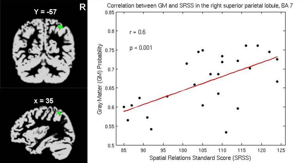
13 Decrease of gray matter volume in 25 patients with cystinosis compared to controls L R y = -37 x = 38 L Right inferior parietal lobule, BA 40, p < 0.001, corrected z = 55


14 Decrease of gray matter volume in 25 patients with cystinosis compared to controls L R y = -32 x = 21 L Right precentral gyrus, BA 4 p < 0.001, corrected z = 66
15 DTI Results White matter integrity is disrupted in parietal white matter bilaterally in the same areas as gray matter volume is diminished


16 Coronal, sagittal, and transverse views of voxels with a significant decrease in fractional anisotropy in the left anterior parietal lobe and postcentral gyrus (global maximum with P <.01, corrected for entire cerebral volume; mm) in 7 children with cystinosis compared with controls. Results are superimposed s on the white matter segment of a spatially normalized T1-weighted MRI.
 in 7 children with cystinosis compared with controls.")
17 Coronal and sagittal views of voxels with a significant decrease in fractional anisotropy in the right anterior parietal lobe and postcentral gyrus g (P <.05, corrected for entire cerebral volume; mm) in 7 children with cystinosis compared with controls. Results are superimposed on the t white matter segment of a spatially normalized T1-weighted MRI.
18 Summary of Brain Structural Differences in Young Children with Cystinosis Gray and white matter volume smaller Largest quantitative difference in right and left parietal lobes (areas thought to direct visual spatial functions)
19 What Causes Differences in Brain Development in Cystinosis? Cause unknown; possibilities include Early cystine accumulation (in utero) with damage to lysosomes Indirect effect of metabolic disturbances Direct effect of gene on some aspect of brain development Effect of medications Longitudinal studies may help to determine which is the most likely cause
20 Impact of Cystinosis on Cognitive Function Children and adults with cystinosis are more likely to demonstrate specific cognitive difficulties than non-cystinotic individuals Visual spatial dysfunction Visual memory dysfunction Attentional problems
21 Cognitive Profiles in Cystinosis Normal intelligence Normal language Normal verbal learning Poor visuomotor skills Deficits in visuospatial processing Deficits in short-term term visual memory Difficulty with visual learning Difficulty with tactile discrimination Normal reading skills Difficulty with math and spelling
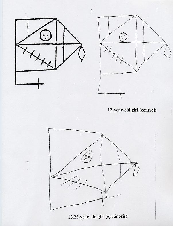
22 Visual Processing in Cystinosis Intact ability to recognize visually presented information Deficit in visual spatial skills (e.g., map-reading) Difficulty with visual memory (i.e., accurately remembering information presented visually)
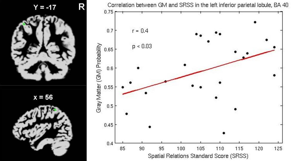
23 Correlation of Brain Structure and Cognitive Function The parietal lobes of the brain are thought to be most involved in visual spatial function Volume of gray matter in the parietal lobes correlates directly with visual spatial function in young cystinosis children
24 1
25 3
26 INTELLIGENCE AND VISUAL PERCEPTION Score IQ VFD Cystinosis Control

27
28
29 Examples
30
31
32
33
34
35
36
37 What does it mean? If the child has spatial problems, you may notice difficulties with: finding his/her way around, drawing or constructing, arithmetic, social studies.
38 How Visual Spatial Problems Affect Learning Math is a very spatially-mediated skill to learn x
39 Visuospatial Difficulties Impacting Arithmetic Not Carrying Direction of Operation
40
41 Although visual spatial and visual memory functions may be impaired, language and auditory processing are intact; this information provides a basis with which to suggest potential remediations if the child is having academic difficulties related to visual spatial dysfunction
42 Remediation Examples MATH: have the student verbalize or talk through the problem; list steps of a process on a study sheet, and allow the student to refer to the sheet; reinforce direction of proceeding with computations. WRITTEN ASSIGNMENTS: use large, lined paper and paper folding; allow student to quietly read aloud written work.
43 Verbal and Visual Learning in Cystinosis
44 VERBAL LEARNING AND MEMORY Words Recalled Trial Control Cystinosis Words Recalled Trial 5 20 Min. Delay Control Cystinosis
45 VISUAL LEARNING AND MEMORY d' Control Cystinosis Trial d' Trial 5 20 Min. Delay Control Cystinosis
46 VISUAL AND VERBAL LEARNING IN CHILDREN AND ADULTS WITH CYSTINOSIS T-Score Trials Child Adult VLMT CVLT
47 VISUAL LEARNING AND MEMORY INCREASED EXPOSURE TIME d' Trial Control Cystinosis d' Trial 5 20 Min. Delay Control Cystinosis
48 *Increasing exposure time to visual stimuli improved performance *Suggests that visual processing problem is due to deficit in visual processing speed *Provides foundation for interventions focused on increasing processing time
49 Take home message Forewarned is forearmed Young children with cystinosis entering formal school, may be at risk for difficulties in these areas Parents can take a proactive approach with schooling from the beginning to help ensure successes
50 Summary Brain development is different in children with cystinosis Differences in brain development correlate with difficulties with visual spatial function Visual spatial difficulties may contribute to learning difficulties Awareness and intervention when appropriate can prevent later problems
51 Intervention Studies Based on our data, an intervention study was designed and conducted (see poster) We could not determine whether the intervention might have helped because of lack of participation and lack of follow-through on the part of participants We are now proposing an intervention study that, if successful, can be incorporated into the classroom with no additional time commitment needed from already over-burdened families
52 Trauner Research Group Lynne Babchuck, M.S.W. Angela Ballantyne Ph.D. Michelle Barney Sunita Bava Kirsten Poehlmann Ph.D. Miriam Sach M.D., Ph.D. Amy Spilkin Ph.D. Rebecca Theilmann Ph.D. Doris Trauner M.D. Jenny Williams
53 Acknowledgments This work is being funded by the NICHD and by the Cystinosis Research Foundation. Previous work has been funded by the Cystinosis Research Network and the National Cystinosis Foundation. We sincerely thank the families who have and are continuing to participate in our research studies.
54 Thank you!!!
Neurological impairment in nephropathic cystinosis: motor coordination deficits
 Pediatr Nephrol (2010) 25:2061 2066 DOI 10.1007/s00467-010-1589-8 ORIGINAL ARTICLE Neurological impairment in nephropathic cystinosis: motor coordination deficits Doris A. Trauner & Jennifer Williams &
CYSTINOSIS AND THE BRAIN
 CYSTINOSIS AND THE BRAIN Doris A. Trauner M.D. Professor, Departments of Neurosciences and Pediatrics University of California San Diego School of Medicine La Jolla, California USA Rady Children s Hospital
Use of Multimodal Neuroimaging Techniques to Examine Age, Sex, and Alcohol-Related Changes in Brain Structure Through Adolescence and Young Adulthood
 American Psychiatric Association San Diego, CA 24 May 2017 Use of Multimodal Neuroimaging Techniques to Examine Age, Sex, and Alcohol-Related Changes in Brain Structure Through Adolescence and Young Adulthood
Brain anatomy tutorial. Dr. Michal Ben-Shachar 459 Neurolinguistics
 Brain anatomy tutorial Dr. Michal Ben-Shachar 459 Neurolinguistics The human brain Left hemisphere Right hemisphere http://www.brainmuseum.org/ Zoom out Zoom in Types of Brain Tissue Gray Matter: Cell
correlates with social context behavioral adaptation.
 REVIEW OF FRONTAL LOBE STRUCTURES Main organization of frontal cortex: 1. Motor area (precentral gyrus). 2. Premotor & supplementary motor areas (immediately anterior to motor area). Includes premotor,
Hemispheric Specialization (lateralization) Each lobe of the brain has specialized functions (Have to be careful with this one.)
 Each lobe of the brain has specialized functions (Have to be careful with this one.)") Cerebral Cortex Principles contralaterality the right half of your brain controls the left half of your body and vice versa. (contralateral control.) Localization of function Specific mental processes
Cerebral Cortex Principles contralaterality the right half of your brain controls the left half of your body and vice versa. (contralateral control.) Localization of function Specific mental processes
Gender Differences in Brain Wiring Connectomes. Arlene R. Taylor PhD Brain References
 Gender Differences in Brain Wiring Connectomes Arlene R. Taylor PhD Brain References www.arlenetaylor.org www.llm.life 8-20-16 Conflict Conflict happens everywhere and it Is expensive Home: contributes
HIV Neurology Persistence of Cognitive Impairment Despite cart
 HIV Neurology Persistence of Cognitive Impairment Despite cart Victor Valcour MD PhD Professor of Medicine Memory and Aging Center, Dept. of Neurology University of California San Francisco, USA 8 th International
Imaging in Pediatric `neurohiv Dr Jackie Hoare Head of Liaison Psychiatry Groote Schuur Hospital, UCT
 Imaging in Pediatric `neurohiv Dr Jackie Hoare Head of Liaison Psychiatry Groote Schuur Hospital, UCT ? Spectrum of Neurocognitive disorders The adult literature on HIV related CNS damage supports a spectrum
FAILURES OF OBJECT RECOGNITION. Dr. Walter S. Marcantoni
 FAILURES OF OBJECT RECOGNITION Dr. Walter S. Marcantoni VISUAL AGNOSIA -damage to the extrastriate visual regions (occipital, parietal and temporal lobes) disrupts recognition of complex visual stimuli
Specific Sulci/Fissures:
 Specific Sulci/Fissures: Central Sulcus Longitudinal Fissure Sylvian/Lateral Fissure Transverse Fissure http://www.bioon.com/book/biology/whole/image/1/1-8.tif.jpg http://www.dalbsoutss.eq.edu.au/sheepbrains_me/human_brain.gif
Supplementary Online Content
 Supplementary Online Content Devenney E, Bartley L, Hoon C, et al. Progression in behavioral variant frontotemporal dementia: a longitudinal study. JAMA Neurol. Published online October 26, 2015. doi:10.1001/jamaneurol.2015.2061.
Human Paleoneurology and the Evolution of the Parietal Cortex
 PARIETAL LOBE The Parietal Lobes develop at about the age of 5 years. They function to give the individual perspective and to help them understand space, touch, and volume. The location of the parietal
Exam 1 PSYC Fall 1998
 Exam 1 PSYC 2022 Fall 1998 (2 points) Briefly describe the difference between a dualistic and a materialistic explanation of brain-mind relationships. (1 point) True or False. George Berkely was a monist.
Homework Week 2. PreLab 2 HW #2 Synapses (Page 1 in the HW Section)
") Homework Week 2 Due in Lab PreLab 2 HW #2 Synapses (Page 1 in the HW Section) Reminders No class next Monday Quiz 1 is @ 5:30pm on Tuesday, 1/22/13 Study guide posted under Study Aids section of website
Homework Week 2 Due in Lab PreLab 2 HW #2 Synapses (Page 1 in the HW Section) Reminders No class next Monday Quiz 1 is @ 5:30pm on Tuesday, 1/22/13 Study guide posted under Study Aids section of website
Functional MRI and Diffusion Tensor Imaging
 Functional MRI and Diffusion Tensor Imaging Andrew Steven March 23, 2018 Ochsner Neuroscience Symposium None Disclosure 1 Objectives Review basic principles of BOLD fmri and DTI. Discuss indications and
Summary of findings from the previous meta-analyses of DTI studies in MDD patients. SDM (39) 221 Left superior longitudinal
 221 Left superior longitudinal") Supplemental Data Table S1 Summary of findings from the previous meta-analyses of DTI studies in MDD patients Study Analysis Method Included studies, n MDD (medicated) HC Results (MDDHC)
Supplemental Data Table S1 Summary of findings from the previous meta-analyses of DTI studies in MDD patients Study Analysis Method Included studies, n MDD (medicated) HC Results (MDDHC)
Gross Organization I The Brain. Reading: BCP Chapter 7
 Gross Organization I The Brain Reading: BCP Chapter 7 Layout of the Nervous System Central Nervous System (CNS) Located inside of bone Includes the brain (in the skull) and the spinal cord (in the backbone)
Cortical Organization. Functionally, cortex is classically divided into 3 general types: 1. Primary cortex:. - receptive field:.
 Cortical Organization Functionally, cortex is classically divided into 3 general types: 1. Primary cortex:. - receptive field:. 2. Secondary cortex: located immediately adjacent to primary cortical areas,
Neuropsychological Evaluation of
 Neuropsychological Evaluation of Alzheimer s Disease Joanne M. Hamilton, Ph.D. Shiley-Marcos Alzheimer s Disease Research Center Department of Neurosciences University of California, San Diego Establish
FRONTAL LOBE. Central Sulcus. Ascending ramus of the Cingulate Sulcus. Cingulate Sulcus. Lateral Sulcus
 FRONTAL LOBE Central Ascending ramus of the Cingulate Cingulate Lateral Lateral View Medial View Motor execution and higher cognitive functions (e.g., language production, impulse inhibition, reasoning
Functional Neuroanatomy. IBRO ISN African Neuroscience School 4-13 th Dec 2014 Nairobi, Kenya
 Functional Neuroanatomy IBRO ISN African Neuroscience School 4-13 th Dec 2014 Nairobi, Kenya What is/are the function(s) of the nervous system? Sensation Perception Visceral activities (Homeostasis) Behavior
Is DTI Increasing the Connectivity Between the Magnet Suite and the Clinic?
 Current Literature In Clinical Science Is DTI Increasing the Connectivity Between the Magnet Suite and the Clinic? Spatial Patterns of Water Diffusion Along White Matter Tracts in Temporal Lobe Epilepsy.
Title:Atypical language organization in temporal lobe epilepsy revealed by a passive semantic paradigm
 Author's response to reviews Title:Atypical language organization in temporal lobe epilepsy revealed by a passive semantic paradigm Authors: Julia Miro (juliamirollado@gmail.com) Pablo Ripollès (pablo.ripolles.vidal@gmail.com)
Excellent Network Courses. Department of Neurology Affiliated hospital of Jiangsu University
 Excellent Network Courses Department of Neurology Affiliated hospital of Jiangsu University Agnosia Visual Agnosia Lissauer (1890) described 2 types: a) Apperceptive Cannot see objects b) Associative Does
P. Hitchcock, Ph.D. Department of Cell and Developmental Biology Kellogg Eye Center. Wednesday, 16 March 2009, 1:00p.m. 2:00p.m.
 Normal CNS, Special Senses, Head and Neck TOPIC: CEREBRAL HEMISPHERES FACULTY: LECTURE: READING: P. Hitchcock, Ph.D. Department of Cell and Developmental Biology Kellogg Eye Center Wednesday, 16 March
Approach to the Child with Developmental Delay
 Approach to the Child with Developmental Delay Arwa Nasir Department of Pediatrics University of Nebraska Medical Center DISCLOSURE DECLARATION Approach to the Child with Developmental Delay Arwa Nasir
CEREBRUM. Dr. Jamila EL Medany
 CEREBRUM Dr. Jamila EL Medany Objectives At the end of the lecture, the student should be able to: List the parts of the cerebral hemisphere (cortex, medulla, basal nuclei, lateral ventricle). Describe
Supplementary appendix
 Supplementary appendix This appendix formed part of the original submission and has been peer reviewed. We post it as supplied by the authors. Supplement to: Schiller R, IJsselstijn H, Hoskote A, et al.
NeuroTracker Published Studies & Research
 NeuroTracker Published Studies & Research Evidence of Relevance in Measurement, Learning and Transfer for Learning and Learning Related Conditions NeuroTracker evolved out of a pure science approach through
Diffusion Tensor Imaging 12/06/2013
 12/06/2013 Beate Diehl, MD PhD FRCP University College London National Hospital for Neurology and Neurosurgery Queen Square London, UK American Epilepsy Society Annual Meeting Disclosure None Learning
Paul Moes, Professor of Psychology - Neuroscience and Education Group - Christian Perspectives in Science - Education Program
 Paul Moes, Professor of Psychology - Neuroscience and Education Group - Christian Perspectives in Science - Education Program Thesis: Secret to the brain is Unity The problem: focused on parts A sample
Quantitative Neuroimaging- Gray and white matter Alteration in Multiple Sclerosis. Lior Or-Bach Instructors: Prof. Anat Achiron Dr.
 Quantitative Neuroimaging- Gray and white matter Alteration in Multiple Sclerosis Lior Or-Bach Instructors: Prof. Anat Achiron Dr. Shmulik Miron INTRODUCTION Multiple Sclerosis general background Gray
Announcement. Danny to schedule a time if you are interested.
 Announcement If you need more experiments to participate in, contact Danny Sanchez (dsanchez@ucsd.edu) make sure to tell him that you are from LIGN171, so he will let me know about your credit (1 point).
Neural correlates of adaptive working memory training in a glycogen storage disease type-iv patient
 CASE STUDY Neural correlates of adaptive working memory training in a glycogen storage disease type-iv patient Kristin Lee 1, Thomas Ernst 1, Gro Løhaugen,3, Xin Zhang 1 & Linda Chang 1 1 Department of
Stuttering Research. Vincent Gracco, PhD Haskins Laboratories
 Stuttering Research Vincent Gracco, PhD Haskins Laboratories Stuttering Developmental disorder occurs in 5% of children Spontaneous remission in approximately 70% of cases Approximately 1% of adults with
Sensorimotor Functioning. Sensory and Motor Systems. Functional Anatomy of Brain- Behavioral Relationships
 Sensorimotor Functioning Sensory and Motor Systems Understanding brain-behavior relationships requires knowledge of sensory and motor systems. Sensory System = Input Neural Processing Motor System = Output
Chapter 2 Test. 1. Evolutionary structures within the are the most primitive. *a. hindbrain b. thalamus c. forebrain d. midbrain e.
 Cognitive Psychology In and Out of the Laboratory 5th Edition Galotti TEST BANK Full clear download (no formatting errors) at: https://testbankreal.com/download/cognitive-psychology-laboratory-5thedition-galotti-test-bank/
Leah Militello, class of 2018
 Leah Militello, class of 2018 Objectives 1. Describe the general organization of cerebral hemispheres. 2. Describe the locations and features of the different functional areas of cortex. 3. Understand
Procedia - Social and Behavioral Sciences 159 ( 2014 ) WCPCG 2014
 WCPCG 2014") Available online at www.sciencedirect.com ScienceDirect Procedia - Social and Behavioral Sciences 159 ( 2014 ) 743 748 WCPCG 2014 Differences in Visuospatial Cognition Performance and Regional Brain Activation
Available online at www.sciencedirect.com ScienceDirect Procedia - Social and Behavioral Sciences 159 ( 2014 ) 743 748 WCPCG 2014 Differences in Visuospatial Cognition Performance and Regional Brain Activation
Psy /16 Human Communication. By Joseline
 Psy-302 11/16 Human Communication By Joseline Lateralization Left Hemisphere dominance in speech production in 95% of right handed and 70% of left handed people Left -> Timing, Sequence of events Right
Chapter 11 summary definitief ineke brands.indd :57:59
 chapter 11 Summary chapter 11 Both type 1 and type 2 diabetes mellitus are associated with altered brain function, a complication referred to as diabetic encephalopathy. Previous studies have shown that
Funding: NIDCF UL1 DE019583, NIA RL1 AG032119, NINDS RL1 NS062412, NIDA TL1 DA
 The Effect of Cognitive Functioning, Age, and Molecular Variables on Brain Structure Among Carriers of the Fragile X Premutation: Deformation Based Morphometry Study Naomi J. Goodrich-Hunsaker*, Ling M.
BINGES, BLUNTS AND BRAIN DEVELOPMENT
 BINGES, BLUNTS AND BRAIN DEVELOPMENT Why delaying the onset of alcohol and other drug use during adolescence is so important Aaron White, PhD Division of Epidemiology and Prevention Research National Institute
DWI assessment of ischemic changes in the fetal brain
 DWI assessment of ischemic changes in the fetal brain Dafi Bergman, 4 th year Medical student in the 4-year program, Sackler school of medicine B.Sc Life and Medical Sciences, Tel Aviv University Supervised
Carlo Semenza (University of Padova) Simple Calculation In The Brain: Evidence From Direct Cortical Electro-Stimulation
 Simple Calculation In The Brain: Evidence From Direct Cortical Electro-Stimulation") Carlo Semenza (University of Padova) Simple Calculation In The Brain: Evidence From Direct Cortical Electro-Stimulation New Approaches To The Neural Basis of Mathematical Cognition. Symposium 9 How do
Carlo Semenza (University of Padova) Simple Calculation In The Brain: Evidence From Direct Cortical Electro-Stimulation New Approaches To The Neural Basis of Mathematical Cognition. Symposium 9 How do
Cognitive Impairment and Magnetic Resonance Changes in Multiple Sclerosis. Background
 Cognitive Impairment and Magnetic Resonance Changes in Multiple Sclerosis Victoria A Levasseur 1,2, Samantha Lancia 1, Gautam Adusumilli 1, Zach Goodman 1, Stuart D. Cook 3, Diego Cadavid 4, Robert T.
Pamela S. Klonoff, PhD Clinical Director Center for Transitional Neuro-Rehabilitation Barrow Neurological Institute, Phoenix, Arizona
 Neuropsychology Pamela S. Klonoff, PhD Clinical Director Center for Transitional Neuro-Rehabilitation Barrow Neurological Institute, Phoenix, Arizona Top Ten Ways to Understand and Cope with a Brain Tumor
Topic 11 - Parietal Association Cortex. 1. Sensory-to-motor transformations. 2. Activity in parietal association cortex and the effects of damage
 Topic 11 - Parietal Association Cortex 1. Sensory-to-motor transformations 2. Activity in parietal association cortex and the effects of damage Sensory to Motor Transformation Sensory information (visual,
2. Area of the brain affected by the seizures.
 Learning Through Storms When discussing learning, we sometimes refer to cognition, or one s ability to think, learn and use information. Seizures can impact cognition, learning and behaviour in a variety
Caring Sheet #11: Alzheimer s Disease:
 CARING SHEETS: Caring Sheet #11: Alzheimer s Disease: A Summary of Information and Intervention Suggestions with an Emphasis on Cognition By Shelly E. Weaverdyck, PhD Introduction This caring sheet focuses
A Healthy Brain. An Injured Brain
 A Healthy Brain Before we can understand what happens when a brain is injured, we must realize what a healthy brain is made of and what it does. The brain is enclosed inside the skull. The skull acts as
CEREBRUM Dr. Jamila Elmedany Dr. Essam Eldin Salama
 CEREBRUM Dr. Jamila Elmedany Dr. Essam Eldin Salama Objectives At the end of the lecture, the student should be able to: List the parts of the cerebral hemisphere (cortex, medulla, basal nuclei, lateral
AUDIOLOGY INFORMATION SERIES ASHA S CONSUMER NEWSLETTER. Hearing Loss and Its Implications for Learning and Communication
 AUDIOLOGY INFORMATION SERIES ASHA S CONSUMER NEWSLETTER Vol. 1 No. 2 2000 Hearing Loss and Its Implications for Learning and Communication Hearing Loss and Children: The Facts and Why They Are Important!
The Central Nervous System I. Chapter 12
 The Central Nervous System I Chapter 12 The Central Nervous System The Brain and Spinal Cord Contained within the Axial Skeleton Brain Regions and Organization Medical Scheme (4 regions) 1. Cerebral Hemispheres
Neuroimaging Findings in Young Drinkers: Does Teenage Drinking Harm the Brain? Susan F. Tapert, Ph.D. University of California, San Diego
 Neuroimaging Findings in Young Drinkers: Does Teenage Drinking Harm the Brain? Susan F. Tapert, Ph.D. University of California, San Diego 2 Overview What is normal adolescence? How do binge drinkers differ?
Thalamus and Sensory Functions of Cerebral Cortex
 Thalamus and Sensory Functions of Cerebral Cortex I: To describe the functional divisions of thalamus. II: To state the functions of thalamus and the thalamic syndrome. III: To define the somatic sensory
Cerebral Cortex 1. Sarah Heilbronner
 Cerebral Cortex 1 Sarah Heilbronner heilb028@umn.edu Want to meet? Coffee hour 10-11am Tuesday 11/27 Surdyk s Overview and organization of the cerebral cortex What is the cerebral cortex? Where is each
MRI-Based Classification Techniques of Autistic vs. Typically Developing Brain
 MRI-Based Classification Techniques of Autistic vs. Typically Developing Brain Presented by: Rachid Fahmi 1 2 Collaborators: Ayman Elbaz, Aly A. Farag 1, Hossam Hassan 1, and Manuel F. Casanova3 1Computer
How We Grow & Change
 How We Grow & Change Neural Development What makes up nerves? Neurons! (single cells) Interesting Facts About Neurons: Average brain has approx 100 billion neurons and we only use 10% (10 billion neurons)!
Disorders of Object and Spatial perception. Dr John Maasch Brain Injury Rehabilitation Service Burwood Hospital.
 Disorders of Object and Spatial perception Dr John Maasch Brain Injury Rehabilitation Service Burwood Hospital. Take Home Message 1 Where there are lesions of the posterior cerebrum and posterior temporal
Advances in Clinical Neuroimaging
 Advances in Clinical Neuroimaging Joseph I. Tracy 1, PhD, ABPP/CN; Gaelle Doucet 2, PhD; Xaiosong He 2, PhD; Dorian Pustina 2, PhD; Karol Osipowicz 2, PhD 1 Department of Radiology, Thomas Jefferson University,
COGNITIVE ALTERATIONS IN CHRONIC KIDNEY DISEASE K K L E E
 COGNITIVE ALTERATIONS IN CHRONIC KIDNEY DISEASE K K L E E Attention Problem Solving Language Cognitive Domains Decision Making Memory Reasoning The Cardiovascular Health Cognition Study shows higher S
Neuropsychology and Metabolic Conditions: The Neurocognitive Profile of FOD/OAA and the benefits of neuropsychological assessment
 Neuropsychology and Metabolic Conditions: The Neurocognitive Profile of FOD/OAA and the benefits of neuropsychological assessment Christopher Boys, PhD, LP Pediatric Neuropsychologist Associate Professor
The Frontal Lobes. Anatomy of the Frontal Lobes. Anatomy of the Frontal Lobes 3/2/2011. Portrait: Losing Frontal-Lobe Functions. Readings: KW Ch.
 The Frontal Lobes Readings: KW Ch. 16 Portrait: Losing Frontal-Lobe Functions E.L. Highly organized college professor Became disorganized, showed little emotion, and began to miss deadlines Scores on intelligence
Henry Molaison. Biography. From Wikipedia, the free encyclopedia
 Henry Molaison From Wikipedia, the free encyclopedia Henry Gustav Molaison (February 26, 1926 December 2, 2008), known widely as H.M., was an American memory disorder patient who had a bilateral medial
49a A&P: Nervous System -! Synaptic Transmission and Central Nervous System
 49a A&P: Nervous System -! Synaptic Transmission and Central Nervous System 49a A&P: Nervous System -! Synaptic Transmission and Central Nervous System! Class Outline" 5 minutes" "Attendance, Breath of
Supplementary Online Material Supplementary Table S1 to S5 Supplementary Figure S1 to S4
 Supplementary Online Material Supplementary Table S1 to S5 Supplementary Figure S1 to S4 Table S1: Brain regions involved in the adapted classification learning task Brain Regions x y z Z Anterior Cingulate
Short Term and Working Memory
 Short Term and Working Memory 793 Short Term and Working Memory B R Postle, University of Wisconsin Madison, Madison, WI, USA T Pasternak, University of Rochester, Rochester, NY, USA ã 29 Elsevier Ltd.
Shape Modeling of the Corpus Callosum for Neuroimaging Studies of the Brain (Part I) Dongqing Chen, Ph.D.
 Dongqing Chen, Ph.D.") The University of Louisville CVIP Lab Shape Modeling of the Corpus Callosum for Neuroimaging Studies of the Brain (Part I) Dongqing Chen, Ph.D. Computer Vision & Image Processing (CVIP) Laboratory Department
The University of Louisville CVIP Lab Shape Modeling of the Corpus Callosum for Neuroimaging Studies of the Brain (Part I) Dongqing Chen, Ph.D. Computer Vision & Image Processing (CVIP) Laboratory Department
BIOL Dissection of the Sheep and Human Brain
 BIOL 2401 Dissection of the Sheep and Human Brain Laboratory Objectives After completing this lab, you should be able to: Identify the main structures in the sheep brain and to compare them with those
Supplemental Information. Direct Electrical Stimulation in the Human Brain. Disrupts Melody Processing
 Current Biology, Volume 27 Supplemental Information Direct Electrical Stimulation in the Human Brain Disrupts Melody Processing Frank E. Garcea, Benjamin L. Chernoff, Bram Diamond, Wesley Lewis, Maxwell
Theory of mind skills are related to gray matter volume in the ventromedial prefrontal cortex in schizophrenia
 Theory of mind skills are related to gray matter volume in the ventromedial prefrontal cortex in schizophrenia Supplemental Information Table of Contents 2 Behavioral Data 2 Table S1. Participant demographics
 Passport control a bit carried away. appreciated the advice forgot to talk to the manager, next thing I know my fmri thankfully, when aroused things back to normal Inattentive impaired children and adolescents:
How do individuals with congenital blindness form a conscious representation of a world they have never seen? brain. deprived of sight?
 How do individuals with congenital blindness form a conscious representation of a world they have never seen? What happens to visual-devoted brain structure in individuals who are born deprived of sight?
Resistance to forgetting associated with hippocampus-mediated. reactivation during new learning
 Resistance to Forgetting 1 Resistance to forgetting associated with hippocampus-mediated reactivation during new learning Brice A. Kuhl, Arpeet T. Shah, Sarah DuBrow, & Anthony D. Wagner Resistance to
MULTI-CHANNEL COMMUNICATION
 INTRODUCTION Research on the Deaf Brain is beginning to provide a new evidence base for policy and practice in relation to intervention with deaf children. This talk outlines the multi-channel nature of
The significance of sensory motor functions as indicators of brain dysfunction in children
 Archives of Clinical Neuropsychology 18 (2003) 11 18 The significance of sensory motor functions as indicators of brain dysfunction in children Abstract Ralph M. Reitan, Deborah Wolfson Reitan Neuropsychology
Neuropsychology in Spina Bifida. Dr Ellen Northcott Clinical Neuropsychologist Kids Rehab, CHW
 Neuropsychology in Spina Bifida Dr Ellen Northcott Clinical Neuropsychologist Kids Rehab, CHW Who are neuropsychologists? Undergraduate Degree (eg. BPsych, BSc, BA) Honours in Psychology Master or Doctor
1 in 68 in US. Autism Update: New research, evidence-based intervention. 1 in 45 in NJ. Selected New References. Autism Prevalence CDC 2014
 Autism Update: New research, evidence-based intervention Martha S. Burns, Ph.D. Joint Appointment Professor Northwestern University. 1 Selected New References Bourgeron, Thomas (2015) From the genetic
10/22/2012. Nature versus nurture Are boys and girls really different? Are differences age dependent? Are there cognitive and behavioral differences?
 Is anything new in the age-old battle between the sexes? DOES GENDER MATTER? The Neurobiology of Gender differences in Learning and Learning Disabilities Dr. Susan R. Grant Lutherville, Maryland,21093
The Adverse Effect of Chemotherapy on the Developing Brain. Ellen van der Plas, PhD Research Fellow at SickKids
 The Adverse Effect of Chemotherapy on the Developing Brain Ellen van der Plas, PhD Research Fellow at SickKids Overview of today s talk Why study cancer survivors? Quick introduction on acute lymphoblastic
NEUROPSYCHOLOGICAL ASSESSMENT S A R A H R A S K I N, P H D, A B P P S A R A H B U L L A R D, P H D, A B P P
 NEUROPSYCHOLOGICAL ASSESSMENT S A R A H R A S K I N, P H D, A B P P S A R A H B U L L A R D, P H D, A B P P NEUROPSYCHOLOGICAL EXAMINATION A method of examining the brain; abnormal behavior is linked to
Timing Impairments and Neural Dysfunction in Basal Ganglia Disorders
 Timing Impairments and Neural Dysfunction in Basal Ganglia Disorders Deborah L. Harrington University of California, San Diego Veterans Affairs San Diego Healthcare System Co-Investigators University of
EFFECTS OF ADHD ON EARLY LEARNING AND ACDEMIC PERFORMANCE 1
 EFFECTS OF ADHD ON EARLY LEARNING AND ACDEMIC PERFORMANCE 1 The Effects of ADHD on Learning and Academic Performance in the Pre- and Elementary School Years. Christopher Kalogeropoulos November 19, 2012
C. Brock Kirwan, Ph.D.
 , Ph.D. Department of Psychology & Neuroscience Center Brigham Young University 1052 Kimball Tower Provo, UT 84602 Phone: (801) 422-2532 kirwan@byu.edu ACADEMIC & RESEARCH POSITIONS Assistant Professor:
Language After Traumatic Brain Injury
 Chapter 7 Language After Traumatic Brain Injury 10/24/05 COMD 326, Chpt. 7 1 1 10/24/05 COMD 326, Chpt. 7 2 http://www.californiaspinalinjurylawyer.com/images/tbi.jpg 2 TBI http://www.conleygriggs.com/traumatic_brain_injury.shtml
HOARE Jacqueline, PHILLIPS Nicole, FOUCHE Jean- Paul, JOSKA John A, MYER Landon, ZAR Heather J STEIN Dan J
 Mental health, cognition and associated brain changes in perinatally infected young adolescents in the Cape Town Adolescent Antiretroviral Cohort (CTAAC) HOARE Jacqueline, PHILLIPS Nicole, FOUCHE Jean-
Neural correlates of memory for object identity and object location: effects of aging
 Neuropsychologia 40 (2002) 1428 1442 Neural correlates of memory for object identity and object location: effects of aging Alessandra Schiavetto a, Stefan Köhler a, Cheryl L. Grady a, Gordon Winocur a,c,
Memory Development. Cognitive Development
 Memory Development Cognitive Development Memory as information storage Memory Why does our memory sometimes fail us? Memory Schachter s Seven Sins of Memory 1. Transience 2. Absent-Mindedness 3. Blocking
By Lauren Stowe, PhD, CCC-SLP & Gina Rotondo, MS, CCC-SLP The Speech Therapy Group
 By Lauren Stowe, PhD, CCC-SLP & Gina Rotondo, MS, CCC-SLP The Speech Therapy Group http://www.acquiredbraininjury.com/interactive brain/interactivebrain.swf 1. Hormones make the science messy 2. Difference
Paediatric HIV -Developmental Aspects. Dr Kirsty Donald Division of Developmental Paediatrics Red Cross Children s Hospital
 Paediatric HIV -Developmental Aspects Dr Kirsty Donald Division of Developmental Paediatrics Red Cross Children s Hospital Normal Development Development occurs with sequential acquisition of skills in
Surgery for Medically Refractory Focal Epilepsy
 Surgery for Medically Refractory Focal Epilepsy Seth F Oliveria, MD PhD The Oregon Clinic Neurosurgery Director of Functional Neurosurgery: Providence Brain and Spine Institute Portland, OR Providence
Cerebrum-Cerebral Hemispheres. Cuneyt Mirzanli Istanbul Gelisim University
 Cerebrum-Cerebral Hemispheres Cuneyt Mirzanli Istanbul Gelisim University The largest part of the brain. Ovoid shape. Two incompletely separated cerebral hemispheres. The outer surface of the cerebral
giessen.de/cms/cognition
 Space to Reason Markus Knauff University of Gießen markus.knauff@psychol.uni giessen.de www.uni giessen.de/cms/cognition For every complex problem, there is an answer that is clear, simple, and wrong.
How do we construct Intelligence tests? Tests must be: Standardized Reliable Valid
 Test Construction How do we construct Intelligence tests? Tests must be: Standardized Reliable Valid Standardization The test must be pre-tested to a representative sample of people and form a normal distribution
The neurolinguistic toolbox Jonathan R. Brennan. Introduction to Neurolinguistics, LSA2017 1
 The neurolinguistic toolbox Jonathan R. Brennan Introduction to Neurolinguistics, LSA2017 1 Psycholinguistics / Neurolinguistics Happy Hour!!! Tuesdays 7/11, 7/18, 7/25 5:30-6:30 PM @ the Boone Center
Brain Imaging Applied to Memory & Learning
 Brain Imaging Applied to Memory & Learning John Gabrieli Department of Brain & Cognitive Sciences Institute for Medical Engineering & Sciences McGovern Institute for Brain Sciences MIT Levels of Analysis
MALATTIA CARDIOVASCOLARE NELL ANZIANO DIABETICO. Diabete e demenza. Enzo Manzato
 MALATTIA CARDIOVASCOLARE NELL ANZIANO DIABETICO Diabete e demenza Enzo Manzato Lancet 2016; 387: 1513 Lancet 2016; 387: 1377 MEN Diabetes Care 2016;39:300 WOMEN Diabetes Care 2016;39:300 Multiple-adjusted
European Prevention of Alzheimer s Dementia (EPAD)
") European Prevention of Alzheimer s Dementia (EPAD) Ron Marcus, MD ISCTM Adaptive Design Workshop February 20, 2018 1 EPAD Goal The European Prevention of Alzheimer's Dementia (EPAD) project aims to develop
European Prevention of Alzheimer s Dementia (EPAD) Ron Marcus, MD ISCTM Adaptive Design Workshop February 20, 2018 1 EPAD Goal The European Prevention of Alzheimer's Dementia (EPAD) project aims to develop