Point-of-Care Ultrasound: An Introduction
|
|
|
- Shawn Hill
- 5 years ago
- Views:
Transcription
1 Point-of-Care Ultrasound: An Introduction Delegation Teaching Package for Registered Respiratory Therapists and Anesthesia Assistants Developed by: Rob Bryan RRT, AA Edited by: Kelly Hassall RRT, FCSRT, MEd This document has been developed by the RTSO with assistance from Mackenzie Health and is intended to serve as a sample of a medical delegation learning package. The RTSO and Mackenzie Health are not responsible for any of its content.
2 Objectives: To discuss the theory behind the use of Point of Care (POC) ultrasound To review the different types of probes and their clinical applications To familiarize the bedside clinician with the POC ultrasound unit Functionality of the controls Optimization of views
3 What are Sound Waves? Sound waves are mechanical vibrations and are measured in Hertz (Hz) Period is the time for one cycle to occur (µs) Hertz represents the number of cycles in a second Size of the amplitude is measured in decibels (bd) Wavelength is the distance between two excitations
4 What is Ultrasound? Ultrasound is a frequency above that which humans can hear, or more than 20,000Hz (20kHz) Therapeutic ultrasound is designed to create heat using mechanical sound waves Diagnostic ultrasound is in the millions of hertz and used to visualize structures
5 Point-Of-Care Applications for Ultrasound Point-of-care (POC) ultrasonography is defined as: ultrasonography brought to the patient and performed by the provider in real time Procedural guidance: the use of ultrasound to visualize anatomical structures to improve success and decrease complications of procedures (i.e. arterial line insertions) Diagnostic assessment: the use of ultrasound to diagnose or rule out certain conditions by examining a particular organ, disease or procedure directly relevant to their area of expertise (i.e. pneumothorax) Screening: the use of ultrasound as a means to identify patients at risk for certain diseases (i.e. abdominal aortic aneurysms)

6 Piezoelectric Effect and Ultrasound First described in 1880 by French physicists Pierre and Jacques Currie who observed the generation of an electrical charge as a result of a mechanical force being applied to certain crystals and materials and described it as the Piezoelectric Effect Paul Langevin later developed piezoelectric materials that can generate and receive mechanical vibration at high frequency known as ultrasound


7 How do we provide POC Ultrasound? Ultrasound acoustic waves are created by deformation of piezoelectric crystals or ceramic materials when stimulated by electrical energy producing high frequency mechanical pulses Ultrasound transducers produce and transmit sound waves and receive reflected sound waves or echoes converting them back into electrical energy that can be interpreted by a computer processor to produce an image
8 Principles of Ultrasound Imaging The density (concentration of medium) and stiffness (resistance of a medium to compress) directly influence the speed acoustic waves move through it Media with higher density will transmit mechanical waves with greater speed than lesser dense media Acoustic Impedance is the difficulty of sound moving through a medium as a result of its density, in practical terms penetration decreases as frequency increases Velocity of ultrasound beams are calculated by: Velocity=Wavelength (mm) x Frequency (Hz)
9 Principles of Ultrasound Imaging Acoustic speed of mechanical waves through different media: MEDIA VELOCITY AIR WATER SOFT TISSUE BONE 331 meters/second 1495 meters/second 1540 meters/second 4080 meters/second

10 Interaction of Ultrasound Waves with Tissue Ultrasound will travel a straight path through homogeneous medium The path ultrasound that travels through in a heterogeneous medium will be altered The relationship between ultrasound and tissue can best be described in terms of reflection, scattering, refraction, and attenuation The interaction of ultrasound waves through the media and the direction in which they travel is complex Note: Even the slightest amount of air between the probe and the scan site will severely degrade the image quality always use a coupling medium (acoustic gel) to maintain image quality.

11 Reflection Occurs when the ultrasound beam rebounds off a tissue interface and a certain amount returns back to the transducer The magnitude of the reflected wave is dependent on the acoustic impedance of the tissue The magnitude of the reflection is also dependent on the angle between the ultrasound beam and the tissue with the optimal reflection occurring at 90º perpendicular orientation The greater the acoustic impedance the greater the reflection, if the impedance is equal there will virtually be no echo or no reflection of the different boundaries.

12 Scattering Is the redirection of sound in any direction by a rough surfaces or heterogeneous mediums particularly on surfaces and boundaries that are smaller than the wavelength


13 Refraction Is a change in sound direction (bending) when crossing the boundary between two media


14 Attenuation During transmission ultrasound strength is progressively reduced as it converts from ultrasound energy to heat and absorbed by surrounding tissue Dependent on frequency and wavelength (higher frequencies are absorbed at a greater rate than lower frequencies) Acoustic impedance is also an influencing factor with higher impedance increasing attenuation of the ultrasound beam

15 Ultrasound Image and Resolution Resolution is the ability to distinguish between two closely related structures and varies directly with the frequency and inversely with the wavelength If two structures are closer then one wavelength apart then they will not seen as separate Higher frequency and shorter wavelengths improve resolution but decrease the penetration of the ultrasound beam

16 Aspects of Spatial Resolution Axial resolution is the ability to distinguish between two structures in the same direction of the acoustic wave Lateral resolution is the ability to distinguish between two adjacent structures that are perpendicular to the acoustic wave

17 Ultrasound frequency affecting resolution Resolution can be improved by increasing frequency and reducing beam width


18 Types of transducers Type Picture Image Clinical use Linear Array Phased Array Curved Array Rectangular shade display Covers the wide of the probe Idea for near field scans, nerves, vessels and soft tissue Frequency : 6-13 MHz Scan depth 6 cm Pie shaped tip towards the transducer Better for visualizing deeper structures and Organs Frequency : 1-5 MHz Scan depth 35 cm Combination of linear and phased array Frequency : 2-5 MHz Scan depth 30 cm Superficial tissue and anatomical landmarks, Musculoskeletal, Nerve, Small Parts, Vascular, Venous Abdominal, Cardiology, Obstetrics, Orbital, TCD Deep nerves, neuraxial imaging, Abdominal, Gynecology, Musculoskeletal, Obstetrics
19 Ultrasound Image Modes Electric signals of echoes are amplified and displayed on a monitor Can be displayed as a conventional imaging, compound image, tissue harmonic imaging (THI), and combined compound-thi imaging Images can be displayed in static form (allowing for measuring distance between two interfaces, tissue or anatomic landmarks) or in real time providing temporal relations of these interfaces Ultrasound Modes include: A-Mode, B-mode, Doppler- Mode, and M-Mode

20 Compound Imaging vs Conventional

21 Ultrasound Image Modes A-Mode is the oldest modality and sends a single pulse with vertical deflection into a medium and waits for a return of signal It is a one dimensional image and produces a series of peaks and valleys in relation to when the ultrasound beam reaches different tissues.

22 Ultrasound Image Modes B-Mode or brightness modulation and is the primary mode used in regional anaesthesia It creates a two dimensional image by simultaneously scanning piezoelectric elements compared to one in A-Mode. Converts amplitudes of echoes into various brightness of gray with horizontal and vertical direction representing real distances in tissue


23 Ultrasound Image Modes Doppler-Mode detects changes in frequency of a sound wave resulting from relative motion between a sound source and the receiver. Colour Doppler colour-codes Doppler movement onto the B-Mode and shows the direction of blood flow with red noting blood flowing towards the probe and blue noting blood flowing away from the probe Blue Away Red Towards

24 Ultrasound Image Modes M-Mode or Motion modulation is B-Mode with a continuous update of the returning echoes forming a sequence of B- Mode images that show change over time Useful in cardiology and obstetrics

25 Successful Imaging Optimizing an image is an essential skill in performing ultrasound guided procedures Involves complex understanding of the ultrasound system/equipment and the use of various transducers Requires in depth knowledge of anatomy, anatomical landmarks and location in the body Appreciation for the characteristics and appearance of tissues and structures viewed under ultrasound (specifically hyperechoic, hypoechoic, and honeycomb patterns and properties)
26 Setting Up for Ultrasound Procedures Steps Considerations S Supplies Gather supplies: ultrasound machine, transducer covers, nerve stimulator and needle, tray, LA drawn and labelled, plug in unit to avoid powering down during procedure C Comfortable positioning A Ambiance N Name and procedure N Nominate transducer Patient, ACT, equipment, ultrasound unit should be strategically position to optimize ergonomics and efficiency and infection control Ultrasound should be on the opposite side of the patient with the screen aimed at the operators eye Dim the lights to view the screen better time-out to ensure correct block, correct site and correct patient including any pt considerations and ensure all the equipment is present and functioning before starting Select a transducer that best suits the block or procedure being done (linear transducer for superficial scans and curved array for deeper scans I Infection control Disinfect the skin to reduce the risk of infection and contamination N Note lateral/medial side on screen G Gain depth Touch the side of the transducer to orientate the medial lateral side of the patient corresponds with the image on the screen Use a coupling gel to reduce the potential for poor image quality related to decreased reflection-absorption rates
27 knobology There are many knobs and functions on the modern POC ultrasound unit and can become overwhelming for the end user to navigate Most companies have programmed settings (or presets) and proprietary software to help optimize the view depending on the type of scanning required Each manufacturer has their own twist on standard features and image enhancement tools that can add to the confusion Some features require the end user to scroll to another page to access part of the menu they need to complete a task
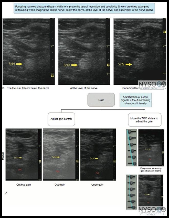
28 Five Essential Function Keys in Ultrasound Key Function TIP Depth Frequency Focusing Gain Doppler First consideration in imaging Dependant on the location of the structure and body habitus Optimal depth is important for focusing Higher frequency ultrasound have a higher rate of absorption and attenuate at shallower depths Lower frequency ultrasound travel deeper but sacrifice resolution Lateral resolution can be adjusted by increasing the frequency or focusing the beam width by narrowing at the level of the nerve Is the ration of output to input electrical power Adjustments amplify small voltages received in the transducer into larger allowing for more information processing and storage Overall gain will affect the whole image Time-Gain compensation will affect the near field or far field specifically Used to detect vascular structures or location and spread of local anaesthetics The target structure should be at the centre of the image Choose the transducer with the best frequency range and depth to visualize the target structure Adjust the focus/focal zone at the same level or 0.5 cm below the target structure Too much gain will blur structures into one another, Useful in far field imaging to better identify structures that are damped by attenuation or acoustic impedance Can be decreased if scattering is a concern Power Doppler is more sensitive in identifying vessels
29

30 artifact- UUUGGHHH!!
31 Optimal Imaging Depth Field Depth Transducer (frequency) Target Site < 2 cm Linear array (6-13 MHz) Wrist, ankle block cm Linear array (6-13 MHz) Interscalene, axillary brachial plexus Linear array (6-13 MHz) Femoral, superclavicular, abdominus plane block (TAP) cm Linear array (6-13 MHz) or Curved array (2-5 MHz) Infraclavicular, popliteal, subgluteal, sciatic nerve Curved array (2-5 MHz) Pundendal, gluteal sciatic nerve, lumbar plexus >10.0 cm Curved array (2-5 MHz) Anterior approach to sciatic nerve
32 References Moore CL, Copel JA. Point-of-Care Ultrasonography. NEJM [internet] [cited 2018 Oct 20];364(8) Available from: URL NEJM.org The New York School of Regional Anesthesia. Ultrasound Guided Techniques [Internet]. New York: The New York School of Regional Anesthesia;2018 [cited 2018 Oct 21]. Available from: Vincent JL, Abraham E, Moore F, Kochanek P, Fink M. Textbook of Critical Care. 7 th Edition. Canada:Elsevier;
Basic Physics of Ultrasound and Knobology
 WELCOME TO UTMB Basic Physics of Ultrasound and Knobology By Daneshvari Solanki, FRCA Laura B. McDaniel Distinguished Professor Anesthesiology and Pain Medicine University of Texas Medical Branch Galveston,
Ultrasound Physics and Knobology Alan Macfarlane. Consultant Anaesthetist Glasgow Royal Infirmary
 Ultrasound Physics and Knobology Alan Macfarlane Consultant Anaesthetist Glasgow Royal Infirmary RAPM 2009; 34: 40-46 Ultrasound Proficiency Understanding US image generation and device operation Image
1 Fundamentals. Basic Definitions and Physics Principles. Fundamentals
 1 To become versed in the language of ultrasonography, it is necessary to review some of the basic principles of physics. The wave physics principles of ordinary (i.e., audible) sound apply to ultrasound
Diagnostic Ultrasound. Sutiporn Khampunnip, M.D.
 Diagnostic Ultrasound Sutiporn Khampunnip, M.D. Definition of Ultrasound Ultrasound is simply sound waves, like audible sound. High-frequency sound and refers to mechanical vibrations above 20 khz. Human
Principles of Ultrasound. Cara C. Prideaux, M.D. University of Utah PM&R Sports Medicine Fellow March 14, 2012
 Principles of Ultrasound Cara C. Prideaux, M.D. University of Utah PM&R Sports Medicine Fellow March 14, 2012 None Disclosures Outline Introduction Benefits and Limitations of US Ultrasound (US) Physics
Ultrasound Physics & Terminology
 Ultrasound Physics & Terminology This module includes the following: Basic physics terms Basic principles of ultrasound Ultrasound terminology and terms Common artifacts seen Doppler principles Terms for
The Physics of Ultrasound. The Physics of Ultrasound. Claus G. Roehrborn. Professor and Chairman. Ultrasound Physics
 The Physics of Ultrasound Pipe Organ 10-8000 Emission Dog 452-1080 Man 85-1100 Spectrum Bat 10,000-120,000 Porpoise 7000-120,000 Claus G. Roehrborn Professor and Chairman 10 20 Cycles per second Reception
Ultrasound Principles cycle Frequency Wavelength Period Velocity
 ! Teresa S. Wu, MD, FACEP Director, EM Ultrasound Program & Fellowship Co-Director, Simulation Based Training Program & Fellowship Associate Program Director, EM Residency Program Maricopa Medical Center
Terminology Tissue Appearance
 By Marc Nielsen, MD Advantages/Disadvantages Generation of Image Ultrasound Machine/Transducer selection Modes of Ultrasound Terminology Tissue Appearance Scanning Technique Real-time Portable No ionizing
Physical Principles of Ultrasound
 Physical Principles of Ultrasound Grateful appreciation to Richard A. Lopchinsky, MD, FACS and Nancy H. Van Name, RDMS, RTR, and MarleneKattaron, RDMS 2000 UIC All Rights Reserved. Course Objectives Identify
Preamble (disclaimer)
") Preamble (disclaimer) PHYSICS AND PRINCIPLES OF HEAD/NECK ULTRASOUND Joseph C. Sniezek, MD FACS LTC, MC, USA Otolaryngology/H&N Surgery Tripler Army Medical Center 1. I am not a physicist 2. ACS has recommended
Preamble (disclaimer) PHYSICS AND PRINCIPLES OF HEAD/NECK ULTRASOUND Joseph C. Sniezek, MD FACS LTC, MC, USA Otolaryngology/H&N Surgery Tripler Army Medical Center 1. I am not a physicist 2. ACS has recommended
Introduction to Ultrasound Guided Region Anesthesia
 Introduction to Ultrasound Guided Region Anesthesia Brian D. Sites, MD Dept of Anesthesiology Dartmouth-Hitchcock Medical Center INTRODUCTION Welcome to Introduction to Ultrasound Guided Regional Anesthesia.
Ultrasound Physics & Doppler
 Ultrasound Physics & Doppler Endocrine University 2018 Mark Lupo, MD, FACE, ECNU Objectives Review the essential components of ultrasound physics in neck sonography Demonstrate the importance of ultrasound
Ultrasound guidance in regional anesthesia has
 Ultrasound and Regional Anesthesia Artifacts and Pitfall Errors Associated With Ultrasound-Guided Regional Anesthesia. Part I: Understanding the Basic Principles of Ultrasound Physics and Machine Operations
Basic of Ultrasound Physics E FAST & Renal Examination. Dr Muhammad Umer Ihsan MBBS,MD, DCH CCPU,DDU1,FACEM
 Basic of Ultrasound Physics E FAST & Renal Examination Dr Muhammad Umer Ihsan MBBS,MD, DCH CCPU,DDU1,FACEM What is Sound? Sound is Mechanical pressure waves What is Ultrasound? Ultrasounds are sound waves
CONTENTS. Test Number cpd Tanya Reynolds (Nat. Dip. Diag. Rad., B. Tech. Diag. Rad., B. Tech. Ultrasound)
") CONTENTS page 1-15 page 16 BASIC 2-DIMENSIONAL ULTRASOUND PRINCIPLES Multiple Choice Test Test Number cpd 41640 Tanya Reynolds (Nat. Dip. Diag. Rad., B. Tech. Diag. Rad., B. Tech. Ultrasound) Tanya is
CONTENTS page 1-15 page 16 BASIC 2-DIMENSIONAL ULTRASOUND PRINCIPLES Multiple Choice Test Test Number cpd 41640 Tanya Reynolds (Nat. Dip. Diag. Rad., B. Tech. Diag. Rad., B. Tech. Ultrasound) Tanya is
Ultrasound Knobology
 Ultrasound Knobology Raj Dasgupta MD, FACP, FCCP, FASSM Assistant Professor of Clinical Medicine Pulmonary / Critical Care / Sleep Medicine University of Southern California (USC) Objectives Physics of
Introduction to Biomedical Imaging
 Alejandro Frangi, PhD Computational Imaging Lab Department of Information & Communication Technology Pompeu Fabra University www.cilab.upf.edu Basic principles. Comparison to X-rays Ultrasound > 20kHz
What is Ultrasound? Resolution Image production Attenuation Imaging modes Ultrasound artifacts... 7
 What is Ultrasound?... 1 Resolution... 3 Image production... 3 Attenuation... 4 Imaging modes... 5 Ultrasound artifacts... 7 0 What is Ultrasound? High frequency sound of frequencies 2-50 MHz is used in
Ultrasound in Anesthesia: Applying Scientific Principles to Clinical Practice
 AANA Journal Course Update for Nurse Anesthetists 3 6 CE Credits* Ultrasound in Anesthesia: Applying Scientific Principles to Clinical Practice Christian R. Falyar, CRNA, DNAP The use of ultrasound as
Sound in medicine. CH.12. Dr.Rajaa أ.م.د. رجاء سهيل جنم جامعة تكريت كلية طب االسنان. General Properties of Sound
 CH.12. Dr.Rajaa Sound in medicine أ.م.د. رجاء سهيل جنم جامعة تكريت كلية Sound : It is the audible waves of frequency between 20 Hz and 20 khz. Infrasound : refers to the sound of frequency below the normal
Diploma of Medical Ultrasonography (DMU) Physical Principles of Ultrasound and Instrumentation Syllabus
 Physical Principles of Ultrasound and Instrumentation Syllabus") Diploma of Medical Ultrasonography (DMU) Physical Principles of Ultrasound and Instrumentation Syllabus Page 1 of 7 11/18 Candidates are expected to cover all of the content of this syllabus when preparing
Diploma of Medical Ultrasonography (DMU) Physical Principles of Ultrasound and Instrumentation Syllabus Page 1 of 7 11/18 Candidates are expected to cover all of the content of this syllabus when preparing
Ultrasonic Testing Level I:
 Ultrasonic Testing Level I: 1- Sound Wave - Introduction - ASNT Level I - Sound Wave Propagation - Velocity / Frequency / Wave Length - Acoustic Impedance - Energy / Intensity 2- Ultrasound Wave Modes
Basics of US Regional Anaesthesia. November 2008
 Basics of US Regional Anaesthesia November 2008 Essential Physics HIGH frequency = great resolution but poor penetration LOW frequency = poor resolution but great penetration Potential Advantages of US
ULTRASOUND IMAGING EE 472 F2018. Prof. Yasser Mostafa Kadah
 ULTRASOUND IMAGING EE 472 F2018 Prof. Yasser Mostafa Kadah www.k-space.org Recommended Textbook Diagnostic Ultrasound: Physics and Equipment, 2nd ed., by Peter R. Hoskins (Editor), Kevin Martin (Editor),
WELCOME! Introduction to Bedside Ultrasound
 WELCOME! Introduction to Bedside Ultrasound TEACHERS University of California-Irvine School of Medicine Nathan Molina nathan.d.molina@gmail.com Trevor Plescia taplescia90@gmail.com Jack Silva jpsilva42@gmail.com
ULTRASOUND. OB/Gyn (Core) Ultrasound PIEZOELECTRIC EFFECT. Principles of Ultrasound Physics and Instrumentation. Nathan Pinkney, BS, CDOS
 Ultrasound PIEZOELECTRIC EFFECT. Principles of Ultrasound Physics and Instrumentation. Nathan Pinkney, BS, CDOS") 1 OB/Gyn (Core) Ultrasound Principles of Ultrasound Physics and Instrumentation Nathan Pinkney, BS, CDOS Philadelphia College of Osteopathic Medicine 2016 ULTRASOUND CATEGORIES OF SOUND INFRASOUND = below
1 OB/Gyn (Core) Ultrasound Principles of Ultrasound Physics and Instrumentation Nathan Pinkney, BS, CDOS Philadelphia College of Osteopathic Medicine 2016 ULTRASOUND CATEGORIES OF SOUND INFRASOUND = below
Dr Emma Chung. Safety first - Physical principles for excellent imaging
 Safety first - Physical principles for excellent imaging Dr Emma Chung Lecturer in Medical Physics, University of Leicester Clinical Scientist, University Hospitals of Leicester NHS Trust Thanks to Caroline
DIGITAL IMAGE PROCESSING IN ULTRASOUND IMAGES
 DIGITAL IMAGE PROCESSING IN ULTRASOUND IMAGES Kamaljeet Kaur Computer Science & Engineering Department Guru Nanak Dev Engg. College, Ludhiana. Punjab-India meetk.89@gmail.com ABSTRACT-- Image processing
Introduction & Physics of ED Ultrasound. Objectives. What? - Limited Studies. Who? - ED Docs
 Introduction & Physics of ED Ultrasound Martine Sargent, MD Ultrasound Director, Assistant Professor UCSF Department of Emergency Medicine San Francisco General Hospital & Trauma Center Objectives Who?
Lesson 03: Sound Wave Propagation and Reflection. This lesson contains 15 slides plus 14 multiple-choice questions.
 Lesson 03: Sound Wave Propagation and Reflection This lesson contains 15 slides plus 14 multiple-choice questions. Accompanying text for the slides in this lesson can be found on pages 8 through 14 in
Supplement (videos)
") Supplement (videos) Ruben s tube (sound): http://www.youtube.com/watch?v=gpcquuwqayw Doppler US (diagnostic use): http://www.youtube.com/watch?v=fgxzg-j_hfw http://www.youtube.com/watch?v=upsmenyoju8 High
Supplement (videos) Ruben s tube (sound): http://www.youtube.com/watch?v=gpcquuwqayw Doppler US (diagnostic use): http://www.youtube.com/watch?v=fgxzg-j_hfw http://www.youtube.com/watch?v=upsmenyoju8 High
Regional Anaesthesia
 Regional Anaesthesia Basic Sciences Anaesthetic Course Dr Vassilis ATHANASSOGLOU FRCA Consultant Anaesthetist and Lecturer in Medicine Oxford University Hospitals NHS Trust, UK Geneva, 5th June 2017 Faculty
INTRODUCTION. Getting the best scan. Choosing a probe. Choosing the frequency
 Getting the best scan Choosing a probe Select the most appropriate probe for the particular scan required. s vary in their: operating frequency range higher ultrasound frequencies provide better discrimination
Lesson 07: Ultrasound Transducers. This lesson contains 73 slides plus 16 multiple-choice questions.
 Lesson 07: Ultrasound Transducers This lesson contains 73 slides plus 16 multiple-choice questions. This lesson was derived from pages 33 through 42 in the textbook: Ultrasound Transducers Ultrasound Transducers
Ultrasound. Principles of Medical Imaging. Contents. Prof. Dr. Philippe Cattin. MIAC, University of Basel. Oct 17th, 2016
 Ultrasound Principles of Medical Imaging Prof. Dr. Philippe Cattin MIAC, University of Basel Contents Abstract 1 Image Generation Echography A-Mode B-Mode M-Mode 2.5D Ultrasound 3D Ultrasound 4D Ultrasound
4.17. RESEARCHING MODELS WITH AN ULTRASONIC ECHOSCOPE
 4.17. RESEARCHING MODELS WITH AN ULTRASONIC ECHOSCOPE Purpose of experiment Determine the main characteristics of ultrasound waves, and the distances and positions of models using an ultrasonic echoscope.
Basic Physics of Ultrasound in Transesophageal Echocardiography
 SPECIAL ARTICLE IJUTPC Basic Physics of Ultrasound in Transesophageal Echocardiography Basic Physics of Ultrasound in Transesophageal Echocardiography 1 Mary Korula, 2 Ravi Hebballi 1 Senior Consultant,
Basic Ultrasound Physics Board Review Questions
 Basic Ultrasound Physics Board Review Questions Sidney K. Edelman, PhD ESP Ultrasound The Woodlands, TX Question 1 What is the wavelength of 2 MHz sound in soft tissue? 1. 1.54 mm 2. 0.75 mm 3. 0.75 cm
Tissue Strain Analytics Virtual Touch Tissue Imaging and Quantification
 Whitepaper Tissue Strain Analytics Virtual Touch Tissue Imaging and Quantification ACUSON S2000 Ultrasound System Answers for life. Page 1 Tissue Strain Analytics: Virtual Touch Tissue Imaging and Quantification
Background & Indications Probe Selection
 Teresa S. Wu, MD, FACEP Director, EM Ultrasound Program & Fellowship Co-Director, Simulation Based Training Program & Fellowship Associate Program Director, EM Residency Program Maricopa Medical Center
3/20/2017. Disclosures. Ultrasound Fundamentals. Ultrasound Fundamentals. Bone Anatomy. Tissue Characteristics
 Disclosures Images of ultrasound equipment in this presentation are not an endorsement Fundamentals of Musculoskeletal Ultrasound Physics and Knobology Shane A. Shapiro, M.D. Assistant Professor Orthopedic
Emergency Medicine Interest Group (EMIG) 2016
 2016") Emergency Medicine Interest Group (EMIG) 2016 Welcome to the flipped classroom (learning objectives summary) for the 2016 Emergency Medicine Interest Group (EMIG) Procedures Workshop. Overview - Tuesday
Emergency Medicine Interest Group (EMIG) 2016 Welcome to the flipped classroom (learning objectives summary) for the 2016 Emergency Medicine Interest Group (EMIG) Procedures Workshop. Overview - Tuesday
4.17. RESEARCHING MODELS WITH AN ULTRASONIC ECHOSCOPE
 4.17. RESEARCHING MODELS WITH AN ULTRASONIC ECHOSCOPE Purpose of experiment Determine the main characteristics of ultrasound waves, and the distances and positions of models using an ultrasonic echoscope.
The 2 nd Cambridge Advanced Emergency Ultrasound Course
 The 2 nd Cambridge Advanced Emergency Ultrasound Course Addenbrooke s Hospital Cambridge Sept 2008 1 2 Faculty! UK! USA! Australia! Toshiba! Emergency Medicine! Radiology 3 Programme! Day 1 Introduction
Confident and Conclusive Diagnosis with Flexible Solutions
 Internal Medicine Confident and Conclusive Diagnosis with Flexible Solutions ALPINION MEDICAL SYSTEMS We are Ultrasound Professionals Ultrasound is widely used as the gold-standard for internal medicine
Concepts of Imaging and Knobology
 Concepts of Imaging and Knobology Pravin Patil, MD FACC FASE Associate Professor of Medicine Director, Cardiovascular Disease Training Program Lewis Katz School of Medicine at Temple University Disclosures
Ultrasound: Past and Present. Lecturer: Dr. John M Hudson, PhD
 Ultrasound: Past and Present Lecturer: Dr. John M Hudson, PhD Disclosures 2 No conflicts of interest to declare Course Outline 3 1. Survey of ultrasound physics & applications 2. (Sep 21) 3. (Sep 28) 4.
What is Ultrasound? What is Ultrasound? B A. Basic Principles of Ultrasound. Basic Principles of Ultrasound. Basic Principles of Ultrasound
 Introduction to Ultrasound Principles Mani Montazemi, RDMS Baylor College of Medicine Division of Maternal-Fetal Medicine Department of Obstetrics and Gynecology Manager, Maternal Fetal Center Imaging
Ultrasound Guided Regional Nerve Blocks
 Ultrasound Guided Regional Nerve Blocks In the country of the blind the one eyed man is King -Deciderius Erasmus (1466-1536) Objectives Benefits of Regional Anesthesia Benefits of US guidance Role of ultrasound
Certificate in Clinician Performed Ultrasound (CCPU) Syllabus. Vascular Access (venous (peripheral and central) and arterial)
 Syllabus. Vascular Access (venous (peripheral and central) and arterial)") Certificate in Clinician Performed Ultrasound (CCPU) Syllabus Vascular Access (venous (peripheral and central) and arterial) Page 1 of 8 04/16 Vascular Access (venous (peripheral and central) and arterial)
Certificate in Clinician Performed Ultrasound (CCPU) Syllabus Vascular Access (venous (peripheral and central) and arterial) Page 1 of 8 04/16 Vascular Access (venous (peripheral and central) and arterial)
Application of Phased Array Radar Theory to Ultrasonic Linear Array Medical Imaging System
 Application of Phased Array Radar Theory to Ultrasonic Linear Array Medical Imaging System R. K. Saha, S. Karmakar, S. Saha, M. Roy, S. Sarkar and S.K. Sen Microelectronics Division, Saha Institute of
Image optimization for critical care US
 Image optimization for critical care US 1 Although we assume you are already familiar with focused US in the ED, it might not hurt to revise the basics: Machines & transducers US appearance of normal tissues
Needle visualization with ZONARE ultrasound systems
 Needle visualization with ZONARE ultrasound systems This material provides a general overview of ultrasound guided needle imaging and techniques and is not intended to replace formal training or education
Medical Imaging. By: Engr. Joseph Ronald Canedo
 Medical Imaging By: Engr. Joseph Ronald Canedo Medical Sonography (Ultrasound) is an ultrasound-based diagnostic imaging technique used to visualize muscles and internal organs, their size, structures
Ultrasound in Peripheral Nerve Interventions
 Ultrasound in Peripheral Nerve Interventions John L. Lin, M.D. Shepherd Center Assistant Clinical Professor Emory University, School of Medicine Outline Ultrasound basics Nerve blocks in physiatric setting
The Essentials Tissue Characterization and Knobology
 The Essentials Tissue Characterization and Knobology Randy E. Moore, DC, RDMS RMSK No relevant financial relationships Ultrasound The New Standard of Care Musculoskeletal sonography has become the standard
Development of Ultrasound Based Techniques for Measuring Skeletal Muscle Motion
 Development of Ultrasound Based Techniques for Measuring Skeletal Muscle Motion Jason Silver August 26, 2009 Presentation Outline Introduction Thesis Objectives Mathematical Model and Principles Methods
Routine Quality Assurance Cookbook
 This Cookbook is a companion guide to the AIUM Routine Quality Assurance (QA) for Diagnostic Ultrasound Equipment document, which outlines the basic QA requirements for AIUM-accredited practices. The Guide
Ultrasound Use in Anaesthesia
 Trainee Name: 1 Ultrasound Use in Anaesthesia Assessments to accompany Workbook for anaesthetic trainees in North Queensland 2010 Authors: Mark Fairley, Emile Kurukchi, Andrew Potter 2 Trainee Name: Ultrasound
Table of contents. Foreword. Preface. 1 Introduction Historical Perspective 00
 Table of contents Foreword Preface 1 Introduction 00 1.1 Historical Perspective 00 2 Fundamentals of musculoskeletal ultrasound 00 2.1 Frequency and wavelength 00 2.2 Generating ultrasound waves 00 2.3
Optimising your Doppler settings for an accurate PI. Alison McGuinness Mid Yorks Hospitals
 Optimising your Doppler settings for an accurate PI Alison McGuinness Mid Yorks Hospitals Applications Both maternal uterine and fetal circulations can be studied with doppler sonography Uterine arteries
Ultrasound Applied Physics
 Ultrasound Applied Physics University of Toronto Department of Medical Imaging Applied Physics Mini-Course #3 2016 Ultrasound Laboratory Manual and Examination Booklet 1/21/2016 Ultrasound Applied Physics
High resolution ultrasound scanner for skin imaging
 High resolution ultrasound scanner for skin imaging Christine Turlat Sales Director Atys medical 17 Parc d Arbora 69510 SOUCIEU EN JARREST Atys company Principle of ultrasound imaging DERMCUP Normal image
Ultrasonography of the Neck as an Adjunct to FNA. Nicole Massoll M.D.
 Ultrasonography of the Neck as an Adjunct to FNA Nicole Massoll M.D. Basic Features of Head and Neck Ultrasound and Anatomy Nicole Massoll M.D. University of Arkansas for Medical Sciences, Little Rock
Chapter 1. Principles of medical ultrasound. Overview. Background history: first steps to the piezo-electric effect.
 Chapter 1 Principles of medical ultrasound GRAHAM ARTHURS, PATRICK HILL AND TREVOR FRANKEL Overview This chapter provides an introduction to the ultrasound process for trainees in anesthesia and other
Diagnostic approach to heart disease
 Diagnostic approach to heart disease Initial work up History Physical exam Chest radiographs ECG Special studies Echocardiography Cardiac catheterization Echocardiography principles Technique of producing
Ultrasound 10/1/2014. Basic Echocardiography for the Internist. Mechanical (sector) transducer Piezoelectric crystal moved through a sector sweep
 transducer Piezoelectric crystal moved through a sector sweep") Ultrasound Basic Echocardiography for the Internist Carol Gruver, MD, FACC UT Erlanger Cardiology Mechanical wave of compression and rarefaction Requires a medium for transmission Ultrasound frequency
Ultrasound Basic Echocardiography for the Internist Carol Gruver, MD, FACC UT Erlanger Cardiology Mechanical wave of compression and rarefaction Requires a medium for transmission Ultrasound frequency
Learning Objectives. Ultrasound for the Primary Care Provider. Portable Ultrasound: Laptops, Tablets, Plug-in Probes, and Pocket devices
 Learning Objectives Ultrasound for the Primary Care Provider Richard Hoppmann, MD, FACP University of South Carolina School of Medicine Assess the main components and functions of a portable ultrasound
TG-128: Quality Assurance for Prostate Brachytherapy Ultrasound
 TG-128: Quality Assurance for Prostate Brachytherapy Ultrasound STEVEN SUTLIEF DOUG PFEIFFER (HEATHER PIERCE, WENGZHENG FENG, JIM KOFLER) AAPM ANNUAL MEETING 2010 Educational Objectives To describe the
Endobronchial Ultrasound
 Endobronchial Ultrasound l Armin Ernst Editors Felix J.F. Herth Endobronchial Ultrasound An Atlas and Practical Guide 13 Editors Armin Ernst, MD Chief, Section of Interventional Pulmonology; Director,
Descriptions of NDT Projects Fall 2004 October 31, 2004
 Descriptions of NDT Projects Fall 2004 October 31, 2004 Introduction There are two separate NDT labs in Magister: ULTRA for ultrasound and EDDY for eddy current. Both labs are equipped with mechanical
Underwater Acoustic Measurements in Megahertz Frequency Range.
 Underwater Acoustic Measurements in Megahertz Frequency Range. Current State and Prospects of Development in Russia Alexander M. Enyakov,, Many medical applications of underwater acoustic measurements
Xperius Ultrasound System Responsive Advanced Simple NEW
 Xperius Ultrasound System Responsive Advanced Simple NEW Reimagining Regional Anesthesia with Ultrasound Imaging Enhancing regional anesthesia procedures by combining more than 300 years of experience
Physics. Norman McDicken Tom Anderson CHAPTER ULTRASOUND. Ultrasound Propagation
 CHPTER 2 Physics Norman McDicken Tom nderson This chapter provides an introduction to the physics of medical ultrasound (US). Several books exist that can be consulted to extend the material presented
Chapter 2 Pitfalls in Musculoskeletal Ultrasound
 Chapter 2 Pitfalls in Musculoskeletal Ultrasound Violeta Maria Vlad MD, PhD Introduction Taking a good ultrasound (US) picture is an art. Interpreting it is a science. This is in fact everything US is
Ultrasound in Medicine
 Ultrasound in Medicine Experimental Equipment for Medical Education Universities Colleges Medical Schools Medical and Med-Technical Training Education can befun! WELCOME TO GAMPT Devices and accessories
Chapter 14. Imaging Artifacts
 Chapter 14 Image Artifacts The complex physical interactions that occur between an ultrasound beam and human anatomy and the intricate and sophisticated technological components of a sonographic imaging
Musculoskeletal Ultrasound: Basics, Utility, and Clinical Applications
 Musculoskeletal Ultrasound: Basics, Utility, and Clinical Applications Andrew Lavigne, MD, FRCPC Physical Medicine and Rehabilitation CSCN Diplomat (EMG) Dip Sport Medicine Eugene Maida, MD, PGY-4 Resident
Breast Imaging Essentials
 Breast Imaging Essentials Module 9 Transcript 2016 ASRT. All rights reserved. Breast Imaging Essentials Module 9 Breast Ultrasound 1. ASRT Animation 2. Welcome Welcome to Module 9 of Breast Imaging Essentials
DOW-RAD, DOW DIAGNOSTIC COMPLEX, DUHS TRAINING PROGRAM HANDBOOK 2013
 DOW-RAD, DOW DIAGNOSTIC COMPLEX, DUHS TRAINING PROGRAM HANDBOOK 2013 CERTIFICATE COURSE INVASCULAR/DOPPLER ULTRASOUND: Introduction: Ultrasound is an evolving technology with wide spectrum application
Ultrasound basics Part 1
 Ultrasound basics Part 1 'Ultrasound enhanced critical care medicine' Rohit Patel, MD University of Florida Health Director, Critical Care Ultrasound Surgical ICU Center for Intensive Care Gainesville,
Pulse-Echo Ultrasound Imaging. Resolution in Ultrasound Imaging. Doppler Ultrasound. Resolution vs Penetration. Medical Imaging (EL582/BE620/GA4426)
") Medical Imaging (EL582/BE620/GA4426) Pulse-Echo Ultrasound Imaging Ultrasound Imaging Lecture 2 Daniel (Dan) Turnbull, Ph.D. Skirball Institute and Dept of Radiology NYU School of Medicine (daniel.turnbull@med.nyu.edu)
Medical Imaging (EL582/BE620/GA4426) Pulse-Echo Ultrasound Imaging Ultrasound Imaging Lecture 2 Daniel (Dan) Turnbull, Ph.D. Skirball Institute and Dept of Radiology NYU School of Medicine (daniel.turnbull@med.nyu.edu)
Focus on your needs. Ultrasound system HS60 SAMSUNG MEDISON CO., LTD. CT-HS60 V1.0-GI-FT EN
 Samsung Medison, an affiliate of Samsung Electronics, is a global medical company founded in 1985. With a mission to bring health and well-being to people's lives, the company manufactures diagnostic ultrasound
S1Stephanie J. Doniger
 Section 1 Ultrasound fundamentals Introduction S1Stephanie J. Doniger Pediatric Emergency Medicine is a relatively new field of medicine developed in the 1980s. Since its inception, several advancements
Ultrasound Guided Injections
 Ultrasound Guided Injection Technique More accurate injections Better Results! 1 Benefits: Increased Level of Certainty ie : really know how accurate PRP/Prolotherapy Avoid damage to articular cartilage
inerve Guide to Nerves 2009
 inerve Guide to Nerves 2009 A guide to self learning and self assessment Context: The following guide is intended to help interpret the sono-anatomy and follow a systematic stepwise approach to the practice
E-CUBE INNO goes where you go
 E-CUBE INNO goes where you go ALPINION MEDICAL SYSTEMS We are Ultrasound Professionals You go where E-CUBE inno goes, ALPINION s E-CUBE inno introduces more innovative technology. The E-CUBE inno is a
ULTRASOUND QA SOLUTIONS. Ensure Accurate Screening, Diagnosis & Monitoring DOPPLER FLOW PHANTOMS MULTI-PURPOSE PHANTOMS TRANSDUCER TEST PHANTOMS
 ULTRASOUND QA SOLUTIONS Ensure Accurate Screening, Diagnosis & Monitoring DOPPLER FLOW PHANTOMS MULTI-PURPOSE PHANTOMS TRANSDUCER TEST PHANTOMS INNOVATORS IN ADVANCED ULTRASOUND TECHNIQUES Gammex is the
ULTRASOUND NOMENCLATURE
 Chapter 1: Ultrasound Nomenclature, Image Orientation, and Basic Instrumentation CYNTHIA SIKOWSKI Ultrasound waves are sound waves that have a frequency exceeding 20,000 Hz. When sound waves are transmitted
Thyroid Ultrasound Physics and Doppler
 Thyroid Ultrasound Physics and Doppler Advanced AACE-ACE US training course 2017 Dev Abraham MD, MRCP(UK), ECNU, FACE Professor of Medicine, University of Utah No Disclosures Natural Ability to see with
ULTRASOUND QA SOLUTIONS. Ensure Accurate Screening, Diagnosis & Monitoring DOPPLER FLOW PHANTOMS MULTI-PURPOSE PHANTOMS TRANSDUCER TEST PHANTOMS
 ULTRASOUND QA SOLUTIONS Ensure Accurate Screening, Diagnosis & Monitoring DOPPLER FLOW PHANTOMS MULTI-PURPOSE PHANTOMS TRANSDUCER TEST PHANTOMS INNOVATORS IN ADVANCED ULTRASOUND TECHNIQUES Gammex is the
Introduction. Cardiac Imaging Modalities MRI. Overview. MRI (Continued) MRI (Continued) Arnaud Bistoquet 12/19/03
 MRI (Continued) Arnaud Bistoquet 12/19/03") Introduction Cardiac Imaging Modalities Arnaud Bistoquet 12/19/03 Coronary heart disease: the vessels that supply oxygen-carrying blood to the heart, become narrowed and unable to carry a normal amount
Introduction Cardiac Imaging Modalities Arnaud Bistoquet 12/19/03 Coronary heart disease: the vessels that supply oxygen-carrying blood to the heart, become narrowed and unable to carry a normal amount
GE Healthcare. LOGIQ E9 XDclear 2.0. Complete Ultrasound HEAD TO TOE OBESE TO THIN NEONATE TO GERIATRIC
 GE Healthcare LOGIQ E9 XDclear 2.0 Complete Ultrasound HEAD TO TOE OBESE TO THIN NEONATE TO GERIATRIC In addition to extraordinary image quality, the system has been further enhanced to meet the needs
SAMSUNG. Mobile excellence. Excellence on the Move ULTRASOUND H60. Ultrasound system HM70A with Plus
 SAMSUNG ULTRASOUND H60 Ultrasound system HM70A with Plus Excellence on the Move Deliver excellence wherever you go A slim and compact ultrasound system HM70A with Plus offers uncompromised image performance
1. Fig. 1 shows data for the intensity of a parallel beam of X-rays after penetration through varying thicknesses of a material
 1. Fig. 1 shows data for the intensity of a parallel beam of X-rays after penetration through varying thicknesses of a material. intensity / MW m 2 thickness / mm 0.91 0.40 0.69 0.80 0.52 1.20 0.40 1.60
Pushing the boundaries
 Samsung Medison is a global leading medical devices company. Founded in 1985, the company now sells cutting-edge medical devices including diagnostic ultrasound, digital X-ray and blood analyzer, around
An Overview of Ultrasound Testing For Lesion Detection in Human Kidney
 Journal of Tomography System & Sensors Application Vol.1, Issue 1, June 2018 An Overview of Ultrasound Testing For Lesion Detection in Human Kidney Aina Fadhilah Abd Rahim 1, Zawin Najah Abd Halim 1, Jaysuman
Premium performance compact ultrasound. Philips CX50 POC CompactXtreme ultrasound system
 Premium performance compact ultrasound Philips CX50 POC CompactXtreme ultrasound system Philips CX50 POC CompactXtreme system for regional anesthesia and pain medicine I need to clearly and quickly see
Flaw Assessment Using Shear wave Phased array Ultrasonic Transducer
 18th World Conference on Nondestructive Testing, 16-20 April 2012, Durban, South Africa Flaw Assessment Using Shear wave Phased array Ultrasonic Transducer Byungsik YOON AUTHOR 1, Hee-Jong LEE CO-AUTHOR