Transcranial Pulsed Ultrasound Stimulates Intact Brain Circuits
|
|
|
- Irene Lewis
- 5 years ago
- Views:
Transcription
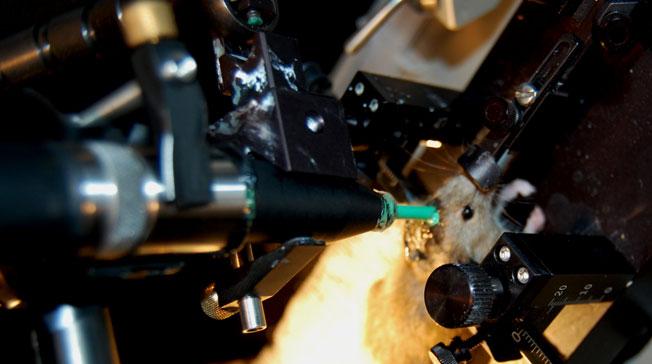
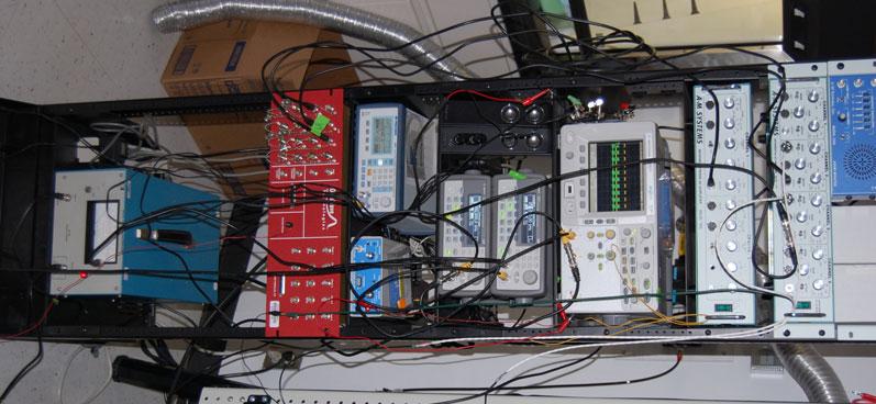
1 Neuron, Volume 66 Supplemental Information Transcranial Pulsed Ultrasound Stimulates Intact Brain Circuits Yusuf Tufail, Alexei Matyushov, Nathan Baldwin, Monica L. Tauchmann, Joseph Georges, Anna Yoshihiro, Stephen I. Helms Tillery, and William J. Tyler SUPPLEMENTAL FIGURE LEGENDS Figure S1 related to Figures 1-7. A general experimental configuration for conducting intact brain circuit stimulation using transcranial pulsed ultrasound is shown. The illustration is meant as a guide to facilitate the set-up of same or similar approaches in other laboratories.
2
3 Figure S2 related to Figure 1. To further outline the strategy for constructing stimulus waveforms a typical low-intensity US waveforms is illustrated. The example US stimulus waveform was constructed using single US pulses (A) containing cycles of.5 MHz US. (B) Fifty US pulses were repeated at a PRF of 2. KHz to produce final stimulus waveforms lasting 25 msec. (C) Multi-dimensional pressure output profiles obtained using a 25 mm planar US transducer alone (top) and with a 5 mm acoustic collimator tapered to a 2 mm output diameter (right) used to differentially target US stimulus waveforms to intact brain regions are illustrated. (D) Normalized pressure profiles obtained with a 25 mm planar transducer (black) and with a tapered 2 mm output diameter (red) collimator are illustrated for X (left) and Y (right) planes. The full-width half-maximum (FWHM) values are illustrated for each plane.
4 Single US Pulse A Hydrophone Voltage Trace (mv) Cycle Pulse; f =.5 MHz Time (msec) ISPTA = 13 mw/cm Time (msec) (m m) ce (mm ).8 FWHM mm 1.89 mm X-axis Distance (mm).8 FWHM mm 1.61 mm Y-axis Distance (mm) ce Dis tan -. Y-a xis Distan 7.5 Dis tan X-axis ) Y-a x ce (mm -. Normalized Pressure Normalized Pressure D Distan X-axis.4 is -7.5 ce (m m ) Normalized Pressure Normalized Pressure ISPPA =.225 W/cm2 5 5 US Pulses; PRF = 2. KHz C PII =.65 mj/cm2 1 ulus Waveform. Hydrophone Voltage (mv) B
5 Figure S3 related to Figure 5. (A) Representative images from coronal sections immunolabeled with antibodies against c-fos. Images are depicted for dorsal cortex of slices rostral and caudal to the stimulus zone, as well as within the stimulus zone (indicated by red). The 2 mm diameter stimulus zone is marked by red arrows for two coronal sections within the stimulus region along the rostral-caudal axis. C-fos + cells are best viewed when using a "magnifying glass" option in the document reader to zoom in on regions of cortex. Within stimulated zones, one can clearly observe a greater density of c-fos + cells. In these regions a rough medial-lateral demarcation zone of positive cells can also be observed, which helps one visualize the path of pulsed US through the tissue. Mean c-fos+ cell densities are plotted for the stimulated hemispheres and zones, as well as the contralateral control hemispheres for the medial-lateral (B) and dorsal-ventral (C) brain axes.


6 A 1. mm Bregma -1. mm Stim Zone Bregma -1.7 mm Stim Zone Bregma -2.2 mm Bregma -4.1 mm 4 *P <.5 C Stim Zone * * * ** * 3 c-fos+ cells/6.25 x 1-2 mm2 Contralateral Control Dorsal ** * 1 2 *P < midline 4 Pial Surface Cortex ** * 1 Depth (mm) c-fos+ cells/6.25 x 1-2 mm2 B Stimulated Hemisphere Corpus Callosum Hippocampus 2 Geniculate Nuclei 3 Thalamic Nuclei VPL VPM 4 Cerebral Peduncle 4 lateral midline 1 distance (mm) 2 3 Subthalamic Nucleus 5 4 lateral 6 Ventral * Stimulated Hemisphere Contralateral Control
7 Figure S4 related to Figure 6. (A) Representative EMG recordings obtained from the left triceps brachii of the same mouse across multiple stimulation trial days in response to stimulation of motor cortex with transcranial pulsed US. The histogram shows the mean EMG amplitudes obtained from mice undergoing multiple repeated trials across days as illustrated above. (B) Electron micrographs illustrate neuropil from the motor cortex of an unstimulated control (left) compared to a US stimulated cortical region (right). Cellular membranes, synapses, dendrites, mitochondria, endoplasmic reticulum, and myelination are observed in these images and do not appear to be different across treatment groups indicating low-intensity transcranial US stimulation produces no gross ultrastructural damage to brain tissue. (C) Histograms illustrating mean rotorod running times (left and middle) and mean wire hang times (right) obtained 24 hr prior to, and 24 hr and 7 d following sham-treatment or US stimulation of motor cortex.


8 A Transcranial US Brain Circuit Stimulation of Right M1 Repeated Across Weeks Control B 1. um 2 µv Left Tricep EMG Response 3 sec Day 7 2 µv Left Tricep EMG Response 3 sec 19,5X Magnification Day EMG Amplitude (µv) d 7d 14d US stim day 17 RPM Control pre 24h 7d post stim 26 RPM pre 24h 7d post stim Hang Time (sec) Figure 3 Rotorod Run Time (sec) 2 µv 3 sec Left Tricep EMG Response Day 14 Performance C Motor pre 24h 7d post stim
9 Figure S5 related to Figure 7. Individual extracellular recording traces obtained in the CA1 s.p. region of intact hippocampus in response to transcranial pulsed US. Traces (black) are illustrated for US-evoked LFP (1-12 Hz; left) and corresponding US-evoked SWP (16-2 Hz; middle) frequency-bands. A 25 msec region of the US-evoked SWP response is expanded (red) to illustrate SWP "ripples" (right) in response to transcranial stimulation of the intact mouse hippocampus with pulsed US.
10 LFP (1-12 Hz) SWP (16-2 Hz) SWP ripples 1 mv 5 µv 1 µv 25 msec region expanded 5 msec 5 msec 25 msec
11 Table S1. Low-intensity transcranial US waveform properties used to stimulate intact mouse motor cortex. PD PRF p r P II I SPTA f (MHz) c/p (msec) (Hz) Np length (sec) (MPa) (mj/cm 2 ) (mw/cm 2 ) MI * * Relatively high-intensity US stimulus waveform used to assess safety
Nature Neuroscience doi: /nn Supplementary Figure 1. Characterization of viral injections.
 Supplementary Figure 1 Characterization of viral injections. (a) Dorsal view of a mouse brain (dashed white outline) after receiving a large, unilateral thalamic injection (~100 nl); demonstrating that
Embryological origin of thalamus
 diencephalon Embryological origin of thalamus The diencephalon gives rise to the: Thalamus Epithalamus (pineal gland, habenula, paraventricular n.) Hypothalamus Subthalamus (Subthalamic nuclei) The Thalamus:
Nature Neuroscience: doi: /nn Supplementary Figure 1. Large-scale calcium imaging in vivo.
 Supplementary Figure 1 Large-scale calcium imaging in vivo. (a) Schematic illustration of the in vivo camera imaging set-up for large-scale calcium imaging. (b) High-magnification two-photon image from
Thalamo-Cortical Relationships Ultrastructure of Thalamic Synaptic Glomerulus
 Central Visual Pathways V1/2 NEUR 3001 dvanced Visual Neuroscience The Lateral Geniculate Nucleus () is more than a relay station LP SC Professor Tom Salt UCL Institute of Ophthalmology Retina t.salt@ucl.ac.uk
Fig.1: A, Sagittal 110x110 mm subimage close to the midline, passing through the cingulum. Note that the fibers of the corpus callosum run at a
 Fig.1 E Fig.1:, Sagittal 110x110 mm subimage close to the midline, passing through the cingulum. Note that the fibers of the corpus callosum run at a slight angle are through the plane (blue dots with
Nature Neuroscience: doi: /nn Supplementary Figure 1. Diverse anorexigenic signals induce c-fos expression in CEl PKC-δ + neurons
 Supplementary Figure 1 Diverse anorexigenic signals induce c-fos expression in CEl PKC-δ + neurons a-c. Quantification of CEl c-fos expression in mice intraperitoneal injected with anorexigenic drugs (a),
Ultrasound Physics & Terminology
 Ultrasound Physics & Terminology This module includes the following: Basic physics terms Basic principles of ultrasound Ultrasound terminology and terms Common artifacts seen Doppler principles Terms for
Introduction to the Central Nervous System: Internal Structure
 Introduction to the Central Nervous System: Internal Structure Objective To understand, in general terms, the internal organization of the brain and spinal cord. To understand the 3-dimensional organization
A Review of Low-Intensity Transcranial Focused Ultrasound for Clinical Applications
 1 A Review of Low-Intensity Transcranial Focused Ultrasound for Clinical Applications Bystritsky A 1, Korb AS 1 Author affiliations: 1 Semel Institute for Neuroscience and Human Behavior, David Geffen
Overview of the Nervous System (some basic concepts) Steven McLoon Department of Neuroscience University of Minnesota
 Steven McLoon Department of Neuroscience University of Minnesota") Overview of the Nervous System (some basic concepts) Steven McLoon Department of Neuroscience University of Minnesota 1 Coffee Hour Tuesday (Sept 11) 10:00-11:00am Friday (Sept 14) 8:30-9:30am Surdyk s
Overview of the Nervous System (some basic concepts) Steven McLoon Department of Neuroscience University of Minnesota 1 Coffee Hour Tuesday (Sept 11) 10:00-11:00am Friday (Sept 14) 8:30-9:30am Surdyk s
Supplementary Materials for
 advances.sciencemag.org/cgi/content/full/1/10/e1500775/dc1 Supplementary Materials for Structural-functional connectivity deficits of neocortical circuits in the Fmr1 /y mouse model of autism Matthias
Transcranial Focused Ultrasound for BOLD fmri Signal Modulation in Humans
 Transcranial Focused Ultrasound for BOLD fmri Signal Modulation in Humans Leo Ai, Jerel K. Mueller, Andrea Grant, Yigitcan Eryaman, and Wynn Legon Abstract Transcranial focused ultrasound (tfus) is an
NMIJ measurement service on ultrasonic field parameters available to demonstrate performance and safety of ultrasonic medical equipment
 NMIJ measurement service on ultrasonic field parameters available to demonstrate performance and safety of ultrasonic medical equipment Masahiro Yoshioka National Metrology Institute of Japan (NMIJ) National
Thalamic control of cortical states
 Supplementary Information Thalamic control of cortical states James F.A. Poulet, Laura M.J. Fernandez, Sylvain Crochet & Carl C.H. Petersen Supplementary Information consists of: 1. Methods 2. Supplementary
Supplemental Information. A Visual-Cue-Dependent Memory Circuit. for Place Navigation
 Neuron, Volume 99 Supplemental Information A Visual-Cue-Dependent Memory Circuit for Place Navigation Han Qin, Ling Fu, Bo Hu, Xiang Liao, Jian Lu, Wenjing He, Shanshan Liang, Kuan Zhang, Ruijie Li, Jiwei
Nature Neuroscience: doi: /nn Supplementary Figure 1
 Supplementary Figure 1 Drd1a-Cre driven ChR2 expression in the SCN. (a) Low-magnification image of a representative Drd1a-ChR2 coronal brain section (n = 2) showing endogenous tdtomato fluorescence (magenta).
EFFECTIVE PARAMETERS FOR ULTRASOUND-INDUCED IN VIVO NEUROSTIMULATION
 http://dx.doi.org/1.116/j.ultrasmedbio.212.9.9 Ultrasound in Med. & Biol., Vol. -, No. -, pp. 1 2, 212 Copyright Ó 212 World Federation for Ultrasound in Medicine & Biology Printed in the USA. All rights
Supplementary Movie Caption
 Supplementary Movie Caption 1. Movie S1. Ultrasound-induced blood focusing in vitro (Fig.2b). 2. Movie S2. Acoustic canalization of blood flow in the gap between two capillaries (Fig. 2d). 3. Movie S3.
Lesson 07: Ultrasound Transducers. This lesson contains 73 slides plus 16 multiple-choice questions.
 Lesson 07: Ultrasound Transducers This lesson contains 73 slides plus 16 multiple-choice questions. This lesson was derived from pages 33 through 42 in the textbook: Ultrasound Transducers Ultrasound Transducers
Nature Neuroscience: doi: /nn Supplementary Figure 1
 Supplementary Figure 1 Atlas representations of the midcingulate (MCC) region targeted in this study compared against the anterior cingulate (ACC) region commonly reported. Coronal sections are shown on
Vision II. Steven McLoon Department of Neuroscience University of Minnesota
 Vision II Steven McLoon Department of Neuroscience University of Minnesota 1 Ganglion Cells The axons of the retinal ganglion cells form the optic nerve and carry visual information into the brain. 2 Optic
Ube3a is required for experience-dependent maturation of the neocortex
 Ube3a is required for experience-dependent maturation of the neocortex Koji Yashiro, Thorfinn T. Riday, Kathryn H. Condon, Adam C. Roberts, Danilo R. Bernardo, Rohit Prakash, Richard J. Weinberg, Michael
Anatomical Substrates of Somatic Sensation
 Anatomical Substrates of Somatic Sensation John H. Martin, Ph.D. Center for Neurobiology & Behavior Columbia University CPS The 2 principal somatic sensory systems: 1) Dorsal column-medial lemniscal system
Neural Recording Methods
 Neural Recording Methods Types of neural recording 1. evoked potentials 2. extracellular, one neuron at a time 3. extracellular, many neurons at a time 4. intracellular (sharp or patch), one neuron at
For more information about how to cite these materials visit
 Author(s): Peter Hitchcock, PH.D., 2009 License: Unless otherwise noted, this material is made available under the terms of the Creative Commons Attribution Non-commercial Share Alike 3.0 License: http://creativecommons.org/licenses/by-nc-sa3.0/
Nature Methods: doi: /nmeth Supplementary Figure 1. Activity in turtle dorsal cortex is sparse.
 Supplementary Figure 1 Activity in turtle dorsal cortex is sparse. a. Probability distribution of firing rates across the population (notice log scale) in our data. The range of firing rates is wide but
Outline of the next three lectures
 Outline of the next three lectures Lecture 35 Anatomy of the human cerebral cortex gross and microscopic cell types connections Vascular supply of the cerebral cortex Disorders involving the cerebral cortex
Sound waves from the auditory environment all combine in the ear canal to form a complex waveform. This waveform is deconstructed by the cochlea with
 1 Sound waves from the auditory environment all combine in the ear canal to form a complex waveform. This waveform is deconstructed by the cochlea with respect to time, loudness, and frequency and neural
SOMATOSENSORY SYSTEMS: Pain and Temperature Kimberle Jacobs, Ph.D.
 SOMATOSENSORY SYSTEMS: Pain and Temperature Kimberle Jacobs, Ph.D. Sensory systems are afferent, meaning that they are carrying information from the periphery TOWARD the central nervous system. The somatosensory
Auditory and Vestibular Systems
 Auditory and Vestibular Systems Objective To learn the functional organization of the auditory and vestibular systems To understand how one can use changes in auditory function following injury to localize
Assessment of a Linear Phased Array Transducer Parameters for Brain Stimulation. M. Salehnia 1,2, H. Ghadiri 1,2,*
 Original Article Assessment of a Linear Phased Array Transducer Parameters for Brain Stimulation M. Salehnia,2, H. Ghadiri,2,* - Department of Medical Physics and Biomedical Engineering, Tehran University
Nature Neuroscience: doi: /nn Supplementary Figure 1
 Supplementary Figure 1 Relative expression of K IR2.1 transcript to enos was reduced 29-fold in capillaries from knockout animals. Relative expression of K IR2.1 transcript to enos was reduced 29-fold
Nature Neuroscience: doi: /nn Supplementary Figure 1
 Supplementary Figure 1 Distribution of GlyT2::eGFP fibers in the mouse thalamus at three different coronal levels. Note the innervation centered in the rostral (CL, PC) and caudal (PF) nuclear groups of
Supplementary Figure 1. ACE robotic platform. A. Overview of the rig setup showing major hardware components of ACE (Automatic single Cell
 2 Supplementary Figure 1. ACE robotic platform. A. Overview of the rig setup showing major hardware components of ACE (Automatic single Cell Experimenter) including the MultiClamp 700B, Digidata 1440A,
Lecture overview. What hypothesis to test in the fly? Quantitative data collection Visual physiology conventions ( Methods )
") Lecture overview What hypothesis to test in the fly? Quantitative data collection Visual physiology conventions ( Methods ) 1 Lecture overview What hypothesis to test in the fly? Quantitative data collection
Lecture overview What hypothesis to test in the fly? Quantitative data collection Visual physiology conventions ( Methods ) 1 Lecture overview What hypothesis to test in the fly? Quantitative data collection
CISC 3250 Systems Neuroscience
 CISC 3250 Systems Neuroscience Levels of organization Central Nervous System 1m 10 11 neurons Neural systems and neuroanatomy Systems 10cm Networks 1mm Neurons 100μm 10 8 neurons Professor Daniel Leeds
S100B+ Cell Density day Spine Formation (%)
") b * CP * HC CP Th DAPI S100B ** 50 40 30 * 20 10 0-10 -20 * * ** 9 6 3 0 0-50 50-100 100-150 150-200 Distance from Prism Face (um) 1-day Spine Formation (%) 1-day Spine Elimination (%) 12 Distance from
b * CP * HC CP Th DAPI S100B ** 50 40 30 * 20 10 0-10 -20 * * ** 9 6 3 0 0-50 50-100 100-150 150-200 Distance from Prism Face (um) 1-day Spine Formation (%) 1-day Spine Elimination (%) 12 Distance from
The effect of anesthetic dose on the motor response induced by low intensity pulsed ultrasound stimulation
 https://doi.org/10.1186/s12868-018-0476-2 BMC Neuroscience RESEARCH ARTICLE Open Access The effect of anesthetic dose on the motor response induced by low intensity pulsed ultrasound stimulation Yi Yuan
Cerebellum. Steven McLoon Department of Neuroscience University of Minnesota
 Cerebellum Steven McLoon Department of Neuroscience University of Minnesota 1 Anatomy of the Cerebellum The cerebellum has approximately half of all the neurons in the central nervous system. The cerebellum
SUPPLEME TARY FIGURE 1 a b c
 Coherent gamma oscillations couple the amygdala and striatum during learning. Popescu, Popa, Pare SUPPLEME TARY FIGURE 1 a b c LG LP LD R LA BL OT BM HF V HF VP RE VP CL PC d PU e 2 mm R f CP HF 2 mm GP
Supplementary Figure 1. Nature Neuroscience: doi: /nn.4547
 Supplementary Figure 1 Characterization of the Microfetti mouse model. (a) Gating strategy for 8-color flow analysis of peripheral Ly-6C + monocytes from Microfetti mice 5-7 days after TAM treatment. Living
Annular Array Transducer and Matched Amplifier for Therapeutic Ultrasound
 ARCHIVES OF ACOUSTICS 35, 4, 653 660 (2010) DOI: 10.2478/v10168-010-0049-6 Annular Array Transducer and Matched Amplifier for Therapeutic Ultrasound Wojciech SECOMSKI, Andrzej NOWICKI, Janusz WÓJCIK, Marcin
Brain anatomy and artificial intelligence. L. Andrew Coward Australian National University, Canberra, ACT 0200, Australia
 Brain anatomy and artificial intelligence L. Andrew Coward Australian National University, Canberra, ACT 0200, Australia The Fourth Conference on Artificial General Intelligence August 2011 Architectures
Introduction to Electrophysiology
 Introduction to Electrophysiology Dr. Kwangyeol Baek Martinos Center for Biomedical Imaging Massachusetts General Hospital Harvard Medical School 2018-05-31s Contents Principles in Electrophysiology Techniques
Organization of the nervous system 2
 Organization of the nervous system 2 Raghav Rajan Bio 334 Neurobiology I August 22nd 2013 1 Orienting within the brain absolute axes and relative axes SUPERIOR (above) ANTERIOR (in front) Anterior/Posterior,
The Cerebellum. Outline. Lu Chen, Ph.D. MCB, UC Berkeley. Overview Structure Micro-circuitry of the cerebellum The cerebellum and motor learning
 The Cerebellum Lu Chen, Ph.D. MCB, UC Berkeley 1 Outline Overview Structure Micro-circuitry of the cerebellum The cerebellum and motor learning 2 Overview Little brain 10% of the total volume of the brain,
Nature Protocols: doi: /nprot Supplementary Figure 1. Effect of different enzymes on CD13 and CD31 populations.
 Supplementary Figure 1 Effect of different enzymes on CD13 and CD31 populations. Comparison of cortex dissociated for 30 minutes with collagenase/dispase digestion (a) or papain (b). Papain degrades the
Strick Lecture 3 March 22, 2017 Page 1
 Strick Lecture 3 March 22, 2017 Page 1 Cerebellum OUTLINE I. External structure- Inputs and Outputs Cerebellum - (summary diagram) 2 components (cortex and deep nuclei)- (diagram) 3 Sagittal zones (vermal,
Our senses provide us with wonderful capabilities. If you had to lose one, which would it be?
 Our senses provide us with wonderful capabilities. If you had to lose one, which would it be? Neurological disorders take away sensation without a choice! http://neuroscience.uth.tmc.edu/s2/chapter04.html
Cerebral Cortex 1. Sarah Heilbronner
 Cerebral Cortex 1 Sarah Heilbronner heilb028@umn.edu Want to meet? Coffee hour 10-11am Tuesday 11/27 Surdyk s Overview and organization of the cerebral cortex What is the cerebral cortex? Where is each
LISC-322 Neuroscience Cortical Organization
 LISC-322 Neuroscience Cortical Organization THE VISUAL SYSTEM Higher Visual Processing Martin Paré Assistant Professor Physiology & Psychology Most of the cortex that covers the cerebral hemispheres is
Nature Neuroscience: doi: /nn Supplementary Figure 1. Trial structure for go/no-go behavior
 Supplementary Figure 1 Trial structure for go/no-go behavior a, Overall timeline of experiments. Day 1: A1 mapping, injection of AAV1-SYN-GCAMP6s, cranial window and headpost implantation. Water restriction
Laboratory Manual for Comparative Anatomy and Physiology Figure 15.1 Transparency Master 114
 Neuron Capillary Astrocyte Microglial cell Neuron Fluid-filled cavity Process of oligodendrocyte Ependymal cells Brain or spinal cord tissue Myelin sheath Nerve fibers Figure 15.1 Transparency Master 114
Brain Rhythms. Objectives
 Brain Rhythms Michael O. Poulter, Ph.D. Professor, Molecular Brain Research Group Robarts Research Institute Depts of Physiology & Pharmacology, Clinical Neurological Sciences Schulich School of Medicine
COGNITIVE SCIENCE 107A. Sensory Physiology and the Thalamus. Jaime A. Pineda, Ph.D.
 COGNITIVE SCIENCE 107A Sensory Physiology and the Thalamus Jaime A. Pineda, Ph.D. Sensory Physiology Energies (light, sound, sensation, smell, taste) Pre neural apparatus (collects, filters, amplifies)
Medical Neuroscience Tutorial
 Pain Pathways Medical Neuroscience Tutorial Pain Pathways MAP TO NEUROSCIENCE CORE CONCEPTS 1 NCC1. The brain is the body's most complex organ. NCC3. Genetically determined circuits are the foundation
Analysis of Transcranial Focused Ultrasound Beam Profile Sensitivity for Neuromodulation of the Human Brain
 Analysis of Transcranial Focused Ultrasound Beam Profile Sensitivity for Neuromodulation of the Human Brain Jerel K. Mueller 1, Wynn Legon 2, and William J. Tyler* 3 1 School of Biomedical Engineering
SUPPLEMENTARY INFORMATION
 Supplementary Figure 1. Normal AMPAR-mediated fepsp input-output curve in CA3-Psen cdko mice. Input-output curves, which are plotted initial slopes of the evoked fepsp as function of the amplitude of the
Lack of GPR88 enhances medium spiny neuron activity and alters. motor- and cue- dependent behaviors
 Lack of GPR88 enhances medium spiny neuron activity and alters motor- and cue- dependent behaviors Albert Quintana, Elisenda Sanz, Wengang Wang, Granville P. Storey, Ali D. Güler Matthew J. Wanat, Bryan
Organization of the nervous system. [See Fig. 48.1]
![Organization of the nervous system. [See Fig. 48.1]](/thumbs/90/103926552.jpg "Organization of the nervous system. [See Fig. 48.1]") Nervous System [Note: This is the text version of this lecture file. To make the lecture notes downloadable over a slow connection (e.g. modem) the figures have been replaced with figure numbers as found
Nervous System [Note: This is the text version of this lecture file. To make the lecture notes downloadable over a slow connection (e.g. modem) the figures have been replaced with figure numbers as found
Cephalization. Nervous Systems Chapter 49 11/10/2013. Nervous systems consist of circuits of neurons and supporting cells
 Nervous Systems Chapter 49 Cephalization Nervous systems consist of circuits of neurons and supporting cells Nervous system organization usually correlates with lifestyle Organization of the vertebrate
TREATMENT-SPECIFIC ABNORMAL SYNAPTIC PLASTICITY IN EARLY PARKINSON S DISEASE
 TREATMENT-SPECIFIC ABNORMAL SYNAPTIC PLASTICITY IN EARLY PARKINSON S DISEASE Angel Lago-Rodriguez 1, Binith Cheeran 2 and Miguel Fernández-Del-Olmo 3 1. Prism Lab, Behavioural Brain Sciences, School of
Department of Cognitive Science UCSD
 Department of Cognitive Science UCSD Verse 1: Neocortex, frontal lobe, Brain stem, brain stem, Hippocampus, neural node, Right hemisphere, Pons and cortex visual, Brain stem, brain stem, Sylvian fissure,
t e c h n i c a l r e p o r t s
 Transcranial focused ultrasound modulates the activity of primary somatosensory cortex in humans Wynn Legon 1, Tomokazu F Sato 1, Alexander Opitz 1,, Jerel Mueller, Aaron Barbour 1, Amanda Williams 1 &
What Cell Make Up the Brain and Spinal Cord
 What Cell Make Up the Brain and Spinal Cord Jennifer LaVail, Ph.D. (http://anatomy.ucsf.edu/pages/lavaillab/index.html) What kinds of cells are these?" Neuron?" Epithelial cell?" Glial cell?" What makes
Lecture VIII. The Spinal Cord, Reflexes and Brain Pathways!
 Reflexes and Brain Bio 3411! Monday!! 1! Readings! NEUROSCIENCE 5 th ed: Review Chapter 1 pp. 11-21;!!Read Chapter 9 pp. 189-194, 198! THE BRAIN ATLAS 3 rd ed:! Read pp. 4-17 on class web site! Look at
Prof. Saeed Abuel Makarem & Dr.Sanaa Alshaarawy
 Prof. Saeed Abuel Makarem & Dr.Sanaa Alshaarawy 1 Objectives By the end of the lecture, you should be able to: Describe the anatomy and main functions of the thalamus. Name and identify different nuclei
Introduction to TMS Transcranial Magnetic Stimulation
 Introduction to TMS Transcranial Magnetic Stimulation Lisa Koski, PhD, Clin Psy TMS Neurorehabilitation Lab Royal Victoria Hospital 2009-12-14 BIC Seminar, MNI Overview History, basic principles, instrumentation
Developmental sequence of brain
 Cerebellum Developmental sequence of brain Fourth week Fifth week Location of cerebellum Lies above and behind the medullar and pons and occupies posterior cranial fossa Location of cerebellum External
UNIT 5 REVIEW GUIDE - NERVOUS SYSTEM 1) State the 3 functions of the nervous system. 1) 2) 3)
 State the 3 functions of the nervous system. 1) 2) 3)") UNIT 5 REVIEW GUIDE - NERVOUS SYSTEM State the 3 functions of the nervous system. Briefly describe the general function(s) of each of the following neuron types: a) SENSORY NEURONS: b) INTERNEURONS: c)
UNIT 5 REVIEW GUIDE - NERVOUS SYSTEM State the 3 functions of the nervous system. Briefly describe the general function(s) of each of the following neuron types: a) SENSORY NEURONS: b) INTERNEURONS: c)
Note: Waxman is very sketchy on today s pathways and nonexistent on the Trigeminal.
 Dental Neuroanatomy Thursday, February 3, 2011 Suzanne Stensaas, PhD Note: Waxman is very sketchy on today s pathways and nonexistent on the Trigeminal. Resources: Pathway Quiz for HyperBrain Ch. 5 and
The mammalian cochlea possesses two classes of afferent neurons and two classes of efferent neurons.
 1 2 The mammalian cochlea possesses two classes of afferent neurons and two classes of efferent neurons. Type I afferents contact single inner hair cells to provide acoustic analysis as we know it. Type
Medial View of Cerebellum
 Meds 5371 System Neuroscience D. L. Oliver CEREBELLUM Anterior lobe (spinal) Posterior lobe (cerebral) Flocculonodular lobe (vestibular) Medial View of Cerebellum 1 Ventral View of Cerebellum Flocculus
Supplementary Figure 1: Kv7 currents in neonatal CA1 neurons measured with the classic M- current voltage-clamp protocol.
 Supplementary Figures 1-11 Supplementary Figure 1: Kv7 currents in neonatal CA1 neurons measured with the classic M- current voltage-clamp protocol. (a), Voltage-clamp recordings from CA1 pyramidal neurons
Tuning properties of individual circuit components and stimulus-specificity of experience-driven changes.
 Supplementary Figure 1 Tuning properties of individual circuit components and stimulus-specificity of experience-driven changes. (a) Left, circuit schematic with the imaged component (L2/3 excitatory neurons)
Motor systems III: Cerebellum April 16, 2007 Mu-ming Poo
 Motor systems III: Cerebellum April 16, 2007 Mu-ming Poo Population coding in the motor cortex Overview and structure of cerebellum Microcircuitry of cerebellum Function of cerebellum -- vestibulo-ocular
Brainstem: Midbrain. 1. Midbrain gross external anatomy 2. Internal structure of the midbrain:
 Brainstem: Midbrain 1. Midbrain gross external anatomy 2. Internal structure of the midbrain: cerebral peduncles tegmentum tectum (guadrigeminal plate) Midbrain Midbrain general features location between
Resonant synchronization of heterogeneous inhibitory networks
 Cerebellar oscillations: Anesthetized rats Transgenic animals Recurrent model Review of literature: γ Network resonance Life simulations Resonance frequency Conclusion Resonant synchronization of heterogeneous
Circuits & Behavior. Daniel Huber
 Circuits & Behavior Daniel Huber How to study circuits? Anatomy (boundaries, tracers, viral tools) Inactivations (lesions, optogenetic, pharma, accidents) Activations (electrodes, magnets, optogenetic)
Suppl. Information Supplementary Figure 1. Strategy/latency analysis of individual mice during maze learning. a,
 Goal-oriented searching mediated by ventral hippocampus early in trial-and-error learning Ruediger, S, Spirig, D., Donato, F., Caroni, P. Suppl. Information Supplementary Figure 1. Strategy/latency analysis
Systems Neuroscience Dan Kiper. Today: Wolfger von der Behrens
 Systems Neuroscience Dan Kiper Today: Wolfger von der Behrens wolfger@ini.ethz.ch 18.9.2018 Neurons Pyramidal neuron by Santiago Ramón y Cajal (1852-1934, Nobel prize with Camillo Golgi in 1906) Neurons
Thalamus and the Internal Capsule
 This power point is made available as an educational resource or study aid for your use only. This presentation may not be duplicated for others and should not be redistributed or posted anywhere on the
Nervous System, Neuroanatomy, Neurotransmitters
 Nervous System, Neuroanatomy, Neurotransmitters Neurons Structure of neurons Soma Dendrites Spines Axon Myelin Nodes of Ranvier Neurons Structure of neurons Axon collaterals 1 Neurons Structure of neurons
Hormonal gain control of a medial preoptic area social reward circuit
 CORRECTION NOTICE Nat. Neurosci. 20, 449 458 (2017) Hormonal gain control of a medial preoptic area social reward circuit Jenna A McHenry, James M Otis, Mark A Rossi, J Elliott Robinson, Oksana Kosyk,
BASAL GANGLIA. Dr JAMILA EL MEDANY
 BASAL GANGLIA Dr JAMILA EL MEDANY OBJECTIVES At the end of the lecture, the student should be able to: Define basal ganglia and enumerate its components. Enumerate parts of Corpus Striatum and their important
Activity Dependent Changes At the Developing Neuromuscular Junction
 Activity Dependent Changes At the Developing Neuromuscular Junction (slides 16, 17 and 18 have been slightly modified for clarity) MCP Lecture 2-3 9.013/7.68 04 Neuromuscular Junction Development 1. Muscle
Somatosensory System. Steven McLoon Department of Neuroscience University of Minnesota
 Somatosensory System Steven McLoon Department of Neuroscience University of Minnesota 1 Course News Dr. Riedl s review session this week: Tuesday (Oct 10) 4-5pm in MCB 3-146B 2 Sensory Systems Sensory
-80 Figure 1. Identification of dopaminergic neurons in. VTA slices (a) Micrographs demonstrate the location of the VTA with
 Micrographs demonstrate the location of the VTA with") Potential (mv) A B C Potential (mv) D E F Biocytin TH Merge R 12 12 1 1 8 G 6 4 8 6 4 H 2 2-2 -2-4 -4-6 -6 Supplemental -8-8 Figure 1. Identification of dopaminergic neurons in -1 VTA slices. -1 (a) Micrographs
Potential (mv) A B C Potential (mv) D E F Biocytin TH Merge R 12 12 1 1 8 G 6 4 8 6 4 H 2 2-2 -2-4 -4-6 -6 Supplemental -8-8 Figure 1. Identification of dopaminergic neurons in -1 VTA slices. -1 (a) Micrographs
Functional Neuroanatomy and Physiology for Movement Disorders
 Functional Neuroanatomy and Physiology for Movement Disorders Chang, Won Seok Division of Stereotactic and Functional Neurosurgery, Department of Neurosurgery, Brain Research Institute, Yonsei University
Cortical Control of Movement
 Strick Lecture 2 March 24, 2006 Page 1 Cortical Control of Movement Four parts of this lecture: I) Anatomical Framework, II) Physiological Framework, III) Primary Motor Cortex Function and IV) Premotor
Nsci 2100: Human Neuroanatomy 2017 Examination 3
 Name KEY Lab Section Nsci 2100: Human Neuroanatomy 2017 Examination 3 On this page, write your name and lab section. On your bubble answer sheet, enter your name (last name, space, first name), internet
PARIETAL LOBE. Vasilios A. Zerris MD, MPH, MSc, FAANS
 PARIETAL LOBE Vasilios A. Zerris MD, MPH, MSc, FAANS Diplomate of the American Board of Neurological Surgery Fellow of the American Association of Neurological Surgeons Professor of Neurosurgery, European
Astrocyte signaling controls spike timing-dependent depression at neocortical synapses
 Supplementary Information Astrocyte signaling controls spike timing-dependent depression at neocortical synapses Rogier Min and Thomas Nevian Department of Physiology, University of Berne, Bern, Switzerland
Biological Bases of Behavior. 8: Control of Movement
 Biological Bases of Behavior 8: Control of Movement m d Skeletal Muscle Movements of our body are accomplished by contraction of the skeletal muscles Flexion: contraction of a flexor muscle draws in a
Nature Neuroscience: doi: /nn.4642
 Supplementary Figure 1 Recording sites and example waveform clustering, as well as electrophysiological recordings of auditory CS and shock processing following overtraining. (a) Recording sites in LC
NS201C Anatomy 1: Sensory and Motor Systems
 NS201C Anatomy 1: Sensory and Motor Systems 25th January 2017 Peter Ohara Department of Anatomy peter.ohara@ucsf.edu The Subdivisions and Components of the Central Nervous System Axes and Anatomical Planes
General Sensory Pathways of the Trunk and Limbs
 General Sensory Pathways of the Trunk and Limbs Lecture Objectives Describe gracile and cuneate tracts and pathways for conscious proprioception, touch, pressure and vibration from the limbs and trunk.
File name: Supplementary Information Description: Supplementary Figures, Supplementary Table and Supplementary References
 File name: Supplementary Information Description: Supplementary Figures, Supplementary Table and Supplementary References File name: Supplementary Data 1 Description: Summary datasheets showing the spatial
Supporting Information
 ATP from synaptic terminals and astrocytes regulates NMDA receptors and synaptic plasticity through PSD- 95 multi- protein complex U.Lalo, O.Palygin, A.Verkhratsky, S.G.N. Grant and Y. Pankratov Supporting
LIMBIC SYSTEM. Dr. Amani A. Elfaki Associate Professor Department of Anatomy
 LIMBIC SYSTEM Dr. Amani A. Elfaki Associate Professor Department of Anatomy Learning Objectives Define the limbic system Identify the parts of the limbic system Describe the circulation of the limbic system