blood mononuclear cells were stained with AAD, annexin, CD3, CD4, CD25, LAP and specific
|
|
|
- Shavonne Lang
- 5 years ago
- Views:
Transcription
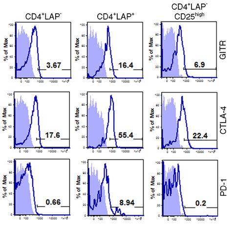
1 Supplementary figure legends Figure 1: Selective expression of regulatory markers on CD4 + LAP + T cells. Human peripheral blood mononuclear cells were stained with AAD, annexin, CD3, CD4, CD25, LAP and specific markers including GITR, CTLA-4 and PD-1. Filled blue profiles represent isotype control; empty profiles represent specific staining for different antibodies on selected populations. The percentage positive cells are indicated in each plot. Results are representative of 3 independent experiments. Figure 2: Suppressive activity of non-activated and activated CD4 + LAP + T cells. A, Proliferation of CFSE labeled responder T cells in absence and presence of CD4 + LAP + T cells. B, Sorted CD4 + LAP + T cells were activated with anti-cd3, anti-cd28 and IL-2 and were stained for expression of LAP. Black histograms represent CD4 + LAP - cells, red histograms represent CD4 + LAP + cells on day0 and blue histograms represent expression of LAP on CD4 + LAP + T cells after 48hrs of activation. C, Suppression mediated by activated or non-activated CD4 + LAP + T cells. Results are shown as mean and SD of four independent experiments. D, Effect of CD4 + LAP + T cells on IFN-γ and IL-2 secretion from responder T cells in a co-culture assay. E, Suppression of responder T cell proliferation mediated by CD4 + LAP + T cells in absence and presence of transwell.
2 Supplementary Figure 1
3 Supplementary Figure 2
4 Supplementary Table 1: List of antibodies used to determine signaling pathways in LAP + T cells Target Phospho-Elk-1 (Ser383) Antibody Phospho-p38 MAPK (Thr180/Tyr182) Phospho-p38 MAPK (Thr180/Tyr182) Phospho-p38 MAP Kinase (Thr180/Tyr182) Antibody Phospho-MKK3/MKK6 (Ser189/207) Antibody Phospho-ATF-2 (Thr71) Antibody Phospho-HSP27 (Ser82) Antibody Phospho-MAPKAPK-2 (Thr334) Antibody Phospho-SEK1/MKK4 (Thr261) Antibody Phospho-SAPK/JNK (Thr183/Tyr185) Antibody Phospho-ATF-2 (Thr71) Antibody Phospho-c-Jun (Ser63) II Antibody Phospho-Akt (Ser473) Antibody Phospho-Akt (Thr308) Antibody Akt Antibody Phospho-GSK-3beta (Ser9) Antibody Phospho-Raf (Ser259) Antibody Phospho-PTEN (Ser380) Antibody Phospho-PDK1 (Ser241) Antibody Phospho-Stat1 (Tyr701) Antibody Phospho-Stat2 (Tyr690) Antibody Phospho-Stat3 (Tyr705) Antibody Phospho-Stat3 (Ser727) Antibody Phospho-Stat5 (Tyr694) Antibody Phospho-Stat6 (Tyr641) Antibody Company Millipore
5 Phospho-c-Raf (Ser338) (56A6) Rabbit mab Phospho-Src Family (Tyr416) Antibody Phospho-Pyk2 (Tyr402) Pyk2 Phospho-MEK1/2 (Ser217/221) Antibody Phospho-MEK1/2 (Ser221) Phospho-p44/42 MAPK (Thr202/Tyr204) Phospho-p44/42 MAP Kinase (Thr202/Tyr204) Antibody Phospho-Erk 1/2 (Thr202/Tyr204) Phospho-p90RSK (Ser380) Antibody Phospho-PKC (pan) (BetaII Ser660) Antibody Phospho-PKCalpha/betaII (Thr638/641) Antibody Phospho-PKCdelta (Thr505) Antibody Phospho-PKCdelta (Ser643) Antibody Phospho-PKD/PKCmu (Ser744/748) Antibody Phospho-PKD/PKCmu (Ser916) Antibody PKD/PKCmu Antibody Phospho-PKCtheta (Thr538) Antibody Phospho-PKCzeta/lambda (Thr410/403) Antibody Phospho-Lck (Tyr505) Antibody LAT (phospho Y191) antibody [E205] Phospho-Zap70 (Y315 + Y319) Phospho-ZAP70 (Tyr493) Phospho-CD19 (Tyr513) Antibody Phospho-Syk (Tyr323) Antibody Phospho-Syk (Tyr525/526) Antibody Syk Antibody Phospho-Zap-70 (Tyr319)/Syk (Tyr352) Antibody Phospho-BLNK (Tyr96) Antibody Phospho-PLCgamma2 (Tyr1217) Antibody Phospho-PLCgamma1 (Tyr771) Antibody Phospho-NFKB2 p100 (Ser866/870) Millipore
6 Phospho-NF-kappaB p105 (Ser933) Antibody Phospho-NF-κB p65 (Ser536) Antibody Phospho-IKKalpha/beta (Ser176/180) Antibody II Phospho-IkappaB-alpha (Ser32) Antibody Phospho-Akt Substrate (RXRXXS/T) (110B7) Rabbit mab Phospho-(Ser) PKC Substrate Antibody Phospho-PKA Substrate (RRXS/T) (100G7) Rabbit mab Phospho-SHIP1 Phospho-(Ser) CDKs Substrate Antibody Phospho-(Ser/Thr) ATM/ATR Substrate Antibody Phospho-IRAK1 (Ser376) Antibody Phospho-TAK1 (Thr184/187) (90C7) Rabbit mab Phospho-IRF-3 (Ser396) Antibody Phospho-IRAK (Thr209) Phospho-Smad2 (Thr220) Phospho-Smad2 (Ser465/467) Phospho-Smad3 (Ser423/425) Phospho-Smad1/5 (ser463/465) Phospho-Smad2 (Ser465/467) Phospho-mTor (Ser2448) Phospho-mTor (Ser 2481) Phospho-MKK7(Ser271/Thr275) Antibody Phospho-Tpl2 (Ser 400) Phospho-PI3K p85 (Tyr458)/p55 (Tyr199) Phospho-MARCKS (Ser ) Phospho-cFos (Thr232) Tpl2 Antibody PI3K p85 mtor Smad2 Smad5 Phospho-FoxO1 (Ser256) Antibody Prosci Prosci
7 Phospho-FoxO1 (Ser319)/FoxO4 (Ser262) Antibody Phospho-FoxO1 (Thr24)/FoxO3a (Thr32) Antibody Phospho-FoxO3a (Ser318/321) Antibody Phospho-FoxO3a (Ser253) Antibody FoxO1 Antibody FoxO3a Antibody FoxO4 Antibody IRAK4 IRAK-M Antibody IRAK antibody p38 MAP Kinase Antibody PKCα Antibody TAK1 Antibody Phospho-Src (Tyr527) Antibody FOXP3 RORgT Phospho-Stat1 (Ser727) Phospho-Stat5 (Tyr694) Acetyl-Stat3 (Lys685) Antibody Phospho-Stat1 (Ser727) Phospho-Stat5 (Tyr694) Phospho-Stat6 (Tyr641) Stat1 Stat2 Stat3 Stat5 Stat6 Smad3 Smad4 Smad6 Phospho-Smad2 (Ser467) Phospho-Smad3 (Ser423/425) Biolegend
8 Src Phospho-Src (Tyr529) Phospho-Src (Tyr418) Non-phospho-Src (Tyr527) IRF3 IRF4 IRF7 IRF8 Phospho-IRF-3 (Ser396) Aryl hydrocarbon Receptor p44/42 MAPK (Erk1/2) TRAF2 TRAF3 NIK Phospho-IKK alpha/beta (Ser176/180) IKK alpha NFkB2 p100/p52 RelB Raptor Rictor MyD88 Antibody Tollip Antibody TAB1 SOCS1 TRAF6 Phospho-Rac1/cdc42 (Ser71) Antibody Phospho-CREB (Ser133) ATF3 MEF2C MEF2C (phospho S387) MEF2C (phospho T300) Proteintech
Supplementary Material. Part I: Sample Information. Part II: Pathway Information
 Supplementary Material Part I: Sample Information Three NPC cell lines, CNE1, CNE2, and HK1 were treated with CYC202. Gene expression of 380 selected genes were collected at 0, 2, 4, 6, 12 and 24 hours
MAPK Pathway
 MAPK Pathway Mitogen-activated protein kinases (MAPK) are proteins that are serine/ threonine specific kinases which are activated by a wide range of stimuli including proinflammatory cytokines, growth
Timing Matters in T Cell Differentiation
 Virtual PI Meeting April 2012 000 p1 001 101 p3 p2 011 100 111 110 Timing Matters in T Cell Differentiation Natasa Miskov-Zivanov University of Pittsburgh 010 Acknowledgements 2 Faeder Lab: Department
Dox. R26-rtTA Tyr-CreERT2. any ink/arf, no rtta (n=8) ink/arf +/+ (n=5) Day 0 Day 11 Day 18 Day 28
 ink/arf +/+ (n=5) Day 0 Day 11 Day 18 Day 28") A 4OHT Dox hraf iip tumors inras ddh 2 O -RT Ink/Arf / Pten l/ l R26-lsl-rtTA Tyr-reERT2 TetO-hRAF V6E Ink/Arf / Pten / R26-rtTA Tyr-reERT2 TetO-hRAF V6E Ink/Arf / Pten / R26-rtTA Tyr-reERT2 TetO-hRAF
A 4OHT Dox hraf iip tumors inras ddh 2 O -RT Ink/Arf / Pten l/ l R26-lsl-rtTA Tyr-reERT2 TetO-hRAF V6E Ink/Arf / Pten / R26-rtTA Tyr-reERT2 TetO-hRAF V6E Ink/Arf / Pten / R26-rtTA Tyr-reERT2 TetO-hRAF
Supplementary Figure 1. IL-12 serum levels and frequency of subsets in FL patients. (A) IL-12
 IL-12") 1 Supplementary Data Figure legends Supplementary Figure 1. IL-12 serum levels and frequency of subsets in FL patients. (A) IL-12 serum levels measured by multiplex ELISA (Luminex) in FL patients before
1 Supplementary Data Figure legends Supplementary Figure 1. IL-12 serum levels and frequency of subsets in FL patients. (A) IL-12 serum levels measured by multiplex ELISA (Luminex) in FL patients before
Bio-Plex Pro Cell Signaling Assays
 Acute Phase Response Cancer Cardiovascular Disease Diabetes Cytokines, Chemokines, Growth Factors Immunology/Inflammation Immunoglobulin Isotyping Cell Signaling Toxicology Bio-Plex Pro Cell Signaling
Signaling Through Immune System Receptors (Ch. 7)
") Signaling Through Immune System Receptors (Ch. 7) 1. General principles of signal transduction and propagation. 2. Antigen receptor signaling and lymphocyte activation. 3. Other receptors and signaling
Signaling Through Immune System Receptors (Ch. 7) 1. General principles of signal transduction and propagation. 2. Antigen receptor signaling and lymphocyte activation. 3. Other receptors and signaling
Understanding Signaling Pathways by Modifying Sensitivity to PLX4720 in B-RAF V600E Melanoma
 Understanding Signaling Pathways by Modifying Sensitivity to PLX4720 in B-RAF V600E Melanoma Muska Hassan NCI-ICBP Summer Fellow Broad Institute of MIT and Harvard: Cancer Program Mentor: Cory Johannessen,
Cancer Biology Products
 Cancer Biology Products Rev. 013008 Apoptosis Cell adhesion 14-3-3 ζ/δ 14-3-3 ζ/δ Akt β-catenin ASK-1 Cutaneous Lymphocyte-associated Antigen (CLA) Dystroglycan-Phosphorylated (Tyr893) EMMPRIN (Neurothelin,
Supplementary figures
 Supplementary figures Supplementary Figure 1 A WT -/- Percentage(%).6.4.2 Peripheral blood Cell number(1 6 ) 4 3 2 1 MLN Cell number(1 7 ) 1 8 6 4 2 Spleen Supplementary Figure 1 No baseline value differences
Improved Stability of the LANCE Ultra Signal in Kinase Assays
 Improved Stability of the LANCE Ultra Signal in Kinase Assays LANCE Ultra is a high throughput screening (HTS) technology platform optimized for homogeneous time-resolved fluorescence resonance energy
Phosphorylation Site Company Cat #
 Supplemental Table 1. Antibodies used for RPPA analysis. Label Protein Phosphorylation Site Company Cat # Used on MDA_CLSS Used on MDA_Pilot 4EBP1 4EBP1 Cell Signaling 9452 No 4EBP1.pS65 4EBP1 S65 Cell
Therapeutic Resistance to HER2 Targeted Therapies. Neil Spector, M.D Duke Cancer Institute Duke University Medical Center
 Therapeutic Resistance to HER2 Targeted Therapies Neil Spector, M.D Duke Cancer Institute Duke University Medical Center Relevant Financial Disclosures Millennium/Takeda Pharmaceuticals (consultant, sponsored
Signal Transduction Pathway Smorgasbord
 Molecular Cell Biology Lecture. Oct 28, 2014 Signal Transduction Pathway Smorgasbord Ron Bose, MD PhD Biochemistry and Molecular Cell Biology Programs Washington University School of Medicine Outline 1.
Supplementary Figure 1 Role of Raf-1 in TLR2-Dectin-1-mediated cytokine expression
 Supplementary Figure 1 Supplementary Figure 1 Role of Raf-1 in TLR2-Dectin-1-mediated cytokine expression. Quantitative real-time PCR of indicated mrnas in DCs stimulated with TLR2-Dectin-1 agonist zymosan
Title: Cytosolic DNA-mediated, STING-dependent pro-inflammatory gene. Fig. S1. STING ligands-mediated signaling response in MEFs. (A) Primary MEFs (1
 Primary MEFs (1") 1 Supporting Information 2 3 4 Title: Cytosolic DNA-mediated, STING-dependent pro-inflammatory gene induction necessitates canonical NF-κB activation through TBK1 5 6 Authors: Abe et al. 7 8 9 Supporting
1 Supporting Information 2 3 4 Title: Cytosolic DNA-mediated, STING-dependent pro-inflammatory gene induction necessitates canonical NF-κB activation through TBK1 5 6 Authors: Abe et al. 7 8 9 Supporting
Growth and Differentiation Phosphorylation Sampler Kit
 Growth and Differentiation Phosphorylation Sampler Kit E 0 5 1 0 1 4 Kits Includes Cat. Quantity Application Reactivity Source Akt (Phospho-Ser473) E011054-1 50μg/50μl IHC, WB Human, Mouse, Rat Rabbit
State of the Art 3: Immunotherapy and Modulators of Apoptosis
 State of the Art 3: Immunotherapy and Modulators of Apoptosis James Finke, PhD - Cleveland Clinic, Immunology Crystal Mackall, MD - NCI, Pediatric Oncology James Mier, MD - BIDMC, Medical Oncology Craig
Supplementary Information. Table of contents
 Supplementary Information Table of contents Fig. S1. Inhibition of specific upstream kinases affects the activity of the analyzed readouts Fig. S2. Down-regulation of INCENP gene induces the formation
Supplementary Figure 1. Normal T lymphocyte populations in Dapk -/- mice. (a) Normal thymic development in Dapk -/- mice. Thymocytes from WT and Dapk
 Normal thymic development in Dapk -/- mice. Thymocytes from WT and Dapk") Supplementary Figure 1. Normal T lymphocyte populations in Dapk -/- mice. (a) Normal thymic development in Dapk -/- mice. Thymocytes from WT and Dapk -/- mice were stained for expression of CD4 and CD8.
Supplementary Figure 1. Normal T lymphocyte populations in Dapk -/- mice. (a) Normal thymic development in Dapk -/- mice. Thymocytes from WT and Dapk -/- mice were stained for expression of CD4 and CD8.
W/T Itgam -/- F4/80 CD115. F4/80 hi CD115 + F4/80 + CD115 +
 F4/8 % in the peritoneal lavage 6 4 2 p=.15 n.s p=.76 CD115 F4/8 hi CD115 + F4/8 + CD115 + F4/8 hi CD115 + F4/8 + CD115 + MHCII MHCII Supplementary Figure S1. CD11b deficiency affects the cellular responses
Overcoming Resistance to HER2 Inhibitors Through State-Specific Kinase Binding
 Supplementary Information Overcoming Resistance to Inhibitors Through State-Specific Kinase Binding Chris J. Novotny 1, Sirkku Pollari 2, Jin H. Park 3, Mark A. Lemmon 3,4, Weijun Shen 2*, Kevan M. Shokat
Supplemental Figure 1. Signature gene expression in in vitro differentiated Th0, Th1, Th2, Th17 and Treg cells. (A) Naïve CD4 + T cells were cultured
 Naïve CD4 + T cells were cultured") Supplemental Figure 1. Signature gene expression in in vitro differentiated Th0, Th1, Th2, Th17 and Treg cells. (A) Naïve CD4 + T cells were cultured under Th0, Th1, Th2, Th17, and Treg conditions. mrna
Supplemental Figure 1. Signature gene expression in in vitro differentiated Th0, Th1, Th2, Th17 and Treg cells. (A) Naïve CD4 + T cells were cultured under Th0, Th1, Th2, Th17, and Treg conditions. mrna
Akt and mtor pathways differentially regulate the development of natural and inducible. T H 17 cells
 Akt and mtor pathways differentially regulate the development of natural and inducible T H 17 cells Jiyeon S Kim, Tammarah Sklarz, Lauren Banks, Mercy Gohil, Adam T Waickman, Nicolas Skuli, Bryan L Krock,
Nature Medicine: doi: /nm.3922
 Title: Glucocorticoid-induced tumor necrosis factor receptor-related protein co-stimulation facilitates tumor regression by inducing IL-9-producing helper T cells Authors: Il-Kyu Kim, Byung-Seok Kim, Choong-Hyun
Planar Waveguides: How Nano Layers Enable to Detect Zepto Moles of Macro Molecules in Pico Liter Spots on Micro Arrays
 Planar Waveguides: How Nano Layers Enable to Detect Zepto Moles of Macro Molecules in Pico Liter Spots on Micro Arrays Dr. Markus Ehrat Zeptosens A Division of Bayer Schweiz AG SSOM Meeting March 16 /17
Supplementary Material
 Supplementary Material The Androgen Receptor is a negative regulator of eif4e Phosphorylation at S209: Implications for the use of mtor inhibitors in advanced prostate cancer Supplementary Figures Supplemental
IMMUNORECEPTOR SIGNALING AS ATOPIC OF RESEARCH has a history of about 30 years. Though the effects of
 Preface IMMUNORECEPTOR SIGNALING AS ATOPIC OF RESEARCH has a history of about 30 years. Though the effects of plant lectins on lymphocyte activation were studied back in the 1960s, the modern era began
D2 inhibits TLR2- initiated 12p40 transcription (-) TLR2 PGN MDP. MyD88 IRAK ECSIT TRAF6 NIK. Smallest unit of PGN muramyl dipeptide IKK.
 TLR2 PGN MDP. MyD88 IRAK ECSIT TRAF6 NIK. Smallest unit of PGN muramyl dipeptide IKK.") D2 inhibits TLR2- initiated 12p40 transcription CARD CARD NOD2 LRR RICK/Rip2 NIK MDP TRAF6 PGN TLR2 MyD88 IRAK ECSIT (-) IKK Smallest unit of PGN muramyl dipeptide IκB NF-κB atanabe et al, 2004 NF-κB IL-12p40
D2 inhibits TLR2- initiated 12p40 transcription CARD CARD NOD2 LRR RICK/Rip2 NIK MDP TRAF6 PGN TLR2 MyD88 IRAK ECSIT (-) IKK Smallest unit of PGN muramyl dipeptide IκB NF-κB atanabe et al, 2004 NF-κB IL-12p40
CD25-PE (BD Biosciences) and labeled with anti-pe-microbeads (Miltenyi Biotec) for depletion of CD25 +
 and labeled with anti-pe-microbeads (Miltenyi Biotec) for depletion of CD25 +") Supplements Supplemental Materials and Methods Depletion of CD25 + T-cells from PBMC. Fresh or HD precultured PBMC were stained with the conjugate CD25-PE (BD Biosciences) and labeled with anti-pe-microbeads
Supplements Supplemental Materials and Methods Depletion of CD25 + T-cells from PBMC. Fresh or HD precultured PBMC were stained with the conjugate CD25-PE (BD Biosciences) and labeled with anti-pe-microbeads
Phospho-AKT Sampler Kit
 Phospho-AKT Sampler Kit E 0 5 1 0 0 3 Kits Includes Cat. Quantity Application Reactivity Source Akt (Ab-473) Antibody E021054-1 50μg/50μl IHC, WB Human, Mouse, Rat Rabbit Akt (Phospho-Ser473) Antibody
2. Appendix Tables legend General Legend applicable for Table S1 to S4 (Page 10)
") Appendix Data The hvps- signalling module counteracts inhibition of the PIK-Akt pathway to maintain mtorc activity and tumour growth Ruzica Bago, Eeva Sommer, Pau Castel, Claire Crafter, Fiona P. Bailey,
Appendix Data The hvps- signalling module counteracts inhibition of the PIK-Akt pathway to maintain mtorc activity and tumour growth Ruzica Bago, Eeva Sommer, Pau Castel, Claire Crafter, Fiona P. Bailey,
LANCE and LANCE Ultra TR-FRET Technology Product List
 LANCE and LANCE Ultra TR-FRET Technology Product List ORDERING INFORMATION Please refer to the following for ordering LANCE TR-FRET and LANCE Ultra TR-FRET products. LANCE TR-FRET and LANCE Ultra TR-FRET
PATHOGEN INNOCUOUS ANTIGEN. No Danger- very low expression of costimulatory ligands Signal One Only
 Harvard-MIT Division of Health Sciences and Technology HST.176: Cellular and Molecular Immunology Course Director: Dr. Shiv illai AICD Naive Activated Effector Memory Activated Effector Naive AICD Activated
INTRACELLULAR SIGNALLING
 INTRACELLULAR SIGNALLING R. Benacka,, MD, PhD Department of Pathophysiology Medical faculty, Safarik University 1. Long distance chemo A. Receptors without enzymatic activity c-amp IP3- dependent c-gmp/no
AACR 101st Annual Meeting 2010, Washington D.C. Experimental and Molecular Therapeutics Section 29; Abstract #3855
 Investigation of the Growth Inhibitory Activity of the MEK Inhibitor ARRY-162 in Combination with Everolimus in a Variety of KRas and PI3K Pathway Mutant Cancers Brian Tunquist, Tyler Risom, Debbie Anderson,
Supplementary Fig. 1 No relative growth advantage of Foxp3 negative cells.
 Supplementary Fig. 1 Supplementary Figure S1: No relative growth advantage of Foxp3 negative cells. itreg were induced from WT (A) or FIR (B) CD4 + T cells. FIR itregs were then removed from the TCR signal
Supplementary Materials for
 www.sciencesignaling.org/cgi/content/full/6/264/rs4/dc1 Supplementary Materials for A Systems Approach for Decoding Mitochondrial Retrograde Signaling Pathways Sehyun Chae, Byung Yong Ahn, Kyunghee Byun,
Think Tank on Molecular Targets: Survival and Death Pathways in Cancer
 Think Tank on Molecular Targets: Survival and Death Pathways in Cancer Oncogenes Induce Cell Proliferation & Cell Death Proliferation Mitogens ONCOGENES Apoptosis Adapted from G Evan Survival Signals Block
G-Protein Signaling. Introduction to intracellular signaling. Dr. SARRAY Sameh, Ph.D
 G-Protein Signaling Introduction to intracellular signaling Dr. SARRAY Sameh, Ph.D Cell signaling Cells communicate via extracellular signaling molecules (Hormones, growth factors and neurotransmitters
PI3K Background. The SignalRx R & D pipeline is shown below followed by a brief description of each program:
 PI3K Background The phosphatidylinositol 3-kinase (PI3K) pathway is a key cell signaling node whose dysregulation commonly results in the transformation of normal cells into cancer cells. The role of PI3K
Table S1: Data-generating Model:
 Table S1: Data-generating Model: Below are the parameters used for the data-generating model (section 1.5). The model was generated using CNORode. The data generating model is included below (figure S1).
Biol403 MAP kinase signalling
 Biol403 MAP kinase signalling The mitogen activated protein kinase (MAPK) pathway is a signalling cascade activated by a diverse range of effectors. The cascade regulates many cellular activities including
Supplementary Figure 1 CD4 + T cells from PKC-θ null mice are defective in NF-κB activation during T cell receptor signaling. CD4 + T cells were
 Supplementary Figure 1 CD4 + T cells from PKC-θ null mice are defective in NF-κB activation during T cell receptor signaling. CD4 + T cells were isolated from wild type (PKC-θ- WT) or PKC-θ null (PKC-θ-KO)
1. Activated receptor tyrosine kinases (RTKs) phosphorylates themselves
 phosphorylates themselves") Enzyme-coupled receptors Transmembrane proteins Ligand-binding domain on the outer surface Cytoplasmic domain acts as an enzyme itself or forms a complex with enzyme 1. Activated receptor tyrosine kinases
Enzyme-coupled receptors Transmembrane proteins Ligand-binding domain on the outer surface Cytoplasmic domain acts as an enzyme itself or forms a complex with enzyme 1. Activated receptor tyrosine kinases
Lecture 7: Signaling Through Lymphocyte Receptors
 Lecture 7: Signaling Through Lymphocyte Receptors Questions to Consider After recognition of its cognate MHC:peptide, how does the T cell receptor activate immune response genes? What are the structural
a 10 4 Link et al. Supplementary Figure 1 Nature Immunology: doi: /ni.1842 Cells per mouse ( 10 5 ) TRPV2KO anti-gr1 anti-gr anti-f4/80
 TRPV2KO anti-gr1 anti-gr anti-f4/80") a 10 4 WT 10 4 TRPV2KO 10 3 10 3 anti-gr1 10 2 10 1 anti-gr1 10 2 10 1 10 0 10 0 10 1 10 2 10 3 10 4 anti-f4/80 42.3 45.2 10 0 10 0 10 1 10 2 10 3 10 4 anti-f4/80 10 4 10 4 40 42.5 anti-cd11b 10 3 10 2
a 10 4 WT 10 4 TRPV2KO 10 3 10 3 anti-gr1 10 2 10 1 anti-gr1 10 2 10 1 10 0 10 0 10 1 10 2 10 3 10 4 anti-f4/80 42.3 45.2 10 0 10 0 10 1 10 2 10 3 10 4 anti-f4/80 10 4 10 4 40 42.5 anti-cd11b 10 3 10 2
I. Top scoring maps. 1.IFN gamma signaling
 I. Top scoring maps 1.IFN gamma signaling Interferon-gamma signaling Interferons (IFNs) are pleiotropic cytokines that mediate anti-viral responses, inhibit proliferation and participate in immune surveillance
Protein SD Units (P-value) Cluster order
 Cluster order") SUPPLEMENTAL TABLE AND FIGURES Table S1. Signature Phosphoproteome of CD22 E12 Transgenic Mouse BPL Cells. T-test vs. Other Protein SD Units (P-value) Cluster order ATPase (Ab-16) 1.41 0.000880 1 mtor
SUPPLEMENTAL TABLE AND FIGURES Table S1. Signature Phosphoproteome of CD22 E12 Transgenic Mouse BPL Cells. T-test vs. Other Protein SD Units (P-value) Cluster order ATPase (Ab-16) 1.41 0.000880 1 mtor
Content Prioritization And Content Entry and Quality Control Process
 Content Prioritization And Content Entry and Quality Control Process The process of data capture begins with the definition of the content module or sub-module to be built (see figure 1). Broadly we define
Receptor mediated Signal Transduction
 Receptor mediated Signal Transduction G-protein-linked receptors adenylyl cyclase camp PKA Organization of receptor protein-tyrosine kinases From G.M. Cooper, The Cell. A molecular approach, 2004, third
SUPPLEMENTARY INFORMATION
 doi: 1.138/nature89 IFN- (ng ml ) 5 4 3 1 Splenocytes NS IFN- (ng ml ) 6 4 Lymph node cells NS Nfkbiz / Nfkbiz / Nfkbiz / Nfkbiz / IL- (ng ml ) 3 1 Splenocytes IL- (ng ml ) 1 8 6 4 *** ** Lymph node cells
Supplemental Materials
 Supplemental Materials Programmed death one homolog maintains the pool size of regulatory T cells by promoting their differentiation and stability Qi Wang 1, Jianwei He 1, Dallas B. Flies 2, Liqun Luo
CLL what do I need to know as an Internist in Taimur Sher MD Associate Professor of Medicine Mayo Clinic
 CLL what do I need to know as an Internist in 218 Taimur Sher MD Associate Professor of Medicine Mayo Clinic Case 1 7 y/o white male for yearly medical evaluation Doing well and healthy Past medical history
* Kyoto Encyclopedia of Genes and Genomes.
 Supplemental Material Complete gene expression data using Affymetrix 3PRIME IVT ID Chip (54,614 genes) and human immature dendritic cells stimulated with rbmasnrs, IL-8 and control (media) has been deposited
Supplemental Table S1: Inhibition of HDAC class I and class II family by CUDC-101 (IC50 in nm)
") Supplemental Table S1: Inhibition of HDAC class I and class II family by CUDC-101 (IC50 in nm) Class I Class II HDAC1 HDAC2 HDAC3 HDAC8 HDAC4 HDAC5 HDAC6 HDAC7 HDAC9 HDAC10 4.5 12.6 9.1 79.8 13.2 11.4
Supplemental Table S1: Inhibition of HDAC class I and class II family by CUDC-101 (IC50 in nm) Class I Class II HDAC1 HDAC2 HDAC3 HDAC8 HDAC4 HDAC5 HDAC6 HDAC7 HDAC9 HDAC10 4.5 12.6 9.1 79.8 13.2 11.4
Muse Assays for Cell Analysis
 Muse Assays for Cell Analysis Multiple Assay Outputs for Cell Analysis Cell Health Cell Signalling Immunology Muse Count & Viability Kit Muse Cell Cycle Kit Muse Annexin V & Dead Cell Kit Muse Caspase
A particular set of insults induces apoptosis (part 1), which, if inhibited, can switch to autophagy. At least in some cellular settings, autophagy se
, which, if inhibited, can switch to autophagy. At least in some cellular settings, autophagy se") A particular set of insults induces apoptosis (part 1), which, if inhibited, can switch to autophagy. At least in some cellular settings, autophagy serves as a defence mechanism that prevents or retards
A particular set of insults induces apoptosis (part 1), which, if inhibited, can switch to autophagy. At least in some cellular settings, autophagy serves as a defence mechanism that prevents or retards
Supplementary Figure 1. IHC and proliferation analysis of pten-deficient mammary tumors
 Wang et al LEGENDS TO SUPPLEMENTARY INFORMATION Supplementary Figure 1. IHC and proliferation analysis of pten-deficient mammary tumors A. Induced expression of estrogen receptor α (ERα) in AME vs PDA
Blocking antibodies and peptides. Rat anti-mouse PD-1 (29F.1A12, rat IgG2a, k), PD-
, PD-") Supplementary Methods Blocking antibodies and peptides. Rat anti-mouse PD-1 (29F.1A12, rat IgG2a, k), PD- L1 (10F.9G2, rat IgG2b, k), and PD-L2 (3.2, mouse IgG1) have been described (24). Anti-CTLA-4 (clone
Supplementary Methods Blocking antibodies and peptides. Rat anti-mouse PD-1 (29F.1A12, rat IgG2a, k), PD- L1 (10F.9G2, rat IgG2b, k), and PD-L2 (3.2, mouse IgG1) have been described (24). Anti-CTLA-4 (clone
SUPPLEMENTARY INFORMATION
 Figure S1. Silver staining and immunoblotting of the purified TAK1 kinase complex. The TAK1 kinase complex was purified through tandem affinity methods (Protein A and FLAG), and aliquots of the purified
Supplemental Information. Human Carboxylesterase 2 Reverses. Obesity-Induced Diacylglycerol Accumulation. and Glucose Intolerance
 Cell Reports, Volume 18 Supplemental Information Human Carboxylesterase 2 Reverses Obesity-Induced Diacylglycerol Accumulation and Glucose Intolerance Maxwell A. Ruby, Julie Massart, Devon M. Hunerdosse,
human epithelial cells were pretreated with control sirna (50 nm) or GSK-3β sirna (50 nm)
 or GSK-3β sirna (50 nm)") GSK3β facilitates IFNγ signaling Supplementary Figure Legends Figure S1. The effects of inhibiting GSK3β on IFNγinduced TNFα expression. A, A549 human epithelial cells were pretreated with control sirna
GSK3β facilitates IFNγ signaling Supplementary Figure Legends Figure S1. The effects of inhibiting GSK3β on IFNγinduced TNFα expression. A, A549 human epithelial cells were pretreated with control sirna
Yong Wu, Ph.D. Division of Cancer Research and Training (DCRT) Charles R. Drew University of Medicine & Science
 Charles R. Drew University of Medicine & Science") Yong Wu, Ph.D. Division of Cancer Research and Training (DCRT) Charles R. Drew University of Medicine & Science Jay Vadgama, Ph.D Chief, Division of Cancer Research and Training Background One in 8 women
Yong Wu, Ph.D. Division of Cancer Research and Training (DCRT) Charles R. Drew University of Medicine & Science Jay Vadgama, Ph.D Chief, Division of Cancer Research and Training Background One in 8 women
Signal transduction of smooth muscle contraction. Smooth muscle contraction. p p. active. MLC 20 MBS PP1 δ M21. active. active. inactive PKC.
 International Corporation life. science. discovery. International Corporation Kinase Assay Kits { Kinase Detection and Inhibitor Screening Assay Kits Signal transduction of smooth muscle contraction Smooth
CPM (x 10-3 ) Tregs +Teffs. Tregs alone ICOS CLTA-4
 Tregs +Teffs. Tregs alone ICOS CLTA-4") A 2,5 B 4 Number of cells (x 1-6 ) 2, 1,5 1, 5 CPM (x 1-3 ) 3 2 1 5 1 15 2 25 3 Days of culture 1/1 1/2 1/4 1/8 1/16 1/32 Treg/Teff ratio C alone alone alone alone CD25 FoxP3 GITR CD44 ICOS CLTA-4 CD127
A 2,5 B 4 Number of cells (x 1-6 ) 2, 1,5 1, 5 CPM (x 1-3 ) 3 2 1 5 1 15 2 25 3 Days of culture 1/1 1/2 1/4 1/8 1/16 1/32 Treg/Teff ratio C alone alone alone alone CD25 FoxP3 GITR CD44 ICOS CLTA-4 CD127
Supplementary material. Supplementary Figure legends
 Supplementary material Supplementary Figure legends Supplementary Figure 1: Senescence-associated proliferation stop in response to oncogenic N-RAS expression Proliferation of NHEM cells without (ctrl.)
Adenosine stimulates the recruitment of endothelial progenitor cells to the ischemic heart
 Adenosine stimulates the recruitment of endothelial progenitor cells to the ischemic heart Involvement of the microrna-150-cxcr4-sdf-1α pathway Emeline Goretti, MSc No conflict of interest Endothelial
BL 424 Chapter 15: Cell Signaling; Signal Transduction
 BL 424 Chapter 15: Cell Signaling; Signal Transduction All cells receive and respond to signals from their environments. The behavior of each individual cell in multicellular plants and animals must be
Supplemental Figure 1
 Supplemental Figure 1 1a 1c PD-1 MFI fold change 6 5 4 3 2 1 IL-1α IL-2 IL-4 IL-6 IL-1 IL-12 IL-13 IL-15 IL-17 IL-18 IL-21 IL-23 IFN-α Mut Human PD-1 promoter SBE-D 5 -GTCTG- -1.2kb SBE-P -CAGAC- -1.kb
Cell Signaling II: A circuitous pursuit
 Cell Signaling II: A circuitous pursuit Joe W. Ramos, Ph.D. joeramos@hawaii.edu www2.hawaii.edu/~joeramos From Genes and the Biology of Cancer, Varmus and Weinberg, 1993 1 Epinephrine binds β adrenergic
Supplementary Materials for
 www.sciencesignaling.org/cgi/content/full/7/322/ra38/dc1 Supplementary Materials for Dynamic Reprogramming of Signaling Upon Met Inhibition Reveals a Mechanism of Drug Resistance in Gastric Cancer Andrea
Signal transduction networks in cancer. Prof. Dr. Thomas Wirth Institute of Physiological Chemistry Ulm University
 Signal transduction networks in cancer Prof. Dr. Thomas Wirth Institute of Physiological Chemistry Ulm University How is a cancer cell characterized? Hanahan/Weinberg: The new hallmarks of cancer (Altered)
Supplementary Information. Tissue-wide immunity against Leishmania. through collective production of nitric oxide
 Supplementary Information Tissue-wide immunity against Leishmania through collective production of nitric oxide Romain Olekhnovitch, Bernhard Ryffel, Andreas J. Müller and Philippe Bousso Supplementary
P-Akt Thr308. T-Akt *** *** Anti-α3 IgG Ctrl
 P-Akt Thr38 P-Akt Thr38 Relative pakt (Thr38) expression (normalized to total Akt) Anti-α3 IgG Anti-α3 IgG V Fig. 1. 3 or 1 integrin blockade effects on Akt Thr38 phosphorylation. Western blotting analysis
Supplementary Fig. 1 p38 MAPK negatively regulates DC differentiation. (a) Western blot analysis of p38 isoform expression in BM cells, immature DCs
 Western blot analysis of p38 isoform expression in BM cells, immature DCs") Supplementary Fig. 1 p38 MAPK negatively regulates DC differentiation. (a) Western blot analysis of p38 isoform expression in BM cells, immature DCs (idcs) and mature DCs (mdcs). A myeloma cell line expressing
Supplementary Fig. 1 p38 MAPK negatively regulates DC differentiation. (a) Western blot analysis of p38 isoform expression in BM cells, immature DCs (idcs) and mature DCs (mdcs). A myeloma cell line expressing
CENTRAL LABORATORY SERVICES
 CENTRAL LABORATORY SERVICES Bridging the Gaps in Biomarker Development and Testing for Global Clinical Trials Biomarkers, Digital Pathology & the Central Lab Presentation Title Presenter Name July 16,
UNIFYING CONCEPTS IN CD28, ICOS AND CTLA4 CO-RECEPTOR SIGNALLING
 UNIFYING CONCEPTS IN CD28, ICOS AND CTLA4 CO-RECEPTOR SIGNALLING Christopher E. Rudd and Helga Schneider Many studies have shown the central importance of the co-receptors CD28, inducible costimulatory
Map kinase signaling pathways and hematologic malignancies
 Review in translational hematology Map kinase signaling pathways and hematologic malignancies Leonidas C. Platanias Introduction Mitogen-activated protein (Map) kinases are widely expressed serine-threonine
Supplementary Figure 1: Func8onal Network Analysis of Kinases Significantly Modulated by MERS CoV Infec8on and Conserved Across All Time Points
 A. B. 8 4 Supplementary Figure : Func8onal Network Analysis of Kinases Significantly Modulated by MERS CoV Infec8on and Conserved Across All Time Points Examined. A) Venn diagram analysis of kinases significantly
Supplementary information. Characterization of c-maf + Foxp3 - Regulatory T Cells Induced by. Repeated Stimulation of Antigen-Presenting B Cells
 Chien 1 Supplementary information Manuscript: SREP-16-42480A Characterization of c-maf + Foxp3 - Regulatory T Cells Induced by Repeated Stimulation of Antigen-Presenting B Cells Chien-Hui Chien 1, Hui-Chieh
Supporting Information
 Supporting Information Natural small molecule FMHM inhibits lipopolysaccharide-induced inflammatory response by promoting TRAF6 degradation via K48-linked polyubiquitination Ke-Wu Zeng 1, Li-Xi Liao 1,
Nature Medicine: doi: /nm.3559
 Supplementary Note 1. A sample alteration report. Each alteration nominated by PHIAL is curated to answer specific fields that are intended to guide physician interpretation. Gene Alteration Patient ID
EGF receptor transactivation is crucial for cholinergic MAP kinase signaling in human keratinocytes
 1st Electronic Conference on Molecular Science EGF receptor transactivation is crucial for cholinergic MAP kinase signaling in human keratinocytes Wymke Ockenga, Sina Kühne, Antje Banning and Ritva Tikkanen
How do pleiotropic kinase hubs mediate specific signaling by TNFR superfamily members?
 Bärbel Schröfelbauer Alexander Hoffmann How do pleiotropic kinase hubs mediate specific signaling by TNFR superfamily members? Authors address Bärbel Schröfelbauer 1, Alexander Hoffmann 1 1 Signaling Systems
A Slfn2 mutation causes lymphoid and myeloid immunodeficiency due to loss of immune cell quiescence
 Supplementary Information A Slfn mutation causes lymphoid and myeloid immunodeficiency due to loss of immune cell quiescence Michael Berger, Philippe Kres, Karine Crozat, Xiaohong Li, Ben A. Croker, Owen
Supplementary Materials for
 www.sciencesignaling.org/cgi/content/full/3/114/ra23/dc1 Supplementary Materials for Regulation of Zap70 Expression During Thymocyte Development Enables Temporal Separation of CD4 and CD8 Repertoire Selection
The Pathologic and Physiologic Role of SOCS-3 in Liver Metabolism. Allison M. Gaudy. Submitted in Partial Fulfillment. of the
 The Pathologic and Physiologic Role of SOCS-3 in Liver Metabolism by Allison M. Gaudy Submitted in Partial Fulfillment of the Requirements for the Degree Doctor of Philosophy Supervised by Professor Dr.
Phospholipase C γ Prof. Graham Carpenter
 Graham Carpenter, h.d. rofessor of Biochemistry Cornelia Crooke Department of Biochemistry Vanderbilt University School of Medicine, Nashville, TN 1 Receptor Tyrosine Kinases GF Extracellular M Intracellular
Supplementary Information
 Supplementary Information Figure S1. Int6 gene silencing efficiency. (A) Western Blot analysis of Int6 expression at different times after sirna transfection. Int6 expression is strongly silenced in Int6
Combined γ- Tocotrienol and Met Inhibitor Treatment Suppresses Mammary Cancer Cell Prolifera;on, Epithelial- to- Mesenchymal Transi;on, and Migra;on
 Combined γ Tocotrienol and Met Inhibitor Treatment Suppresses Mammary Cancer Cell Prolifera;on, Epithelial to Mesenchymal Transi;on, and Migra;on Paul W. Sylvester, Ph.D. Pfizer Endowed Professor of Pharmacy
Compound Screening Libraries
 MedChem Express Compound Screening Libraries Optimized for drug screening and new indication research Bioactive Compound Library FDA-Approved Drug Library Anti-Cancer Compound Library Kinase Inhibitor
EXPERIMENTAL CELL RESEARCH 312 (2006) available at
 available at") available at www.sciencedirect.com www.elsevier.com/locate/yexcr Research Article Sodium arsenite accelerates TRAIL-mediated apoptosis in melanoma cells through upregulation of TRAIL-R1/R2 surface levels
available at www.sciencedirect.com www.elsevier.com/locate/yexcr Research Article Sodium arsenite accelerates TRAIL-mediated apoptosis in melanoma cells through upregulation of TRAIL-R1/R2 surface levels
T cell maturation. T-cell Maturation. What allows T cell maturation?
 T-cell Maturation What allows T cell maturation? Direct contact with thymic epithelial cells Influence of thymic hormones Growth factors (cytokines, CSF) T cell maturation T cell progenitor DN DP SP 2ry
This information is current as of November 17, 2018.
 This information is current as of November 17, 2018. Supplementary Material References Subscription Permissions Email Alerts T Cell-Signaling Network Analysis Reveals Distinct Differences between CD28
Supplementary Table 1. Characterization of HNSCC PDX models established at MSKCC
 Supplementary Table 1. Characterization of HNSCC PDX models established at MSKCC Supplementary Table 2. Drug content and loading efficiency estimated with F-NMR and UV- Vis Supplementary Table 3. Complete
Supplementary Figure 1. Enhanced detection of CTLA-4 on the surface of HIV-specific
 SUPPLEMENTARY FIGURE LEGEND Supplementary Figure 1. Enhanced detection of CTLA-4 on the surface of HIV-specific CD4 + T cells correlates with intracellular CTLA-4 levels. (a) Comparative CTLA-4 levels
Human Immunodeficiency Virus Type-1 Myeloid Derived Suppressor Cells Inhibit Cytomegalovirus Inflammation through Interleukin-27 and B7-H4
 Human Immunodeficiency Virus Type-1 Myeloid Derived Suppressor Cells Inhibit Cytomegalovirus Inflammation through Interleukin-27 and B7-H4 Ankita Garg, Rodney Trout and Stephen A. Spector,,* Department
Signaling. Dr. Sujata Persad Katz Group Centre for Pharmacy & Health research
 Signaling Dr. Sujata Persad 3-020 Katz Group Centre for Pharmacy & Health research E-mail:sujata.persad@ualberta.ca 1 Growth Factor Receptors and Other Signaling Pathways What we will cover today: How
Radiobiology: : A changing science. ca Classical Radiobiology. Phenomenology. Modelling. Understanding of Mechanisms. today
 Modern Radiobiology State of the Art and Future Perspectives H. Peter Rodemann Division of Radiobiology and Molecular Environmental Research Dept. of Radiation Oncology Eberhard-Karls-University Tübingen