ErbB4 migrazione II parte
|
|
|
- Eugene Williamson
- 5 years ago
- Views:
Transcription
1 ErbB4 migrazione II parte Control SVZ cells prefer to migrate on the NRG1 type III substrate the substrate preference of the neuroblasts migrating out of the SVZ explant was evaluated SVZ cells had a strong preference for COS cells expressing NRG1 type III, but not for mock-transfected cells or COS cells expressing NRG1 type I NRG1-III NRG1-I NRG1 type III at the cell surface may provide a permissive, guidance substratum for neuroblast migration towards the OB 1

2 NRG1 type I is a chemoattractant for SVZ cells in vitro SVZ explants were placed adjacent to COS cell aggregates expressing NRG1 type I or mock-transfected COS cell aggregates SVZ cells were attracted towards the NRG1 type I source Although the ability to serve as a moderate chemoattractant in vitro is notable, the in situ hybridization profiles of NRG1 types I and II do not identify an obvious source for this factor in the OB. Come definireste le sostanze rilasciate da queste diverse sorgenti? The top colored circles represent aggregates of cells secreting putative regulators of migration. The small blue circles represent explants, and the larger light-blue circles represent the migrating cells. The size of the circle signifies the number of cells; their location relative to the inner circle shows the preferred direction of migration. The orange line shows the separation between the distal and proximal hemispheres; the dashed lines facilitate the comparison between control and experimental points. The triangle on the right depicts an expected concentration gradient of the molecules. 2
3 Model of the quantitative and qualitative differences in the effects of attractants, repellents, inhibitors, and inducers on migration from explants Defects in OB interneurons in ErbB4-deficient mice to determine whether the defects observed in the RMS of ErbB4-null mice led to any changes in the generation and placement of interneurons in the OB, the olfactory interneurons present in the mutant mice were characterized, as these cells are believed to arise from SVZ neural stem cells. 3


4 CALBINDIN serves as a marker for interneurons primarily located in the glomerular region. When compared with controls, mice had fewer calbindin expressing neurons, with 35% fewer positive cells detected in the glomerular layer. The process arborization of the remaining neurons was retarded, suggesting that these cells may differ functionally from their normal counterparts. 4

 calretinine positive interneurons in the")
 showing a")
5 CALRETININ, a calcium binding protein, is another interneuronal marker, less restricted in its laminar expression and normally detected throughout the layers of the bulb. Mutant mice had fewer (15% fewer) calretinine positive interneurons in the glomerular layers. GAD65 is a general interneuronal marker (glutamic acid decarboxylase-65) showing a 37% decrease in the glomerular layer. Taken together, these findings suggested that the loss of ErbB4 led to a reduction in specific subset of interneurons. 5


6 Migration of newly generated cells to the periglomerular layer of the OB is disrupted in ErbB4-deficient mice Erbb4 lox/+ hgfap-cre Erbb4 lox/- hgfap-cre it has been speculated that neuroblasts migrating tangentially in the RMS switch to a radial mode of migration when they reach the OB and start to disperse towards distinct layers the accumulation of cells at the centre of OB, near the region where the RMS ends, is consistent with the concept that in the absence of ErbB4 signalling, migrating neuroblasts may have additional deficits in transiting from a tangential to radial mode of migration in the OB whether this switch in migratory mode requires radial glial guides is unclear, as the number of radial glia in the adult is very low 6
7 SUMMARY the protein-tyrosine kinase receptor ErbB4 is expressed by the tangentially migrating neuroblasts in the RMS the loss of ErbB4 leads to the formation of an aberrant RMS ErbB4 mutant neuroblasts in the RMS have a slower rate of migration and deficits in orientation these defects are correlated with an altered distribution and differentiation of interneurons in the mature OB these findings imply that ErbB4 has a role in RMS neuroblast migration and olfactory interneuronal placement 7


8 Schematic summary of the progression of tangential migration of ErbB4-positive interneurons from the ventral to the dorsal telencephalon of rats during development. ErbB4-positive cells appear in the MGE as early as E13 and then migrate via the LGE into the lateral parts of the cerebral cortex at E15 E16. By E17, ErbB4-positive cells have reached the medial parts of the cortex. They begin to enter the hippocampal primordium at E18. After E20, they migrate deeply into the hippocampal primordium. CTX, cerebral cortex; HP, hippocampus, MGE, medial ganglionic eminence, LGE, lateral ganglionic eminence. 8
 Nrg1-CRD is expressed throughout the LGE, from the subventricular zone to the developing striatal mantle the developing cortex - the target of the migrating")
 Semaphorin3A (Sema3A) and Semaphorin3F (Sema3F)")

9 Complementary expression of Nrg1 isoforms and ErbB4 in the developing telencephalon during the period of interneuron migration to the cortex (E 13.5) Nrg1-CRD is expressed throughout the LGE, from the subventricular zone to the developing striatal mantle the developing cortex - the target of the migrating ErbB4 + interneurons - specifically expressed the diffusible form of the Nrg1 gene, Nrg1-Ig the analysis of adjacent sections revealed that the tangentially migrating ErbB4 + cells follow a corridor through the LGE that is Nrg1- CRD + (and lacks Semaphorin expression) Semaphorin3A (Sema3A) and Semaphorin3F (Sema3F) were found to be expressed in the striatal mantle where they create an inhibitory territory that migrating cortical interneurons avoid in their way toward the cortex ErbB4-expressing interneurons reach the cortex through a cellular corridor expressing Nrg1- CRD avoiding the striatal mantle due to Sema3A/3F-mediated chemorepulsion 9


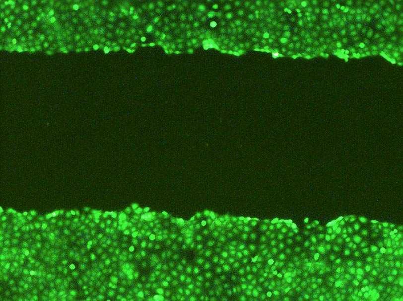
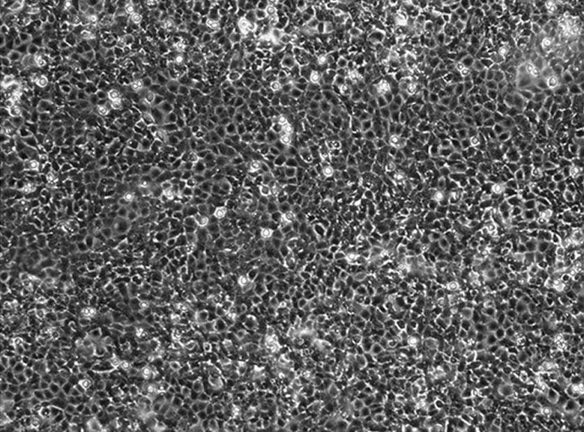
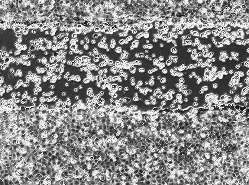
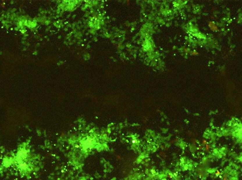
10 Stripe Choice Assay Piastre confluenti di cellule esprimenti la NRG1NRG1-IIIIII-β3 Cell Tracker Green graffi ogni 2 mm mock dopo la piastratura delle mock 24h dopo la piastratura delle mock 10
11 MGE explants +RFP Quantificazione: A: numero cellule rosse (MGE) / Area cells verdi (NRG1-III III-β3) B: numero cellule rosse (MGE) / Area cells incolori (mock) 24h dopo la piastratura delle cellule MGE MGE-Derived Cells Prefer a Nrg1-CRD-Expressing Substrate in the Stripe Choice Assay Slides were coated with alternating stripes of nontransfected COS cells (dark gray dots) and either mock-transfected or Nrg1-CRD-transfected COS cells (red dots). Dissociated E13.5 MGE cells (green dots) were plated on top of the stripe carpets, and their distribution was studied 24 hr later. 11
 or Nrg1-Igtransfected (B) COS cell aggregates.")
 and Nrg1-Ig (right) transfected COS cells.")
12 NRG1-Ig is a chemoattractant for MGE-derived neurons Medial ganglionic eminence (MGE) explants from the telencephalon of E13.5 embryos were cultured in Matrigel adjacent to mock-transfected (A) or Nrg1-Igtransfected (B) COS cell aggregates. ECTOPIC EXPRESSION OF NRG1-Ig REDIRECTS THE MIGRATION OF MGE-DERIVED CELLS IN SLICE CULTURES Coronal slice through the telencephalon with cell aggregates formed with control (left) and Nrg1-Ig (right) transfected COS cells. DiI-labeled cells (arrowheads) from both the ipsilateral and contralateral MGE (asterisk) migrate ectopically in a ventrolateral direction towards the COS cell aggregate expressing NRG1-Ig. 12
13 - different isoforms of neuregulin-1 are expressed in the developing cortex and in the route that migrating interneurons follow toward the cortex, whereas a population of the migrating interneurons express ErbB4, a receptor for neuregulin-1 the different isoforms of neuregulin-1 - type III and type I- act respectively as short- and long- range attractants for migrating interneurons - perturbing ErbB4 function in vitro decreases the number of interneurons MGE that tangentially migrate to the cortex - in vivo, loss of neuregulin-1/erbb4 signalling causes an alteration in the tangential migration of cortical interneurons - these observations provide evidence that neuregulin-1 and its ErbB4 receptor directly control neuronal migration in the nervous system 13
ErbB4 migrazione I parte. 3- ErbB4- NRG1
 ErbB4 migrazione I parte 3- ErbB4- NRG1 1 In rodent brains postnatal neuronal migration is evident in three main areas: the cerebellum (CB), the hippocampus (Hipp) and the rostral migratory stream (RMS).
Cell Migration II: CNS Cell Migration. Steven McLoon Department of Neuroscience University of Minnesota
 Cell Migration II: CNS Cell Migration Steven McLoon Department of Neuroscience University of Minnesota 1 Hey! The major concepts discussed relative to neural crest cell migration apply to cell migration
Cell Migration II: CNS Cell Migration. Steven McLoon Department of Neuroscience University of Minnesota
 Cell Migration II: CNS Cell Migration Steven McLoon Department of Neuroscience University of Minnesota 1 Course News Coffee Hour Wednesday (Oct 18) 9:00-10:00am Surdyk s Café in Northrop Auditorium Stop
During Brain Development Final Destinations for Neurons and Glia Get Separated from Germinal Niches
 During Brain Development Final Destinations for Neurons and Glia Get Separated from Germinal Niches Progenitors are Contained within Unique Domains and Tangentially Fixed. EMBRYO ADULT Migratory Behavior
During Brain Development Final Destinations for Neurons and Glia Get Separated from Germinal Niches
 During Brain Development Final Destinations for Neurons and Glia Get Separated from Germinal Niches Two mayor forms of neuronal migration: Radial and Tangential Leber & Sanes, 95 How do young neurons actually
Receptor tyrosine kinase ErbB4 modulates neuroblast migration and placement in the adult forebrain
 Receptor tyrosine kinase ErbB4 modulates neuroblast migration and placement in the adult forebrain E S Anton 1,8, H T Ghashghaei 1,8, Janet L Weber 2, Corey McCann 1, Tobias M Fischer 2, Isla D Cheung
Prss56, a novel marker of adult neurogenesis in the mouse brain. - Supplemental Figures 1 to 5- Brain Structure and Function
 Prss56, a novel marker of adult neurogenesis in the mouse brain - Supplemental Figures 1 to 5- Brain Structure and Function Alexandre Jourdon 1,2, Aurélie Gresset 1, Nathalie Spassky 1, Patrick Charnay
Primary Mouse Cerebral Cortex Neurons V: 80% TE: 70%
 Primary Mouse Cerebral Cortex Neurons V: 80% TE: 70% Pictures: 9 days after electroporation Red: MAP2 Blue: GFAP Green: GFP The cells were from Embryonic Day 14 Mouse Cerebral Cortex Primary Mouse Hippocampal
Neurodevelopment II Structure Formation. Reading: BCP Chapter 23
 Neurodevelopment II Structure Formation Reading: BCP Chapter 23 Phases of Development Ovum + Sperm = Zygote Cell division (multiplication) Neurogenesis Induction of the neural plate Neural proliferation
Young neurons born in the developing or adult brain have to
 Permissive corridor and diffusible gradients direct medial ganglionic eminence cell migration to the neocortex Hynek Wichterle*, Manuel Alvarez-Dolado, Lynda Erskine, and Arturo Alvarez-Buylla *The Rockefeller
Short- and Long-Range Attraction of Cortical GABAergic Interneurons by Neuregulin-1
 Neuron, Vol. 44, 251 261, October 14, 2004, Copyright 2004 by Cell Press Short- and Long-Range Attraction of Cortical GABAergic Interneurons by Neuregulin-1 Nuria Flames, 1 Jason E. Long, 2 Alistair N.
4/18/2011. Physiology 67 Lecture on Neural Development
 Physiology 67 Lecture on Neural Development 1 2 3 4 5 6 Neural cell categories After the ectodermal tissue has folded into the neural tube, another series of signaling interactions determine the type of
The neuron. Biocytin labeled pyramidal neuron recorded in piriform cortex
 The neuron Biocytin labeled pyramidal neuron recorded in piriform cortex Discovery of the neuron (A) Reticularist Doctrine (B) Neuron Doctrine Exception.GAP JUNCTIONS between neurons Neuronal shape Dendrites
Development and specification of GABAergic cortical interneurons
 Kelsom and Lu Cell & Bioscience 2013, 3:19 Cell & Bioscience REVIEW Development and specification of GABAergic cortical interneurons Corey Kelsom and Wange Lu * Open Access Abstract GABAergic interneurons
mir-7a regulation of Pax6 in neural stem cells controls the spatial origin of forebrain dopaminergic neurons
 Supplemental Material mir-7a regulation of Pax6 in neural stem cells controls the spatial origin of forebrain dopaminergic neurons Antoine de Chevigny, Nathalie Coré, Philipp Follert, Marion Gaudin, Pascal
Are Both Embryonic Migratory Pathways Preserved in the Adult Brain Cerebral Cortex?
 Prague Medical Report / Vol. 107 (2006) No. 1, p. 71 80 71) Are Both Embryonic Migratory Pathways Preserved in the Adult Brain Cerebral Cortex? Šimonová Z., Dutt J. Department of Neuroscience of the Institute
Broad Integration of Expression Maps and Co-Expression Networks Compassing Novel Gene Functions in the Brain
 Supplementary Information Broad Integration of Expression Maps and Co-Expression Networks Compassing Novel Gene Functions in the Brain Yuko Okamura-Oho a, b, *, Kazuro Shimokawa c, Masaomi Nishimura b,
The neuron. Biocytin labeled pyramidal neuron recorded in piriform cortex
 The neuron Biocytin labeled pyramidal neuron recorded in piriform cortex Discovery of the neuron (A) Reticularist Doctrine (B) Neuron Doctrine Exception.GAP JUNCTIONS between neurons Neuronal shape Dendrites
Department of Cognitive Science UCSD
 Department of Cognitive Science UCSD Verse 1: Neocortex, frontal lobe, Brain stem, brain stem, Hippocampus, neural node, Right hemisphere, Pons and cortex visual, Brain stem, brain stem, Sylvian fissure,
Nature Neuroscience: doi: /nn Supplementary Figure 1. MADM labeling of thalamic clones.
 Supplementary Figure 1 MADM labeling of thalamic clones. (a) Confocal images of an E12 Nestin-CreERT2;Ai9-tdTomato brain treated with TM at E10 and stained for BLBP (green), a radial glial progenitor-specific
Genesis of cerebellar interneurons and the prevention of neural DNA damage require XRCC1.
 Genesis of cerebellar interneurons and the prevention of neural DNA damage require XRCC1. Youngsoo Lee, Sachin Katyal, Yang Li, Sherif F. El-Khamisy, Helen R. Russell, Keith W. Caldecott and Peter J. McKinnon.
Development of the Nervous System. Leah Militello, class of 2018
 Development of the Nervous System Leah Militello, class of 2018 Learning Objectives 1. Describe the formation and fate of the neural tube and neural crest including timing and germ layer involved. 2. Describe
Neuronal migration in the adult brain: are we there yet?
 Neuronal migration in the adult brain: are we there yet? H. Troy Ghashghaei*, Cary Lai and E. S. Anton* Abstract The generation and targeting of appropriate numbers and types of neurons to where they are
Development of the Nervous System 1 st month
 Development of the Nervous System 1 st month day 1 - fertilization of egg day 6 - uterine implantation day 18 - trilaminar (3-layered) disc (blastoderm, embryo) ectoderm (dorsal) - nervous system and skin
Supplementary Figure 1
 Supplementary Figure 1 Kif1a RNAi effect on basal progenitor differentiation Related to Figure 2. Representative confocal images of the VZ and SVZ of rat cortices transfected at E16 with scrambled or Kif1a
Cell Birth and Death. Chapter Three
 Cell Birth and Death Chapter Three Neurogenesis All neurons and glial cells begin in the neural tube Differentiated into neurons rather than ectoderm based on factors we have already discussed If these
Potential Treatment and Current Research in Phelan-McDermid Syndrome. 11/16/2016 Frambu Center for Rare Disorders
 Potential Treatment and Current Research in Phelan-McDermid Syndrome 11/16/2016 Frambu Center for Rare Disorders Genetics is Complicated! Deletion 22q13: Therapies Under Investigation Intranasal insulin
Supplementary Figures
 Supplementary Figures Supplementary Figure 1. nrg1 bns101/bns101 embryos develop a functional heart and survive to adulthood (a-b) Cartoon of Talen-induced nrg1 mutation with a 14-base-pair deletion in
Inner ear development Nervous system development
 Upcoming Sessions April 22: Nervous System Development Lecture April 24: Reviews of Axonal Pathfinding in Sensory Systems April 29: Inner Ear Development Lecture May 1: May 6: May 8: Auditory System Pathfinding
Development of the Central Nervous System
 Development of the Central Nervous System an ongoing process, through adolescence and maybe even adult hood? the nervous system is plastic Experience plays a key role Dire consequences when something goes
Transient neuronal populations are required to guide callosal axons: a role for semaphorin 3C.
 Transient neuronal populations are required to guide callosal axons: a role for semaphorin 3C. Mathieu Niquille, Sonia Garel, Fanny Mann, Jean-Pierre Hornung, Belkacem Otsmane, Sébastien Chevalley, Carlos
The Zinc Finger Transcription Factor Sp8 Regulates the Generation and Diversity of Olfactory Bulb Interneurons
 Neuron 49, 503 516, February 16, 2006 ª2006 Elsevier Inc. DOI 10.1016/j.neuron.2006.01.018 The Zinc Finger Transcription Factor Sp8 Regulates the Generation and Diversity of Olfactory Bulb Interneurons
Brain Development III
 Brain Development III Neural Development In the developing nervous system there must be: 1. The formation of different regions of the brain. 2. The ability of a neuron to differentiate. 3. The ability
EGFRs mediate chemotactic migration in the developing telencephalon
 Development 128, 4203-4216 (2001) Printed in Great Britain The Company of Biologists Limited 2001 DEV9789 4203 EGFRs mediate chemotactic migration in the developing telencephalon Damira Caric, Heather
Nature Neuroscience: doi: /nn Supplementary Figure 1. Splenic atrophy and leucopenia caused by T3 SCI.
 Supplementary Figure 1 Splenic atrophy and leucopenia caused by T3 SCI. (a) Gross anatomy of representative spleens from control and T3 SCI mice at 28 days post-injury. (b and c) Hematoxylin and eosin
Huntington s Disease & MARY ET BOYLE, PH.D. DEPARTMENT OF COGNITIVE SCIENCE
 Huntington s Disease & Early Nervous System Development MARY ET BOYLE, PH.D. DEPARTMENT OF COGNITIVE SCIENCE UCSD The cups fell to the floor with a crash. Was this the alarm signal? Or was it forgetting
Introduction. Materials and Methods
 4240 The Journal of Neuroscience, May 15, 2003 23(10):4240 4250 Multiple Cell Populations in the Early Postnatal Subventricular Zone Take Distinct Migratory Pathways: A Dynamic Study of Glial and Neuronal
Nature Neuroscience: doi: /nn Supplementary Figure 1. Confirmation that optogenetic inhibition of dopaminergic neurons affects choice
 Supplementary Figure 1 Confirmation that optogenetic inhibition of dopaminergic neurons affects choice (a) Sample behavioral trace as in Figure 1d, but with NpHR stimulation trials depicted as green blocks
Supplementary information. Nkx2.1 regulates the generation of telencephalic astrocytes during embryonic
 Supplementary information Nkx2.1 regulates the generation of telencephalic astrocytes during embryonic development Shilpi Minocha 1*, Delphine Valloton 1*, Yvan Arsenijevic 2, Jean-René Cardinaux 3, Raffaella
Shh signaling guides spatial pathfinding of raphespinal tract axons by multidirectional repulsion
 ORIGINAL ARTICLE Cell Research (2012) 22:697-716. 2012 IBCB, SIBS, CAS All rights reserved 1001-0602/12 $ 32.00 www.nature.com/cr npg Shh signaling guides spatial pathfinding of raphespinal tract axons
Functional Development of Neuronal Networks in Culture -An in vitro Assay System of Developing Brain for Endocrine Disruptors
 Functional Development of Neuronal Networks in Culture -An in vitro Assay System of Developing Brain for Endocrine Disruptors Masahiro Kawahara and Yoichiro Kuroda Tokyo Metropolitan Institute for Neuroscience
Etv4 BU EMM Etv5 BC MMM UI-M-BH3- Elk1 BC MMM
 Supplementary Tables In situ hybridization probes Genbank Gene Accession Number Clone ID Cat. No Etv1 BC005645 PCR Etv4 BU053662 5062538 EMM1002-5544031 Etv5 BC034680 4036564 MMM1013-7511524 Fev AI876263
CXCR4 and CXCR7 Have Distinct Functions in Regulating Interneuron Migration
 Article CXCR4 and CXCR7 Have Distinct Functions in Regulating Interneuron Migration Yanling Wang, 1, * Guangnan Li, 2,5 Amelia Stanco, 1,5 Jason E. Long, 3 Dianna Crawford, 4 Gregory B. Potter, 1 Samuel
Plasticity of Cerebral Cortex in Development
 Plasticity of Cerebral Cortex in Development Jessica R. Newton and Mriganka Sur Department of Brain & Cognitive Sciences Picower Center for Learning & Memory Massachusetts Institute of Technology Cambridge,
Lhx6 and Lhx8 Coordinately Induce Neuronal Expression of Shh that Controls the Generation of Interneuron Progenitors
 Article Lhx6 and Lhx8 Coordinately Induce Neuronal Expression of Shh that Controls the Generation of Interneuron Progenitors Pierre Flandin, 1 Yangu Zhao, 2 Daniel Vogt, 1 Juhee Jeong, 1 Jason Long, 1,3
Gene co-expression networks in the mouse, monkey, and human brain July 16, Jeremy Miller Scientist I
 Gene co-expression networks in the mouse, monkey, and human brain July 16, 2013 Jeremy Miller Scientist I jeremym@alleninstitute.org Outline 1. Brief introduction to previous WGCNA studies in brain 2.
NIH Public Access Author Manuscript Science. Author manuscript; available in PMC 2011 September 1.
 NIH Public Access Author Manuscript Published in final edited form as: Science. 2010 February 26; 327(5969): 1145 1148. doi:10.1126/science.1183962. Cortical Plasticity Induced by Inhibitory Neuron Transplantation
Emx2 patterns the neocortex by regulating FGF positional signaling
 Emx2 patterns the neocortex by regulating FGF positional signaling Tomomi Fukuchi-Shimogori and Elizabeth A Grove Presented by Sally Kwok Background Cerebral cortex has anatomically and functionally distinct
Supplementary figure 1: LII/III GIN-cells show morphological characteristics of MC
 1 2 1 3 Supplementary figure 1: LII/III GIN-cells show morphological characteristics of MC 4 5 6 7 (a) Reconstructions of LII/III GIN-cells with somato-dendritic compartments in orange and axonal arborizations
Supplemental Information. Otic Mesenchyme Cells Regulate. Spiral Ganglion Axon Fasciculation. through a Pou3f4/EphA4 Signaling Pathway
 Neuron, Volume 73 Supplemental Information Otic Mesenchyme Cells Regulate Spiral Ganglion Axon Fasciculation through a Pou3f4/EphA4 Signaling Pathway Thomas M. Coate, Steven Raft, Xiumei Zhao, Aimee K.
Radial glia in the ventral telencephalon
 REVIEW ARTICLE Radial glia in the ventral telencephalon Miguel Turrero Garcıa and Corey C. Harwell Department of Neurobiology, Harvard Medical School, Boston, MA, USA Correspondence C. C. Harwell, Department
Anterior Olfactory Nucleus
 A Anterior Olfactory Nucleus PETER C. BRUNJES, KURT R. ILLIG Department of Psychology, University of Virginia Charlottesville, VA, USA Synonyms Anterior olfactory cortex Definition The primary component
Nature Neuroscience: doi: /nn Supplementary Figure 1
 Supplementary Figure 1 Atlas representations of the midcingulate (MCC) region targeted in this study compared against the anterior cingulate (ACC) region commonly reported. Coronal sections are shown on
Contralateral migration of oculomotor neurons is regulated by Slit/Robo signaling
 Bjorke et al. Neural Development (2016) 11:18 DOI 10.1186/s13064-016-0073-y RESEARCH ARTICLE Open Access Contralateral migration of oculomotor neurons is regulated by Slit/Robo signaling Brielle Bjorke
Endothelial cell-derived GABA signaling modulates neuronal migration and postnatal behavior
 ORIGINAL ARTICLE Cell Research (2017) :1-28. www.nature.com/cr Endothelial cell-derived GABA signaling modulates neuronal migration and postnatal behavior Suyan Li 1, 2 *, Peeyush Kumar T 1, 2 *, Sampada
Zhu et al, page 1. Supplementary Figures
 Zhu et al, page 1 Supplementary Figures Supplementary Figure 1: Visual behavior and avoidance behavioral response in EPM trials. (a) Measures of visual behavior that performed the light avoidance behavior
PSY 215 Lecture #5 (01/26/2011) (Anatomy of the Brain) Dr. Achtman PSY 215. Lecture 5 Anatomy of the Brain Chapter 4, pages 86-96
 (Anatomy of the Brain) Dr. Achtman PSY 215. Lecture 5 Anatomy of the Brain Chapter 4, pages 86-96") Corrections: none needed PSY 215 Lecture 5 Anatomy of the Brain Chapter 4, pages 86-96 Announcements: Reminder: The first midterm is in one week! Everyone is encouraged to start studying (recommend 30/night
Corrections: none needed PSY 215 Lecture 5 Anatomy of the Brain Chapter 4, pages 86-96 Announcements: Reminder: The first midterm is in one week! Everyone is encouraged to start studying (recommend 30/night
Divergent functions of the proneural genes Mash1 and Ngn2 in the specification of neuronal subtype identity
 Divergent functions of the proneural genes Mash1 and Ngn2 in the specification of neuronal subtype identity Carlos M. Parras, 1,4 Carol Schuurmans, 1,3,4 Raffaella Scardigli, 1 Jaesang Kim, 2 David J.
Mammalian Cerebral Cortex: Embryonic Development and Cytoarchitecture
 Mammalian Cerebral Cortex: Embryonic Development and Cytoarchitecture 2 The prenatal developmental of the mammalian cerebral cortex, including that of humans, is characterized by two sequential and interrelated
NIH Public Access Author Manuscript Trends Mol Med. Author manuscript; available in PMC 2012 August 1.
 NIH Public Access Author Manuscript Published in final edited form as: Trends Mol Med. 2011 August ; 17(8): 452 462. doi:10.1016/j.molmed.2011.03.003. The Contribution of GABAergic Dysfunction to Neurodevelopmental
Lecture 22: A little Neurobiology
 BIO 5099: Molecular Biology for Computer Scientists (et al) Lecture 22: A little Neurobiology http://compbio.uchsc.edu/hunter/bio5099 Larry.Hunter@uchsc.edu Nervous system development Part of the ectoderm
Lori Sussel 1, *, Oscar Marin 1, Shioko Kimura 2 and John L. R. Rubenstein 1, SUMMARY
 Development 126, 3359-3370 (1999) Printed in Great Britain The Company of Biologists Limited 1999 DEV8607 3359 Loss of Nkx2.1 homeobox gene function results in a ventral to dorsal molecular respecification
MIDTERM EXAM 1 COGNITIVE SCIENCE 107A
 MIDTERM EXAM 1 COGNITIVE SCIENCE 107A FALL 2011 Name: Points: / 100 PID: I. SHORT ANSWERS (6 points each for a total of 30 points) 1. Describe two contributions made by Ramon y Cajal (1852-1934) in terms
CNS Developmental. Anke van Eekelen, PhD. Telethon Institute for Child Health Research
 CNS Developmental Anke van Eekelen, PhD Telethon Institute for Child Health Research (Some slides are modified versions of Prof. Alan Harvey s Neuroscience lecture at ANHB and Dr. Joanne Britto s Dev Neuroscience
Regional Distribution of Cortical Interneurons and Development of Inhibitory Tone Are Regulated by Cxcl12/ Cxcr4 Signaling
 The Journal of Neuroscience, January 30, 2008 28(5):1085 1098 1085 Development/Plasticity/Repair Regional Distribution of Cortical Interneurons and Development of Inhibitory Tone Are Regulated by Cxcl12/
Embryonic MGE Cells as a Treatment for Epilepsy December 1, 2012
 Embryonic MGE Cells as a Treatment for Epilepsy December 1, 2012 Scott C. Baraban, PhD University of California, San Francisco American Epilepsy Society Annual Meeting Disclosure Name of Commercial Interest
Nature Protocols: doi: /nprot Supplementary Figure 1. Effect of different enzymes on CD13 and CD31 populations.
 Supplementary Figure 1 Effect of different enzymes on CD13 and CD31 populations. Comparison of cortex dissociated for 30 minutes with collagenase/dispase digestion (a) or papain (b). Papain degrades the
SUPPLEMENTARY INFORMATION
 SUPPLEMENTARY INFORMATION doi:10.1038/nature11306 Supplementary Figures Supplementary Figure 1. Basic characterization of GFP+ RGLs in the dentate gyrus of adult nestin-gfp mice. a, Sample confocal images
Preferential Origin and Layer Destination of GAD65-GFP Cortical Interneurons
 Preferential Origin and Layer Destination of GAD65-GFP Cortical Interneurons Guillermina López-Bendito 1, Katherine Sturgess 1, Ferenc Erdélyi 2, Gábor Szabó 2, Zoltán Molnár 1 and Ole Paulsen 3 1 Department
Evidence of Common Progenitors and Patterns of Dispersion in Rat Striatum and Cerebral Cortex
 The Journal of Neuroscience, May 15, 2002, 22(10):4002 4014 Evidence of Common Progenitors and Patterns of Dispersion in Rat Striatum and Cerebral Cortex Christopher B. Reid 1,2 and Christopher A. Walsh
Recipes for Making Human Interneurons from Stem Cells Require Multiple Factors, Careful Timing, and Long Maturation Periods
 Current Literature In Basic Science Recipes for Making Human Interneurons from Stem Cells Require Multiple Factors, Careful Timing, and Long Maturation Periods Directed Differentiation and Functional Maturation
BRAIN DEVELOPMENT I: ESTABLISHMENT OF BASIC ARCHITECTURE. Thomas Marino, Ph.D.
 BRAIN DEVELOPMENT I: ESTABLISHMENT OF BASIC ARCHITECTURE Thomas Marino, Ph.D. Development of the Brain I. Competencies: Upon completion of this section of the course, the student must be able to: 1. Understand
OPTO 5320 VISION SCIENCE I
 OPTO 5320 VISION SCIENCE I Monocular Sensory Processes of Vision: Color Vision Mechanisms of Color Processing . Neural Mechanisms of Color Processing A. Parallel processing - M- & P- pathways B. Second
Lecture 42: Final Review. Martin Wessendorf, Ph.D.
 Lecture 42: Final Review Martin Wessendorf, Ph.D. Lecture 33 cortex Heilbronner 5 lobes of the cortex Lateral view (left side) Mid-saggital view (right side) Cellular organization of cortex White matter
Nature Neuroscience: doi: /nn Supplementary Figure 1. Neuron class-specific arrangements of Khc::nod::lacZ label in dendrites.
 Supplementary Figure 1 Neuron class-specific arrangements of Khc::nod::lacZ label in dendrites. Staining with fluorescence antibodies to detect GFP (Green), β-galactosidase (magenta/white). (a, b) Class
Sonic hedgehog contributes to oligodendrocyte specification in the mammalian forebrain
 Development 128, 527-540 (2001) Printed in Great Britain The Company of Biologists Limited 2001 DEV1621 527 Sonic hedgehog contributes to oligodendrocyte specification in the mammalian forebrain Susana
Healthy Brain Development: Protective and Risk Factors
 Healthy Brain Development: Protective and Risk Factors Bryce Geeraert PhD Candidate Developmental Neuroimaging Lab January 31, 2018 About me BSc Psychology, Biomedical Engineering PhD candidate Interest
Basal Ganglia General Info
 Basal Ganglia General Info Neural clusters in peripheral nervous system are ganglia. In the central nervous system, they are called nuclei. Should be called Basal Nuclei but usually called Basal Ganglia.
Supplementary Figure 1. Nature Neuroscience: doi: /nn.4547
 Supplementary Figure 1 Characterization of the Microfetti mouse model. (a) Gating strategy for 8-color flow analysis of peripheral Ly-6C + monocytes from Microfetti mice 5-7 days after TAM treatment. Living
Investigating the role of EphAl ephrin-a signalling during trigeminal ganglion axon guidance
 Investigating the role of EphAl ephrin-a signalling during trigeminal ganglion axon guidance A thesis submitted for the degree of Doctor of Philosophy Molecular and Biomedical Science (Discipline of Genetics),
Fig.9.2. Structure of embryonic brain
 T Chapter 9 Development of Ectodermal Organs he ectoderm gives rise to 3 separate cell populations: neural(plate) ectoderm, neural crest cells, and epiderm (general body ectoderm). A primordium (anlage)
Supplementary Figure S1: Pial and periventricular vascular networks and the dual GABA neuron streams of the early embryonic mouse telencephalon
 Supplementary Figure S1: Pial and periventricular vascular networks and the dual GABA neuron streams of the early embryonic mouse telencephalon a) The profile of GABA neurons arriving at the pallial-subpallial
Nature Neuroscience: doi: /nn Supplementary Figure 1. Large-scale calcium imaging in vivo.
 Supplementary Figure 1 Large-scale calcium imaging in vivo. (a) Schematic illustration of the in vivo camera imaging set-up for large-scale calcium imaging. (b) High-magnification two-photon image from
Dlx1&2-Dependent Expression of Zfhx1b (Sip1, Zeb2) Regulates the Fate Switch between Cortical and Striatal Interneurons
 Regulates the Fate Switch between Cortical and Striatal Interneurons") Article Dlx1&2-Dependent Expression of Zfhx1b (Sip1, Zeb2) Regulates the Fate Switch between Cortical and Striatal Interneurons Gabriel L. McKinsey, 1,2 Susan Lindtner, 1 Brett Trzcinski, 3 Axel Visel,
Article Dlx1&2-Dependent Expression of Zfhx1b (Sip1, Zeb2) Regulates the Fate Switch between Cortical and Striatal Interneurons Gabriel L. McKinsey, 1,2 Susan Lindtner, 1 Brett Trzcinski, 3 Axel Visel,
CISC 3250 Systems Neuroscience
 CISC 3250 Systems Neuroscience Levels of organization Central Nervous System 1m 10 11 neurons Neural systems and neuroanatomy Systems 10cm Networks 1mm Neurons 100μm 10 8 neurons Professor Daniel Leeds
Supplementary Figure 1: Signaling centers contain few proliferating cells, express p21, and
 Supplementary Figure 1: Signaling centers contain few proliferating cells, express p21, and exclude YAP from the nucleus. (a) Schematic diagram of an E10.5 mouse embryo. (b,c) Sections at B and C in (a)
Telencephalic cells take a tangent: non-radial migration in the mammalian forebrain
 review Telencephalic cells take a tangent: non-radial migration in the mammalian forebrain Joshua G. Corbin, Susana Nery and Gord Fishell Developmental Genetics Program and the Department of Cell Biology,
Nature Neuroscience doi: /nn Supplementary Figure 1. Characterization of viral injections.
 Supplementary Figure 1 Characterization of viral injections. (a) Dorsal view of a mouse brain (dashed white outline) after receiving a large, unilateral thalamic injection (~100 nl); demonstrating that
Outline. Outline. Background Literature and Slides 10/23/2013. The Schizophrenia Research Forum online: Molecules, Neural Networks and Behavior
 Background Literature and Slides Molecules, Neural Networks and Behavior Roberto Fernández Galán, PhD Assistant Professor of Neurosciences Mt. Sinai Health Care Foundation Scholar Alfred P. Sloan Research
The neurvous system senses, interprets, and responds to changes in the environment. Two types of cells makes this possible:
 NERVOUS SYSTEM The neurvous system senses, interprets, and responds to changes in the environment. Two types of cells makes this possible: the neuron and the supporting cells ("glial cells"). Neuron Neurons
Lesson 33. Objectives: References: Chapter 16: Reading for Next Lesson: Chapter 16:
 Lesson 33 Lesson Outline: Nervous System Structure and Function Neuronal Tissue Supporting Cells Neurons Nerves Functional Classification of Neuronal Tissue Organization of the Nervous System Peripheral
Interneuron dysfunction in psychiatric disorders
 Nature Reviews Neuroscience AOP, published online 18 January 2012; doi:10.1038/nrn3155 REVIEWS Interneuron dysfunction in psychiatric disorders Oscar Marín Abstract Schizophrenia, autism and intellectual
Tlx3 and Tlx1 are post-mitotic selector genes determining glutamatergic over GABAergic cell fates
 Tlx3 and Tlx1 are post-mitotic selector genes determining glutamatergic over GABAergic cell fates L. Cheng, A. Arata, R. Mizuguchi, Y. Qian, A. Karunaratne, P.A. Gray, S. Arata, S. Shirasawa, M. Bouchard,
Multipolar Migration: The Third Mode of Radial Neuronal Migration in the Developing Cerebral Cortex
 9996 The Journal of Neuroscience, November 5, 2003 23(31):9996 10001 Brief Communication Multipolar Migration: The Third Mode of Radial Neuronal Migration in the Developing Cerebral Cortex Hidenori Tabata
Distinct Origins of Neocortical Projection Neurons and Interneurons In Vivo
 Distinct Origins of Neocortical Projection Neurons and Interneurons In Vivo Stewart A. Anderson 2, Christine E. Kaznowski 1, Carrie Horn, John L.R. Rubenstein and Susan K. McConnell 1 Department of Psychiatry,
a 0,8 Figure S1 8 h 12 h y = 0,036x + 0,2115 y = 0,0366x + 0,206 Labeling index Labeling index ctrl shrna Time (h) Time (h) ctrl shrna S G2 M G1
 Time (h) ctrl shrna S G2 M G1") (GFP+ BrdU+)/GFP+ Labeling index Labeling index Figure S a, b, y =,x +, y =,x +,,,,,,,, Time (h) - - Time (h) c d S G M G h M G S G M G S G h Time of BrdU injection after electroporation (h) M G S G M
(GFP+ BrdU+)/GFP+ Labeling index Labeling index Figure S a, b, y =,x +, y =,x +,,,,,,,, Time (h) - - Time (h) c d S G M G h M G S G M G S G h Time of BrdU injection after electroporation (h) M G S G M
Supplementary Figure 1. EC-specific Deletion of Snail1 Does Not Affect EC Apoptosis. (a,b) Cryo-sections of WT (a) and Snail1 LOF (b) embryos at
 Cryo-sections of WT (a) and Snail1 LOF (b) embryos at") Supplementary Figure 1. EC-specific Deletion of Snail1 Does Not Affect EC Apoptosis. (a,b) Cryo-sections of WT (a) and Snail1 LOF (b) embryos at E10.5 were double-stained for TUNEL (red) and PECAM-1 (green).
Supplementary Figure 1. EC-specific Deletion of Snail1 Does Not Affect EC Apoptosis. (a,b) Cryo-sections of WT (a) and Snail1 LOF (b) embryos at E10.5 were double-stained for TUNEL (red) and PECAM-1 (green).
Tangential migration of neurons in the developing cerebral cortex
 Development 121, 2165-2176 (1995) Printed in Great Britain The Company of Biologists Limited 1995 2165 Tangential migration of neurons in the developing cerebral cortex Nancy A. O Rourke*, Daniel P. Sullivan,
Neocortex. Cortical Structures in the Brain. Neocortex Facts. Laminar Organization. Bark-like (cortical) structures: Shepherd (2004) Chapter 12
 structures: Shepherd (2004) Chapter 12") Neocortex Shepherd (2004) Chapter 12 Rodney Douglas, Henry Markram, and Kevan Martin Instructor: Yoonsuck Choe; CPSC 644 Cortical Networks Cortical Structures in the Brain Bark-like (cortical) structures:
Neocortex Shepherd (2004) Chapter 12 Rodney Douglas, Henry Markram, and Kevan Martin Instructor: Yoonsuck Choe; CPSC 644 Cortical Networks Cortical Structures in the Brain Bark-like (cortical) structures:
Carl P. Wonders* and Stewart A. Anderson
 The origin and specification of cortical interneurons Carl P. Wonders* and Stewart A. Anderson Abstract GABA-containing interneurons are crucial to both the development and function of the cerebral cortex.