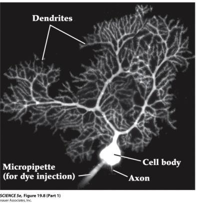
The neuron. Biocytin labeled pyramidal neuron recorded in piriform cortex
|
|
|
- Christal Boone
- 5 years ago
- Views:
Transcription


1 The neuron Biocytin labeled pyramidal neuron recorded in piriform cortex

2 Discovery of the neuron

3

4

5 (A) Reticularist Doctrine (B) Neuron Doctrine Exception.GAP JUNCTIONS between neurons
6 Neuronal shape Dendrites Soma Pyramidal neuron (multipolar) Axon

7

8

9


10
11
12
13
14
15 Functional classification of neurons Projection neurons Local circuit neurons

16 Neurons can also be identified by immunocytochemistry


")
17 Various NEURONAL TYPES in the mammalian brain Purkinje cell Pyramidal neuron Granular neuron (interneuron)


18 Morphology and neuronal types in the cerebellum Molecular/functional heterogeneity of Purkinje cells?

19 Morphology and distribution of pyramidal neurons in the neocortex These pyramidal n. never project axons to targets outside the telencephalon Molyneaux et al., 2007 These include Pyramidal neurons of the largest size, which are located in deep-layer V and extend projections to the brainstem and spinal cord


20 Neocortical interneurons diversity Huang et al., 2007
21 Easy neuronal classification Structural classification: Unipolar, bipolar, multipolar, more Functional classification: projection (inter)neurons local circuit (inter)neurons excytatory (neurotransmitters: Glutamate, etc.) inhibitory (neurotransm.: GABA, glycine, etc.)

22 The problem of neuronal classification and subtype identification. We need to classify different neuronal types in order to speak a common language with other neuroscientists and in order to understand the complexity of brain function HOW should we classify neurons? By morphology? By functional features? By expression markers? How do we put together information from different approaches? for discussion see Yuste, 2005
 solution would be")


23 The problem of neuronal classification The obvious (but not the easiest) solution would be COMBINING different approaches in the same experimental model. An example: ABSTRACT (1) Whole-cell electrophysiological recording on cortical slices + (2) intracellular injection of byocytin in recorded neurons (for later recognition and morphological analysis) + (3) fluorescence immunocytochemistry for selected markers (calcium-binding proteins: parvalbumin, calbindin and calretinin)

24 The phenotype of BC-injected/electrophysiologically-recorded interneurons is determined by immunocytochemistry Zaitsev et al.,


25 The combination of intracellularinjection techniques and immunocytochemistry suggests that the same phenotypic marker is expressed by interneurons with different morphologies Basket c. Chandelier c. Do different morphologies indicate different functional features? Ascending arbors c. Descending arbors c. Vertically oriented c. Zaitsev et al., 2005
 of interneurons are obtained: FS (Fast Spiking) and non-fs.")
26 Data were processed using CLUSTER ANALYSIS: correlation between electrophysiological properties and expression of specific Ca + - binding proteins When cells are grouped based only on electrophysiological properties, two main groups (= clusters) of interneurons are obtained: FS (Fast Spiking) and non-fs. These two clusters do show significant differences in Ca + -binding protein content Zaitsev et al., 2005

27 CONCLUSIONS: parvalbumin-expressing interneurons are exclusively FS calretinin- and calbindin-expressing interneurons are mainly non-fs multiple morphologies can correspond to a single functionally-defined phenotype FS neuron PARVALBUMIN phenotype basket cells chandelier cells non-fs neuron CALRETININ phenotype CALBINDIN phenotype vertically-oriented cells cell with ascending arbors cell with discending arbors

28 Electrophysiological and gene expression profiling of neuronal cell types Yano et al., 2006

29 another way to classify cortical interneurons is....to consider their developmental origin: Gelman & Marin, 2010


30 Cortical development Vitalis & Rossier, 2010

 and striatum, respectively Coronal section (indicated in a) illustrating the major")
31 Primary routes of interneuron migration during cortical development. Faux et al, 2012 Cortical interneurons born in the MGE and CGE in the ventral telencephalon follow tangential migratory paths into the developing cortex. Once within the cortical wall, cells disperse before entering the cortical plate and reside in a final position. The LGE-derived neurons migrate rostrally and ventrally into the olfactory bulb (OB) and striatum, respectively Coronal section (indicated in a) illustrating the major routes of tangential migration through the embryonic telencephalon. Interneurons migrate from the MGE (M) and traverse the LGE (L) whilst avoiding the striatum (Str).

-")
32 MGE-originating interneurons CGE-originating interneurons FS, parvalbumin(somato statin)-positive cells non-fs, calretinin(vip, NPY)- positive cells


33 In utero fate-mapping of cortical interneurons... Ventricular and subventricular zones of the MGE and CGE were dissected from mouse embryos expressing EGFP under the b-actin promoter (b-actin EGFP mice) and transplanted in the appropriate regions of wild-type host-embryos ( homotypic transplants) Butt et al., followed by

 neurons originate from CGE This means that different subtypes of interneurons are generated at different spatial positions (=")
34 electrophysiological, immunocytochemical and morphological riconstruction of mature interneurons in young-adult mice Butt et al., 2007 CONCLUSIONS: parvalbumin-expressing/fs/basketshaped neurons originate from MGE calretinin-expressing/non-fs/bipolar (vertically-oriented?) neurons originate from CGE This means that different subtypes of interneurons are generated at different spatial positions (= from different progenitors) CLASSIFYING neurons BY THEIR ORIGIN could be a new method of neuronal classification


35 Cortical interneuron diversity largely emerges from spatially segregated progenitor cells with distinct transcriptional profiles Gelman & Marin, 2010

 Various transcription factors are expressed in distinct patterns throughout the subpallial germinal zones.")

36 IN SUMMARY: Origins and diversity of neocortical interneurons (A) Neocortical interneurons are derived from progenitor cells located in the proliferative zones of the ventral telencephalon, specifically within the medial ganglionic eminence (MGE) and caudal ganglionic eminence (CGE). A small proportion is produced in the preopticarea (PoA). (B) Various transcription factors are expressed in distinct patterns throughout the subpallial germinal zones. (C) Neocortical interneurons are highly diverse and can be defined based on morphology, neurochemical expression, electrophysiological properties, and subcellular synaptic targeting specificity. Sultan et al., 2013 Frontiers in Cellular Neuroscience Volume 7, Article 221

37 Using Fluorescence Activated Cell Sorting to Examine Cell-Type-Specific Gene Expression in Rat Brain Tissue Figure 3. Neurons, astrocytes, and microglia sorted from a male hippocampus. The hippocampus from one male rat was dissociated and stained with the antibodies for CD11b, GLT1 and Thy1 and sorted using a FACS machine. (A) Cells were first sorted based on their forward and side scatter from all possible events. This gate is called P1 (population 1). (B) Next, single cells, also called singlets, were sorted based on their size from the doublets or larger clumps of cells. This gate is called P2. (C) Third, the single cells were gated as either APC-CD11b positive (CD11b+ gate, P4) or APC-CD11b negative (CD11b- gate, P3). (D) APC-CD11b negative cells were subsequently sorted into PE-GLT1 positive cells (GLT1+ gate, P6) and FITC-Thy1 positive cells (Thy1+ gate, P5). The breakdown of all events and all gates was generated from the FACS software depicted in a table which is presented on the right.
38 Figure 5. Real-time PCR analysis of cell-typespecific genes from sorted cells. Neurons (green bars), astrocytes (red bars) and microglia (blue bars) were sorted based on the protocol described above and mrna was extracted for confirmation of celltype-specific gene expression. (A) Iba1 is a calcium binding protein expressed exclusively in microglia sorted from the male hippocampus. (B) GFAP is a filament protein expressed predominantly in astrocytes sorted from the male hippocampus (C) NR1 is a ubiquitous subunit of the NMDA glutamatergic receptor that was expressed predominantly on neurons sorted from the male hippocampus. (D) Iba1 was also expressed exclusively on microglia sorted from the male cerebellum. (E) Interestingly, GFAP was not expressed in any of the cell types sorted from the male cerebellum. (F) The NR1 subunit of the NMDA receptor was also expressed predominantly on neurons sorted from the male cerebellum. Figure 6. Real-time PCR analysis of calbindin expressed in sorted neural cells. Cells sorted using FACS can be used to analyze cell-typespecific gene expression. (A) Neurons (green bars) expressed significantly more Calbindin than either astrocytes (red bars) or microglia (blue bars) sorted from the male hippocampus. (B) Neurons sorted from the male cerebellum expressed significantly higher levels of Calbindin than either astrocytes or microglia sorted from the cerebellum, but also significantly higher levels than the neurons sorted from the hippocampus.

39 Cell types in the mouse cortex and hippocampus revealed by single-cell RNA-seq* Zeisel et al., 2015 Science 347: Fig. 1 Molecular census of somatosensory S1 cortex and hippocampus CA1 by unbiased sampling and single-cell RNA-seq. (A) Workflow for obtaining and analyzing single-cell RNA-seq from juvenile mouse cortical cells, from dissection to singlecell RNA-seq and biclustering. (B) Visualization of nine major classes of cells using t-distributed stochastic neighbor embedding (tsne). Each dot is a single cell, and cells are laid out to show similarities. Colored contours correspond to the nine clusters in (A) and fig. S3. Expression of known markers is shown using the same layout (blue, no expression; white, 1% quantile; red, 99% quantile). (C) Hierarchical clustering analysis on 47 subclasses. Bar plots show number of captured cells in CA1 and S1, number of detected polya+ RNA molecules per cell, and total number of genes detected per cell. * RNA-Seq (RNA sequencing), uses next-generation sequencing (NGS) to reveal and quantify the whole transcriptome in a biological sample. See Mutz et al., 2013 (Current Opinion in Biotechnolog, 24:22 30) for review of the technique
. Layer-specific expression shown by in situ hybridization (Allen Brain Atlas).")
40 Fig. 2 Neuron subclasses in the somatosensory cortex Zeisel et al., 2015 (A) Subclasses of pyramidal neurons in the somatosensory cortex (S1) identified by BackSPIN clustering. Bar plots show mean expression of selected known and novel markers (error bars show standard deviations). Layer-specific expression shown by in situ hybridization (Allen Brain Atlas). S1PyrL23, layer II-III; S1PyrL4, layer IV; S1PyrL5a, layer Va; S1PyrL5, layer V; S1PyrL6, layer VI; S1PyrL6b, layer VIb; S1PyrDL, deep layers; ClauPyr, claustrum. (B) Identification of interneuron subclasses. Bar plots show selected known and novel markers. Fraction of S1/CA1 cells is depicted at bottom: blue, S1; yellow, CA1; white, flow-sorted Htr3a+ cells from S1. (C) Immunohistochemistry demonstrating the existence and localization of novel PAX6+/5HT3aEGFP+ interneurons, Int11. Bar plots show the layer distribution of these neurons. (D) Intrinsic electrophysiology and morphology of PAX6+ interneurons in S1 layer I, identified by post hoc staining.
 Immunohistochemistry for glial fibrillary acidic protein (red, Astro1) and MFGE8 (green, Astro2). Scale bar, 50 mm.")
41 Zeisel et al., 2015 Fig. 3 Characterization of glial subclasses. (A) Two types of astrocytes (Astro1 and Astro2) identified by common and distinct markers. (B) Immunohistochemistry for glial fibrillary acidic protein (red, Astro1) and MFGE8 (green, Astro2). Scale bar, 50 mm. (C) Genes showing expression restricted to microglia (Mgl), perivascular macrophages (Pvm), and peritoneal macrophages (Pmac). Error bars show standard deviations. (D) Cartoon illustrating the morphology and localization of microglia and perivascular macrophages. (E) Immunostaining for AIF1 (previously known as Iba-1, blue) marking microglia, and for MRC1 (green) and LYVE1 (red) marking perivascular macrophages. Asterisk, a microglia cell. Arrow, a perivascular macrophage aligned to a vessel (not stained). Scale bar, 20 mm. (F) Heat map showing progressive changes in gene expression along oligodendrocyte differentiation, illustrated below. (G) Single-molecule RNA FISH for Itpr2 and Cnksr3 mark a strict subset of oligodendrocytes (as identified by Plp1). Scale bar, 11 mm.
The neuron. Biocytin labeled pyramidal neuron recorded in piriform cortex
 The neuron Biocytin labeled pyramidal neuron recorded in piriform cortex Discovery of the neuron (A) Reticularist Doctrine (B) Neuron Doctrine Exception.GAP JUNCTIONS between neurons Neuronal shape Dendrites
ErbB4 migrazione II parte
 ErbB4 migrazione II parte Control SVZ cells prefer to migrate on the NRG1 type III substrate the substrate preference of the neuroblasts migrating out of the SVZ explant was evaluated SVZ cells had a strong
Cell Migration II: CNS Cell Migration. Steven McLoon Department of Neuroscience University of Minnesota
 Cell Migration II: CNS Cell Migration Steven McLoon Department of Neuroscience University of Minnesota 1 Hey! The major concepts discussed relative to neural crest cell migration apply to cell migration
Cell Migration II: CNS Cell Migration. Steven McLoon Department of Neuroscience University of Minnesota
 Cell Migration II: CNS Cell Migration Steven McLoon Department of Neuroscience University of Minnesota 1 Course News Coffee Hour Wednesday (Oct 18) 9:00-10:00am Surdyk s Café in Northrop Auditorium Stop
Prss56, a novel marker of adult neurogenesis in the mouse brain. - Supplemental Figures 1 to 5- Brain Structure and Function
 Prss56, a novel marker of adult neurogenesis in the mouse brain - Supplemental Figures 1 to 5- Brain Structure and Function Alexandre Jourdon 1,2, Aurélie Gresset 1, Nathalie Spassky 1, Patrick Charnay
Nature Neuroscience: doi: /nn Supplementary Figure 1. MADM labeling of thalamic clones.
 Supplementary Figure 1 MADM labeling of thalamic clones. (a) Confocal images of an E12 Nestin-CreERT2;Ai9-tdTomato brain treated with TM at E10 and stained for BLBP (green), a radial glial progenitor-specific
mir-7a regulation of Pax6 in neural stem cells controls the spatial origin of forebrain dopaminergic neurons
 Supplemental Material mir-7a regulation of Pax6 in neural stem cells controls the spatial origin of forebrain dopaminergic neurons Antoine de Chevigny, Nathalie Coré, Philipp Follert, Marion Gaudin, Pascal
What Cell Make Up the Brain and Spinal Cord
 What Cell Make Up the Brain and Spinal Cord Jennifer LaVail, Ph.D. (http://anatomy.ucsf.edu/pages/lavaillab/index.html) What kinds of cells are these?" Neuron?" Epithelial cell?" Glial cell?" What makes
Genesis of cerebellar interneurons and the prevention of neural DNA damage require XRCC1.
 Genesis of cerebellar interneurons and the prevention of neural DNA damage require XRCC1. Youngsoo Lee, Sachin Katyal, Yang Li, Sherif F. El-Khamisy, Helen R. Russell, Keith W. Caldecott and Peter J. McKinnon.
Supplementary figure 1: LII/III GIN-cells show morphological characteristics of MC
 1 2 1 3 Supplementary figure 1: LII/III GIN-cells show morphological characteristics of MC 4 5 6 7 (a) Reconstructions of LII/III GIN-cells with somato-dendritic compartments in orange and axonal arborizations
Neocortex. Cortical Structures in the Brain. Neocortex Facts. Laminar Organization. Bark-like (cortical) structures: Shepherd (2004) Chapter 12
 structures: Shepherd (2004) Chapter 12") Neocortex Shepherd (2004) Chapter 12 Rodney Douglas, Henry Markram, and Kevan Martin Instructor: Yoonsuck Choe; CPSC 644 Cortical Networks Cortical Structures in the Brain Bark-like (cortical) structures:
Neocortex Shepherd (2004) Chapter 12 Rodney Douglas, Henry Markram, and Kevan Martin Instructor: Yoonsuck Choe; CPSC 644 Cortical Networks Cortical Structures in the Brain Bark-like (cortical) structures:
During Brain Development Final Destinations for Neurons and Glia Get Separated from Germinal Niches
 During Brain Development Final Destinations for Neurons and Glia Get Separated from Germinal Niches Progenitors are Contained within Unique Domains and Tangentially Fixed. EMBRYO ADULT Migratory Behavior
Development and specification of GABAergic cortical interneurons
 Kelsom and Lu Cell & Bioscience 2013, 3:19 Cell & Bioscience REVIEW Development and specification of GABAergic cortical interneurons Corey Kelsom and Wange Lu * Open Access Abstract GABAergic interneurons
Neurodevelopment II Structure Formation. Reading: BCP Chapter 23
 Neurodevelopment II Structure Formation Reading: BCP Chapter 23 Phases of Development Ovum + Sperm = Zygote Cell division (multiplication) Neurogenesis Induction of the neural plate Neural proliferation
Neurobiology. Cells of the nervous system
 Neurobiology Cells of the nervous system Anthony Heape 2010 1 The nervous system Central nervous system (CNS) Peripheral nervous system (PNS) 2 Enteric nervous system (digestive tract, gall bladder and
CYTOARCHITECTURE OF CEREBRAL CORTEX
 BASICS OF NEUROBIOLOGY CYTOARCHITECTURE OF CEREBRAL CORTEX ZSOLT LIPOSITS 1 CELLULAR COMPOSITION OF THE CEREBRAL CORTEX THE CEREBRAL CORTEX CONSISTS OF THE ARCHICORTEX (HIPPOCAMPAL FORMA- TION), PALEOCORTEX
SUPPLEMENTARY FIG. S2. Representative counting fields used in quantification of the in vitro neural differentiation of pattern of dnscs.
 Supplementary Data SUPPLEMENTARY FIG. S1. Representative counting fields used in quantification of the in vitro neural differentiation of pattern of anpcs. A panel of lineage-specific markers were used
Gene co-expression networks in the mouse, monkey, and human brain July 16, Jeremy Miller Scientist I
 Gene co-expression networks in the mouse, monkey, and human brain July 16, 2013 Jeremy Miller Scientist I jeremym@alleninstitute.org Outline 1. Brief introduction to previous WGCNA studies in brain 2.
Functional Development of Neuronal Networks in Culture -An in vitro Assay System of Developing Brain for Endocrine Disruptors
 Functional Development of Neuronal Networks in Culture -An in vitro Assay System of Developing Brain for Endocrine Disruptors Masahiro Kawahara and Yoichiro Kuroda Tokyo Metropolitan Institute for Neuroscience
Supplementary Figure 1. ACE robotic platform. A. Overview of the rig setup showing major hardware components of ACE (Automatic single Cell
 2 Supplementary Figure 1. ACE robotic platform. A. Overview of the rig setup showing major hardware components of ACE (Automatic single Cell Experimenter) including the MultiClamp 700B, Digidata 1440A,
Nervous system part 1. Danil Hammoudi.MD
 Nervous system part 1 Danil Hammoudi.MD The central nervous system (CNS) is formed by : the brain spinal cord. These elements are enclosed within the skull and spinal vertebral canal. They are covered
Zhu et al, page 1. Supplementary Figures
 Zhu et al, page 1 Supplementary Figures Supplementary Figure 1: Visual behavior and avoidance behavioral response in EPM trials. (a) Measures of visual behavior that performed the light avoidance behavior
Supplemental Figures Supplemental Figure 1:
 Supplemental Figures Supplemental Figure 1: Representative FACS data showing Concurrent Brain cell type Acquisition using either Percoll PLUS (top row) or myelin removal beads (bottom two rows). Debris
Plasticity of Cerebral Cortex in Development
 Plasticity of Cerebral Cortex in Development Jessica R. Newton and Mriganka Sur Department of Brain & Cognitive Sciences Picower Center for Learning & Memory Massachusetts Institute of Technology Cambridge,
Primary Mouse Cerebral Cortex Neurons V: 80% TE: 70%
 Primary Mouse Cerebral Cortex Neurons V: 80% TE: 70% Pictures: 9 days after electroporation Red: MAP2 Blue: GFAP Green: GFP The cells were from Embryonic Day 14 Mouse Cerebral Cortex Primary Mouse Hippocampal
Supplementary information. Nkx2.1 regulates the generation of telencephalic astrocytes during embryonic
 Supplementary information Nkx2.1 regulates the generation of telencephalic astrocytes during embryonic development Shilpi Minocha 1*, Delphine Valloton 1*, Yvan Arsenijevic 2, Jean-René Cardinaux 3, Raffaella
Supplementary Figure 1
 Supplementary Figure 1 Genetic labeling of microglia Male and female 2-3 month-old CreERT2;R26-tdTomato mice or CreERT2;R26-tdTomato;Iba1-eGFP transgenic mice were treated with 1x, 2x (48 h apart), or
Nature Neuroscience: doi: /nn Supplementary Figure 1
 Supplementary Figure 1 Quantification of myelin fragments in the aging brain (a) Electron microscopy on corpus callosum is shown for a 18-month-old wild type mice. Myelin fragments (arrows) were detected
Embryonic MGE Cells as a Treatment for Epilepsy December 1, 2012
 Embryonic MGE Cells as a Treatment for Epilepsy December 1, 2012 Scott C. Baraban, PhD University of California, San Francisco American Epilepsy Society Annual Meeting Disclosure Name of Commercial Interest
Development of the Nervous System 1 st month
 Development of the Nervous System 1 st month day 1 - fertilization of egg day 6 - uterine implantation day 18 - trilaminar (3-layered) disc (blastoderm, embryo) ectoderm (dorsal) - nervous system and skin
Biology 218 Human Anatomy
 Chapter 17 Adapted form Tortora 10 th ed. LECTURE OUTLINE A. Overview of the Nervous System (p. 537) 1. The nervous system and the endocrine system are the body s major control and integrating centers.
Supplementary Figure 1. Nature Neuroscience: doi: /nn.4547
 Supplementary Figure 1 Characterization of the Microfetti mouse model. (a) Gating strategy for 8-color flow analysis of peripheral Ly-6C + monocytes from Microfetti mice 5-7 days after TAM treatment. Living
Cells of the Nervous System
 Cells of the Nervous System Layout of the Nervous System Central Nervous System (CNS) Brain (in the skull) Spinal Cord (in the spine) Interprets sensory input, initiates movement, and mediates complex
COGNITIVE SCIENCE 107A MIDTERM EXAM 1 - FALL Name: PID: Total Pts: /100pts
 COGNITIVE SCIENCE 107A MIDTERM EXAM 1 - FALL 2009 Name: PID: Total Pts: /100pts I. SHORT ANSWERS (5 points each for a total of 30 points) 1. Label the three meningeal layers in the following diagram. Describe
Radial glia in the ventral telencephalon
 REVIEW ARTICLE Radial glia in the ventral telencephalon Miguel Turrero Garcıa and Corey C. Harwell Department of Neurobiology, Harvard Medical School, Boston, MA, USA Correspondence C. C. Harwell, Department
During Brain Development Final Destinations for Neurons and Glia Get Separated from Germinal Niches
 During Brain Development Final Destinations for Neurons and Glia Get Separated from Germinal Niches Two mayor forms of neuronal migration: Radial and Tangential Leber & Sanes, 95 How do young neurons actually
Supplementary Information
 Supplementary Information Title Degeneration and impaired regeneration of gray matter oligodendrocytes in amyotrophic lateral sclerosis Authors Shin H. Kang, Ying Li, Masahiro Fukaya, Ileana Lorenzini,
CISC 3250 Systems Neuroscience
 CISC 3250 Systems Neuroscience Levels of organization Central Nervous System 1m 10 11 neurons Neural systems and neuroanatomy Systems 10cm Networks 1mm Neurons 100μm 10 8 neurons Professor Daniel Leeds
Neurons vs. glia. Traditionally, glia have been viewed as passive cells that help to maintain the function of neurons.
 GLIA Neurons vs. glia The defining characteristic of a neuron is its ability to transmit rapid electrical signals in the form of action potentials. All other neural cells that lack this property are broadly
Announcement. Danny to schedule a time if you are interested.
 Announcement If you need more experiments to participate in, contact Danny Sanchez (dsanchez@ucsd.edu) make sure to tell him that you are from LIGN171, so he will let me know about your credit (1 point).
CSE511 Brain & Memory Modeling. Lect03: Intro to Neuroscience
 CSE511 Brain & Memory Modeling CSE511 Brain & Memory Modeling Lect02: BOSS Discrete Event Simulator Lect03: Intro to Neuroscience Chapter 1 of Purves et al., 4e Larry Wittie Computer Science, StonyBrook
Integration of GABAergic Interneurons into Cortical Cell Assemblies: Lessons from Embryos and Adults
 Integration of GABAergic Interneurons into Cortical Cell Assemblies: Lessons from Embryos and Adults Giorgia Bartolini, 1,2 Gabriele Ciceri, 1,2 and Oscar Marín 1, * 1 Instituto de Neurociencias, Consejo
Are Both Embryonic Migratory Pathways Preserved in the Adult Brain Cerebral Cortex?
 Prague Medical Report / Vol. 107 (2006) No. 1, p. 71 80 71) Are Both Embryonic Migratory Pathways Preserved in the Adult Brain Cerebral Cortex? Šimonová Z., Dutt J. Department of Neuroscience of the Institute
In vivo reprogramming reactive glia into ipscs to produce new neurons in the
 In vivo reprogramming reactive glia into ipscs to produce new neurons in the cortex following traumatic brain injury Xiang Gao 1, Xiaoting Wang 1, Wenhui Xiong 1, Jinhui Chen 1, * 1 Spinal Cord and Brain
Cognitive Neuroscience Structure and Function
 Phylogeny of the cortex Cognitive Neuroscience Structure and Function The neocortex of mammals developed out of the primordial neopallium, which, like that of certain present-day amphibians, consisted
Nervous System. Electrical Signals.III Signal Transmission at Synapses Neurotransmitters.V Neural Circuits.VI
 Nervous System Overview.I Histology.II Electrical Signals.III Signal Transmission at Synapses Neurotransmitters.V Neural Circuits.VI Repairs.VII Pathology.VIII.IV 1 Controls and integrates all body activities
Broad Integration of Expression Maps and Co-Expression Networks Compassing Novel Gene Functions in the Brain
 Supplementary Information Broad Integration of Expression Maps and Co-Expression Networks Compassing Novel Gene Functions in the Brain Yuko Okamura-Oho a, b, *, Kazuro Shimokawa c, Masaomi Nishimura b,
Brain Development III
 Brain Development III Neural Development In the developing nervous system there must be: 1. The formation of different regions of the brain. 2. The ability of a neuron to differentiate. 3. The ability
Conserved properties of dentate gyrus neurogenesis across postnatal development revealed by single-cell RNA sequencing
 SUPPLEMENTARY INFORMATION Resource https://doi.org/10.1038/s41593-017-0056-2 In the format provided by the authors and unedited. Conserved properties of dentate gyrus neurogenesis across postnatal development
Nervous system. Dr. Rawaa Salim Hameed
 Nervous system Dr. Rawaa Salim Hameed Central nervous system (CNS) CNS consists of the brain (cerebrum, cerebellum, and brainstem) and spinal cord CNS is covered by connective tissue layers, the meninges
April 29, Neurophysiology. Chul-Kyu Park, Ph.D. Assistant Professor Department of Physiology, Graduate School of Medicine, Gachon University,
 April 29, 2016 Neurophysiology Chul-Kyu Park, Ph.D. Assistant Professor Department of Physiology, Graduate School of Medicine, Gachon University, Cells in the brain Neurons glia 1. Astrocytes 2. Microglia
Supplementary Materials for
 www.sciencesignaling.org/cgi/content/full/10/487/eaag2476/dc1 Supplementary Materials for Gene expression profiles of brain endothelial cells during embryonic development at bulk and single-cell levels
Supplementary Figure 1. SDS-FRL localization of CB 1 in the distal CA3 area of the rat hippocampus. (a-d) Axon terminals (t) in stratum pyramidale
 Axon terminals (t) in stratum pyramidale") Supplementary Figure 1. SDS-FRL localization of CB 1 in the distal CA3 area of the rat hippocampus. (a-d) Axon terminals (t) in stratum pyramidale (b) show stronger immunolabeling for CB 1 than those in
Supplementary Figure 1. SDS-FRL localization of CB 1 in the distal CA3 area of the rat hippocampus. (a-d) Axon terminals (t) in stratum pyramidale (b) show stronger immunolabeling for CB 1 than those in
Shift 1, 8 July 2018, 09:30-13:00
 Shift 1, 8 July 2018, 09:30-13:00 CNS patterning A001-A014 Stem cells: basic biology and postnatal neurogenesis - part I Development of neural systems: Molecular and genetic characterisationa Epigenetic
Carl P. Wonders* and Stewart A. Anderson
 The origin and specification of cortical interneurons Carl P. Wonders* and Stewart A. Anderson Abstract GABA-containing interneurons are crucial to both the development and function of the cerebral cortex.
When cells are already maximally potentiated LTP is occluded.
 When cells are already maximally potentiated LTP is occluded. Stein, V et al., (2003) J Neurosci, 23:5503-6606. Also found in Rat Barrel Cortex Ehrlich & Malinow (2004) J. Neurosci. 24:916-927 Over-expression
Nature Immunology: doi: /ni.3412
 Supplementary Figure 1 Gata1 expression in heamatopoietic stem and progenitor populations. (a) Unsupervised clustering according to 100 top variable genes across single pre-gm cells. The two main cell
Chapter 2. The Cellular and Molecular Basis of Cognition Cognitive Neuroscience: The Biology of the Mind, 2 nd Ed.,
 Chapter 2. The Cellular and Molecular Basis of Cognition Cognitive Neuroscience: The Biology of the Mind, 2 nd Ed., M. S. Gazzaniga, R. B. Ivry, and G. R. Mangun, Norton, 2002. Summarized by B.-W. Ku,
Chapter 2. The Cellular and Molecular Basis of Cognition
 Chapter 2. The Cellular and Molecular Basis of Cognition Cognitive Neuroscience: The Biology of the Mind, 2 nd Ed., M. S. Gazzaniga,, R. B. Ivry,, and G. R. Mangun,, Norton, 2002. Summarized by B.-W. Ku,
Supplementary Figure 1
 Supplementary Figure 1 Kif1a RNAi effect on basal progenitor differentiation Related to Figure 2. Representative confocal images of the VZ and SVZ of rat cortices transfected at E16 with scrambled or Kif1a
Nerve tissue & the Nervous System
 Nerve tissue & the Nervous System The human nervous system, by far the most complex system in the body, is formed by a network of many billion nerve cells (neurons), all assisted by many more supporting
Simulating inputs of parvalbumin inhibitory interneurons onto excitatory pyramidal cells in piriform cortex
 Simulating inputs of parvalbumin inhibitory interneurons onto excitatory pyramidal cells in piriform cortex Jeffrey E. Dahlen jdahlen@ucsd.edu and Kerin K. Higa khiga@ucsd.edu Department of Neuroscience
Cerebral Cortex 1. Sarah Heilbronner
 Cerebral Cortex 1 Sarah Heilbronner heilb028@umn.edu Want to meet? Coffee hour 10-11am Tuesday 11/27 Surdyk s Overview and organization of the cerebral cortex What is the cerebral cortex? Where is each
Spatial profiles of inhibition in piriform cortex. Anne-Marie Oswald Department of Neuroscience, University of Pittsburgh
 Spatial profiles of inhibition in piriform cortex Anne-Marie Oswald Department of Neuroscience, University of Pittsburgh amoswald@pitt.edu Spatial representation of odors Olfactory Bulb Odor Maps Olfactory
Lecture 22: A little Neurobiology
 BIO 5099: Molecular Biology for Computer Scientists (et al) Lecture 22: A little Neurobiology http://compbio.uchsc.edu/hunter/bio5099 Larry.Hunter@uchsc.edu Nervous system development Part of the ectoderm
Embryological origin of thalamus
 diencephalon Embryological origin of thalamus The diencephalon gives rise to the: Thalamus Epithalamus (pineal gland, habenula, paraventricular n.) Hypothalamus Subthalamus (Subthalamic nuclei) The Thalamus:
File name: Supplementary Information Description: Supplementary Figures, Supplementary Table and Supplementary References
 File name: Supplementary Information Description: Supplementary Figures, Supplementary Table and Supplementary References File name: Supplementary Data 1 Description: Summary datasheets showing the spatial
PHY3111 Mid-Semester Test Study. Lecture 2: The hierarchical organisation of vision
 PHY3111 Mid-Semester Test Study Lecture 2: The hierarchical organisation of vision 1. Explain what a hierarchically organised neural system is, in terms of physiological response properties of its neurones.
Nature Protocols: doi: /nprot Supplementary Figure 1. Effect of different enzymes on CD13 and CD31 populations.
 Supplementary Figure 1 Effect of different enzymes on CD13 and CD31 populations. Comparison of cortex dissociated for 30 minutes with collagenase/dispase digestion (a) or papain (b). Papain degrades the
MOLECULAR AND CELLULAR NEUROSCIENCE
 MOLECULAR AND CELLULAR NEUROSCIENCE BMP-218 November 4, 2014 DIVISIONS OF THE NERVOUS SYSTEM The nervous system is composed of two primary divisions: 1. CNS - Central Nervous System (Brain + Spinal Cord)
Nature Methods: doi: /nmeth Supplementary Figure 1. Activity in turtle dorsal cortex is sparse.
 Supplementary Figure 1 Activity in turtle dorsal cortex is sparse. a. Probability distribution of firing rates across the population (notice log scale) in our data. The range of firing rates is wide but
A Cxcl12-Cxcr4 Chemokine Signaling Pathway Defines
 Supplemental Data A Cxcl12-Cxcr4 Chemokine Signaling Pathway Defines the Initial Trajectory of Mammalian Motor Axons Ivo Lieberam, Dritan Agalliu, Takashi Nagasawa, Johan Ericson, and Thomas M. Jessell
TGF-β Signaling Regulates Neuronal C1q Expression and Developmental Synaptic Refinement
 Supplementary Information Title: TGF-β Signaling Regulates Neuronal C1q Expression and Developmental Synaptic Refinement Authors: Allison R. Bialas and Beth Stevens Supplemental Figure 1. In vitro characterization
Cellular components of CNS
 Cellular components of CNS Cellular components of CNS Neurons Glial cells: Astrocytes (including radial glia), oligodendrocytes, microglia, ependymal cells Epithelial cells of choroid plexus Endothelial
NIH Public Access Author Manuscript Science. Author manuscript; available in PMC 2011 September 1.
 NIH Public Access Author Manuscript Published in final edited form as: Science. 2010 February 26; 327(5969): 1145 1148. doi:10.1126/science.1183962. Cortical Plasticity Induced by Inhibitory Neuron Transplantation
Computational Approaches to Transcriptome Signatures in the Human Brain
 Computational Approaches to Transcriptome Signatures in the Human Brain Agilent Technologies eseminar February 18, 2016 Mike Hawrylycz, Ph.D. ALLEN Adult Human Atlas Online tools An Anatomic Transcriptional
Supplemental Information. Menin Deficiency Leads to Depressive-like. Behaviors in Mice by Modulating. Astrocyte-Mediated Neuroinflammation
 Neuron, Volume 100 Supplemental Information Menin Deficiency Leads to Depressive-like Behaviors in Mice by Modulating Astrocyte-Mediated Neuroinflammation Lige Leng, Kai Zhuang, Zeyue Liu, Changquan Huang,
Development of the Nervous System. Leah Militello, class of 2018
 Development of the Nervous System Leah Militello, class of 2018 Learning Objectives 1. Describe the formation and fate of the neural tube and neural crest including timing and germ layer involved. 2. Describe
Supplementary Figure 1
 Supplementary Figure 1 The average sigmoid parametric curves of capillary dilation time courses and average time to 50% peak capillary diameter dilation computed from individual capillary responses averaged
Chapter 2. Investigation into mir-346 Regulation of the nachr α5 Subunit
 15 Chapter 2 Investigation into mir-346 Regulation of the nachr α5 Subunit MicroRNA s (mirnas) are small (< 25 base pairs), single stranded, non-coding RNAs that regulate gene expression at the post transcriptional
Long-Term, Selective Gene Expression in Developing and Adult Hippocampal Pyramidal Neurons Using Focal In Utero Electroporation
 The Journal of Neuroscience, May 9, 2007 27(19):5007 5011 5007 Toolbox Editor s Note: Toolboxes are intended to briefly highlight a new method or a resource of general use in neuroscience or to critically
Nervous System, Neuroanatomy, Neurotransmitters
 Nervous System, Neuroanatomy, Neurotransmitters Neurons Structure of neurons Soma Dendrites Spines Axon Myelin Nodes of Ranvier Neurons Structure of neurons Axon collaterals 1 Neurons Structure of neurons
Human Histology The Nervous System. Dr. Rawaa Salim Hameed
 Human Histology The Nervous System Dr. Rawaa Salim Hameed The organization of the nervous system Anatomically, the nervous system is divided into:- Neurohistology Structurally, nerve tissue consists of
Unique functional properties of somatostatin-expressing GABAergic neurons in mouse barrel cortex
 Supplementary Information Unique functional properties of somatostatin-expressing GABAergic neurons in mouse barrel cortex Luc Gentet, Yves Kremer, Hiroki Taniguchi, Josh Huang, Jochen Staiger and Carl
Jonas Rose bird brains
 Jonas Rose bird brains Bird brains BIRDS? Who are they What tricks can they do What do their brains look like What don t we know yet What are the implications Bird evolution Birds are dinosaurs Henry Huxley
Neurotransmitter Systems I Identification and Distribution. Reading: BCP Chapter 6
 Neurotransmitter Systems I Identification and Distribution Reading: BCP Chapter 6 Neurotransmitter Systems Normal function of the human brain requires an orderly set of chemical reactions. Some of the
Nervous Tissue Mediates Perception and Response *
 OpenStax-CNX module: m46057 1 Nervous Tissue Mediates Perception and Response * OpenStax This work is produced by OpenStax-CNX and licensed under the Creative Commons Attribution License 3.0 By the end
Overview of the Nervous System (some basic concepts) Steven McLoon Department of Neuroscience University of Minnesota
 Steven McLoon Department of Neuroscience University of Minnesota") Overview of the Nervous System (some basic concepts) Steven McLoon Department of Neuroscience University of Minnesota 1 Coffee Hour Tuesday (Sept 11) 10:00-11:00am Friday (Sept 14) 8:30-9:30am Surdyk s
Overview of the Nervous System (some basic concepts) Steven McLoon Department of Neuroscience University of Minnesota 1 Coffee Hour Tuesday (Sept 11) 10:00-11:00am Friday (Sept 14) 8:30-9:30am Surdyk s
Summarized by B.-W. Ku, E. S. Lee, and B.-T. Zhang Biointelligence Laboratory, Seoul National University.
 Chapter 2. The Cellular l and Molecular Basis of Cognition Cognitive Neuroscience: The Biology of the Mind, 3 rd Ed., M. S. Gazzaniga, R. B. Ivry, and G. R. Mangun, Norton, 2008. Summarized by B.-W. Ku,
Nature Neuroscience: doi: /nn Supplementary Figure 1. Distribution of starter cells for RV-mediated retrograde tracing.
 Supplementary Figure 1 Distribution of starter cells for RV-mediated retrograde tracing. Parcellation of cortical areas is based on Allen Mouse Brain Atlas and drawn to scale. Thick white curves, outlines
Nervous System. Master controlling and communicating system of the body. Secrete chemicals called neurotransmitters
 Nervous System Master controlling and communicating system of the body Interacts with the endocrine system to control and coordinate the body s responses to changes in its environment, as well as growth,
Dorsal Cochlear Nucleus. Amanda M. Lauer, Ph.D. Dept. of Otolaryngology-HNS
 Dorsal Cochlear Nucleus Amanda M. Lauer, Ph.D. Dept. of Otolaryngology-HNS May 30, 2016 Overview Structure Response properties Hypothesized roles in hearing Review of VCN-DCN circuits and projections Structure
Nervous system is the most complex system in our body. It is formed by a network of more than 100 million nerve cells (neurons) assisted by many more
 assisted by many more") Nervous system Nervous system is the most complex system in our body. It is formed by a network of more than 100 million nerve cells (neurons) assisted by many more glial cells. Devoid from connective
Nervous system Nervous system is the most complex system in our body. It is formed by a network of more than 100 million nerve cells (neurons) assisted by many more glial cells. Devoid from connective
Supplementary Figure S1: Tanycytes are restricted to the central/posterior hypothalamus
 Supplementary Figure S1: Tanycytes are restricted to the central/posterior hypothalamus a: Expression of Vimentin, GFAP, Sox2 and Nestin in anterior, central and posterior hypothalamus. In the anterior
Supplemental Information. Aryl Hydrocarbon Receptor Controls. Monocyte Differentiation. into Dendritic Cells versus Macrophages
 Immunity, Volume 47 Supplemental Information Aryl Hydrocarbon Receptor Controls Monocyte Differentiation into Dendritic Cells versus Macrophages Christel Goudot, Alice Coillard, Alexandra-Chloé Villani,
B108 BC Neurons and Glial Cells *
 OpenStax-CNX module: m62431 1 B108 BC Neurons and Glial Cells * Melodye Gold Based on Human Biology Chapter 17.2: Neurons and Glial Cells by OpenStax Willy Cushwa This work is produced by OpenStax-CNX
Tlx3 and Tlx1 are post-mitotic selector genes determining glutamatergic over GABAergic cell fates
 Tlx3 and Tlx1 are post-mitotic selector genes determining glutamatergic over GABAergic cell fates L. Cheng, A. Arata, R. Mizuguchi, Y. Qian, A. Karunaratne, P.A. Gray, S. Arata, S. Shirasawa, M. Bouchard,
Introduction to Computational Neuroscience
 Introduction to Computational Neuroscience Lecture 7: Network models Lesson Title 1 Introduction 2 Structure and Function of the NS 3 Windows to the Brain 4 Data analysis 5 Data analysis II 6 Single neuron
ErbB4 migrazione I parte. 3- ErbB4- NRG1
 ErbB4 migrazione I parte 3- ErbB4- NRG1 1 In rodent brains postnatal neuronal migration is evident in three main areas: the cerebellum (CB), the hippocampus (Hipp) and the rostral migratory stream (RMS).
Medial View of Cerebellum
 Meds 5371 System Neuroscience D. L. Oliver CEREBELLUM Anterior lobe (spinal) Posterior lobe (cerebral) Flocculonodular lobe (vestibular) Medial View of Cerebellum 1 Ventral View of Cerebellum Flocculus