Knockdown of Malic Enzyme 2 Suppresses Lung Tumor Growth, Induces Differentiation and Impacts PI3K/AKT Signaling
|
|
|
- Moses Nelson
- 5 years ago
- Views:
Transcription
1 SUPPLEMENTARY INFORMATION Knockdown of Malic Enzyme 2 Suppresses Lung Tumor Growth, Induces Differentiation and Impacts PI3K/AKT Signaling Jian-Guo Ren 1, Pankaj Seth 1, Clary B. Clish 2, Pawel K. Lorkiewicz 3,4, Richard M. Higashi 3,4, Andrew N. Lane 3,5, Teresa W.-M. Fan 3,4,5 and Vikas P. Sukhatme 1 * 1 Divisions of Interdisciplinary Medicine and Biotechnology, Hematology-Oncology and Nephrology, Beth Israel Deaconess Medical Center (BIDMC) and Harvard Medical School, 330 Brookline Avenue, Boston, MA 02215; and 2 Metabolite Profiling Initiative, The Broad Institute of MIT and Harvard, 7 Cambridge Center, Cambridge, MA Center for Regulatory and Environmental Analytical Metabolomics, 4 Department of Chemistry, and 5 J. G. Brown Cancer Center, University of Louisville, Louisville, KY These authors made equal contributions. * Correspondence: Vikas P. Sukhatme MD PhD Beth Israel Deaconess Medical Center. 330 Brookline Avenue, Gryzmish 602, Boston, MA Tel: Fax: vsukhatm@bidmc.harvard.edu Running title: ME2 modulates lung tumor growth
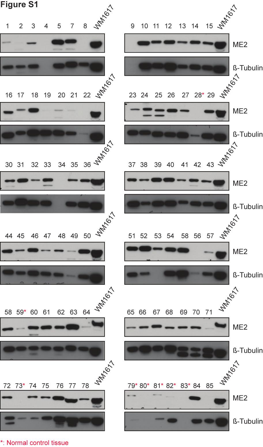
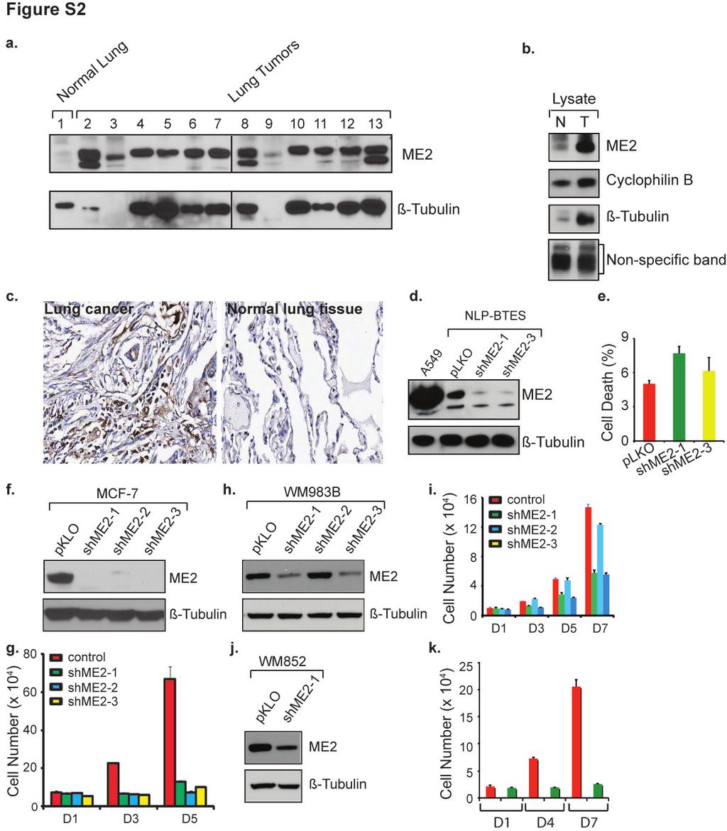
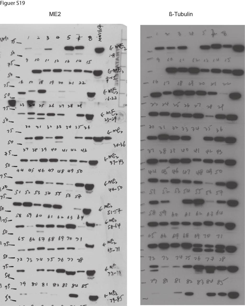
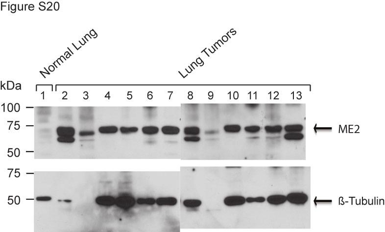
2 SUPPLEMENTAL TABLES AND FIGURE LEGENDS. Table S1. Characteristics of the study samples from tumor patients. Table S2. Characteristics of the study samples from lung tumor patients. Table S3. Characteristics of the study samples from melanoma patients. Figure S1. ME2 expression in solid tumor samples. Human cancer tissues lysed in RIPA buffer were purchased from Protein Biotechnologies and analyzed for ME2 and β-tubulin expression by Western blotting. Equal amounts of protein were loaded. A549 was used as a positive control and loaded in each membrane. The full-length blots are presented in Supplemental Figure S19. Figure S2. ME2 expression in lung cancer and the effects on Normal lung primaery, breast cancer and melanoma cell proliferation of independent shrnas targeting ME2. a, Human lung cancer tissues lysed in RIPA buffer were purchased from Protein Biotechnologies and analyzed for ME2 and β-tubulin expression by Western blotting. Equal amounts of protein were loaded. The blots showed here are from two different gels that have been run under the same experiment conditions and exposed on the same film. The full-length blots are presented in Supplemental Figure S20. b, Paired normal and tumor lung tissues from the same patient were analyzed for ME2, cyclophilin B and β- tubulin expression. Equal amounts of protein were loaded and the non-specific bands were used to show the similar loading. The full-length blots are presented in
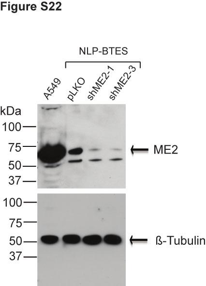
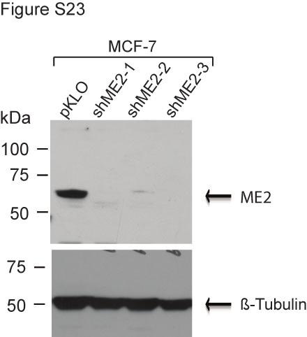
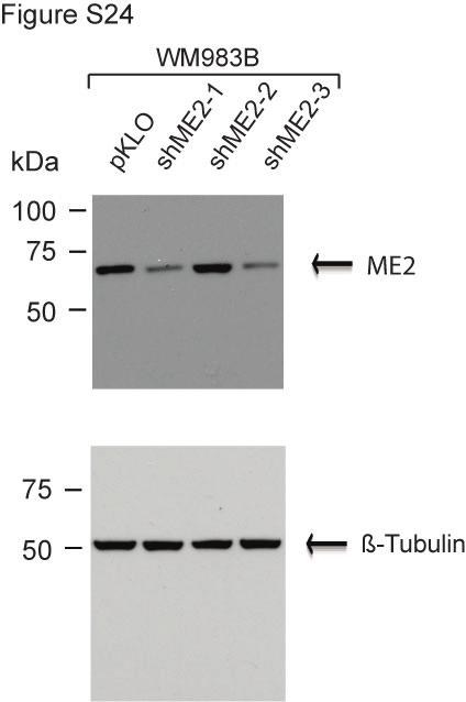
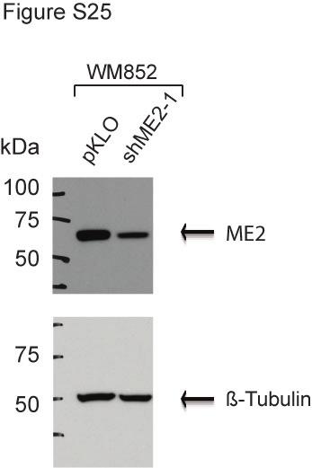
3 Supplemental Figure S21. c, A representative image from the tissue array stained with ME2 antibody. d, Knockdown of ME2 in normal lung primary cells. Western blot analysis using an ME2 antibody of lysate from pools of normal lung primary epithelial cells transduced with two independent ME2 shrna lentiviruses, following 5 days growth without any selection. Data are representative of two independent experiments. The fulllength blots are presented in Supplemental Figure S22. e, The effects of knockdown of ME2 on cell death in normal lung primary cells. f, Western blot analysis using an ME2 antibody of lysate from pools of MCF-7 cells transduced with three independent ME2 shrna lentiviruses, following selection of puromycin for 10 days. Data are representative of at least three independent experiments. All three pools showed marked ME2 silencing. The full-length blots are presented in Supplemental Figure S23. g, Cell proliferation in MCF-7 cells transduced with the indicated shrna lentiviral constructs as described in f. Data are representative of three independent experiments. h, Western blot analysis using an ME2 antibody of lysate from pools of WM983B cells transduced with three independent ME2 shrna lentiviruses, following selection of puromycin for 10 days. Data are representative of at least three independent experiments. The full-length blots are presented in Supplemental Figure S24. i, Cell proliferation in WM983B cells transduced with the indicated shrna lentiviral constructs as described in h. Data are representative of three independent experiments. j, Western blot analysis using an ME2 antibody of lysate from pool of WM852 cells transduced with ME2 shrna lentiviruses, following selection of puromycin for 10 days. Data are representative of at least three independent experiments. The full-length blots are presented in Supplemental Figure S25.
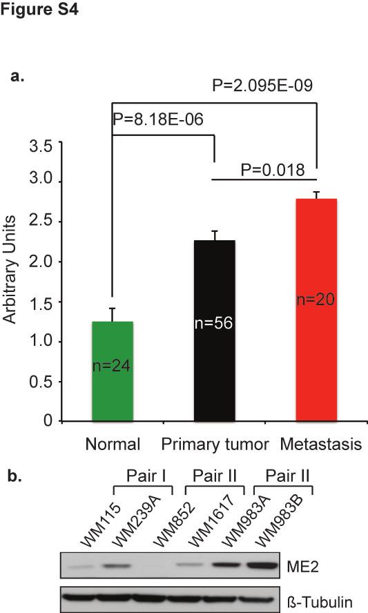
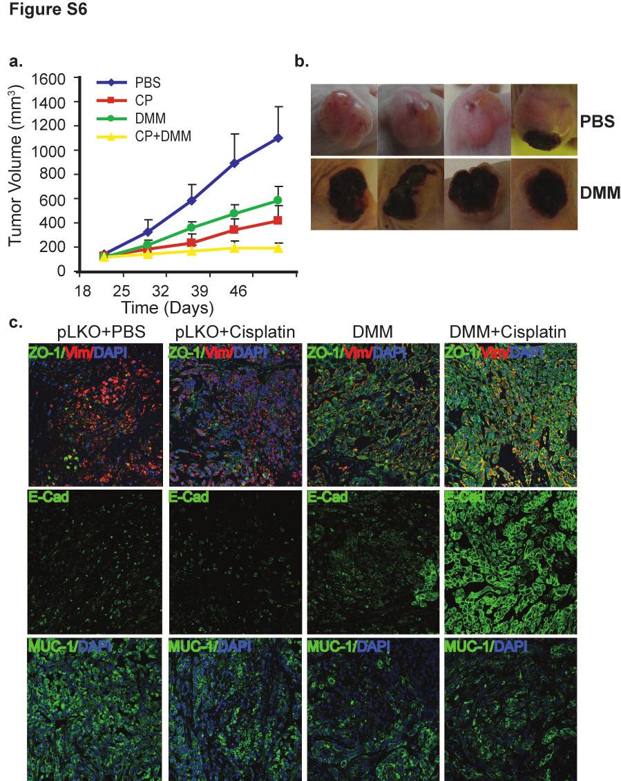
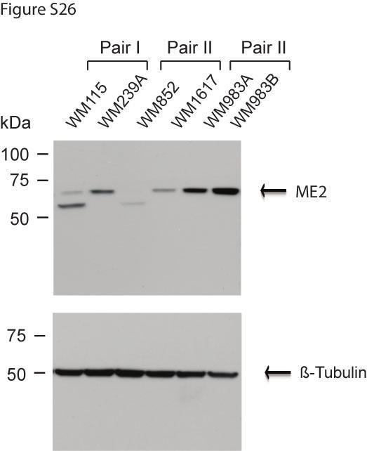
4 k, Cell proliferation in WM852 cells transduced with the indicated shrna lentiviral constructs as described in j. Data are representative of three independent experiments. Figure S3. Tissue array analysis of ME2 expression in 8 types of human solid tumors. Figure S4. ME2 expression in human melanoma analyzed by tissue array and Western blot. a, Tissue array analysis of ME2 expression. b, ME2 levels in 3-paired primary and metastatic melanoma cell lines (primary melanoma cell lines from same patient). The full-length blots are presented in Supplemental Figure S26. Figure S5. DMM selectively kills tumor cells. a, DMM treatment induces ROS production in both normal and tumor lung cells. ROS was detected by flow cytometry using CM-H 2 DCFDA. b, DMM selectively kills lung tumor cells compared to normal lung epithelial cells and normal human fibroblasts. Cell death in A549 cells was analyzed by Annexin V reagent. Each histogram is representative of three experiments. Figure S6. DMM synergistically inhibits cisplatin-treated tumor growth. a, Cisplatin (CP) and DMM combination treatment synergistically inhibits A549 tumor growth. Approximately 5 x 10 6 control plko cells were subcutaneously implanted into female athymic nude mice and treated with DMM, cisplatin or DMM/cisplatin combination as described in Materials and Methods. Tumor sizes were measured and calculated. b, Direct injection of DMM into xenograft tumors causes tumor shrinkage and ulceration. c,
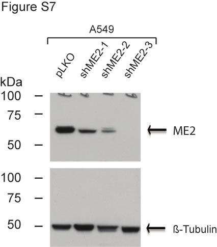
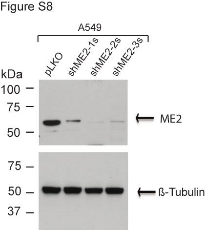
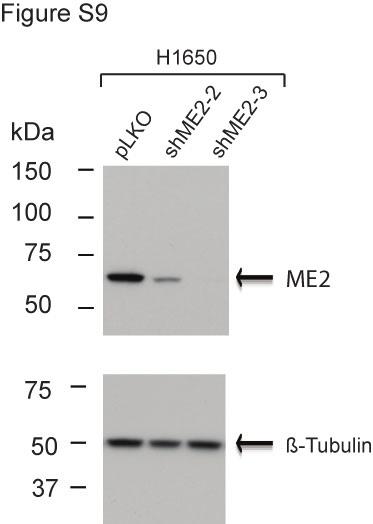
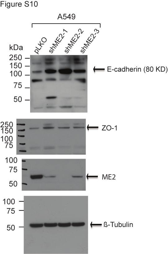
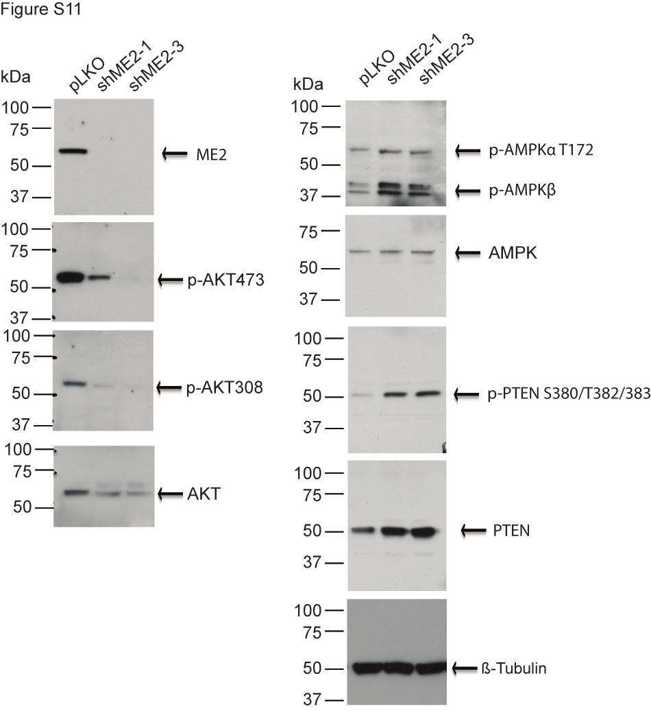
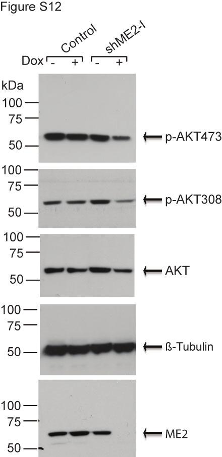
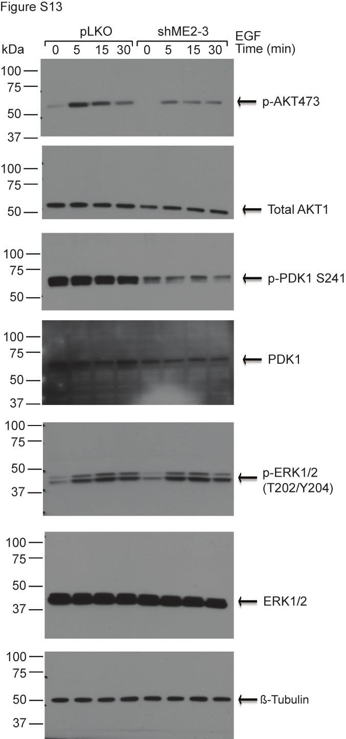
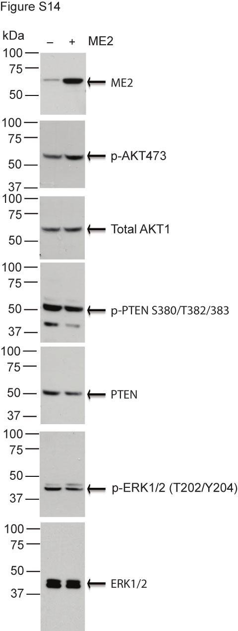
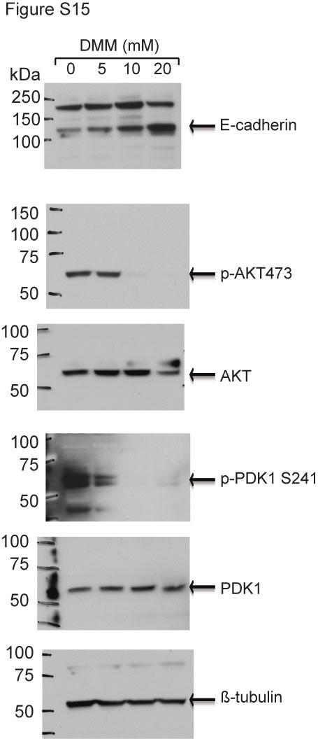
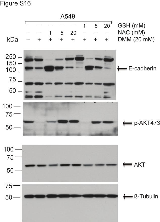
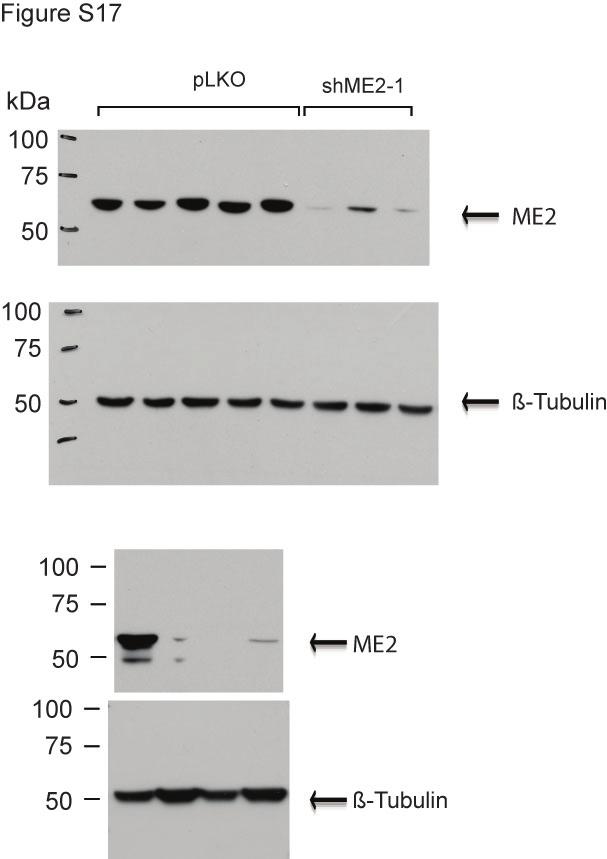
5 The combination of DMM and cisplatin treatment inhibits mucin-1 expression and increases expression of the differentiation markers E-cadherin and ZO-1 in vivo. Figure S7. Full length blots of Figure 1a. Figure S8. Full length blots of Figure 1c. Figure S9. Full length blots of Figure 1e. Figure S10. Full length blots of Figure 2b. Figure S11. Full length blots of Figure 3a. Figure S12. Full length blots of Figure 3c. Figure S13. Full length blots of Figure 3e. Figure S14. Full length blots of Figure 3g. Figure S15. Full length blots of Figure 4g. Figure S16. Full length blots of Figure 5d. Figure S17. Full length blots of Figure 6b.
6 Figure S18. Full length blots of Figure 6d. Figure S19. Full length blots of Figure S1. Figure S20. Full length of Figure S2a Figure S21. Full length blots of Figure S2b Figure S22. Full length blots of Figure S2d Figure S23. Full length blots of Figure S2f. Figure S24. Full length blots of Figure S2h. Figure S25. Full length blots of Figure S2j Figure S26. Full length blots of Figure S4b
7 Table S1 Samples Diagnosis Grade Stage TNM Sex Age Stomach Cancer: 2 Adenocarcinoma 3 II T3N0M0 M 52 3 Mucinous Adenocarcinoma n/a II n/a F 56 4 Infiltrating carcinoma n/a n/a n/a M 42 5 Infiltrating carcinoma n/a n/a n/a M 75 Larynx Cancer: 7 Squamous cell carcinoma 2 n/a n/a F 75 8 Squamous cell carcinoma 2 III T4N0Mx M 71 9 Squamous cell carcinoma 1 II T2N0Mx M 55 Uterus/Ovary Cancer: 10 Endometrial adenocarcinoma 1 IIIc T2N1M0 F Endometrial adenocarcinoma 2 Ic T1N0M0 F Endometrial adenocarcinoma 4 n/a n/a F Uterine Leiomyosarcoma 1 n/a n/a F 40 Bladder Cancer: 12 Transitional cell carcinoma n/a I T1bNxM0 F Papillary transitional cell carcinoma 1 II T2NxM0 M Transitional cell carcinoma 2 II T2NxM0 M Squamous cell carcinoma 3 II T2NxM0 M Adenocarcinoma 3 II T2mNxM 0 M 74 Skin cancer: 79 Normal skin n/a n/a n/a M Squamous cell carcinoma 2 III T2N1M0 F Basal cell carcinoma n/a n/a T3N0Mx F n/a 19 Keratinizing squamous cell carcinoma 3 II T2N0M0 F Squamous cell carcinoma 2 n/a n/a F Granular cell tumor n/a n/a n/a F 53 Keratinizing squamous cell 22 carcinoma n/a n/a T2N0M0 M Malignant fibrous histiocytoma 3 n/a n/a M Sebaceous adenocarcinoma n/a n/a n/a F 78 Lung Cancer: 25 Papillary Adenocarcinoma 2 IIIA T3N0M0 M Adeno-Squamous Cell Carcinoma 2 I T2N0M0 M Large Cell Carcinoma 3 III T3NxM0 M Normal tissue n/a n/a n/a n/a n/a 29 Small Cell Carcinoma 3 IIB T2N1M0 M Squamous Cell Carcinoma 2 I T2N0M0 M 45
8 31 Squamous Cell Carcinoma 3 n/a n/a M Squamous Cell Carcinoma 2 I T1N0M0 M Adenocarcinoma 2 II T2N0Mx F Adenocarcinoma 3 n/a n/a F Small Cell Carcinoma n/a II n/a F Spindle Cell Carcinoma n/a I T2N0M0 F 25 Breast Cancer: 37 Ductal Carcinoma 2 IIIB T2N1M0 F Ductal Carcinoma 2 IIB T2N1M0 F Ductal Carcinoma 3 IIA T2N0M0 F Ductal Carcinoma 3 IIB T2N1M0 F Mucinous Carcinoma 1 II T2N1Mx F Lobular Carcinoma n/a III T3N2M0 F Metaplastic Carcinoma 3 IV T4N1bMx F Intraductal Carcinoma 2 IIA T2N0M0 F 41 Colon Cancer: 45 Adenocarcinoma 2 I T2N0M0 M Adenocarcinoma 1 II T3N0M0 M Adenocarcinoma 2 II T3N0M0 M Adenocarcinoma 3 II T3N0M0 M Adenocarcinoma 3 II T4NxM0 M Mucinous Adenocarcinoma 2 III T3N1M0 M Adenocarcinoma 3 IIIA T2N1M0 M Multipolyposis n/a n/a n/a M Lymphoma n/a n/a n/a M non-hodgkin Lymphoma n/a n/a n/a M Leiomyosarcoma 2 I T2N0M0 M Papillary Adenocarcinoma 2 III T3N0M0 F 56 Kidney Cancer: 57 Papillary renal cell carcinoma 1 n/a n/a M Renal clear cell carcinoma 1 n/a n/a F Renal cysts n/a n/a n/a n/a n/a 60 Renal clear cell carcinoma 3 III T2N0Mx M Renal clear cell carcinoma 2 II n/a F Adenocarcinoma 3 II T2N0M0 M 37 Liver Cancer: 63 Trabecular adenocarcinoma 2 n/a n/a M Hepatocellular carcinoma 2 II T2N0Mx M Hepatocellular carcinoma 3 IIIa T3N0Mx M Hepatocellular carcinoma 3 IIIa T3N0M0 F Hepatocellular carcinoma fibrolamellar 2 II T2N0Mx M Gastrointestinal stromal carcinoma 1 II T3N0Mx F Normal liver n/a n/a n/a M 50
9 Prostate Cancer: 69 Benign prostatic fibrosis n/a n/a n/a M Normal prostate n/a n/a n/a M Prostatic adenoma n/a n/a n/a M Prostatic hyperplasia n/a n/a n/a M Prostatic fibroma n/a n/a n/a M 68 Thyroid Cancer: 73 Benign colloid nodules n/a n/a n/a F Papillary carcinoma n/a n/a n/a F Papillary carcinoma 1 n/a n/a F Medullary carcinoma 2 III T3N1Mx F 38 1 Papillary carcinoma 2 III T2N1M0 F 24 Brain tumpr 82 total protein normal brain - human n/a n/a n/a n/a n/a 84 total protein tumor brain - human n/a n/a n/a n/a n/a pancreatic tissue 83 total protein normal pancreas - human n/a n/a n/a n/a n/a 85 total protein pancreas tumor - human n/a n/a n/a n/a n/a
10 Table S2 Samples Diagnosis Grade Stage TNM Sex Age 1 Normal lung n/a n/a n/a M n/a 2 Adenocarcinoma 3 n/a n/a F 70 3 Large Cell Carcinoma 3 III T3N0Mx M 70 4 Spindle Cell Carcinoma n/a I T2N0M0 F 25 5 Small Cell Carcinoma n/a II n/a F 56 6 Squamous Cell Carcinoma 2 I T1N0M0 M 58 7 Squamous Cell Carcinoma 3 n/a n/a M 37 8 Adenocarcinoma 2 II T2N0Mx F 51 9 Squamous Cell Carcinoma 2 I T2N0M0 M Small Cell Carcinoma 3 IIB T2N1M0 M Large Cell Carcinoma 3 III T3NxM0 M Adeno-Squamous Cell Carcinoma 2 I T2N0M0 M Papillary Adenocarcinoma 2 IIIA T3N0M0 M 42
11 Table S3 No. Age Sex Organ Pathology diagnosis TNM Type 1 80 F Skin Malignant melanoma of head and neck T4N1M0 Malignant 2 60 M Skin Malignant melanoma of left buttocks T4N1M0 Malignant 3 16 F Skin Malignant melanoma of back T4N0M0 Malignant 4 74 M Skin Malignant melanoma of left foot T4N0M0 Malignant 5 55 M Skin Malignant melanoma of sole T4N0M0 Malignant 6 51 M Skin Malignant melanoma of shoulder T4N0M0 Malignant 7 63 F Skin Malignant melanoma of left foot T4N0M0 Malignant 8 46 F Skin Malignant melanoma of thigh T4N0M0 Malignant 9 32 F Skin Malignant melanoma (sparse) of right leg T4N0M0 Malignant M Skin Malignant melanoma of left heel T3N2M1 Malignant M Skin Malignant melanoma of right sole T2N0M0 Malignant M Skin Malignant melanoma of left shoulder T4N0M0 Malignant M Skin Malignant melanoma of left leg T3N0M0 Malignant F Skin Malignant melanoma of left sole T4N0M0 Malignant M Skin Malignant melanoma of left shoulder T4N0M0 Malignant F Skin Malignant melanoma of left sole T4N0M0 Malignant F Skin Malignant melanoma of left thumb T4N0M0 Malignant F Skin Malignant melanoma of left buttocks T4N0M0 Malignant M Skin Malignant melanoma of right abdominal wall T4N1M0 Malignant F Skin Malignant melanoma of left heel T3N0M0 Malignant M Skin Malignant melanoma of right chest wall T4N0M0 Malignant F Skin Malignant melanoma of anus T4N0M0 Malignant F Skin Malignant melanoma of left sole T1N0M0 Malignant F Skin Malignant melanoma of right thumb T4N0M0 Malignant F Skin Malignant melanoma of chest wall T4N0M0 Malignant M Skin Malignant melanoma of heel T4N0M0 Malignant F Skin Malignant melanoma of right toe T4N0M0 Malignant F Vulva Malignant melanoma T4N0M0 Malignant M Skin Malignant melanoma of right heel T4N1M1 Malignant M Skin Malignant melanoma of left buttocks T4N1M0 Malignant M Skin Malignant melanoma of back T3N0M0 Malignant F Skin Malignant melanoma of left thigh T2N0M0 Malignant M Skin Malignant melanoma of left oxter T4N0M0 Malignant F Skin Malignant melanoma of left foot T4N0M0 Malignant M Skin Malignant melanoma of right thigh T4N0M0 Malignant M Skin Malignant melanoma (sparse) of left buttocks T4N0M0 Malignant M Skin Malignant melanoma of scalp T4N0M0 Malignant M Skin Malignant melanoma of perianal T4N0M0 Malignant F Skin Malignant melanoma of right thumb T3N0M0 Malignant F Vulva Malignant melanoma T4N0M0 Malignant F Vulva Malignant melanoma T1N0M0 Malignant F Skin Malignant melanoma of right cheek T4N0M0 Malignant F Skin Malignant melanoma (sparse) with necrosis of right thumb T4N0M0 Malignant F Skin Malignant melanoma of right upper arm T2N0M0 Malignant M Skin Malignant melanoma of right sole T4N0M0 Malignant F Skin Malignant melanoma of left leg T3N0M0 Malignant M Skin Malignant melanoma of right sole T4N0M0 Malignant M Skin Malignant melanoma with necrosis of right thumb T4N0M0 Malignant M Skin Malignant melanoma of right medial malleolus T4N0M0 Malignant F Skin Malignant melanoma of left heel T4N0M0 Malignant M Skin Malignant melanoma of left foot T4N2M0 Malignant M Skin Malignant melanoma of right buttocks T4N0M0 Malignant M Skin Malignant melanoma of left sole T4N0M0 Malignant M Skin Malignant melanoma of right sole T2N0M0 Malignant M Skin Malignant melanoma of scalp T4N0M0 Malignant F Skin Malignant melanoma of scalp T4N0M0 Malignant F Lymph node Metastatic malignant melanoma of right groin - Metastasis M Lymph node Metastatic malignant melanoma of right groin - Metastasis F Lymph node Metastatic malignant melanoma with necrosis of left groin - Metastasis M Lymph node Metastatic malignant melanoma of right oxter - Metastasis M Lymph node Metastatic malignant melanoma of right groin - Metastasis
12 62 47 F Lymph node Metastatic malignant melanoma of right oxter - Metastasis M Lymph node Metastatic malignant melanoma of right neck - Metastasis F Lymph node Metastatic malignant melanoma of right groin - Metastasis M Lymph node Metastatic malignant melanoma of right oxter - Metastasis M Lymph node Metastatic malignant melanoma of left preauricula - Metastasis M Lymph node Metastatic malignant melanoma of right neck - Metastasis F Lymph node Metastatic malignant melanoma of left groin - Metastasis F Lymph node Metastatic malignant melanoma of left groin - Metastasis F Lymph node Metastatic malignant melanoma of right groin - Metastasis F Lymph node Metastatic malignant melanoma of groin - Metastasis F Lymph node Metastatic malignant melanoma of groin - Metastasis F Lymph node Metastatic malignant melanoma of right groin - Metastasis F Lymph node Metastatic malignant melanoma of groin - Metastasis F Lymph node Metastatic malignant melanoma of neck - Metastasis F Lymph node Metastatic malignant melanoma of left groin - Metastasis M Skin Pigmented mole (hyperplasia of squamous epithelium ) - Benign F Skin Atypical melanophoric nevus of left shoulder - Benign F Skin Intradermal nevus of left abdominal wall - Benign F Skin Intradermal nevus of chest wall - Benign F Skin Compound nevus of right waist - Benign M Skin Compound nevus of left leg - Benign M Skin Intradermal nevus of left shoulder - Benign M Skin Intradermal nevus of right cheek - Benign F Skin Atypical melanophoric nevus of right dorsum of foot - Benign 86 2 F Skin Intradermal nevus of frontal region - Benign 87 6 M Skin Compound nevus of left face - Benign 88 5 Mon. M Skin Intradermal nevus of face - Benign M Skin Intradermal nevus (sparse) of scalp - Benign M Skin Intradermal nevus of back - Benign 91 2 F Skin Intradermal nevus of left leg - Benign M Skin Intradermal nevus of face - Benign M Skin Intradermal nevus of left thigh - Benign F Skin Compound nevus of right buttocks - Benign M Skin Malignant transformation of junctional nevus of abdominal wall - Benign M Skin Compound nevus of scalp - Benign 97 7 F Skin Junctional nevus of right forearm - Benign M Skin Compound nevus of upper arm - Benign M Skin Sebaceous nevus (hyperplasia of squamous epithelium) of face - Benign M Skin Sebaceous nevus of left elbow - Benign 101 Tissue marker
13
14
15
16
17
18
19
20
21
22
23
24
25
26
27
28
29

30
31
32
33
34
35
36
37
38
SHN-1 Human Digestive Panel Test results
 SHN-1 Human Digestive Panel Test results HN-30 tongue HN-24 salivary gland HN-12 larynx HN-28 esophagus HN-29 stomach HN-20 pancreas HN-13 liver HN-14 gall bladder HN-27-1 duodenum HN-27-2 ileum HN-27-3
List of Available TMAs in the PRN
 TMA RPCI_BrainCa01 RPCI_BrCa03 RPCI_BrCa04 RPCI_BrCa05 RPCI_BrCa0 RPCI_BrCa07 RPCI_BrCa08 RPCI_BrCa15 RPCI_BrCa1 RPCI_BrCa17 RPCI_BrCa18 RPCI_BrCa19 RPCI_BrCa20 RPCI_BrCa21 RPCI_BrCa24 RPCI_BrCa25 RPCI_BrCa2
Table 1. Clinicopathological features of melanoma samples (shown in Fig. 2) exhibiting higher XYZ mrna levels.
 exhibiting higher XYZ mrna levels.") Sheikh MS et al., Privileged communication. Probe: N T N T N T N T N T N T Table 1. Clinicopathological features of melanoma samples (shown in Fig. 2) exhibiting higher mrna levels. Patient No. Age (yrs)
Sheikh MS et al., Privileged communication. Probe: N T N T N T N T N T N T Table 1. Clinicopathological features of melanoma samples (shown in Fig. 2) exhibiting higher mrna levels. Patient No. Age (yrs)
ENCR RECOMMENDATIONS
 E N C R EUROPEAN NETWORK OF CANCER REGISTRIES (ENCR) ENCR RECOMMENDATIONS Non-Melanoma Skin Cancers Members of the Working Group: Dr T. Davies, East Anglian Cancer Registry, Cambridge, UK Mrs M. Page,
Table of Contents. Preface xi. Acknowledgments xiii. Part I Overview of the Diagnostic Process 1. 1 Overview of Grading and Staging 3
 Table of Contents Preface xi Acknowledgments xiii Part I Overview of the Diagnostic Process 1 1 Overview of Grading and Staging 3 Identification of the process 3 Identification of tumor types 5 Grading
Epithelial tumors. Dr. F.F. Khuzin, PhD Dr. M.O. Mavlikeev
 Epithelial tumors Dr. F.F. Khuzin, PhD Dr. M.O. Mavlikeev Epithelial tumors Tumors from the epithelium are the most frequent among tumors. There are 2 group features of these tumors: The presence in most
performed to help sway the clinician in what the appropriate diagnosis is, which can substantially alter the treatment of management.
 Hello, I am Maura Polansky at the University of Texas MD Anderson Cancer Center. I am a Physician Assistant in the Department of Gastrointestinal Medical Oncology and the Program Director for Physician
H&E, IHC anti- Cytokeratin
 Cat No: OVC2281 - Ovary cancer tissue array Lot# Cores Size Cut Format QA/QC OVC228101 228 1.1mm 4um 12X19 H&E, IHC anti- Cytokeratin Recommended applications: For Research use only. RNA or protein ovary
Outcomes Report: Accountability Measures and Quality Improvements
 Outcomes Report: Accountability Measures and Quality Improvements The FH Memorial Medical Center s Cancer Committee ensures that patients with cancer are treated according to the nationally accepted measures.
Benign Mimics of Malignancy in Breast Pathology
 Arthur Purdy Stout Society of Surgical Pathologists Companion Meeting Benign Mimics of Malignancy in Breast Pathology Stuart J. Schnitt, M.D. Beth Israel Deaconess Medical Center and Harvard Medical School,
Tumour Structure and Nomenclature. Paul Edwards. Department of Pathology and Cancer Research UK Cambridge Institute, University of Cambridge
 Tumour Structure and Nomenclature Paul Edwards Department of Pathology and Cancer Research UK Cambridge Institute, University of Cambridge Malignant Metastasis Core idea of cancer Normal Cell Slightly
SOMAPLEX REVERSE PHASE PROTEIN MICROARRAY HUMAN KIDNEY TUMOR & NORMAL TISSUE
 SOMAPLEX REVERSE PHASE PROTEIN MICROARRAY HUMAN KIDNEY TUMOR & NORMAL TISSUE 45 CLINICAL CASES SERIAL DILUTION MULTIPLE PROTEIN CONCENTRATION QUANTITATIVE ASSAY PRODUCT NUMBER: PM1-001-N SOMAPLEX REVERSE
Breast pathology. 2nd Department of Pathology Semmelweis University
 Breast pathology 2nd Department of Pathology Semmelweis University Breast pathology - Summary - Benign lesions - Acute mastitis - Plasma cell mastitis / duct ectasia - Fat necrosis - Fibrocystic change/
LYMPHATIC DRAINAGE AXILLARY (MOSTLY) INTERNAL MAMMARY SUPRACLAVICULAR
 INTERNAL MAMMARY SUPRACLAVICULAR") BREAST LYMPHATIC DRAINAGE AXILLARY (MOSTLY) INTERNAL MAMMARY SUPRACLAVICULAR HISTOLOGY LOBE: (10 in whole breast) LOBULE: (many per lobe) ACINUS/I, aka ALVEOLUS/I: (many per lobule) DUCT(S): INTRA- or
BREAST LYMPHATIC DRAINAGE AXILLARY (MOSTLY) INTERNAL MAMMARY SUPRACLAVICULAR HISTOLOGY LOBE: (10 in whole breast) LOBULE: (many per lobe) ACINUS/I, aka ALVEOLUS/I: (many per lobule) DUCT(S): INTRA- or
Case year female. Routine Pap smear
 Case 1 57 year female Routine Pap smear Diagnosis? 1. Atypical glandular cells of unknown significance (AGUS) 2. Endocervical AIS 3. Endocervical adenocarcinoma 4. Endometrial adenocarcinoma 5. Adenocarcinoma
Basement membrane in lobule.
 Bahram Memar, MD Basement membrane in lobule. Normal lobule-luteal phase Normal lobule-follicular phase Lactating breast Greater than 95% are adenocarcinomas in situ carcinomas and invasive carcinomas.
Neoplasia part I. Dr. Mohsen Dashti. Clinical Medicine & Pathology nd Lecture
 Neoplasia part I By Dr. Mohsen Dashti Clinical Medicine & Pathology 316 2 nd Lecture Lecture outline Review of structure & function. Basic definitions. Classification of neoplasms. Morphologic features.
Other Sites. Table 2 Continued. MPH Rules 11/8/07. NAACCR Webinar Series 1
 MPH s 11/8/07 Other s 1 Table 2 Continued Use this two-page table to select combination histology codes. Compare the terms in the diagnosis to the terms in Columns 1 and 2. If the terms match, code the
Cancer Program Report 2014
 Cancer Program Report 2014 Queen of the Valley Hospital St Joseph Health Queen of the Valley Hospital - 2014 Site Table Site Total Class Sex Group Cases Analytic NonAn M F 0 I II ALL SITES 661 494 167
Contents. Basic Ultrasound Principles and Terminology. Ultrasound Nodule Characteristics
 Contents Basic Ultrasound Principles and Terminology Basic Ultrasound Principles... 1 Ultrasound System... 2 Linear Transducer for Superficial Images and Ultrasound-Guided FNA... 3 Scanning Planes... 4
3/25/2019. Rare uterine cancers ~3% Leiomyosarcoma Carcinosarcoma (MMMT) Endometrial Stromal Sarcomas Aggressive tumors High Mortality Rates
 Endometrial Stromal Sarcomas Aggressive tumors High Mortality Rates") J. Anthony Rakowski D.O., F.A.C.O.O.G. MSU SCS Board Review Coarse Rare uterine cancers ~3% Leiomyosarcoma Carcinosarcoma (MMMT) Endometrial Stromal Sarcomas Aggressive tumors High Mortality Rates Signs
J. Anthony Rakowski D.O., F.A.C.O.O.G. MSU SCS Board Review Coarse Rare uterine cancers ~3% Leiomyosarcoma Carcinosarcoma (MMMT) Endometrial Stromal Sarcomas Aggressive tumors High Mortality Rates Signs
Macmillan Publications
 S1 S2 S3 S3 S3 S4 S5 S6 S7 S8 S8 S9 S10 S11 S11 S12 S13 S14 S15 S17 S18 S19 Bladder Cancer: Non-Invasive, Invasive and Advanced Bone Cancer: Primary, Secondary Colon Cancer, Anal Cancer, Rectal Cancer
NEOPLASIA-I CANCER. Nam Deuk Kim, Ph.D.
 NEOPLASIA-I CANCER Nam Deuk Kim, Ph.D. 1 2 Tumor in the hieroglyphics of the Edwin Smith papyrus (1,600 B.C., Breasted s translation 1930) 3 War on Cancer (National Cancer Act, 1971) 4 Cancer Acts in Korea
Benign and malignant epithelial lesions: Seborrheic keratosis: A common benign pigmented epidermal tumor occur in middle-aged or older persons more
 Benign and malignant epithelial lesions: Seborrheic keratosis: A common benign pigmented epidermal tumor occur in middle-aged or older persons more common on the trunk; but extremities, head and neck are
Cancer: recent advances and implications for underwriting
 Cancer: recent advances and implications for underwriting Robert Rubens Select 74 Bristol 25 February 2010 Agenda Epidemiology - changing mortality Evidence-base for underwriting breast cancer ovarian
Laboratory for Quantitative Medicine Technical Report #2 April 10, Equation Parameters
 Laboratory for Quantitative Medicine Technical Report #2 April 10, 2009 Equation Parameters This report summarizes all the equation parameters used by the web-based calculators, and provides information
Appendix 4: WHO Classification of Tumours of the pancreas 17
 S3.01 The WHO histological tumour type must be recorded. CS3.01a The histological type of the tumour should be recorded based on the current WHO classification 17 (refer to Appendices 4-7). Appendix 4:
Gross appearance of nodular hyperplasia in material obtained from suprapubic prostatectomy. Note the multinodular appearance and the admixture of
 Tiền liệt tuyến Tiền liệt tuyến Gross appearance of nodular hyperplasia in material obtained from suprapubic prostatectomy. Note the multinodular appearance and the admixture of solid and microcystic areas.
Educational Cases EQA November T.J. Palmer Raigmore Hospital Inverness
 Educational Cases EQA November 2013 T.J. Palmer Raigmore Hospital Inverness Case 2 Clinical Details Dob 11 February 1951 PMH: 1964 Extraction of 45 aet 13 yr 1966 Cyst between 44 and 46 enucleated 1973
Oncology 101. Cancer Basics
 Oncology 101 Cancer Basics What Will You Learn? What is Cancer and How Does It Develop? Cancer Diagnosis and Staging Cancer Treatment What is Cancer? Cancer is a group of more than 100 different diseases
Learning Objectives. ! At the end of the presentation,students will be able to:
 Learning Objectives! At the end of the presentation,students will be able to: Define Breast Cancer Identify the controllable and uncontrollable risk factors of breast cancer Recognize the 4 stages of breast
Lesion Imaging Characteristics Mass, Favoring Benign Circumscribed Margins Intramammary Lymph Node
 Lesion Imaging Characteristics Mass, Favoring Benign Circumscribed Margins Intramammary Lymph Node Oil Cyst Mass, Intermediate Concern Microlobulated Margins Obscured Margins Mass, Favoring Malignant Indistinct
ab Skin tumor tissue array, 75 cases, 150 samples (1.1mm)
") ab178285 Skin tumor tissue array, 75 cases, 150 samples (1.1mm) nstructions for Use Designed for HC or SH based protein or RNA tissue profiling in various skin tumors. This product is for research use
ab178285 Skin tumor tissue array, 75 cases, 150 samples (1.1mm) nstructions for Use Designed for HC or SH based protein or RNA tissue profiling in various skin tumors. This product is for research use
ACRIN 6666 Therapeutic Surgery Form
 S1 ACRIN 6666 Therapeutic Surgery Form 6666 Instructions: Complete a separate S1 form for each separate area of each breast excised with the intent to treat a cancer (e.g. each lumpectomy or mastectomy).
Breast Pathology. Breast Development
 Breast Pathology Lecturer: Hanina Hibshoosh, M.D. Reading: Kumar, Cotran, Robbins, Basic Pathology, 6th Edition, pages 623-635 Breast Development 5th week - thickening of the epidermis - milk line 5th
DATA REQUEST RESPONSE- XRT AND BRACHYTHERAPY
 Date: 10 th April 2018 DATA REQUEST RESPONSE- XRT AND BRACHYTHERAPY Request: 1. Utilization Data of Overseas Beam Therapy and Brachytherapy 2. Diagnoses Data of Overseas Claims for Beam Therapy and Brachytherapy
Take Home Quiz 1 Please complete the quiz below prior to the session. Use the Multiple Primary and Histology Rules
 Take Home Quiz 1 Please complete the quiz below prior to the session. Use the Multiple Primary and Histology Rules Case 1 72 year old white female presents with a nodular thyroid. This was biopsied in
SIRT6 histone deacetylase functions as a potential oncogene in human melanoma -
 SIRT6 histone deacetylase functions as a potential oncogene in human melanoma - Garcia-Peterson et al Supplementary Figure S:Tissue microarray (TMA) staining and organization. A) Diagram of the TMA indicating
Cutaneous metastases. Thaddeus Mully. University of California, San Francisco Professor, Departments of Pathology and Dermatology
 Cutaneous metastases Thaddeus Mully University of California, San Francisco Professor, Departments of Pathology and Dermatology DISCLOSURE OF RELATIONSHIPS WITH INDUSTRY Thaddeus Mully Course C005 Essential
Contents 1 The Windows of Susceptibility to Breast Cancer 2 The So Called Pre-Neoplastic Lesions and Carcinoma In Situ
 Contents 1 The Windows of Susceptibility to Breast Cancer... 1 1.1 Introduction... 1 1.2 Risk Factor and Etiological Agents... 2 1.3 The Concept of the Windows of Susceptibility to Carcinogenesis... 5
GASTROINTESTINAL IMAGING STUDY GUIDE
 GASTROINTESTINAL IMAGING STUDY GUIDE Pharynx Diverticula Foreign bodies Trauma o Motility Disorders Esophagus Diverticula Trauma Esophagitis Barrett esophagus Rings, webs, and strictures Varices Benign
Mody. AIS vs. Invasive Adenocarcinoma of the Cervix
 Common Problems in Gynecologic Pathology Michael T. Deavers, M.D. Houston Methodist Hospital, Houston, Texas Common Problems in Gynecologic Pathology Adenocarcinoma in-situ (AIS) of the Cervix vs. Invasive
AJCC 7th Edition Handbook Errata as of 9/21/10
 5 81 Larynx ICD-O-3 Topography Codes Delete C32.3 Laryngeal cartilage 5 81 Larynx ICD-O-3 Topography Codes Add an asterisk after C32.8 5 81 Larynx ICD-O-3 Topography Codes Add an asterisk after C32.9 5
MEDICAL POLICY Gene Expression Profiling for Cancers of Unknown Primary Site
 POLICY: PG0364 ORIGINAL EFFECTIVE: 04/22/16 LAST REVIEW: 07/26/18 MEDICAL POLICY Gene Expression Profiling for Cancers of Unknown Primary Site GUIDELINES This policy does not certify benefits or authorization
Case #1: 75 y/o Male (treated and followed by prostate cancer oncology specialist ).
.") SOLID TUMORS WORKSHOP Cases for review Prostate Cancer Case #1: 75 y/o Male (treated and followed by prostate cancer oncology specialist ). January 2009 PSA 4.4, 20% free; August 2009 PSA 5.2; Sept 2009
SOLID TUMORS WORKSHOP Cases for review Prostate Cancer Case #1: 75 y/o Male (treated and followed by prostate cancer oncology specialist ). January 2009 PSA 4.4, 20% free; August 2009 PSA 5.2; Sept 2009
incidence rate x 100,000/year
 Tier R=rare C=common Cancer Entity European crude and age adjusted incidence by cancer, years of diagnosis 2000 and 2007 Analisys based on 83 population-based cancer registries * applying the European
Icd 10 code metastatic adenocarcinoma endometrial
 Icd 10 code metastatic adenocarcinoma endometrial 1-10-2017 Free, official coding info for 2018 ICD-10-CM D07.0 - includes detailed rules, notes, synonyms, ICD-9-CM conversion,. 2018 ICD-10-CM Diagnosis
04/10/2018. Intraductal Papillary Neoplasms Of Breast INTRADUCTAL PAPILLOMA
 Intraductal Papillary Neoplasms Of Breast Savitri Krishnamurthy MD Professor of Pathology Deputy Division Head The University of Texas MD Anderson Cancer Center 25 th Annual Seminar in Pathology Pittsburgh,
Pitfalls in thyroid tumor pathology. Prof.Valdi Pešutić-Pisac MD, PhD
 Pitfalls in thyroid tumor pathology Prof.Valdi Pešutić-Pisac MD, PhD Too many or... Tumour herniation through a torn capsule simulating capsular invasion fibrous capsule with a sharp discontinuity, suggestive
Cerebral Parenchymal Lesions: I. Metastatic Neoplasms
 Chapter 4 Cerebral Parenchymal Lesions: I. Metastatic Neoplasms After one has reasonably ruled out the possibility of a nonneoplastic diagnosis (see Chap. 3), one is left with considering a diagnosis of
America, Hershey, PA Australia, Melbourne, VIC Europe, Munich
 America, Hershey, PA Australia, Melbourne, VIC Europe, Munich info@vivopharm.com www.vivopharm.com Bladder MB-9-luc 2 Mouse urinary bladder carcinoma yes yes yes C57BL/6 2 Bone UMR-106 Rat osteosarcoma
Icd 10 code met renal cell
 Icd 10 code met renal cell Search 1-10-2015 Free, official coding info for 2018 ICD - 10 -CM N16 - includes detailed rules, notes, synonyms, ICD -9-CM conversion, index and annotation crosswalks, DRG.
Video Microscopy Tutorial 8
 Video Microscopy Tutorial 8 Common and Uncommon Lesions of the Liver Gladwyn Leiman, MD There are no disclosures necessary. Common and Uncommon Lesions in Liver FNA Gladwyn Leiman University of Vermont
Cancer Fundamentals. Julie Randolph-Habecker, Ph.D. Director, Experimental Histopathology Shared Resource
 Cancer Fundamentals Julie Randolph-Habecker, Ph.D. Director, Experimental Histopathology Shared Resource Cancer Overview Leading cause of death in US 1.2 million diagnosed each year More common after age
-1- Pathology Department (code: 0605) Final Exam for Third year students Date: Time allowed: 2 hours. Paper II (75 marks).
 Final Exam for Third year students Date: Time allowed: 2 hours. Paper II (75 marks).") -1- BENHA UNIVERSITY FACULTY OF MEDICINE Pathology Department (code: 0605) Final Exam for Third year students Date: 28-5-2011 Time allowed: 2 hours. Paper II (75 marks). Please note that this question
-1- BENHA UNIVERSITY FACULTY OF MEDICINE Pathology Department (code: 0605) Final Exam for Third year students Date: 28-5-2011 Time allowed: 2 hours. Paper II (75 marks). Please note that this question
Interventions for non-metastatic squamous cell carcinoma of the skin: a systematic review and pooled analysis of observational studies
 Web appendix 2: SEARCH STRATEGIES Interventions for non-metastatic squamous cell carcinoma of the skin: a systematic review and pooled analysis of observational studies MEDLINE 1. exp epidemiologic studies/
Diagnostic Cytology of Cancer Cases
 Diagnostic Cytology of Cancer Cases Somporn Techangamsuwan Companion Animal Cancer Research Unit (CAC-RU) Department of Pathology, Faculty of Veterinary Science, Chulalongkorn University 1 Tumor or Non-tumor
SCOPE OF PRACTICE PGY-5
 Recognize normal cytomorphology of cells derived from the respiratory, gastrointestinal, and genitourinary tracts, and body fluid (Cerebrospinal fluid, pleural and peritoneal fluid) Recognize normal cytomorphology
The Panel Approach to Diagnostics. Lauren Hopson International Product Specialist Cell Marque Corporation
 The Panel Approach to Diagnostics Lauren Hopson International Product Specialist Cell Marque Corporation Cell Marque Rocklin, California About Cell Marque: IVD primary antibody manufacturer Distributors
Neoplasms of the Canine, Feline and Lemur Liver:
 Neoplasms of the Canine, Feline and Lemur Liver: Classification and Prognosis Annual Seminar of the French Society of Veterinary Pathology John M. Cullen VMD PhD DACVP North Carolina State University Primary
Abid Irshad, MD Director Breast Imaging. Medical University of South Carolina Charleston
 Abid Irshad, MD Director Breast Imaging Medical University of South Carolina Charleston Cases Financial disclosure: I or my family have no financial interest related to the material discussed in this presentation
Differential Diagnosis of Oral Masses. Palatal Lesions
 Differential Diagnosis of Oral Masses Palatal Lesions Palatal Masses Periapical Abscess Torus Palatinus Mucocele Lymphoid Hyperplasia Adenomatous Hyperplasia Benign Salivary Neoplasms Malignant Salivary
A215- Urinary bladder cancer tissues
 A215- Urinary bladder cancer tissues (formalin fixed) For research use only Specifications: No. of cases: 45 Tissue type: Urinary bladder cancer tissues No. of spots: 2 spots from each cancer case (90
Supplementary Figure 1. Double-staining immunofluorescence analysis of invasive colon and breast cancers. Specimens from invasive ductal breast
 Supplementary Figure 1. Double-staining immunofluorescence analysis of invasive colon and breast cancers. Specimens from invasive ductal breast carcinoma (a) and colon adenocarcinoma (b) were staining
Neuroendocrine Carcinoma. Lebanon Neuroendocrine Neoplasms of H&N Nov /7/2011. Broad Classification:
 H&N Neuroendocrine Neoplasms: Classification and Diagnostic Considerations Adel K. El-Naggar, M.D., Ph.D. The University of Texas MD Anderson Cancer Center, Houston, Texas Broad Classification: A. Epithelial:
Cancer of lymph nodes survival rate
 Cancer of lymph nodes survival rate Search 14-3-2016 Survival rates of pancreatic cancer are based on outcomes of people who've had the disease. Find the survival rates for pancreatic cancer here. 18-12-2017
Salivary Gland Cytology
 Salivary Gland Cytology Diagnostic challenges and potential pitfalls Tarik M. Elsheikh, MD Professor and Medical Director Anatomic Pathology Cleveland Clinic FNA Salivary Gland Lesions Indications Distinguish
Supporting Information
 Supporting Information Vaira et al. 10.1073/pnas.0907676107 0h 24h 48h PTEN pathway Expression 0h 24h 48h Time MAP Kinase Signaling Pathway Expression 0h 24h 48h Time Collagen Expression Fig. S1. Differential
- Selected Tumors of the Skin Appendages - Primary vs. Metastasis
 - Selected Tumors of the Skin Appendages - Primary vs. Metastasis Napa Valley 2018 Victor G. Prieto, MD, PhD Chair of Pathology UT MD Anderson Cancer Center vprieto@mdanderson.org Napa Valley in May Introduction
New Developments in Immunohistochemistry for Gynecologic Pathology
 New Developments in Immunohistochemistry for Gynecologic Pathology Michael T. Deavers, M.D. Professor, Departments of Pathology and Gynecologic Oncology Immunohistochemistry in Gynecologic Pathology Majority
Induction of Erythroid Differentiation in Human Erythroleukemia Cells by Depletion of Malic Enzyme 2
 Induction of Erythroid Differentiation in Human Erythroleukemia Cells by Depletion of Malic Enzyme 2 The Harvard community has made this article openly available. Please share how this access benefits
Nuclear medicine in oncology. 1. Diagnosis 2. Therapy
 Nuclear medicine in oncology 1. Diagnosis 2. Therapy Diagnosis - Conventional methods - Nonspecific radiopharmaceuticals cumulating in tumours - Specific radiopharmaceuticals (receptor- and immunoscintigraphy)
Test Bank for Robbins and Cotran Pathologic Basis of Disease 9th Edition by Kumar
 Link full download: http://testbankair.com/download/test-bank-for-robbins-cotran-pathologic-basis-of-disease-9th-edition-bykumar-abbas-and-aster Test Bank for Robbins and Cotran Pathologic Basis of Disease
Introduction. Results. Discussion. Histopathologic and immunohistochemical findings. Results. conclusions,
 1/5 2/5 Carcinoma distinctive carcinoma. form erysipeloides (CE), metastasis. which clinically Itfrom has resembles been termed erysipelas, is an uncommon, but may extend It164 toclassically back, presents
Desmoplastic Melanoma R/O BCC. Clinical Information. 74 y.o. man with lesion on left side of neck r/o BCC
 R/O BCC Sabine Kohler, M.D. Professor of Pathology and Dermatology Dermatopathology Service Stanford University School of Medicine Clinical Information 74 y.o. man with lesion on left side of neck r/o
2011 to 2015 New Cancer Incidence Truman Medical Center - Hospital Hill
 Number of New Cancers Truman Medical Center Hospital Hill Cancer Registry 2015 Statistical Summary Incidence In 2015, Truman Medical Center diagnosed and/or treated 406 new cancer cases. Four patients
Outcomes Report: Accountability Measures and Quality Improvements
 Outcomes Report: Accountability Measures and Quality Improvements The s Cancer Committee ensures that patients with cancer are treated according to the nationally accepted measures. Because we are an accredited
Human cell line list Production of Exosome Standards - Cell Lysates
 Human cell line list 2016 - Production of Exosome Standards - Cell Lysates 380 peripheral blood leukemia, pre-b cell 1301 blood leukemia, acute lymphoblastic, T cell 5637 bladder carcinoma 8305C thyroid
BREAST PATHOLOGY. Fibrocystic Changes
 BREAST PATHOLOGY Lesions of the breast are very common, and they present as palpable, sometimes painful, nodules or masses. Most of these lesions are benign. Breast cancer is the 2 nd most common cause
UICC TNM 8 th Edition Errata
 UICC TNM 8 th Edition Errata ions are in italics Page 28 Oropharynx p16 positive Pathological Stage II,T2 N2 M0 T3 N0,N1 M0 Stage II,T2 N2 M0 T3,T4 N0,N1 M0 Page 61 Oesophagus Adenocarcinoma Pathological
LN04 - Lymphoma Tissue Microarray
 Reveal Biosciences offers Histochemical Staining, Immunohistochemistry (IHC), In Situ Hybridization (ISH), Whole Slide Imaging, and Quantitative Image Analysis on any TMA LN04 - Lymphoma Tissue Microarray
UICC TNM 8 th Edition Errata
 UICC TNM 8 th Edition Errata ions are in italics Head and Neck Tumours Pages 20, p27, p34, p38, p41, and p49 ly pn2a Metastasis in a single ipsilateral lymph node, less than 3cm in greatest dimension with
FINALIZED SEER SINQ QUESTIONS
 0076 Source 1: WHO Class CNS Tumors pgs: 33 MP/H Rules/Histology--Brain and CNS: What is the histology code for a tumor originating in the cerebellum and extending into the fourth ventricle described as
Effective January 1, 2018 ICD O 3 codes, behaviors and terms are site specific
 Effective January 1, 2018 codes, behaviors and terms are site specific /N 8551/3 Acinar adenocarcinoma (C34. _) Lung primaries diagnosed prior to 1/1/2018 use code 8550/3 For prostate (all years) see 8140/3
Radiology Pathology Conference
 Radiology Pathology Conference Nadia F. Yusaf, M.D. PGY-3 1/29/2010 Presentation material is for education purposes only. All rights reserved. 2010 URMC Radiology Page 1 of 90 Case 1 60 year- old man presents
Whitney A. High, MD, JD, MEng
 ADS Dermatopathology Meeting 2014 Selected Adnexal Tumors Whitney A. High, MD, JD, MEng Associate Professor, Dermatology & Pathology Director of Dermatopathology (Dermatology) University of Colorado School
Radiation Oncology Study Guide
 Radiation Oncology Study Guide For the Initial CertificationQualifying (Computer-Based) Examination General and Radiation Oncology This examination is designed to assess your understanding of the entire
2018 ICD-O-3 Updates in Table Format with Annotation for Reference
 Status Histology Description (this may be preferred term or a synonym) Report Comments New term 8010 3 Urachal carcinoma (C65.9, C66.9, C67._, C68._) New term 8013 3 Combined large cell neuroendocrine
Effective January 1, 2018 ICD O 3 codes, behaviors and terms are site specific
 Effective January 1, 2018 codes, behaviors and terms are site specific Status /N 8010/3 Urachal carcinoma (C65.9, C66.9, C67. _, C68._) 8013/3 Combined large cell neuroendocrine carcinoma (C34. _, C37.9)
Staging and Treatment Update for Gynecologic Malignancies
 Staging and Treatment Update for Gynecologic Malignancies Bunja Rungruang, MD Medical College of Georgia No disclosures 4 th most common new cases of cancer in women 5 th and 6 th leading cancer deaths
CURRICULUM FOR THE BREAST PATHOLOGY ROTATION UNIVERSITY OF FLORIDA DEPARTMENT OF PATHOLOGY
 CURRICULUM FOR THE BREAST PATHOLOGY ROTATION UNIVERSITY OF FLORIDA DEPARTMENT OF PATHOLOGY JULY, 2003 The following is a conceptual curriculum and set of guidelines for Pathology Residents on the Breast
Outline 11/2/2017. Pancreatic EUS-FNA general aspects. Cytomorphologic features of solid neoplasms/lesions of the pancreas
 ENDOSCOPIC ULTRASOUND GUIDED-FINE NEEDLE ASPIRATION CYTOLOGY OF PANCREAS Khalid Amin M.D. Assistant Professor Department of Laboratory Medicine and Pathology University of Minnesota Outline Pancreatic
Radiology Pathology Conference
 Radiology Pathology Conference Sharlin Johnykutty,, MD, Cytopathology Fellow Sara Majewski, MD, Radiology Resident Friday, August 28, 2009 Presentation material is for education purposes only. All rights
Quiz. b. 4 High grade c. 9 Unknown
 Quiz 1. 10/11/12 CT scan abdomen/pelvis: Metastatic liver disease with probable primary colon malignancy. 10/17/12 Colonoscopy with polypectomy: Adenocarcinoma of sigmoid colon measuring at least 6 mm
Proposed All Wales Vulval Cancer Guidelines. Dr Amanda Tristram
 Proposed All Wales Vulval Cancer Guidelines Dr Amanda Tristram Previous FIGO staging FIGO Stage Features TNM Ia Lesion confined to vulva with
NFP Process Centers 95 SWG DCR
 NFP NFP Process Centers 8. ungs, Posterior Back 17. Emotional (R) 18. Mental Body () 6. Heart / Center & eft 7. ungs, Anterior Chest 12. Kidneys / Back 14. Upper Arms 15. Forearms 9. Spleen / eft Side
Test Bank for Robbins and Cotran Pathologic Basis of Disease 9th Edition by Kumar
 Link full download:https://getbooksolutions.com/download/test-bank-for-robbinsand-cotran-pathologic-basis-of-disease-9th-edition-by-kumar Test Bank for Robbins and Cotran Pathologic Basis of Disease 9th
Disclosure of Relevant Financial Relationships
 Squamous entities of the thyroid: Reactive to Neoplastic Michelle D. Williams Associate Professor Dept of Pathology, Head & Neck Section University of Texas MD Anderson Cancer Center Disclosure of Relevant
Submission of samples. Cytology of Lumps and Bumps. Evaluation of samples. Use caution interpreting. Criteria of malignancy.
 Submission of samples Cytology of Lumps and Bumps Paul Avery VMD, PhD, DACVP paul.avery@colostate.edu Air dry only No wet fixation using formalin or ethanol Stain 1-2 on-site to evaluate quality Send all
Normal endometrium: A, proliferative. B, secretory.
 Normal endometrium: A, proliferative. B, secretory. Nội mạc tử cung Nội mạc tử cung Cyclic changes in endometrium.. Approximate relationship of useful microscopic changes. Arias-Stella reaction in endometrial