INTRODUCTION. leading to cell death or carcinogenesis. DNA damage blocks normal cellular processes
|
|
|
- Kristian Wheeler
- 6 years ago
- Views:
Transcription
1 INTRODUCTION DNA damage and types of repair DNA is subject to various types of damage that can impair cellular function leading to cell death or carcinogenesis. DNA damage blocks normal cellular processes such as replication and transcription [1, 2]. Should DNA damage persist it can have catastrophic consequences for the cell and for the organism including mutagenesis, cancer, and cell death (i.e. in neurons). DNA damage may come from internal as well as external sources and is associated with various types of genetic diseases and cancers. Therefore DNA repair provides a mechanism to restore DNA nucleotide sequences to the native state. Several DNA repair pathways have been identified including base excision repair (BER), mismatch repair (MMR) and nucleotide excision repair (NER). The type of lesion will dictate which pathway is utilized for repair. For example an N7-methyl guanine residue can only be removed by BER, while a benzopyrene guanine adduct residue can only be removed by NER [3]. Nucleotide Excision Repair Nucleotide Excision Repair (NER) is a versatile DNA repair pathway because of its ability to repair a wide range of nucleotide adducts [4]. NER is responsible for the repair of bulky DNA damage lesions including cyclobutane pyrimidine dimers (CPDs) and 6-4 pyrimidine pyrimidone photoproducts (6-4PPs) caused by UV radiation [3, 5]. NER repairs platinum DNA adducts that are formed by cancer chemotherapeutic drugs such as cisplatin and carboplatin [1, 6, 7]. UV radiation and platinum compounds cause covalent linkage of adjacent thymine or guanine residues respectively. Aromatic 1
2 hydrocarbons, as found in cigarette smoke, cause guanine adducts that also require NER for their removal. The steps involved in NER include recognition of DNA damage, excising the lesion as a single stranded oligomer, resynthesizing the excised region of DNA, and finally ligating the newly repaired DNA strand [5, 8]. NER is divided into two distinct sub-pathways: transcription-coupled repair (TC-NER), which affects the transcribed strand in genes that are expressed, and global genomic repair (G-NER), which is responsible for removing DNA damage from the overall genome. These pathways differ in the early recognition step of DNA damage, but utilize the same proteins in physically excising the lesion and restoring the DNA sequence. TC-NER is the repair of lesions on DNA that is simultaneously undergoing transcription. While TC-NER only occurs in a small percent of the genome it is nonetheless critical to cell survival. DNA damage recognition in TC-NER occurs when transcription is stalled by the arrest of RNA polymerase II as it encounters the site of DNA damage [9, 10]. The stalled polymerase is thought to trigger the recruitment of repair proteins including Cockayne syndrome complementation group A and B (CSA and CSB) [11]. Defects in TC-NER result in genetic diseases such as Cockayne Syndrome (CS). CS is a rare autosomal recessive genetic disorder that is characterized by severe sunlight (UV) sensitivity, growth and developmental defects, and neurological disease [12-14]. Patients with CS are not cancer prone but typically die at an early age due to neurological problems [15]. Mutations in either the CSA or CSB gene are seen in over 90% of CS patients and result in defective TC-NER [12, 16]. CS cells are defective in TC-NER and highly sensitive to UV-radiation and typically undergo apoptosis if exposed to UV-radiation. 2
3 G-NER repairs damaged DNA throughout the entire genome. Patients that are defective in G-NER suffer from a rare autosomal recessive disease known as xeroderma pigmentosum (XP). Patients with XP are unable to carry out G-NER resulting in sunlight sensitivity, predisposition to skin, lung, liver, and bladder cancers and in some cases neurological disorders [17-19]. Seven human genetic complementation groups (XPA through XPG) have been identified for xeroderma pigmentosum, all of which are defective in NER. Sugasawa et al. have proposed a multistep damage recognition mechanism for G-NER that is currently accepted as the mechanism for G-NER [20]. G- NER is initiated by the recognition of the DNA lesion by XPC. XPC then recruits XPA, RPA and XPG to the site of damage. The XPA and XPG proteins help to form a preincision complex which is stabilized by RPA. TFIIH, a helicase, unwinds the DNA near the site of DNA damage. XPF and XPG are endonucleases that cut the damaged DNA strand 5 and 3 of the lesion ends respectively. The excision releases a fragment that is about nucleotides in length. DNA polymerase δ or pol ε fills in the excised gap. The new fragment is then ligated by DNA ligase I completing the repair [20, 21]. Figure 1 depicts the general mechanism for global genomic nucleotide excision repair (G-NER). 3
.")

4 Figure 1. Basic mechanism for nucleotide excision DNA repair. In G-NER XPC is required for early lesion recognition (in TC-NER CSA and CSB substitute for XPC). In either subpathway, after XPA and TFIIH are recruited, the damaged DNA strand is excised by XPF and XPG releasing a mer oligonucleotide. The excised gap is filled in by DNA polymerase or and ligated by DNA Ligase I to complete DNA repair. The diagram shows a guanine diadduct such as produced by the chemotherapy drug cisplatin. 4
5 XPC One of the most common complementation groups of XP seen in patients is that of XPC [22]. XPC patients show defective G-NER but normal TC-NER. XPC patients exhibit sensitivity to UV radiation and a dramatically increased risk of skin cancer [19]. In addition, somatically-acquired mutations in XPC have been associated with the poor prognosis of patients with Nonsmall Cell Lung Carcinoma (NSCLC) [23]. Specific allelic variants of XPC are associated with increased risk of colorectal cancer [11]. Decreased XPC expression has also been shown to correlate with bladder cancer malignancy and its resistance to cisplatin treatment [24]. As mentioned, the XPC protein is required for early DNA damage recognition and has been shown to be the initiator of G-NER [25, 26]. The XPC step is the rate limiting step in G-NER. XPC has been shown to exhibit a strong binding affinity for damaged DNA with affinity for UV-damaged DNA and lesions that cause helical distortions [20, 27, 28]. Cells that lack XPC exhibit little or no NER [29, 30] (Figure 2). Studies involving XPC knockout mice showed that these mice are viable and develop normally; however, they exhibit increased sensitivity to UV light and are highly susceptible to skin and lung cancer similar to XPC patients [30-35]. 5
6 Figure 2. XPC is rate-limiting and is required for NER DNA repair. The purpose of this experiment was to verify the NER defective phenotype of the mice obtained from commercial sources. The mice originated from Allen Bradley s laboratory prior to their commercial distribution [30]. Bone marrow pooled from three untreated 10-week-old wild type (WT) or knockout Xpc ( / ) mice were used to illustrate that XPC is required for DNA repair. XPC protein was below detection by western blot in Xpc / mice bone marrow (inset). Removal of DNA damage was determined by culturing WT and Xpc / cells in cytokine-containing medium for 15 hours. Cells were irradiated with 20 J/m nm UV-radiation in 1mm depth of phosphate-buffered saline and returned to tissue culture. Time points were taken at 0, 4, 8 and 16 hours and assayed using an antibody to 6
7 UV-induced 6-4 photoproducts. Xpc / bone marrow did not remove DNA lesions after 16 hours, whereas, WT cells removed 80% of the lesions by 16 hours (P < 0.02 by t-test). The data confirms the known rate-limiting and required role of XPC in G-NER. The assay measures mostly G-NER [36]. 7
8 Regulation of XPC Studies have shown that XPC is required for the recognition of DNA damage in G-NER and for the recruitment of DNA repair factors to the site of damage. Given the importance of XPC, relatively little is known about how XPC is regulated. At least one level of regulation occurs at the transcriptional level by transcription factor/tumor suppressor p53 [37]. Studies conducted by Ford et al. revealed that Xpc is inducible in a time and dose dependent manner following UV irradiation in cells that expressed WT p53, while induction was not seen in cells that lacked p53. Further investigations revealed a p53 binding element in the Xpc promoter region and showed evidence for transcription regulation of Xpc by p53 [37, 38]. XPC protein regulation was explored in studies by Wang et al. where they investigated post translational modifications of XPC. Previous studies showed evidence of higher molecular weight forms of XPC after UV irradiation by western blot. Later XPC was shown to be modified by sumoylation and by ubiquitination [39]. The studies by Wang et al. involved mutating XPC at lysine 655 to alanine (K655A). The K655A mutation was shown to block sumoylation, ubiquitination, and degradation of XPC [40]. The data also showed that XPC degradation occurs prior to the recruitment of XPG in the multistep NER process. Other studies provided evidence for the regulation of XPC by XPE (also known as DDB2) which is an ubiquitin ligase. Studies by El-Mahdy et al. showed that the E3 ubiquitin ligase cullin 4a (Cul4a) mediates the degradation of DDB2 and that this event is associated with the regulation of XPC damage recognition in NER [41]. 8
9 Regulation of XPC by ubiquitination The ubiquitin polypeptide has been linked to the regulation of many proteins. One or more ubiquitin peptides are covalently attached to target proteins typically leading to proteosomal degradation. Ubiquitin is covalently attached to target proteins by activating an E1 ubiquitin-activating enzyme, an E2 ubiquitin-conjugating enzyme, and an E3 ubiquitin-protein ligase [42, 43]. The ubiquitin pathway has been implicated in DNA repair and has been found to be required for optimal NER [44-46]. The ubiquitinproteosome pathway and the NER pathway combine to promote DNA repair in UV damaged cells [46-52]. It is commonly thought that the cell cycle suspends its activity to allow time for DNA repair to occur and that DNA repair machinery is in some way removed prior to entering S-phase. It is feasible to think that upon RB phosphorylation the cyclin E protein is activated and thus acts as a signal for the ubiquination of XPC. Previous studies involving the CDT1 checkpoint and cyclin E may provide insight into the mechanism of XPC ubiquitination. The CDT1 cell cycle checkpoint is first phosporylated by cyclin E and then ubiquitinated by Cul4a [53-56]. Studies have also shown that XPC is post-translationally modified by ubiquitination [39]. While CUL4a has been identified as an E3 ubiquitin ligase used to target XPC [41], other ubiquitin ligases may be involved in this process. The MDM2 protein is a known E3 ubiquitin ligase and promotes the ubiquitination of both p53 and RB, however, its role and involvement in DNA repair has not been established. 9
10 DNA Repair and the Cell Cycle It has long been thought that p53 in its native wildtype (WT) form protects cells from DNA damage in part by trans-activating downstream genes whose products are mediators of cell cycle arrest responses. The p21/waf1/cip1 gene (p21) is a prominent example [57]. After DNA damage, p53 induces p21 which is a cyclin dependent kinase inhibitor. Inhibition of cyclin-dependent kinase(s) causes cell cycle arrest. It is widely held that cell cycle arrest(s) protect cells by preventing their S phase entry and thereby prevents replication of damaged DNA templates. However, the findings that Xpc was a p53 regulated gene suggested an active role for repair and not merely a passive protective role that is cell cycle arrest. It would seem therefore, that the relationship between DNA repair and cell cycle arrest would be important to study. Early studies showed that p53 could mediate protection from UV -radiation at the cell survival level and that p53, via its downstream genes, promoted DNA repair [58, 59]. However as p53 regulates at least 100 genes, the finding by Ford that XPC was p53 regulated was a key finding. Other p53 regulated proteins i.e., Gadd45 also contribute to NER [60, 61]. There are probably additional factors besides p53 that regulate XPC expression and/or function. It is with this in mind that we further investigated XPC s regulation and involvement with the cell cycle. In line with the idea that p21 and cell cycle arrest could promote DNA repair, a tetracycline-inducible p21 promoted DNA repair of a UV-damage reporter gene [62]. Moreover, cells null for both p21 alleles were defective in repair of UV-damage using similar experimental approaches [62, 63]. The cell lines used in these studies lacked functional p53, thus, p21 was able to bypass a requirement for p53 in promoting DNA 10
11 repair. However, as no known biochemical contribution by p21 to DNA repair has been forthcoming, it is likely that any contribution by p21 to DNA repair and protection from DNA damage is a cellular effect and not one that can be demonstrated biochemically. Attempts to show an effect of p21 on NER in vitro showed either no effect [64] or an inhibitory effect [65] in contrast to the cell based studies cited above. Because of the conflicting studies of the possible role of p21 in promoting NER, and because XPC is a required biochemical constituent of NER, we focused on XPC regulation in relation to NER and in relation to the RB-mediated cell cycle checkpoint, independent of p53 and p21. The latter factors have been studied in detail elsewhere and the contrasting nature of these studies suggests that a simplified approach is needed [58, 60, 62, 63]. DNA Repair and Cell Cycle Checkpoints The cell cycle is composed of four main phases G0, G1, S, M and G2 which are essential to the ordered proliferation of the cell. The process is tightly regulated. Many studies show a delay in the G1 phase of the cell cycle after DNA damage that allows time for DNA repair prior to S phase where DNA replication occurs [66-71]. DNA damage can cause the cell cycle to arrest or may result in cell death. It is thought that in most instances as DNA damage is encountered, the cell cycle is stalled to complete the repair process before continuing into DNA replication and cell division. Otherwise, continued proliferation of cells with damaged DNA could lead to mutations and carcinogenesis. Therefore, cell cycle checkpoints are critical to responding to DNA damage and to repairing DNA. 11
12 Cell cycle checkpoints are crucial in preventing the replication of damaged DNA. In addition to allowing time for DNA repair to take place, cell cycle checkpoints ensure that the cell has carried out the biochemical events needed to proceed to the next stage in the cycle [2, 72-74]. The G1/S cell cycle checkpoint (Figure 3) is particularly important because it governs S phase entry [75]. Studies conducted by Marini et al. showed that checkpoint activation in G0/G1 was dependent on the recognition and processing of damaged lesions by G-NER [76]. Known checkpoints occur to facilitate the transition into G1/S, S, and G2/M; once the cells pass a checkpoint they are restricted from returning to the previous cell cycle phase. The regulation of the G1/S restriction point involves several proteins. One of the main proteins involved in G1/S is the retinoblastoma protein (RB). RB exists in an under-phosphorylated state for most of G1, however, as the cycle transitions from G1 to S phase RB is phosphorylated at multiple residues allowing the release of E2F and progression of the cell cycle into S phase. RB acts as a mediator of the G1 to S phase transition and is required to for G1/S arrest response to DNA damage [77]. Cyclindependent kinases (CDKs) specifically phosphorylate RB and promote S phase entry. In addition, microarray data exploring the role of XPC in cisplatin treated fibroblasts and XPC defective human cell lines revealed that genes involved in the cell cycle and cell proliferation were induced when cells were treated with cisplatin [78]. These studies revealed that cisplatin DNA damage resulted in cell cycle arrest and in the activation of the p53 pathway. 12
13 Regulation of the cell cycle and XPC by p53 DNA damage-induced G1 arrest has been associated with the p53 tumor suppressor protein [79]. In normal cells, the p53 pathway is activated by DNA damage to cause cell cycle arrest. However, most cancer cells are either p53-null or carry p53 mutations meaning that they are defective in mounting a response to DNA damage. As stated before, XPC is also now known to be regulated by p53 [37]. Therefore theoretically a large percentage of human cancers, by lacking a DNA damage response and Xpc induction may be sensitive to DNA damaging agents commonly used in chemotherapy like cisplatin and carboplatin [63]. However, current studies conducted herein indicate that even p53 null cells can regulate XPC in concert with a G1/S RBmediated checkpoint response. The finding that p53 regulates the DNA repair protein XPC [37] suggests a more active role of the G1 checkpoint in promoting DNA repair, not merely allowing time for repair but actually increasing the availability of the rate-limiting DNA repair protein XPC. Our data indicates a role for RB in stabilizing XPC even in the absence of p53. MDM2 on the other hand, seems to destabilize XPC, which makes sense after repair is complete (usually 3-4 hours). These data add a new level of regulation of the G1/S checkpoint in relation to DNA repair. 13
14 Figure 3. Diagram of the G1/S checkpoint. Normally cycling cells are driven by cyclin dependent kinases (CDKs) which phosphorylate RB and thus move cells into S phase. Rb is thought of as a gatekeeper of the G1/S transition. After DNA damage, Rb mediates p53/p21-mediated cell cycle arrest [57, 58, 80, 81]. p21 (also called Cip1 or Waf1) is an inhibitor of CDKs. Thus, RB phosphorylation is inhibited after DNA damage and cells arrest in G1. It has long been thought that G1 arrest mediated by p53, p21, and Rb, facilitates DNA repair or allows time for repair. After DNA repair is complete, MDM2, also a p53-regulated protein, relieves the G1 arrest and promotes S phase entry and acts as a negative regulator of both RB and p53. 14
15 RB and DNA repair Although the relationship between the RB protein and the cell cycle is known, little is known about RB and DNA repair. In studies conducted by Harrington et al., Rb -/- mouse embryos were defective in cell cycle arrest after treatment with chemotherapeutic drugs and γ and UV-irradiation, implicating a role for RB in DNA damage response [77]. Studies also suggest that loss or inactivation of RB should sensitize cells to DNA damage and that RB plays a role in determining cell fate after DNA damage [82]. Other investigations have suggested other roles for RB in response to UV damage [83]. Recent studies of the E2F1 transcription factor, a downstream effector of RB, showed that E2F1 accumulates at sites of DNA damage and binds to the XPC gene promoter after UV irradiation [84, 85]. E2F1 may transcriptionally or posttranscriptionally regulate XPC although the effects on XPC may be positive or negative. Further exploration of the relationship between RB and DNA repair is needed to determine its role in the repair process. It is with this in mind that studies herein investigate the DNA repair protein XPC and the involvement of RB and G1/S cyclins in XPC regulation. We hypothesize that key G1/S proteins, including RB and G1/S cyclins, are involved in the regulation of XPC in the context of the G1/S cell cycle checkpoint. 15
16 MATERIALS AND METHODS Cell lines H1299 human lung carcinoma cells and Saos2 human osteosarcoma cells were cultured in DMEM 4500mg/L glucose supplemented with 10% fetal bovine serum (FBS) and 1% L-glutamine and gentamicin. Cells were plated for transfection as described below at sub-confluency. H1299 and Saos2 carry deletions in both p53 alleles and do not express p53 protein [86]. Extensive characterization as part of the National Cancer Institute (NCI) anticancer drug screen showed absence of functional p53 and absence of the G1/S checkpoint in these cell lines [86]. Cell transfection and plasmids Cells were evenly seeded at 50% confluency onto cell culture dishes and incubated at 37 C overnight to allow adhesion. Cells were transiently transfected using either Fugene HD transfection reagent (Roche Applied Science, Indianapolis, IN, USA) or Turbofect transfection reagent (Fermentas Life Science, Ontario, Canada) per manufacturer s protocols. Transfection mixtures were added to each plate drop-wise. The plate was then gently rocked back and forth by hand to achieve even distribution. The cells were incubated for 48 hours at 37 C before collection. All plasmids, with the exception of shrna plasmids, were pcmv3 derivatives and encoded full-length cdnas driven by the CMV promoter. Plasmids used for shrna studies were prs derivatives driven by the U6 promoter. Empty pcmv3 or pcmv6 (Origene, Rockville, MD) plasmids were used as vector controls. Full-length cdna encoding Xpc, Rb, or cyclin E 16
17 were in pcmv6 (Origene). RB mutants driven by CMV plasmids were a gift from Dr. Nick Dyson, Massachusetts General Hospital. Additional plasmids used throughout are listed in Table 1. PLASMID DETAILS SOURCE pcmv6-xl5 Empty Vector Origene (Rockville, MD) Xpc Xpc cdna in pcmv6-xl5 Origene Mdm2 Mdm2 cdna in pcmv6-xl5 Origene Rb Rb cdna in pcmv6-xl5 Origene pcmv3 Empty Vector Dr. M.B. Kastan cyclin E cyclin E ORF in pcmv3 Dr. M.L. Smith [87] cyclin D1 cyclin D ORF in pcmv3 Dr. Phil Hinds prs Empty Vector Origene Cul4a Cul4a DN Mdm2 shrna Constructs against Cul4A in prs plasmid Cul4a dominant negative in pcmv3 shrna Constructs against Mdm2 in prs plasmid Origene Addgene (Cambridge, MA) Origene Table 1. Plasmids used in H1299 and Saos cell transfection experiments. The pcmv3 and pcmv6-xl5 plasmids were used as vector controls. The Xpc cdna in pcmv6- XL5 and Rb cdna in pcmv6-xl5 were used to carry out XPC, RB co-transfections 17
18 and throughout most experiments. The amount of plasmid used was optimized for each experiment ranging from 0.1-1µg. Immunoblotting Samples were lysed by directly adding 2x SDS loading dye (1.5g SDS, 2% bromophenol blue, 1.23g DTT, dissolved in 10ml of water) and collected by scraping and transferred into 1.5ml eppendorf tubes. The samples were boiled for approximately 30 minutes and 15-20µl of sample were added to precast 4-20% Tris-Glycine gels (Invitrogen, Carlsbad, CA) and run at volts. Proteins were transferred to nitrocellulose membranes at 5 volts (<10) amps overnight. Nitrocellulose membranes were blocked with 4% nonfat milk buffer prior to probing with primary and secondary antibodies. Primary antibodies to XPC A-5 mouse monoclonal or XPC H-300 rabbit polyclonal (Santa Cruz Biotechnology, Santa Cruz, CA, USA) were used at concentrations of 0.1-1µg/ml in most experiments. Anti-β actin rabbit polyclonal (Thermo Scientific, Waltham, MA, USA), anti-β galactosidase mouse monoclonal BG-02 (Abcam, Cambridge, MA, USA), and other additional antibodies were used as indicated in figure legends and in Table 2. Secondary antibodies were peroxidase conjugates from Santa Cruz. Immunoblots were detected by ECL (Pierce Thermo Scientific, Rockville, IL, USA) and exposed to Kodak Biomax XAR film (Sigma Aldrich, St. Louis, MO, USA). 18
19 Western blot antibodies PRIMARY ANTIBODY CLONE SOURCE XPC A-5 mouse monoclonal Santa Cruz Biotechnology XPC H-300 rabbit polyclonal Santa Cruz Biotechnology XPC D10 mouse monoclonal Santa Cruz Biotechnology XPC 3.26 mouse monoclonal Santa Cruz Biotechnology RB XZ55 and X2104 mouse monoclonals Calbiochem RB AF11 mouse monoclonal Calbiochem cyclin E HE12 mouse monoclonal Calbiochem cullin 4a Rabbit polyclonal Abcam hhr23b Rabbit polyclonal Abcam biotin BN-34 mouse monoclonal Sigma-Aldrich ubiquitin PD41 mouse monoclonal Santa Cruz Biotechnology β-galactosidase BG-02 mouse monoclonal Abcam β-actin Rabbit polyclonal Thermo Scientific PCNA PC10 mouse monoclonal Calbiochem Table 2. Western blot antibodies used in immunoblotting H1299 and Saos2 cellular extracts. Primary antibodies in Table 2 were used at various concentrations, generally 0.1-1µg/ml to ensure the optimization of Western blots to detect their respective proteins. 19
20 Recombinant proteins The following recombinant proteins were used as controls in several western blot and immunoprecipitation experiments: RECOMBINANT PROTEIN DETAILS SOURCE XPC partial protein with GST tag Abnova XPC MDM MDM2 Ring domain full length baculovirus construct Full length recombinant protein with GST Mdm2 C-terminal RING domain Dr. Suk-Hee Lee Novus Biological Biomol International Table 3. Recombinant proteins were used as controls for immunoblots and immunoprecipitation experiments. Immunoblots and immunprecipitation experiments were conducted as described herein. Immunoprecipitation (IP) Cells were harvested and lysed in immunoprecipitation (IP) lysis buffer containing 1% Triton X-100 and protease inhibitors. Samples were then transferred to a 1.5ml microcentrifuge tube and incubated on ice 30 minutes and pelleted via microcentrifugation. The supernatant was transferred to a new tube containing 40-50µl of protein A plus protein G agarose beads and 1µl/µg of XPC clone D-10 antibody (Santa Cruz). Rb antibody XZ155 was used in some experiments as indicated. The mixture was then allowed to incubate at 4 C on a rotator overnight. After incubation the samples were 20
21 microcentrifuged for 30 seconds to pellet the beads. Beads were washed 4 times with lysis buffer. Beads were resuspended in 50µl of 2X SDS loading buffer and boiled prior to loading immunoprecipitated protein onto 4-20% Tris-Glycine gels and immunoblotting. Host Cell Reactivation The host cell reactivation assay (HCR) is used to measure DNA repair in cells (Figure 4). HCR is an established assay in use in several laboratories [58, 88, 89]. A plasmid encoding a chloramphenicol acetyltransferase (CAT) reporter gene was damaged by cisplatin to produce 1 to 5 adducted bases per plasmid molecule. NER-defective fibroblasts from XPC patients are defective in reactivating and thereby expressing the reporter gene. NER-proficient cells repair the gene and thereby express the reporter [36]. The ability of the host cells to repair the damaged plasmid is reflected in reporter gene expression. Activity encoded by the damaged reporter gene is determined by ELISA, and is a reflection of the DNA repair capacity of the host cell. One advantage of the assay is that only the damaged plasmid and not the cells receive DNA damage. Thus, cellular effects e.g. apoptosis are not an issue. 21
.")
22 Figure 4. Diagram of host cell reactivation assay. The HCR assay measures DNA repair by restoring a plasmid that encodes a reporter gene (CAT). The reporter gene has been inactivated due to induced damage to the plasmid caused by platinum adducts (indicated by X). The ability of the host cell to repair the damaged plasmid is determined by reporter gene expression. Cells that are defective in NER are unable to repair the damaged plasmid and therefore unable to express the reporter gene. However, NER competent cells are capable of repairing the damage and express the reporter CAT reporter gene indicating the host cells ability to repair DNA damage. 22
23 CAT ELISA The chloramphinicol acetyltransferase enzyme-linked immunosorbent assay (CAT ELISA) immunoassay (Roche Applied Science, Indianapolis, IN, USA) was used for the quantitative determination of the CAT enzyme. Briefly, equivalent protein concentrations of lysed cell extracts were added to an anti-cat-coated 96-well microplate and refrigerated overnight. Microplate wells were then washed and a digoxigenin-labeled antibody to CAT (anti-cat-dig) was added to wells at room temperature for at least 1 hour. Wells were then washed and an antibody to digoxigenin conjugated to peroxidase (anti-dig-pod) was added for 1 hour. The peroxidase substrate ABTS was added after washing. The plate was then incubated and read at 405 nm in a Tecan Spectra plate reader at multiple time intervals. Pulse Chase Analysis After H1299 cells were transfected with Xpc and Rb plasmids, cellular proteins were labeled for 4 hours with an L-methionine analog that can be subsequently labeled with biotin (Invitrogen, Carlsbad, CA, USA). The free label was removed by placing the cells in complete medium and cells were harvested after 0, 4, and 8 hours. XPC was immunoprecipitated as described above and then labeled by biotinylation (Invitrogen). The L-methionine analog has a reactive azide group that is then covalently modified by an alkyne-biotin reagent (Invitrogen) and detected using a mouse monoclonal anti-biotin antibody (Sigma, St. Louis, MO, USA). 23
24 Statistical Analysis All data are presented as mean or relative units + standard error. Data were analyzed as indicated in figure legends using one way ANOVA or paired t-test with Microsoft Office Excel 2007 (Seattle, WA) or Graph Pad Prism (San Diego, CA). 24
25 RESULTS XPC protein expression and DNA repair activity in the presence of RB In an effort to investigate the relationship between the G1/S cell cycle checkpoint and DNA repair, our study focused on the retinoblastoma protein RB, a known mediator of the G1/S checkpoint, and its association with the XPC DNA repair protein. In order to determine the effects of RB on XPC, we chose H1299 human lung cancer cells to carry out most experiments. H1299 cells were chosen because they have low endogenous XPC, thereby making it easier to detect transfected XPC in transient expression experiments. Additionally, these cells are p53 null [86]. To determine the effectiveness of our plasmids and the efficiency of our transfections H1299 cells were transfected with pcmv-rb, pcmv-xpc, or empty plasmid and protein expression was observed. The pcmv-rb and pcmv-xpc plasmids showed appreciable expression of their respective proteins when introduced to H1299 cells compared to empty CMV plasmid (Figure 5A). We then combined pcmv-rb and pcmv-xpc and transfected them into H1299 cells. Empty pcmv plasmid was used to keep the amount of plasmid DNA constant across all the dishes, while pcmv-rb and pcmv-xpc plasmids alone were transfected as controls. When combined with pcmv-rb, the abundance of XPC protein was markedly increased (Figure 5B, lane 8) when compared to empty plasmid or the Xpc or Rb plasmid alone. Most importantly, combining pcmv-rb with pcmv-xpc markedly enhanced DNA repair of a cisplatin-damaged reporter plasmid, pcmv-cat (Figure 5C, lane 8). Thus, the co-transfection of Rb together with Xpc significantly (p= ) 25
26 enhanced the DNA repair activity of XPC as measured by host cell reactivation assay. Interestingly, increasing the amount of XPC plasmid alone did not affect the DNA repair activity of the cells (Figure 5A, lane 4 compared to Figure 5C, lane 4), indicating that XPC exists in an inactive form that can be activated in combination with RB. Data herein (Figure 5A and C control lanes) provide evidence that, despite increases in XPC expression, its DNA repair capacity does not increase. Our data shows DNA repair is increased with the addition of RB (Figure 5C) implicating a level of regulation that does not rely on transcription. 26

27 120 kda XPC kda Rb - C 27
28 Figure 5. RB enhances XPC-mediated host cell reactivation of a cisplatin-damaged reporter gene. A) Plasmids were transfected at indicated amounts. The total amount of plasmid DNA was kept constant by adding empty CMV plasmid where necessary. H1299 cells have low endogenous XPC. RB and XPC were encoded by the respective plasmids and were readily detected by western blot (lanes 2 and 4). B) Mixing RB and XPC plasmids resulted in a clear increase in XPC protein (western blot, lane 8). XPC was detected with antibody A5, while RB was detected with antibody AF-11. Actin served as a loading control. C) Host cell reactivation (HCR) corresponding to the same lanes as in A and B. Neither RB alone, nor XPC alone (bar graph, lanes 2 and 4 respectively) resulted in increased HCR. When combined (bar graph, lane 8) a significant increase in HCR was observed (P= ). CAT activity was measured by ELISA as previously described in materials and methods. 28
29 RB activation of XPC does not require ongoing protein synthesis It is likely that the observed enhancement of XPC-mediated DNA repair is due to 1) interaction of XPC with RB and/or 2) activating modifications of XPC protein that are triggered by interaction with RB. A third possibility is that RB enhanced the translation of XPC protein. However, if this were the case, it would result in increased activation of the XPC protein observed in Figure 5C lane 8. As can be seen in Figure 5A, simply increasing the amount of XPC to 1µg did not lead to a functional increase in XPC DNA repair activity as indicated and measured by host-cell reactivation (Figure 5A, lane 4 compared to Figure 5C, lane 4). We conducted experiments with cycloheximide to block protein synthesis to address this possibility and observed that co-transfection with RB led to increased XPC levels in the presence of cycloheximide (Figure 6A). Therefore, the observed enhancement of XPC by RB does not require ongoing protein synthesis. To determine whether Rb affected the half-life of XPC we conducted pulsechase experiments, in which cells co-transfected with Rb and Xpc were labeled with an L-methionine analog to label methionine residues during a 4-hour pulse according to manufacturer s instructions (Invitrogen). Cells were returned to complete medium with excess unlabeled L-methionine ( chase ) and were harvested 0, 4, or 8 hrs later. The cotransfection of Rb extended the half-life of XPC beyond 8 hrs, compared to <4 hrs in the control transfectants (Figure 6B). Thus, RB appeared to increase the half-life of XPC. 29
30 XPC and Rb Co-Immunoprecipitation Given these results, we hypothesized a possible protein-protein interaction between RB and XPC. We used XPC and RB recombinant proteins to detect a possible interaction between RB and XPC, but failed to detect an interaction (results not shown). One possibility was that the interaction required protein folding concurrent with translation. If this were the case, the two proteins would interact when co-expressed in the same cell. When mixed as full-length purified proteins, they would not show an apparent interaction. To investigate this likelihood, H1299 cells were co-transfected with XPC and Rb and immune complexes containing RB were probed for the presence of XPC and vice-versa. XPC was detected in RB immune complexes, and RB was detected in XPC immune complexes (Figure 7). Thus, there is a putative interaction between Rb and XPC in H1299 cells. RB deletion mutants We attempted to map the interaction domain of RB with XPC using a series of RB deletion mutants for co-transfection experiments. The idea was that we could map the domain by determining which mutants did or did not stabilize XPC. RB deletion mutants were transfected into p53null/rb null Saos2 cells for these experiments (Figure 8). Mutant 1 contains a deletion in the N terminus domain of the RB protein from amino acid Mutant 2 (Δ ) contains deletions involving the E2F and MDM2 binding domains. While mutant 3 contains a deletion beginning with the cyclin/cdk docking site KXLKXL deleting amino acids All mutants were compared to full length RB protein [90]. 30
 was added for 15 hrs. Western blots show accumulation of XPC protein where RB was co-transfected with XPC (lanes 4 and 5).")
31 -anti-biotin Labeled XPC Figure 6. RB stabilizes XPC in the presence of cycloheximide or in pulse-chase experiments. A) H1299 cells were transfected as in Figure 5 with plasmids encoding XPC and RB. After 48 hrs in culture, cycloheximide (CHX; 10µM) was added for 15 hrs. Western blots show accumulation of XPC protein where RB was co-transfected with XPC (lanes 4 and 5). Labled XPC was detected by anti-xpc monoclonal antibody A5. Actin served as a loading control because of its long half life. B) Metabolic labeling of XPC with an L-methionine analog. Cells were transfected with XPC and RB plasmids as indicated, then labeled ( pulsed ) for 4 hours after a 48 hr culture incubation. The label 31
32 was removed and cells returned to complete medium and chased for 0, 4, or 8 hrs. Immunoprecipitated XPC was detected using an anti-biotin antibody. Accumulated XPC is evident in lanes that were co-transfected with RB (lanes 5 and 6). 32
33 RB deletion mutants We attempted to map the interaction domain of RB with XPC using a series of RB deletion mutants for co-transfection experiments. The idea was that we could map the domain by determining which mutants did or did not stabilize XPC. RB deletion mutants were transfected into p53null/rb null Saos2 cells for these experiments (Figure 8). Mutant 1 contains a deletion in the N terminus domain of the RB protein from amino acid Mutant 2 (Δ ) contains deletions involving the E2F and MDM2 binding domains. While mutant 3 contains a deletion beginning with the cyclin/cdk docking site KXLKXL deleting amino acids All mutants were compared to full length RB protein [90]. Surprisingly, each of the three deletion mutants failed to stabilize XPC (Figure 9). Each of the mutants are partially defective in G1 cell cycle arrest, thus, perhaps a G1 arrest is required. Alternatively, each of the mutants may be improperly folded such that they cannot interact with XPC. Each of the mutants is expressed similar to wildtype prb (Figure 9) but we cannot exclude a defect in protein folding. XPC and G1/S cyclins As RB is a key mediator of the G1/S transition in cells, we tested whether the effect of stabilizing XPC was linked to the cell cycle role of RB, or whether it could be a novel function of RB. To explore this prospect we co-transfected a CMV plasmid encoding the G1/S cyclin, cyclin E, together with RB and XPC-encoding plasmids in to Saos2 cells. Cyclin E serves as the signal for S-phase entry by phosphorylating RB at the G1/S transition. The addition of Cyclin E completely abolished the stabilization of XPC 33
34 by RB (Figure 10, lane 8). Thus, it is likely that RB in its underphosphorylated G1 form interacts with and stabilizes XPC, and that RB in its cyclin E-phosphorylated form does not contribute to the stabilization of XPC. The simplest hypothesis to explain the data is that RB protects XPC from degradation and that cyclin E reverses that protection from degradation. The proteosome inhibitor MG132 rescued XPC from cyclin E-mediated turnover in H1299 cells transfected with cyclin E (Figure 11). Cyclin E, as expected, decreased DNA repair in host-cell reactivation assays again suggesting that cyclin E plays a negative role in regulating XPC (Figure 11). Thus, it is likely that XPC is tightly linked to the G1/S cell cycle checkpoint by proteosome-mediated stabilization and destabilization mechanisms. 34


,")
35 blot RB blot XPC Figure 7. Putative interaction between RB and XPC. H1299 cells were transfected with Rb and Xpc plasmids. Immune complexes containing RB or XPC were probed on Western blots to detect possible interaction. Left panel, immune complexes obtained using a negative control (C), MOPC21 IgG, or anti-rb monoclonal antibodies XZ104 and XZ155; right panel, immune complexes obtained using a negative control, 35
36 nonimmune rabbit IgG, or anti-xpc rabbit polyclonal antibody H300. The upper left panel was probed by western blot with polyclonal rabbit anti-xpc. The upper right panel was probed by western blot with monoclonal anti-rb clone AF-11. Lower left and right panels were probed with RB and XPC respectively to serve as additional controls. 36
37 RB Figure 8. Structure of RB and Rb deletion mutants. Wildtype Rb contains 928 amino acid residues. The LXCXE-binding (pocket) domain begins at amino acid 394 and contains two subdomains (A and B), and an intervening spacer (S). The C-terminus also interacts with E2F, a downstream effector of the RB family, and MDM2, a p53 regulated protein and ubiquitin ligase. A cyclin/cdk docking site (KXLKXL) is also present in the C terminus beginning at amino acid residue 870. Three deletion mutants (a gift from Dr. Nick Dyson) were used in co-transfection experiments. Mutant 1 contains a deletion in the N terminus from amino acid Deletion mutant 2 (Δ ) contains deletions involving the E2F and MDM2 binding domains. Mutant 3 contains a deletion beginning with the cyclin/cdk docking site KXLKXL deleting amino acids
. Each of the mutants failed to stabilize XPC (lanes 2, 3, 4). Beta galactosidase served as a co-transfection control.")
38 120- Figure 9. RB mutants fail to stabilize XPC. Wildtype RB or three deletion mutants were co-transfected together with XPC as previously described. Saos2 cells were used. Robust stabilization of XPC was observed by wildtype RB (lane 1). Each of the mutants failed to stabilize XPC (lanes 2, 3, 4). Beta galactosidase served as a co-transfection control. Actin served as a loading control. XPC was detected by anti-xpc monoclonal antibody A5. 38
 RB stabilization of XPC suggesting that stabilization of XPC is related to the cell cycle role of")

39 Figure 10. Cyclin E counteracts (wild type) RB stabilization of XPC suggesting that stabilization of XPC is related to the cell cycle role of RB. Saos2 cells were transfected as previously with the indicated plasmids, again keeping the total amount of plasmid DNA constant. Co-transfection of cyclin E alone or in the presence of RB and/or XPC strongly decreased XPC protein expression suggesting that cyclin E counteracts any stabilizing effect of RB on XPC. Beta galactosidase served as a co-transfection control. 39
40 Actin served as a loading control. XPC was detected by anti-xpc monoclonal antibody A5. RB was detected by anti-rb monoclonal anti-rb clone AF-11 (data not shown). Cyclin E was detected using monoclonal antibody HE-12 (Calbiochem). 40
41 * * Figure 11. Ubiquitin ligase inhibitor MG-132 rescues XPC from cyclin E-triggered degradation. Cyclin E inhibited XPC expression (left panel) and host-cell reactivation (HCR; right panel; p=0.004 comparing lanes 1 and 2 via paired t-test). MG-132 was added (10µM) 15 hrs prior to cell lysis. MG-132 restored XPC and HCR to control levels. H1299 cells were utilized for both panels. 41
42 XPC ubiquitination Previous studies have shown evidence for the regulation of XPC by posttranslational modifications including ubiquitination [34]. To determine whether Cyclin E is involved in the negative regulation of XPC by serving as a signal for ubiquitination, we transfected H1299 cells with Xpc, Cul4a-dominant negative (DN), cyclin D1 and cyclin E plasmids. Immune complexes were probed on Western blots with anti-xpc and anti-ubiquitin to detect ubiquitination of XPC. Ubiquitinated Xpc is detected in the complex containing cyclin E (Figure 12, lane 4). Ubiquitinated Xpc was not observed in lane 2 where Cul4a-DN was used as a control as studies have shown that XPC is regulated by E3 ubiquitin ligase or lane 3 (an additional control) containing the G1/S mediator cyclin D1. These data indicate that Cyclin E may be involved in the negative regulation of XPC and may act as a signal for the ubiquitination and subsequent degradation of XPC upon S-phase entry. It is possible that the phosphorylation of RB by cyclin E upon s-phase entry in turn signals the disassociation of XPC from DNA. 42
 to detect possible XPC ubiquitination.")
43 XPC + Figure 12. Cyclin E may act as a signal for the ubiquitination of XPC. Immune complexes of transfected cell lysates were probed on Western blots using an antiubiquitin mouse monoclonal antibody PD41 (Santa Cruz Biotechnology) to detect possible XPC ubiquitination. Ubiquitinated Xpc is detected in lane 4 in the presence of cyclin E, however, ubiquitinated Xpc was not detected in control lanes. 43
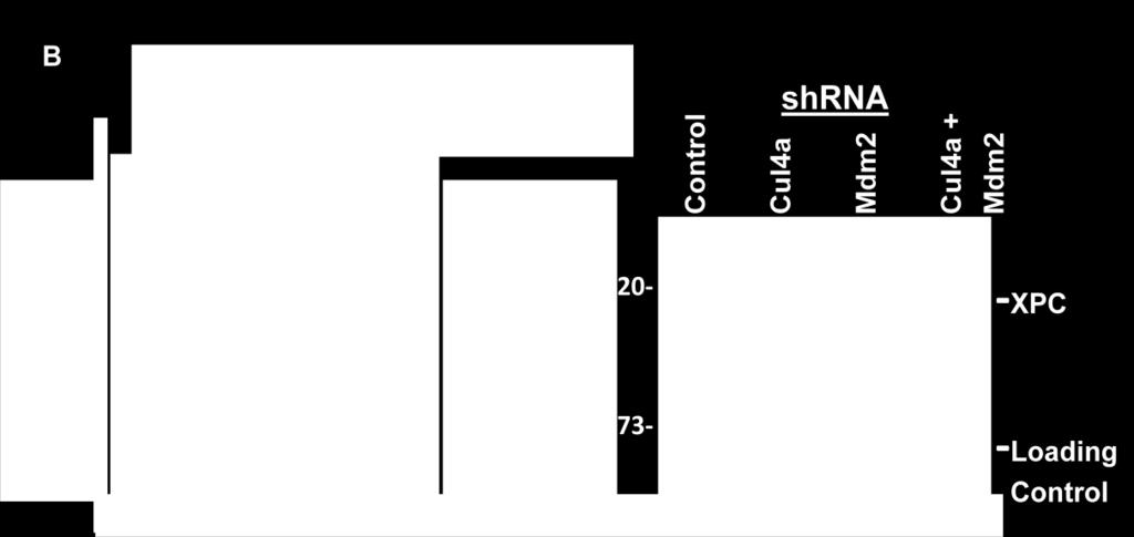
44 XPC and MDM2 While it is known that XPC is modified by E3 ubiquitin ligase Cul4a, multiple ubiquitin ligases are probably involved in the negative regulation of XPC. The MDM2 protein is a known E3 ubiquitin ligase. The involvement of MDM2 in p53 protein regulation and the identification of an MDM2 binding site within the structure of the RB protein (see Figure 8) have made it a protein of interest in the negative regulation of XPC. However, any role for MDM2 in DNA repair and XPC regulation is not known. To explore the relationship between XPC and MDM2, H1299 cells were transfected with empty plasmids or those containing XPC, MDM2 or both. Western blot analysis revealed that XPC steady state level decreased in the presence of MDM2 (Figure 13A lanes 3 and 4) indicating that MDM2 may suppress XPC steady state levels. Conversely, when MDM2 is knocked down by shrna a modest increase in XPC expression was observed (Figure 13B). To determine the functional implications of these data, DNA repair was measured by host-cell reactivation in the presence of XPC, MDM2 or both and also in the presence of Cul4a (positive control) and Mdm2 shrna (lanes correlate with western blots). When E3 ubiquitin ligases Cul4a and MDM2 were knocked down by shrna DNA repair activity increased significantly (Figure 13C, p= < 0.05). These preliminary data provide evidence that MDM2 may be involved in the negative regulation of XPC possibly as an E3 ubiquitin ligase targeting XPC for degradation. Further investigations are needed to explore this possibility. 44

45 A B shrna shrna 45
46 Figure 13. MDM2 may be involved in the negative regulation of XPC. A) H1299 human lung carcinoma cells were cultured and transfected with either an empty plasmid or plasmids containing XPC or MDM2 cdna. Western blots of co-transfected cells reveal that XPC expression decreased in the presence of MDM2. B) p53 null H1299 cells were cultured and transfected with either an empty plasmid or a plasmid containing Cul4a or MDM2 shrna. Western blot analysis showed that XPC expression increased when MDM2 is knocked down. C) DNA repair activity is increased when MDM2 is knocked down. DNA repair was measured by chloramphinicol acetyltransferase (CAT) assay in the presence of XPC, MDM2 or both, and also when Cul4a (positive control) and Mdm2 were knocked down by shrna. When either Cul4a or MDM2 were modestly knocked down by shrna DNA repair increased significantly p= < This increase in XPC expression and DNA repair activity indicate that MDM2 may be involved in the negative regulation of XPC. 46
47 DISCUSSION One important aspect of the G1/S checkpoint is that p53 transcriptionally activates Xpc [37]. This finding suggested that p53 and XPC could actively promote NER, in cells that carry functional p53. However, the data herein suggests an additional level of regulation that does not rely upon p53. We used human lung cancer H1299 cells, which carry deletions in both p53 alleles and have low endogenous levels of XPC and we still observed a significant increase in the cell s DNA repair capacity as measured by host-cell reactivation (HCR) when combined with RB (Figure 5B and C, lanes 8). One often ignored feature of the HCR assays that we and others have used extensively is that it measures NER of a damaged reporter gene. The cells do not receive direct DNA damage. In this assay, we detected a latent XPC protein that did not promote HCR (Figure 5A and C, lanes 4). When combined with RB, XPC was in some way activated for HCR (Figure 5B and C, lanes 8). While most human tumors have defects in the p53 or Rb pathways the data implies that if either p53 or Rb is intact, NER mechanisms are capable of repairing damaged DNA. Given evidence that RB may interact with XPC (Figure 7), it is likely that the interaction of XPC with RB promotes NER at least as measured by HCR. Post-translational modification(s) of XPC in the presence of RB is in evidence (Figure 5B, lane 8). Several studies showed that p21/waf1/cip1 promoted NER in p53-defective cell lines although the mechanistic basis was not known [62, 63, 91-93]. The view was that simply arresting the cell cycle by p21 promoted NER. Our data provide a mechanistic insight, that p21 would lead to accumulation of underphosphorylated RB, which would in 47
48 turn stabilize XPC in a form that is active for NER (Figure 5B and C, lanes 8). Our finding that cyclin E destabilized XPC and led to a decrease in HCR is also consistent with the published studies on p21 (Figure 11). The data represented herein suggests a model for both positive and negative regulation of XPC (Figure 14). Our data shows that XPC exists in functionally active and inactive forms. The data shows that upon DNA damage XPC is then stabilized by the underphosphorylated form of RB and correlates with DNA repair events of the G1/S phase of the cell cycle. The data also show that G1/S cyclins abrogate XPC expression and function which likely corresponds to RB phosphorylation and S-phase entry and that cyclin E may provide a signal for XPC ubiquitination. Perhaps a parallel is suggested by the Cdt1 cell cycle checkpoint protein which is first phosporylated by cyclin E and then ubiquitinated by Cul4a. The cellular model used for the experiments herein is atypical of DNA repair studies. Most studies involving DNA repair mechanisms center around biochemical and cell free systems. Our use of a cellular model of DNA repair is likely more reflective of the signaling mechanisms that actually occur in the cell. The majority of XPC and Rb experiments used an over expression model, whereas, preliminary data involving MDM2 utilized overexpression and the knockdown of MDM2 as a cellular model. Additional studies utilizing a knock down or inducible system of expression may give added insight into the mechanism of action of XPC regulation in the context of the cell cycle. In response to DNA damage, XPC protein is phosphorylated, ubiquitinated, and SUMOylated, however, the amino acid residues involved and the net effects on XPC function are not well-known [37, 39, 94]. Presumably, some post-translational 48
49 modifications promote XPC-mediated NER while others inhibit XPC-mediated NER. The data presented in this study supports the long acknowledged theory that NER occurs readily in G1 but cannot occur in S-phase. Therefore, protein modifications and proteinprotein interactions likely restrict XPC function to G1 while inactivating XPC upon S- phase entry. The XPC protein exists in a heterotrimeric complex that includes the hhr23b and centrin 2 proteins. The hhr23b protein acts to stimulate and stabilize XPC, while the centrin 2 protein plays an important role in centrosome duplication [95-97]. The yeast homologue for hhr23b, Rad23, provided a link between DNA repair and the ubiquitin pathway that led to the discovery that XPC is modified by ubiquitination [48]. Western blot analysis collected from this study (data not shown) indicated that transfecting H1299 cells with the hhr23b protein does not have a noticeable effect on XPC expression. While the XPC protein has been shown to be modified by the ubiquitin ligase Cul4a, most proteins can be poly-ubiquitinated and modified by several ubiquitin ligases [98-99]. The relationship between MDM2 and G1/S mediators along with its involvement in the regulation of the p53 and RB tumor suppressor proteins have made it a likely player in the negative regulation of XPC. MDM2 is an ubiquitin ligase for both RB and p53, and may also act as an ubiquitin ligase for XPC. Our data indicates that MDM2 affects XPC expression and that knocking down MDM2 increased the DNA repair capacity of XPC as measured by HCR (Figure 13) thereby suggesting that MDM2 may play a role in the negative regulation of XPC and possibly act as an E3 ubiquitin ligase for XPC. 49
 and active (oval) form of XPC DNA repair protein.")
50 Cyclin E Figure 14. Schematic of a possible mechanism of XPC regulation in the context of the cell cycle. Data herein reveals a functionally inactive (rectangle) and active (oval) form of XPC DNA repair protein. These data suggest that upon DNA damage XPC is then stabilized by RB and that occurrence correlates with the DNA repair events of the G1 phase of the cell cycle. These data also show that G1/S cyclins, primarily cyclin E (cyclin D data not shown) abrogate XPC expression which likely corresponds to RB phosphorylation and S-phase entry. Data also reveals that MDM2 may signal ubiquitination of XPC in some way. 50
XPC DNA REPAIR PROTEIN REGULATION IN THE CONTEXT OF THE G1/S CELL CYCLE CHECKPOINT
 XPC DNA REPAIR PROTEIN REGULATION IN THE CONTEXT OF THE G1/S CELL CYCLE CHECKPOINT Tabitha M. Hardy Submitted to the faculty of the University Graduate School in partial fulfillment of the requirements
Part-4. Cell cycle regulatory protein 5 (Cdk5) A novel target of ERK in Carb induced cell death
 A novel target of ERK in Carb induced cell death") Part-4 Cell cycle regulatory protein 5 (Cdk5) A novel target of ERK in Carb induced cell death 95 1. Introduction The process of replicating DNA and dividing cells can be described as a series of coordinated
Part-4 Cell cycle regulatory protein 5 (Cdk5) A novel target of ERK in Carb induced cell death 95 1. Introduction The process of replicating DNA and dividing cells can be described as a series of coordinated
A Hepatocyte Growth Factor Receptor (Met) Insulin Receptor hybrid governs hepatic glucose metabolism SUPPLEMENTARY FIGURES, LEGENDS AND METHODS
 Insulin Receptor hybrid governs hepatic glucose metabolism SUPPLEMENTARY FIGURES, LEGENDS AND METHODS") A Hepatocyte Growth Factor Receptor (Met) Insulin Receptor hybrid governs hepatic glucose metabolism Arlee Fafalios, Jihong Ma, Xinping Tan, John Stoops, Jianhua Luo, Marie C. DeFrances and Reza Zarnegar
A Hepatocyte Growth Factor Receptor (Met) Insulin Receptor hybrid governs hepatic glucose metabolism Arlee Fafalios, Jihong Ma, Xinping Tan, John Stoops, Jianhua Luo, Marie C. DeFrances and Reza Zarnegar
HCC1937 is the HCC1937-pcDNA3 cell line, which was derived from a breast cancer with a mutation
 SUPPLEMENTARY INFORMATION Materials and Methods Human cell lines and culture conditions HCC1937 is the HCC1937-pcDNA3 cell line, which was derived from a breast cancer with a mutation in exon 20 of BRCA1
HIV-1 Virus-like Particle Budding Assay Nathan H Vande Burgt, Luis J Cocka * and Paul Bates
 HIV-1 Virus-like Particle Budding Assay Nathan H Vande Burgt, Luis J Cocka * and Paul Bates Department of Microbiology, Perelman School of Medicine at the University of Pennsylvania, Philadelphia, USA
Multistep nature of cancer development. Cancer genes
 Multistep nature of cancer development Phenotypic progression loss of control over cell growth/death (neoplasm) invasiveness (carcinoma) distal spread (metastatic tumor) Genetic progression multiple genetic
RayBio KinaseSTAR TM Akt Activity Assay Kit
 Activity Assay Kit User Manual Version 1.0 March 13, 2015 RayBio KinaseSTAR TM Akt Activity Kit Protocol (Cat#: 68AT-Akt-S40) RayBiotech, Inc. We Provide You With Excellent Support And Service Tel:(Toll
7.012 Quiz 3 Answers
 MIT Biology Department 7.012: Introductory Biology - Fall 2004 Instructors: Professor Eric Lander, Professor Robert A. Weinberg, Dr. Claudette Gardel Friday 11/12/04 7.012 Quiz 3 Answers A > 85 B 72-84
The functional investigation of the interaction between TATA-associated factor 3 (TAF3) and p53 protein
 and p53 protein") THESIS BOOK The functional investigation of the interaction between TATA-associated factor 3 (TAF3) and p53 protein Orsolya Buzás-Bereczki Supervisors: Dr. Éva Bálint Dr. Imre Miklós Boros University of
THESIS BOOK The functional investigation of the interaction between TATA-associated factor 3 (TAF3) and p53 protein Orsolya Buzás-Bereczki Supervisors: Dr. Éva Bálint Dr. Imre Miklós Boros University of
Lecture 10. G1/S Regulation and Cell Cycle Checkpoints. G1/S regulation and growth control G2 repair checkpoint Spindle assembly or mitotic checkpoint
 Lecture 10 G1/S Regulation and Cell Cycle Checkpoints Outline: G1/S regulation and growth control G2 repair checkpoint Spindle assembly or mitotic checkpoint Paper: The roles of Fzy/Cdc20 and Fzr/Cdh1
Supplementary Figure 1. MAT IIα is Acetylated at Lysine 81.
 IP: Flag a Mascot PTM Modified Mass Error Position Gene Names Score Score Sequence m/z [ppm] 81 MAT2A;AMS2;MATA2 35.6 137.28 _AAVDYQK(ac)VVR_ 595.83-2.28 b Pre-immu After-immu Flag- WT K81R WT K81R / Flag
Biochemistry 201: DNA repair January 24, 26, 2000 Gilbert Chu
 1) Why study DNA repair? Biochemistry 201: DNA repair January 24, 26, 2000 Gilbert Chu The genome is assaulted by a myriad of different agents that cause DNA damage. Sources of damage can be both exogenous
SUPPLEMENTARY INFORMATION. Supplementary Figures S1-S9. Supplementary Methods
 SUPPLEMENTARY INFORMATION SUMO1 modification of PTEN regulates tumorigenesis by controlling its association with the plasma membrane Jian Huang 1,2#, Jie Yan 1,2#, Jian Zhang 3#, Shiguo Zhu 1, Yanli Wang
Cancer Biology How a cell responds to DNA Damage
 1 Cancer Biology How a cell responds to DNA Damage Jann Sarkaria Department of Oncology Mayo Clinic 2 EDUCATIONAL GOALS How proteins can transmit signals to each other. The definition of a tumor suppressor
ab E3 Ligase Auto- Ubiquitilylation Assay Kit
 ab139469 E3 Ligase Auto- Ubiquitilylation Assay Kit Instructions for Use For testing ubiquitin E3 ligase activity through assessment of their ability to undergo auto-ubiquitinylation This product is for
Regulators of Cell Cycle Progression
 Regulators of Cell Cycle Progression Studies of Cdk s and cyclins in genetically modified mice reveal a high level of plasticity, allowing different cyclins and Cdk s to compensate for the loss of one
supplementary information
 DOI: 10.1038/ncb1875 Figure S1 (a) The 79 surgical specimens from NSCLC patients were analysed by immunohistochemistry with an anti-p53 antibody and control serum (data not shown). The normal bronchi served
Chapt 15: Molecular Genetics of Cell Cycle and Cancer
 Chapt 15: Molecular Genetics of Cell Cycle and Cancer Student Learning Outcomes: Describe the cell cycle: steps taken by a cell to duplicate itself = cell division; Interphase (G1, S and G2), Mitosis.
SUPPLEMENTARY INFORMATION
 Supplementary Figures Supplementary Figure S1. Binding of full-length OGT and deletion mutants to PIP strips (Echelon Biosciences). Supplementary Figure S2. Binding of the OGT (919-1036) fragments with
Soft Agar Assay. For each cell pool, 100,000 cells were resuspended in 0.35% (w/v)
") SUPPLEMENTARY MATERIAL AND METHODS Soft Agar Assay. For each cell pool, 100,000 cells were resuspended in 0.35% (w/v) top agar (LONZA, SeaKem LE Agarose cat.5004) and plated onto 0.5% (w/v) basal agar.
SUPPLEMENTARY MATERIAL AND METHODS Soft Agar Assay. For each cell pool, 100,000 cells were resuspended in 0.35% (w/v) top agar (LONZA, SeaKem LE Agarose cat.5004) and plated onto 0.5% (w/v) basal agar.
Recombinant Protein Expression Retroviral system
 Recombinant Protein Expression Retroviral system Viruses Contains genome DNA or RNA Genome encased in a protein coat or capsid. Some viruses have membrane covering protein coat enveloped virus Ø Essential
Figure S1. Generation of inducible PTEN deficient mice and the BMMCs (A) B6.129 Pten loxp/loxp mice were mated with B6.
 B6.129 Pten loxp/loxp mice were mated with B6.") Figure S1. Generation of inducible PTEN deficient mice and the BMMCs (A) B6.129 Pten loxp/loxp mice were mated with B6.129-Gt(ROSA)26Sor tm1(cre/ert2)tyj /J mice. To induce deletion of the Pten locus,
Figure S1. Generation of inducible PTEN deficient mice and the BMMCs (A) B6.129 Pten loxp/loxp mice were mated with B6.129-Gt(ROSA)26Sor tm1(cre/ert2)tyj /J mice. To induce deletion of the Pten locus,
Supplementary information. MARCH8 inhibits HIV-1 infection by reducing virion incorporation of envelope glycoproteins
 Supplementary information inhibits HIV-1 infection by reducing virion incorporation of envelope glycoproteins Takuya Tada, Yanzhao Zhang, Takayoshi Koyama, Minoru Tobiume, Yasuko Tsunetsugu-Yokota, Shoji
Supplementary Figure 1. Normal T lymphocyte populations in Dapk -/- mice. (a) Normal thymic development in Dapk -/- mice. Thymocytes from WT and Dapk
 Normal thymic development in Dapk -/- mice. Thymocytes from WT and Dapk") Supplementary Figure 1. Normal T lymphocyte populations in Dapk -/- mice. (a) Normal thymic development in Dapk -/- mice. Thymocytes from WT and Dapk -/- mice were stained for expression of CD4 and CD8.
Supplementary Figure 1. Normal T lymphocyte populations in Dapk -/- mice. (a) Normal thymic development in Dapk -/- mice. Thymocytes from WT and Dapk -/- mice were stained for expression of CD4 and CD8.
Islet viability assay and Glucose Stimulated Insulin Secretion assay RT-PCR and Western Blot
 Islet viability assay and Glucose Stimulated Insulin Secretion assay Islet cell viability was determined by colorimetric (3-(4,5-dimethylthiazol-2-yl)-2,5- diphenyltetrazolium bromide assay using CellTiter
7.012 Problem Set 6 Solutions
 Name Section 7.012 Problem Set 6 Solutions Question 1 The viral family Orthomyxoviridae contains the influenza A, B and C viruses. These viruses have a (-)ss RNA genome surrounded by a capsid composed
Supporting Online Material Material and Methods References Supplemental Figures S1, S2, and S3
 Supporting Online Material Material and Methods References Supplemental Figures S1, S2, and S3 Sarbassov et al. 1 Material and Methods Materials Reagents were obtained from the following sources: protein
BIO360 Fall 2013 Quiz 1
 BIO360 Fall 2013 Quiz 1 1. Examine the diagram below. There are two homologous copies of chromosome one and the allele of YFG carried on the light gray chromosome has undergone a loss-of-function mutation.
Molecular biology :- Cancer genetics lecture 11
 Molecular biology :- Cancer genetics lecture 11 -We have talked about 2 group of genes that is involved in cellular transformation : proto-oncogenes and tumour suppressor genes, and it isn t enough to
CANCER. Inherited Cancer Syndromes. Affects 25% of US population. Kills 19% of US population (2nd largest killer after heart disease)
") CANCER Affects 25% of US population Kills 19% of US population (2nd largest killer after heart disease) NOT one disease but 200-300 different defects Etiologic Factors In Cancer: Relative contributions
CANCER Affects 25% of US population Kills 19% of US population (2nd largest killer after heart disease) NOT one disease but 200-300 different defects Etiologic Factors In Cancer: Relative contributions
Protocol for Gene Transfection & Western Blotting
 The schedule and the manual of basic techniques for cell culture Advanced Protocol for Gene Transfection & Western Blotting Schedule Day 1 26/07/2008 Transfection Day 3 28/07/2008 Cell lysis Immunoprecipitation
Supplementary Figure 1.TRIM33 binds β-catenin in the nucleus. a & b, Co-IP of endogenous TRIM33 with β-catenin in HT-29 cells (a) and HEK 293T cells
 and HEK 293T cells") Supplementary Figure 1.TRIM33 binds β-catenin in the nucleus. a & b, Co-IP of endogenous TRIM33 with β-catenin in HT-29 cells (a) and HEK 293T cells (b). TRIM33 was immunoprecipitated, and the amount of
Supplementary Figure 1.TRIM33 binds β-catenin in the nucleus. a & b, Co-IP of endogenous TRIM33 with β-catenin in HT-29 cells (a) and HEK 293T cells (b). TRIM33 was immunoprecipitated, and the amount of
Cell Cycle, Mitosis, and Microtubules. LS1A Final Exam Review Friday 1/12/07. Processes occurring during cell cycle
 Cell Cycle, Mitosis, and Microtubules LS1A Final Exam Review Friday 1/12/07 Processes occurring during cell cycle Replicate chromosomes Segregate chromosomes Cell divides Cell grows Cell Growth 1 The standard
The Schedule and the Manual of Basic Techniques for Cell Culture
 The Schedule and the Manual of Basic Techniques for Cell Culture 1 Materials Calcium Phosphate Transfection Kit: Invitrogen Cat.No.K2780-01 Falcon tube (Cat No.35-2054:12 x 75 mm, 5 ml tube) Cell: 293
Oncolytic virus strategy
 Oncolytic viruses Oncolytic virus strategy normal tumor NO replication replication survival lysis Oncolytic virus strategy Mechanisms of tumor selectivity of several, some of them naturally, oncolytic
Supplementary data Supplementary Figure 1 Supplementary Figure 2
 Supplementary data Supplementary Figure 1 SPHK1 sirna increases RANKL-induced osteoclastogenesis in RAW264.7 cell culture. (A) RAW264.7 cells were transfected with oligocassettes containing SPHK1 sirna
Introduction to Genetics
 Introduction to Genetics Table of contents Chromosome DNA Protein synthesis Mutation Genetic disorder Relationship between genes and cancer Genetic testing Technical concern 2 All living organisms consist
Supplementary Appendix
 Supplementary Appendix This appendix has been provided by the authors to give readers additional information about their work. Supplement to: Choi YL, Soda M, Yamashita Y, et al. EML4-ALK mutations in
Problem Set 8 Key 1 of 8
 7.06 2003 Problem Set 8 Key 1 of 8 7.06 2003 Problem Set 8 Key 1. As a bright MD/PhD, you are interested in questions about the control of cell number in the body. Recently, you've seen three patients
Chapter 9. Cells Grow and Reproduce
 Chapter 9 Cells Grow and Reproduce DNA Replication DNA polymerase Addition of a nucleotide to the 3 end of a growing strand Use dntps as substrate Release of pyrophosphate Initiation of Replication Replication
TFEB-mediated increase in peripheral lysosomes regulates. Store Operated Calcium Entry
 TFEB-mediated increase in peripheral lysosomes regulates Store Operated Calcium Entry Luigi Sbano, Massimo Bonora, Saverio Marchi, Federica Baldassari, Diego L. Medina, Andrea Ballabio, Carlotta Giorgi
Figure S1 Time-dependent down-modulation of HER3 by EZN No Treatment. EZN-3920, 2 μm. Time, h
 Figure S1 Time-dependent down-modulation of HER3 by EZN-392 HE ER3 mrna A, %Contr rol 12 No Treatment EZN-392, 2 μm 1 8 6 4 2 2 8 24 Time, h Figure S2. Specific target down-modulation by HER3 (EZN-392)
Tumor suppressor genes D R. S H O S S E I N I - A S L
 Tumor suppressor genes 1 D R. S H O S S E I N I - A S L What is a Tumor Suppressor Gene? 2 A tumor suppressor gene is a type of cancer gene that is created by loss-of function mutations. In contrast to
Problem Set 5 KEY
 2006 7.012 Problem Set 5 KEY ** Due before 5 PM on THURSDAY, November 9, 2006. ** Turn answers in to the box outside of 68-120. PLEASE WRITE YOUR ANSWERS ON THIS PRINTOUT. 1. You are studying the development
For the rapid, sensitive and accurate quantification of Ras in various samples
 ab128504 Ras Assay Kit Instructions for Use For the rapid, sensitive and accurate quantification of Ras in various samples This product is for research use only and is not intended for diagnostic use.
Cell Lysis Buffer. Catalog number: AR0103
 Cell Lysis Buffer Catalog number: AR0103 Boster s Cell Lysis Buffer is a ready-to-use Western blot related reagent solution used for efficient extraction of total soluble protein in nondenatured state
Supplementary Fig. 1. GPRC5A post-transcriptionally down-regulates EGFR expression. (a) Plot of the changes in steady state mrna levels versus
 Plot of the changes in steady state mrna levels versus") Supplementary Fig. 1. GPRC5A post-transcriptionally down-regulates EGFR expression. (a) Plot of the changes in steady state mrna levels versus changes in corresponding proteins between wild type and Gprc5a-/-
Supplementary Fig. 1. GPRC5A post-transcriptionally down-regulates EGFR expression. (a) Plot of the changes in steady state mrna levels versus changes in corresponding proteins between wild type and Gprc5a-/-
RAS Genes. The ras superfamily of genes encodes small GTP binding proteins that are responsible for the regulation of many cellular processes.
 ۱ RAS Genes The ras superfamily of genes encodes small GTP binding proteins that are responsible for the regulation of many cellular processes. Oncogenic ras genes in human cells include H ras, N ras,
Introduction to Cancer Biology
 Introduction to Cancer Biology Robin Hesketh Multiple choice questions (choose the one correct answer from the five choices) Which ONE of the following is a tumour suppressor? a. AKT b. APC c. BCL2 d.
Human Oxidized LDL ELISA Kit (MDA-LDL Quantitation), General
, General") Human Oxidized LDL ELISA Kit (MDA-LDL Quantitation), General For the detection and quantitation of human OxLDL in plasma, serum or other biological fluid samples Cat. No. KT-959 For Research Use Only.
Human Oxidized LDL ELISA Kit (MDA-LDL Quantitation), General For the detection and quantitation of human OxLDL in plasma, serum or other biological fluid samples Cat. No. KT-959 For Research Use Only.
BCHM3972 Human Molecular Cell Biology (Advanced) 2013 Course University of Sydney
 2013 Course University of Sydney") BCHM3972 Human Molecular Cell Biology (Advanced) 2013 Course University of Sydney Page 2: Immune Mechanisms & Molecular Biology of Host Defence (Prof Campbell) Page 45: Infection and Implications for Cell
BCHM3972 Human Molecular Cell Biology (Advanced) 2013 Course University of Sydney Page 2: Immune Mechanisms & Molecular Biology of Host Defence (Prof Campbell) Page 45: Infection and Implications for Cell
supplementary information
 Figure S1 Nucleotide binding status of RagA mutants. Wild type and mutant forms of MycRagA was transfected into HEK293 cells and the transfected cells were labeled with 32 Pphosphate. MycRagA was immunoprecipitated
Supplementary Information Supplementary Fig. 1. Elevated Usp9x in melanoma and NRAS mutant melanoma cells are dependent on NRAS for 3D growth.
 Supplementary Information Supplementary Fig. 1. Elevated Usp9x in melanoma and NRAS mutant melanoma cells are dependent on NRAS for 3D growth. a. Immunoblot for Usp9x protein in NRAS mutant melanoma cells
Supplementary Data 1. Alanine substitutions and position variants of APNCYGNIPL. Applied in
 Supplementary Data 1. Alanine substitutions and position variants of APNCYGNIPL. Applied in Supplementary Fig. 2 Substitution Sequence Position variant Sequence original APNCYGNIPL original APNCYGNIPL
Spring 2015 Module 2 System Engineering and Protein Founda<ons
 20.109 Spring 2015 Module 2 System Engineering and Protein Founda
Human Apolipoprotein A1 EIA Kit
 A helping hand for your research Product Manual Human Apolipoprotein A1 EIA Kit Catalog Number: 83901 96 assays 1 Table of Content Product Description 3 Assay Principle 3 Kit Components 3 Storage 4 Reagent
SUPPLEMENT. Materials and methods
 SUPPLEMENT Materials and methods Cell culture and reagents Cell media and reagents were from Invitrogen unless otherwise indicated. Antibiotics and Tet-certified serum were from Clontech. In experiments
Supplementary Information
 Supplementary Information Supplementary Figure 1. CD4 + T cell activation and lack of apoptosis after crosslinking with anti-cd3 + anti-cd28 + anti-cd160. (a) Flow cytometry of anti-cd160 (5D.10A11) binding
MicroRNA sponges: competitive inhibitors of small RNAs in mammalian cells
 MicroRNA sponges: competitive inhibitors of small RNAs in mammalian cells Margaret S Ebert, Joel R Neilson & Phillip A Sharp Supplementary figures and text: Supplementary Figure 1. Effect of sponges on
Mammalian Membrane Protein Extraction Kit
 Mammalian Membrane Protein Extraction Kit Catalog number: AR0155 Boster s Mammalian Membrane Protein Extraction Kit is a simple, rapid and reproducible method to prepare cellular protein fractions highly
SUPPLEMENTARY INFORMATION
 SUPPLEMENTARY INFORMATION FOR Liver X Receptor α mediates hepatic triglyceride accumulation through upregulation of G0/G1 Switch Gene 2 (G0S2) expression I: SUPPLEMENTARY METHODS II: SUPPLEMENTARY FIGURES
p47 negatively regulates IKK activation by inducing the lysosomal degradation of polyubiquitinated NEMO
 Supplementary Information p47 negatively regulates IKK activation by inducing the lysosomal degradation of polyubiquitinated NEMO Yuri Shibata, Masaaki Oyama, Hiroko Kozuka-Hata, Xiao Han, Yuetsu Tanaka,
NF-κB p65 (Phospho-Thr254)
") Assay Biotechnology Company www.assaybiotech.com Tel: 1-877-883-7988 Fax: 1-877-610-9758 NF-κB p65 (Phospho-Thr254) Colorimetric Cell-Based ELISA Kit Catalog #: OKAG02015 Please read the provided manual
Assay Biotechnology Company www.assaybiotech.com Tel: 1-877-883-7988 Fax: 1-877-610-9758 NF-κB p65 (Phospho-Thr254) Colorimetric Cell-Based ELISA Kit Catalog #: OKAG02015 Please read the provided manual
Supplemental Information
 Supplemental Information Tobacco-specific Carcinogen Induces DNA Methyltransferases 1 Accumulation through AKT/GSK3β/βTrCP/hnRNP-U in Mice and Lung Cancer patients Ruo-Kai Lin, 1 Yi-Shuan Hsieh, 2 Pinpin
SUPPLEMENTAL FIGURE LEGENDS
 SUPPLEMENTAL FIGURE LEGENDS Supplemental Figure S1: Endogenous interaction between RNF2 and H2AX: Whole cell extracts from 293T were subjected to immunoprecipitation with anti-rnf2 or anti-γ-h2ax antibodies
SUPPLEMENTARY INFORMATION
 SUPPLEMENTARY INFORMATION doi:1.138/nature9814 a A SHARPIN FL B SHARPIN ΔNZF C SHARPIN T38L, F39V b His-SHARPIN FL -1xUb -2xUb -4xUb α-his c Linear 4xUb -SHARPIN FL -SHARPIN TF_LV -SHARPINΔNZF -SHARPIN
SUPPLEMENTARY INFORMATION
 doi:10.1038/nature11700 Figure 1: RIP3 as a potential Sirt2 interacting protein. Transfected Flag-tagged Sirt2 was immunoprecipitated from cells and eluted from the Sepharose beads using Flag peptide.
Regulation of cell cycle. Dr. SARRAY Sameh, Ph.D
 Regulation of cell cycle Dr. SARRAY Sameh, Ph.D Control of cell cycle: Checkpoints Are the cell cycle controls mechanisms in eukaryotic cells. These checkpoints verify whether the processes at each phase
a b G75 G60 Sw-2 Sw-1 Supplementary Figure 1. Structure predictions by I-TASSER Server.
 a b G75 2 2 G60 Sw-2 Sw-1 Supplementary Figure 1. Structure predictions by I-TASSER Server. a. Overlay of top 10 models generated by I-TASSER illustrates the potential effect of 7 amino acid insertion
Construction of a hepatocellular carcinoma cell line that stably expresses stathmin with a Ser25 phosphorylation site mutation
 Construction of a hepatocellular carcinoma cell line that stably expresses stathmin with a Ser25 phosphorylation site mutation J. Du 1, Z.H. Tao 2, J. Li 2, Y.K. Liu 3 and L. Gan 2 1 Department of Chemistry,
Data Sheet TIGIT / NFAT Reporter - Jurkat Cell Line Catalog #60538
 Data Sheet TIGIT / NFAT Reporter - Jurkat Cell Line Catalog #60538 Background: TIGIT is a co-inhibitory receptor that is highly expressed in Natural Killer (NK) cells, activated CD4+, CD8+ and regulatory
Supplementary Figure 1: si-craf but not si-braf sensitizes tumor cells to radiation.
 Supplementary Figure 1: si-craf but not si-braf sensitizes tumor cells to radiation. (a) Embryonic fibroblasts isolated from wildtype (WT), BRAF -/-, or CRAF -/- mice were irradiated (6 Gy) and DNA damage
Overview of the Expressway Cell-Free Expression Systems. Expressway Mini Cell-Free Expression System
 Overview of the Expressway Cell-Free Expression Systems The Expressway Cell-Free Expression Systems use an efficient coupled transcription and translation reaction to produce up to milligram quantities
Cover Page. The handle holds various files of this Leiden University dissertation.
 Cover Page The handle http://hdl.handle.net/1887/20360 holds various files of this Leiden University dissertation. Author: Eijk, Patrick van Title: Nucleotide excision repair in yeast Date: 2012-12-13
STAT3 (py705)/ Pan STAT3 (Human/Mouse/Rat) ELISA Kit
/ Pan STAT3 (Human/Mouse/Rat) ELISA Kit") STAT3 (py705)/ Pan STAT3 (Human/Mouse/Rat) ELISA Kit Catalog Number KA2176 96 assays Version: 02 Intended for research use only www.abnova.com Table of Contents Introduction... 3 Principle of the Assay...
STAT3 (py705)/ Pan STAT3 (Human/Mouse/Rat) ELISA Kit Catalog Number KA2176 96 assays Version: 02 Intended for research use only www.abnova.com Table of Contents Introduction... 3 Principle of the Assay...
Cells and reagents. Synaptopodin knockdown (1) and dynamin knockdown (2)
 and dynamin knockdown (2)") Supplemental Methods Cells and reagents. Synaptopodin knockdown (1) and dynamin knockdown (2) podocytes were cultured as described previously. Staurosporine, angiotensin II and actinomycin D were all obtained
Supplemental Methods Cells and reagents. Synaptopodin knockdown (1) and dynamin knockdown (2) podocytes were cultured as described previously. Staurosporine, angiotensin II and actinomycin D were all obtained
SUPPLEMENTARY INFORMATION
 Supplementary Discussion The cell cycle machinery and the DNA damage response network are highly interconnected and co-regulated in assuring faithful duplication and partition of genetic materials into
Supplementary Figure 1
 Supplementary Figure 1 Supplementary Figure 1. Neither the activation nor suppression of the MAPK pathway affects the ASK1/Vif interaction. (a, b) HEK293 cells were cotransfected with plasmids encoding
TRAF6 ubiquitinates TGFβ type I receptor to promote its cleavage and nuclear translocation in cancer
 Supplementary Information TRAF6 ubiquitinates TGFβ type I receptor to promote its cleavage and nuclear translocation in cancer Yabing Mu, Reshma Sundar, Noopur Thakur, Maria Ekman, Shyam Kumar Gudey, Mariya
Supplementary Figure 1 Role of Raf-1 in TLR2-Dectin-1-mediated cytokine expression
 Supplementary Figure 1 Supplementary Figure 1 Role of Raf-1 in TLR2-Dectin-1-mediated cytokine expression. Quantitative real-time PCR of indicated mrnas in DCs stimulated with TLR2-Dectin-1 agonist zymosan
Disorders of Cell Growth & Neoplasia. Lecture 4 Molecular basis of cancer
 General Pathology VPM 152 Disorders of Cell Growth & Neoplasia Lecture 4 Molecular basis of cancer Enrique Aburto Apr 2010 Skin tumor in a 10-year-old Rottweiler. Considering the external appearance and
TITLE: The Role of hcdc4 as a Tumor Suppressor Gene in Genomic Instability Underlying Prostate Cancer
 AD Award Number: TITLE: The Role of hcdc4 as a Tumor Suppressor Gene in Genomic Instability Underlying Prostate Cancer PRINCIPAL INVESTIGATOR: Audrey van Drogen, Ph.D. CONTRACTING ORGANIZATION: Sidney
Prognostic value of DNA repair based stratification of hepatocellular carcinoma
 Prognostic value of DNA repair based stratification of hepatocellular carcinoma Zhuo Lin 1, 2 #, Shi-Hao Xu 3 #, Hai-Qing Wang 4, Yi-Jing Cai 1, 2, Li Ying 3, Mei Song 1, 2, Yu-Qun Wang 1, 2, Shan-Jie
Study of different types of ubiquitination
 Study of different types of ubiquitination Rudi Beyaert (rudi.beyaert@irc.vib-ugent.be) VIB UGent Center for Inflammation Research Ghent, Belgium VIB Training Novel Proteomics Tools: Identifying PTMs October
7.06 Cell Biology EXAM #3 April 24, 2003
 7.06 Spring 2003 Exam 3 Name 1 of 8 7.06 Cell Biology EXAM #3 April 24, 2003 This is an open book exam, and you are allowed access to books and notes. Please write your answers to the questions in the
Supplementary Information
 Supplementary Information mediates STAT3 activation at retromer-positive structures to promote colitis and colitis-associated carcinogenesis Zhang et al. a b d e g h Rel. Luc. Act. Rel. mrna Rel. mrna
Molecular Biology (BIOL 4320) Exam #2 May 3, 2004
 Exam #2 May 3, 2004") Molecular Biology (BIOL 4320) Exam #2 May 3, 2004 Name SS# This exam is worth a total of 100 points. The number of points each question is worth is shown in parentheses after the question number. Good
Molecular Biology (BIOL 4320) Exam #2 May 3, 2004 Name SS# This exam is worth a total of 100 points. The number of points each question is worth is shown in parentheses after the question number. Good
Regulation of the Cell Cycle
 Regulation of the Cell Cycle 21 I. OVERVIEW Quiescent differentiated cell / can be induced to re-enter the active cell cycle. urvival Cell division Apoptosis 1 Daughter cells Apoptic cell enescent cell
HEK293FT cells were transiently transfected with reporters, N3-ICD construct and
 Supplementary Information Luciferase reporter assay HEK293FT cells were transiently transfected with reporters, N3-ICD construct and increased amounts of wild type or kinase inactive EGFR. Transfections
Supplementary Data Table of Contents:
 Supplementary Data Table of Contents: - Supplementary Methods - Supplementary Figures S1(A-B) - Supplementary Figures S2 (A-B) - Supplementary Figures S3 - Supplementary Figures S4(A-B) - Supplementary
Online Data Supplement. Anti-aging Gene Klotho Enhances Glucose-induced Insulin Secretion by Upregulating Plasma Membrane Retention of TRPV2
 Online Data Supplement Anti-aging Gene Klotho Enhances Glucose-induced Insulin Secretion by Upregulating Plasma Membrane Retention of TRPV2 Yi Lin and Zhongjie Sun Department of physiology, college of
MTC-TT and TPC-1 cell lines were cultured in RPMI medium (Gibco, Breda, The Netherlands)
") Supplemental data Materials and Methods Cell culture MTC-TT and TPC-1 cell lines were cultured in RPMI medium (Gibco, Breda, The Netherlands) supplemented with 15% or 10% (for TPC-1) fetal bovine serum
Supplemental data Materials and Methods Cell culture MTC-TT and TPC-1 cell lines were cultured in RPMI medium (Gibco, Breda, The Netherlands) supplemented with 15% or 10% (for TPC-1) fetal bovine serum
SUPPLEMENTARY INFORMATION
 Complete but curtailed T-cell response to very-low-affinity antigen Dietmar Zehn, Sarah Y. Lee & Michael J. Bevan Supp. Fig. 1: TCR chain usage among endogenous K b /Ova reactive T cells. C57BL/6 mice
Cell Cycle. Trends in Cell Biology
 Cell Cycle Trends in Cell Biology Cell Cycle The orderly sequence of events by which a cell duplicates its contents and divides into two Daughter Cells Activities of a cell from one cell division to the
Genomic instability. Amin Mahpour
 Genomic instability Amin Mahpour 1 Some questions to ponder What is Genomic instability? What factors contribute to the genomic integrity? How we identify these aberrations? 2 PART I: MOLECULAR BIOLOGY
Early cell death (FGF) B No RunX transcription factor produced Yes No differentiation
 B No RunX transcription factor produced Yes No differentiation") Solution Key - Practice Questions Question 1 a) A recent publication has shown that the fat stem cells (FSC) can act as bone stem cells to repair cavities in the skull, when transplanted into immuno-compromised
Solution Key - Practice Questions Question 1 a) A recent publication has shown that the fat stem cells (FSC) can act as bone stem cells to repair cavities in the skull, when transplanted into immuno-compromised
STAT3 (py705) (Human/Mouse/Rat) ELISA Kit
 (Human/Mouse/Rat) ELISA Kit") STAT3 (py705) (Human/Mouse/Rat) ELISA Kit Catalog Number KA2175 96 assays Version: 01 Intended for research use only www.abnova.com I. INTRODUCTION STAT3 (py705) (Human/Mouse/Rat) ELISA (Enzyme-Linked
STAT3 (py705) (Human/Mouse/Rat) ELISA Kit Catalog Number KA2175 96 assays Version: 01 Intended for research use only www.abnova.com I. INTRODUCTION STAT3 (py705) (Human/Mouse/Rat) ELISA (Enzyme-Linked
Cancer. Questions about cancer. What is cancer? What causes unregulated cell growth? What regulates cell growth? What causes DNA damage?
 Questions about cancer What is cancer? Cancer Gil McVean, Department of Statistics, Oxford What causes unregulated cell growth? What regulates cell growth? What causes DNA damage? What are the steps in
CELL CYCLE MOLECULAR BASIS OF ONCOGENESIS
 CELL CYCLE MOLECULAR BASIS OF ONCOGENESIS Summary of the regulation of cyclin/cdk complexes during celll cycle Cell cycle phase Cyclin-cdk complex inhibitor activation Substrate(s) G1 Cyclin D/cdk 4,6
Mouse Cathepsin B ELISA Kit
 GenWay Biotech, Inc. 6777 Nancy Ridge Drive San Diego, CA 92121 Phone: 858.458.0866 Fax: 858.458.0833 Email: techline@genwaybio.com http://www.genwaybio.com Mouse Cathepsin B ELISA Kit Catalog No. GWB-ZZD154