Emergency U/S: Minimum Imaging Guidelines
|
|
|
- Monica Powers
- 5 years ago
- Views:
Transcription
1 Emergency U/S: Minimum Imaging Guidelines Table of contents: Diagnostic imaging guidelines: orta iliary ladder retention Cardiac DVT FST O transabdominal O transvaginal Ocular Renal Soft Tissue Thoracic Ultrasound-guided procedure imaging guidelines: bscess rthrocentesis Central line Paracentesis Pericardiocentesis Peripheral IV Thoracentesis
 caliper measurement (high in")
 caliper measurement (just above")
...fig D How you might label the images: 1.")

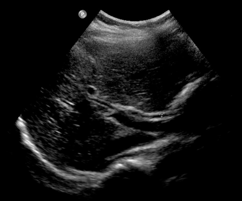
2 Minimum orta Imaging Guidelines: 1. orta transverse HIGH with anterior to posterior (P) caliper measurement (high in the mid-epigastrium)...fig 2. orta transverse MID...Fig 3. orta transverse LOW with anterior to posterior (P) caliper measurement (just above the bifurcation)...fig C Normal aorta diameter is generally considered < 3cm 4. orta in longitudinal (sagittal)...fig D How you might label the images: 1. O TR HIGH 2. O TR MID 3. O TR LOW 4. O LONG Note: My preference is that you also obtain video clips of the transverse and longitudinal aorta...though this is not necessary for billing/documentation...(click here to link to normal aorta videos) C D

 +/- calipers...fig C 4. Gallbladder transverse LOW (fundus) +/- calipers...fig D 5.")

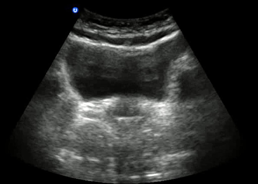
3 Minimum Gallbladder Imaging Guidelines: 1. Gallbladder in longitudinal...fig 2. Gallbladder transverse HI (near the neck) +/- calipers...fig Calipers should be on the liver side...normal, though debatable, is generally considered < 4mm Only one of the transverse views needs to have the G wall measured 3. Gallbladder transverse MID (body) +/- calipers...fig C 4. Gallbladder transverse LOW (fundus) +/- calipers...fig D 5. Common bile duct measurement (calipers) in transverse or longitudinal (inside to inside wall)...figs E & F Normal is generally considered < 4mm...or 1mm per decade of life 6. Liver in transverse...fig G How you might label the images: 1. G LONG 2. G TR (reasonable to use this same label for the high, mid, and low portions) 3. CD 4. LIVER Note: My preference is that you also obtain video clips of the G long and short views...though this is not necessary for billing/ documentation...(click here to link to normal biliary videos) C D

4 E F

5 Minimum Urinary ladder Imaging Guidelines: 1. ladder in transverse with caliper measurements...fig Measuring anterior/posterior and right-to-left dimensions of the bladder 2. ladder in longitudinal with caliper measurements...fig Measuring cranial-to-caudal dimension of the bladder YOU SHOULD USE THE VOLUME CLCULTION ON THE MCHINE, OTHERWISE [X x Y x Z] How you might label the images: 1. L TR 2. L LONG Note: My preference is that you also obtain video clips of the urinary bladder in either transverse or longitudinal...though this is not necessary for billing/documentation...(click here to link to normal bladder videos)


6 Minimum Cardiac Imaging Guidelines: The 4 views (+ IVC) of the cardiac study should be a series of VIDEOS rather than images...the images below are simply to give you an idea of what to look for when performing cardiac imaging (for examples of cardiac videos, click here) Videos to obtain: 1. Subxiphoid view...fig 2. Parasternal long axis view...fig 3. Parasternal short axis view...fig C 4. pical 4 chamber view...fig D 5. IVC view in longitudinal...fig E How you might label the videos: 1. SX 2. PSL 3. PSS 4. 4CH 5. IVC **t a minimum, try to obtain 3 of the 4 listed CRDIC views C D E
...fig 2.")
...fig C **Note.")

7 Minimum Lower Extremity DVT Imaging Guidelines: Images to obtain (for each leg): 1. Dual screen of the Common Femoral Vein without/with compression (calipers over compressed vein)...fig 2. Dual screen of the Superficial Femoral Vein (mid-thigh) without/with compression (calipers over compressed vein)...fig 3. Dual screen of the Popliteal Vein without/with compression (calipers over compressed vein)...fig C **Note...Figure is the best example of the way the exam should be documented, with dual screen and calipers How you might label the images: 1. CFV 2. SFV 3. PV Note: My preference is that you also obtain video clips of the upper thigh and popliteal fossa with dynamic compression...though this is not necessary for billing/documentation...(click here to link to normal venous compression videos) C
...Fig 2. Subxiphoid view (save video clip).")

8 Minimum FST Imaging Guidelines: 1. RUQ (one or more views to show pleural space, subphrenic space, Morison s, and inferior pole of the kidney)...fig 1. LUQ (one or more views to show pleural space, subphrenic space, splenorenal space, and inferior pole of the kidney; this may not be possible in the LUQ)...Fig 2. Subxiphoid view (save video clip)...fig C (lternative if SX not available: Parasternal long axis...click her for example) 3. Suprapubic view in transverse...fig D **You should log the PTX portion of the study under Thoracic, though do not bill separately for the PTX study How you might label the images: 1. RUQ 2. LUQ 3. SX 4. SP (for suprapubic) Note: My preference is that you also obtain video clips of the 4 views (the cardiac view SHOULD have a video)...though this is not necessary for billing/documentation...(click here to link to normal FST videos) C D


 3. R OV 4.")

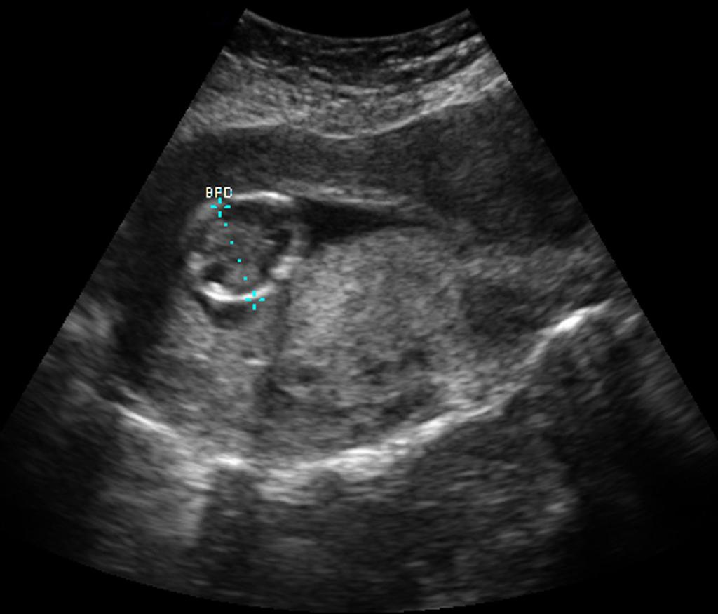
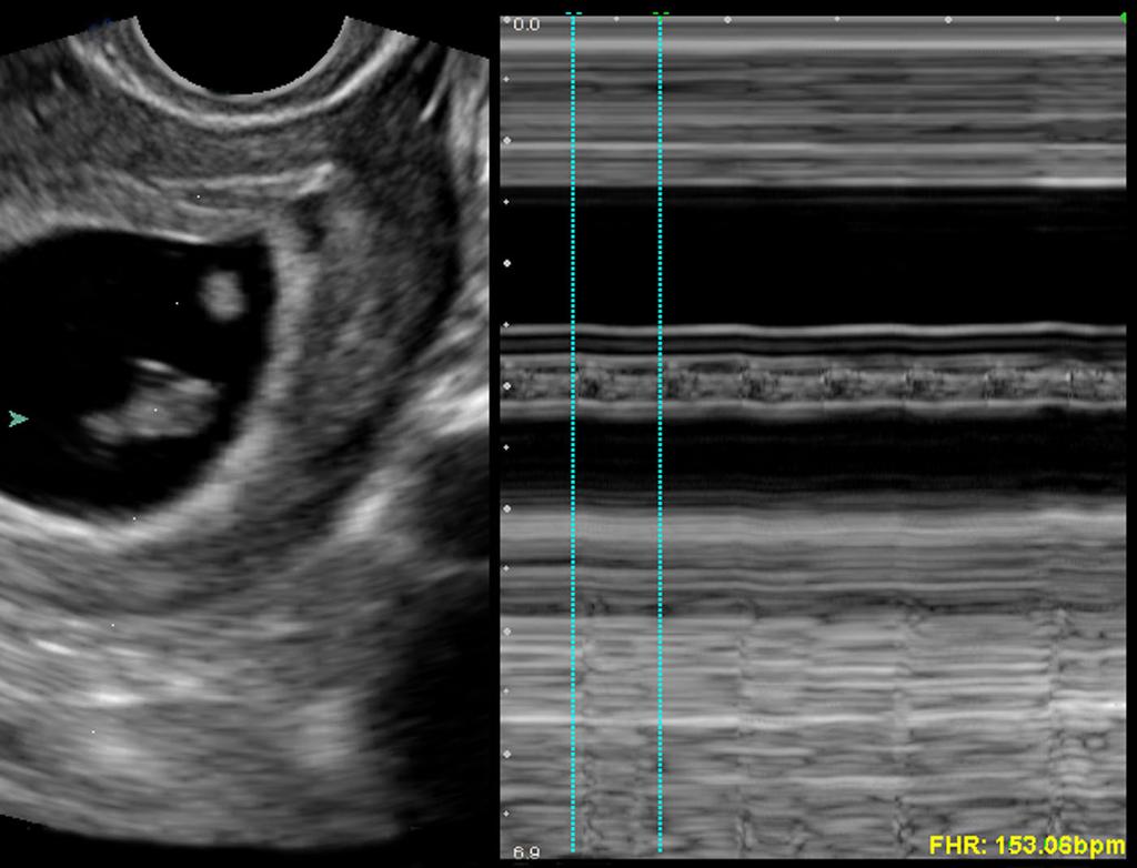
9 Minimum O Transabdominal U/S Imaging Guidelines: 1. Uterus in longitudinal...fig 2. Uterus in transverse HI...Fig 3. Uterus in transverse MID...Fig C 4. Uterus in transverse LOW...Fig D 5. Right ovary with calipers (if seen)...fig E 6. Left ovary with calipers (if seen)...fig E 7. Gestational age measurement using calcs : Crown rump length...fig F iparietal diameter...fig G 8. Fetal heart rate (in M-mode)...Fig H How you might label the images: 1. UT LONG 2. UT TR (reasonable to use this same label for the high, mid, and low portions) 3. R OV 4. L OV Note: My preference is that you also obtain video clips of the uterus in transverse and longitudinal, as well as each ovary that is visualized...though this is not necessary for billing/documentation (click here to link to normal TUS O videos) C D


10 E F G H


11 Minimum O Transvaginal U/S Imaging Guidelines: 1. Uterus in longitudinal...fig 2. Uterus in transverse HI...Fig 3. Uterus in transverse MID...Fig C 4. Uterus in transverse LOW...Fig D 5. Right ovary with calipers (if seen)...fig E 6. Left ovary with calipers (if seen)...fig E 7. Gestational age measurement using calcs : Crown rump length (if you re able to measure PD, it probably shouldn t be doing a transvaginal...just saying)...fig F 8. Fetal heart rate (in M-mode)...Fig G How you might label the images: 1. UT LONG 2. UT TR (reasonable to use this same label for the high, mid, and low portions) 3. R OV 4. L OV Note: My preference is that you also obtain video clips of the uterus in transverse and longitudinal, as well as each ovary that is visualized...though this is not necessary for billing/documentation...(click here to link to normal TVUS O videos) C D


12 E F G

 Note: My preference is that you also obtain a video clip of the orbit")
13 Minimum Ocular Imaging Guidelines: 1. 1 view of the globe to include the lens and retina...fig 2. Measurement of the optic nerve sheath diameter...fig 3mm from the retina, then measure across How you might label the images: 1. R/L GLOE 2. ONSD (optic nerve sheath diameter) Note: My preference is that you also obtain a video clip of the orbit (click here to link to normal eye videos)...though this is not necessary for billing/documentation
 transverse HI +/- calipers...fig C 3.")
 transverse LOW +/- calipers...fig C 5. Urinary bladder in transverse...fig D & E (male and female) 6.")

14 1. Right kidney in longitudinal...fig 1. Left kidney in longitudinal...fig Minimum Renal Imaging Guidelines: 2. Right or left kidney (depending on symptomatic side) transverse HI +/- calipers...fig C 3. Right or left kidney (depending on symptomatic side) transverse MID +/- calipers...fig C 4. Right or left kidney (depending on symptomatic side) transverse LOW +/- calipers...fig C 5. Urinary bladder in transverse...fig D & E (male and female) 6. Urinary bladder in longitudinal...figs F & G (male and female) **Note that I ve only included one figure to represent the transverse images...you will need to document all 3 levels How you might label the images: 1. R/L K LONG 2. R/L K TR (reasonable to use this same label for the high, mid, and low portions) 3. L TR 4. L LONG Note: My preference is that you also obtain video clips of the kidneys and bladder...though this is not necessary for billing/ documentation...(click here to link to normal Renal videos) C D


15 E F G
 or cellulitis (Fig ) or")
 should be obtained in 2 orthogonal planes (with")


16 Minimum Soft Tissue Imaging Guidelines: 1. ny cellulitis or foreign body (Fig ) or cellulitis (Fig ) or fracture (Fig C) or other soft tissue finding like a lymph node (Fig D) should be obtained in 2 orthogonal planes (with consideration of caliper measurements as indicated)...which means 2 images at least... Note: My preference is that you also obtain video clips of any soft tissue pathology...though this is not necessary for billing/ documentation...(click here to link to normal soft tissue videos) C D

 2. M-mode if looking for pneumothorax.")
...though this is not necessary for billing/ documentation.")
17 Minimum Thoracic Imaging Guidelines: 1. Still image of affected lung...figs - ( coronal view of the lung, with the probe placed along the lateral thoracic wall, allows visualization of the diaphragm and is an additional recommended image...fig C) 2. M-mode if looking for pneumothorax...fig D Note: My preference is that you also obtain video clips of the lung exam(s)...though this is not necessary for billing/ documentation...(click here to link to normal thoracic videos) C D


18 Minimum U/S-Guided bscess Drainage Imaging Guidelines: 1. ONE image of the abscess, preferably with caliper measurements (not necessary to do orthogonal planes)...figs -D Note: My preference is that you also obtain a video clip of the abscess...though this is not necessary for billing/documentation... (click here to link to abscess drainage videos) C D

19 Minimum U/S-Guided rthrocentesis Imaging Guidelines: 1. ONE image of the joint effusion, preferably with caliper measurements (not necessary to do orthogonal planes)...fig Note: My preference is that you also obtain a video clip of the joint effusion...though this is not necessary for billing/ documentation...(click here to link to arthrocentesis videos)

20 Minimum U/S-Guided Central Line Imaging Guidelines: 1. ONE image of the catheter or needle in the vessel, either in transverse or longitudinal...fig Note: You might try to get a video, always nice to have those for lectures...again, this is not necessary for billing/documentation... (click here to link to central line videos)


21 Minimum U/S-Guided Peripheral Line Imaging Guidelines: 1. ONE image of the catheter or needle in the vessel, either in transverse or longitudinal...figs - Note: You might try to get a video, always nice to have those for lectures...again, this is not necessary for billing/documentation... (click here to link to peripheral line videos)

22 Minimum U/S-Guided Paracentesis Imaging Guidelines: 1. ONE image of the ascites (not necessary to do orthogonal planes)...fig Note: My preference is that you also obtain a video clip of the ascites...though this is not necessary for billing/documentation... (click here to link to paracentesis videos)
.")
23 Minimum U/S-Guided Pericardiocentesis Imaging Guidelines: 1. ONE image of the pericardial effusion (not necessary to do orthogonal planes)...fig Note: You also need to obtain a video clip of the pericardial effusion (the drainage should really occur WHILE using dynamic ultrasound guidance)...though this is not necessary for billing/documentation...(click here to link to pericardiocentesis videos)
24 Minimum U/S-Guided Thoracentesis Imaging Guidelines: 1. ONE image of the pleural efffusion (not necessary to do orthogonal planes)...fig Note: My preference is that you also obtain a video clip of the pleural effusion...though this is not necessary for billing/ documentation...(click here to link to thoracentesis videos)
Point-of-Care Ultrasound Guide for Landmarks, Recording, and Report Content. TJUH/MHD EM Ultrasound Division 2012
 Point-of-Care Ultrasound Guide for Landmarks, Recording, and Report Content TJUH/MHD EM Ultrasound Division 2012 Table of Contents 1 - Objectives 2 - Procedural 3 - AAA 4 - Abdominal OB 5 - Transvaginal
Index. Note: Page numbers of article titles are in boldface type.
 Note: Page numbers of article titles are in boldface type. A Abscess(es) localization of for incision and drainage ultrasound-guided, 288 290 musculoskeletal sonographic appearance of, 256 periorbital
L o o k L i s t e n F e e l S c a n. Your Pocus Cards For Your Every Day Scanning.
 L o o k L i s t e n F e e l S c a n Your Pocus Cards For Your Every Day Scanning E-FAST Extended Focused Assessment by Sonography in Trauma Subcostal Heart View Pleural Sliding on M-mode (Sea-shore sign)
Emergency Ultrasound Rotation Handbook Policies and Expectations
 Emergency Ultrasound Rotation Handbook Policies and Expectations Medical University of South Carolina Division of Emergency Medicine Geoffrey Hayden, MD, RDMS, Ultrasound Director & Fellowship Director
Point of Care Ultrasound (PoCUS)
") Point of Care Ultrasound (PoCUS) Competency Assessment Forms AORTA Competency A Focussed Assessment of the Aorta (AAA) Guidance Please follow this guidance as closely as possible to ensure consistency
Point of Care Ultrasound (PoCUS) Competency Assessment Forms AORTA Competency A Focussed Assessment of the Aorta (AAA) Guidance Please follow this guidance as closely as possible to ensure consistency
Emergency Ultrasound Standard Reporting Guidelines
 Emergency Ultrasound Standard Reporting Guidelines October 2011 *heterogenous corrected to heterogeneous on pages 9 & 11, January 2016 Emergency Ultrasound Standard Reporting Guidelines: Introduction and
Focused Assessment Sonography of Trauma (FAST) Scanning Protocol
 Scanning Protocol") Focused Assessment Sonography of Trauma (FAST) Scanning Protocol Romolo Gaspari CHAPTER 3 GOAL OF THE FAST EXAM Demonstrate free fluid in abdomen, pleural space, or pericardial space. EMERGENCY ULTRASOUND
Focused Assessment Sonography of Trauma (FAST) Scanning Protocol Romolo Gaspari CHAPTER 3 GOAL OF THE FAST EXAM Demonstrate free fluid in abdomen, pleural space, or pericardial space. EMERGENCY ULTRASOUND
Archiving in Qpath Defining Adequate
 General Archiving Information for QPath Users As you become familiar with Qpath and how to archive your clips you will want to be sure you are capturing good quality clips for review. The properly captured,
LOWER EXTREMITY VENOUS COMPRESSION ULTRASOUND. CPT Stacey Good, DO Emergency Medicine Ultrasound Fellow Madigan Army Medical Center
 LOWER EXTREMITY VENOUS COMPRESSION ULTRASOUND CPT Stacey Good, DO Emergency Medicine Ultrasound Fellow Madigan Army Medical Center Learning Objectives Setup and patient positioning for optimizing success
Abdominal Ultrasonography
 Abdominal Ultrasonography David A. Masneri, DO, FACEP, FAAEM Assistant Professor of Emergency Medicine Assistant Director, Emergency Medicine Residency Medical Director, Operational Medicine Division Center
Advanced Bedside Ultrasound Course for Primary Care Clinicians MUSE 2.0
 M U S E McGill UltraSound Evaluation Program Advanced Bedside Ultrasound Course for Primary Care Clinicians MUSE 2.0 Table of Contents Course description... 2 Introduction... 2 Accreditation... 2 Course
My Patient Has Abdominal Pain PoCUS of the Biliary Tract and the Urinary Tract
 My Patient Has Abdominal Pain PoCUS of the Biliary Tract and the Urinary Tract Objectives PoCUS for Biliary Disease PoCUS for Renal Colic PoCUS for Urinary Retention Biliary Disease A patient presents
Focused Assessment with Sonography in Trauma (FAST) UC Irvine School of Medicine
 UC Irvine School of Medicine") Focused Assessment with Sonography in Trauma (FAST) UC Irvine School of Medicine Purpose of FAST exam Quickly evaluate patient s status in emergency situations Blunt or penetrating trauma Visualize fluid
Focused Assessment with Sonography in Trauma (FAST) UC Irvine School of Medicine Purpose of FAST exam Quickly evaluate patient s status in emergency situations Blunt or penetrating trauma Visualize fluid
The faculty will include physicians with international reputations as outstanding ultrasound educators.
 Ultrasound Courses Course Description Whether you re a beginner or a seasoned sonographer, this year s AAEM pre-conference ultrasound course will be worth your time. We will be offering a half day course
Internal Medicine Ultrasound Curriculum Outline. Mike Wagner, MD, FACP,
 Internal Medicine Ultrasound Curriculum Outline Mike Wagner, MD, FACP, RDMS @sonointernist Before Curriculum Implementation Bolus Training No Image QA outside of the ED No formal requirements Competency
Ultrasound (2 weeks,) v PGY 1 & 3
 v PGY 1 & 3") Ultrasound (2 weeks,) v 11.14.10 PGY 1 & 3 PLEASE READ THESE GUIDELI ES CAREFULLY. YOU ARE RESPO SIBLE FOR FAMILIARITY WITH THIS CO TE T A D FOR ACHIEVI G THE GOALS OF THE ROTATIO. PLEASE CO TACT A ULTRASOU
Ultrasound (2 weeks,) v 11.14.10 PGY 1 & 3 PLEASE READ THESE GUIDELI ES CAREFULLY. YOU ARE RESPO SIBLE FOR FAMILIARITY WITH THIS CO TE T A D FOR ACHIEVI G THE GOALS OF THE ROTATIO. PLEASE CO TACT A ULTRASOU
Emergency Ultrasound Educational Objectives
 Emergency Ultrasound Educational Objectives 1) Basics of Technique A) Machine Operation Administrative Demonstrate basic workflow ordering, performing, reporting studies Explain how to enter pt info if
The Human Body: An Overview of Anatomy. Anatomy. Physiology. Anatomy - Study of internal and external body structures
 C H A P T E R 1 The Human Body: An Orientation An Overview of Anatomy Anatomy The study of the structure of the human body Physiology The study of body function Anatomy - Study of internal and external
Abdomen and Retroperitoneum Ultrasound Protocols
 Abdomen and Retroperitoneum Ultrasound Protocols Reviewed By: Anna Ellermeier, MD Last Reviewed: March 2018 Contact: (866) 761-4200, Option 1 **NOTE for all examinations: 1. If documenting possible flow
Introduction to Anatomical Terms. Packet #3
 Introduction to Anatomical Terms Packet #3 Directional Terms Directional terms describe the positions of structures relative to other structures or locations in the body. Introduction Superior vs. Inferior
ICCUME All Domain recommendations Montreal, October 14,
 Domain 1 (Scope) ICCUME All Domain recommendations Montreal, October 14, 2017 10 1.1 Goal: The ICC will produce consensus recommendations on An integrated ultrasound curriculum for undergraduate medical
AAFP s Point of Care Ultrasound Recommended Curriculum Guidelines & The SonoSim Ultrasound Training Solution
 AAFP s Point of Care Ultrasound Recommended Curriculum Guidelines & The SonoSim Ultrasound Training Solution In 2018, the American Academy of Family Physicians (AAFP) released recommended guidelines for
Manual of Emergency and Critical Care Ultrasound
 Manual of Emergency and Critical Care Ultrasound Second Edition Manual of Emergency and Critical Care Ultrasound Second Edition Vicki E. Noble MD, RDMS, FACEP Director, Division of Emergency Ultrasound,
Human Anatomy Key Points Unit 1/ Study Guide
 Human Anatomy Key Points Unit 1/ Study Guide I. Anatomy and Physiology a. Anatomy 1. Means cutting apart (dissection) 2. Study of the body and the relationships of its parts to each other. 3. Dissection
Anatomy & Physiology Ch 1: The Human Body Worksheet
 Anatomy & Physiology Ch 1: The Human Body Worksheet 1. The structures of the body are organized in successively larger and more complex structures. Fill in the blanks with the correct terms for these increasingly
ASSOC. PROF. DR. SADIK GİRİŞGİN Necmettin Erbakan Uni. Meram Medicine School
 ASSOC. PROF. DR. SADIK GİRİŞGİN Necmettin Erbakan Uni. Meram Medicine School Portable Probe variety Real time imaging Cheap No radiation Repeatable Cost effective 1988 1971 1990 2001 2008 FAST First case
Guide to Small Animal Vascular Imaging using the Vevo 770 Micro-Ultrasound System
 Guide to Small Animal Vascular Imaging using the Vevo 770 Micro-Ultrasound System January 2007 Objectives: After completion of this module, the participant will be able to accomplish the following: Understand
Introduction. Background Evidence System of examination Diagnoses & Variants Final actions Limitation of the examination
 Rule in DVT Introduction Background Evidence System of examination Diagnoses & Variants Final actions Limitation of the examination BACKGROUND Common presentation Influence initial management NICE Guidelines
SECTION 1. Selected Diagnosic and Therapeutic Techniques of Emergency Medicine
 1 SECTION 1 Selected Diagnosic and Therapeutic Techniques of Emergency Medicine Chapter 1 Dignostic Techniques Emergency Ultrasound KEY CONCEPTS EUS is the simultaneous performance and interpretation of
CAEP Emergency Ultrasound Committee- Curriculum Working Group Members. Vancouver General Hospital. Lions Gate Hospital. Royal Columbian Hospital
 Appendix A CAEP Emergency Ultrasound Committee- Curriculum Working Group Members Daniel Kim Donna Lee Maja Stachura Justin Ahn Oron Frenkel Vancouver General Hospital Vancouver General Hospital Lions Gate
EFAST. Extended Focussed Assessment with Sonography for Trauma. Ultrasound Logbook. Name
 EFAST Extended Focussed Assessment with Sonography for Trauma Ultrasound Logbook ame Contents EFAST Accreditation Requirements 25 Abdominal Aorta Report Forms 3 Formative Assessments 1 Summative Assessment
Ex. 1 :Language of Anatomy
 Collin College BIOL 2401 : Human Anatomy & Physiology Ex. 1 :Language of Anatomy The Anatomical Position Used as a reference point when referring to specific areas of the human body Body erect Head and
Basics of Interventional Radiology Coding 2018
 Basics of Interventional Radiology Coding 2018 Prepared and Published By: MedLearn Publishing A Division of MedLearn Media, Inc. 445 Minnesota Street, Suite 514 St. Paul, MN 55101 1-800-252-1578 medlearnmedia.com
Certificate in Clinician Performed Ultrasound (CCPU) Syllabus. Extended Focussed Abdominal Scan for Trauma (E-FAST)
 Syllabus. Extended Focussed Abdominal Scan for Trauma (E-FAST)") Certificate in Clinician Performed Ultrasound (CCPU) Syllabus Extended Focussed Abdominal Scan for Trauma (E-FAST) Page 1 of 6 01/17 ACN 001 679 161 ABN 64 001 679 Extended Focussed Abdominal Scan for
Certificate in Clinician Performed Ultrasound (CCPU) Syllabus Extended Focussed Abdominal Scan for Trauma (E-FAST) Page 1 of 6 01/17 ACN 001 679 161 ABN 64 001 679 Extended Focussed Abdominal Scan for
January Details of the fee code revisions can be found highlighted in Schedule A, attached.
 Government of Newfoundland and Labrador Department of Health and Community Services January 2018 18-01 TO: RE: ALL FEE-FOR-SERVICE PHYSICIANS CHANGES TO DOPPLER ULTRASOUND FEE CODES The Department of Health
BIO 137 Human Anatomy & Physiology I. Laboratory Manual. Laboratory #1: Measurements, Body Organization and Anatomical Systems
 BIO 137 Human Anatomy & Physiology I Laboratory Manual Laboratory #1: Measurements, Body Organization and Anatomical Systems Lab Exercise 1 Measurements Body Organization Body Systems What you need to
Perioperative Ultrasonography Ehab Farag, MD, FRCA Hesham Elsharkawy David G. Anthony, M.D.
 Perioperative Ultrasonography Ehab Farag, MD, FRCA Hesham Elsharkawy David G. Anthony, M.D. Cleveland Clinic, Cleveland OH 1 Complications during central venous catheterization (CVC) occur 2% -15% of the
Basics of Interventional Radiology Coding 2017
 Basics of Interventional Radiology Coding 2017 Prepared and Published By: MedLearn Publishing A Division of Panacea Healthcare Solutions, Inc. 287 East Sixth Street, Suite 400 St. Paul, MN 55101 1-800-252-1578
CHAPTER 2 Terms Pertaining to the Body as a Whole
 CHAPTER 2 Terms Pertaining to the Body as a Whole OBJECTIVES 1. Define terms that apply to the structural organization of the body. 2. Identify the body cavities and the organs contained within the cavities.
Chapter 1: Introduction to the Human Body Test Bank
 Chapter 1: Introduction to the Human Body Test Bank MULTIPLE CHOICE 1. What is the branch of science that studies how the body functions? a. Anatomy b. Histology c. Pathology d. Physiology 2. Which word
Extended FAST Exam. Goal of Trauma Care. Golden Hour of Trauma
 Extended FAST Exam Goal of Trauma Care Golden Hour of Trauma Best INITIAL screening modality in trauma efast 2014 LLSA Article (ACEP Policy Statement) Level B Recommendation: In hemodynamically unstable
Abdominal Ultrasound
 Abdominal Ultrasound Imaging Control Buttons Depth The organ imaged should take up 3/4 of the screen Frequency = Penetration Use high frequencies (harmonics) for fluid filled and superficial structures
Objectives. The Extended FAST Exam. Focused Assessment e With Sonography In. Trauma (FAST)
") Northern California Emergency Ultrasound Course Objectives The Extended FAST Exam Rimon Bengiamin, MD, RDMS UC SF Discuss the components of the EFAST exam Evaluate the utility of the EFAST Review how to
Northern California Emergency Ultrasound Course Objectives The Extended FAST Exam Rimon Bengiamin, MD, RDMS UC SF Discuss the components of the EFAST exam Evaluate the utility of the EFAST Review how to
Introduction to The Human Body
 1 Introduction to The Human Body FOCUS: The human organism is often examined at seven structural levels: chemical, organelle, cell, tissue, organ, organ system, and the organism. Anatomy examines the structure
Abdominal ultrasound:
 Abdominal ultrasound: Non-traumatic acute abdomen Wittanee Na-ChiangMai, MD Department of Radiology ChiangMai University 26/04/2017 Contents Technique of examination Normal anatomy Emergency conditions
Fig. A.1. Frontal. plane. Transverse. plane. Sagittal plane. Copyright McGraw-Hill Education. Permission required for reproduction or display.
 Fig. A.1 Frontal plane Transverse plane Sagittal plane McGraw-Hill Education/Joe DeGrandis Fig. A.2 (a) Sagittal section (b) Frontal section (c) Transverse section Table A.1 Fig. A.3 Cephalic r. (head)
POINT OF CARE ULTRASOUND - Venous US for DVT
 POINT OF CARE ULTRASOUND - Venous US for DVT The diagnosis of deep venous thrombosis (DVT) using ultrasound in the emergency department. DVT US is easy to perform and can be usually be completed in less
Basic Training. ISUOG Basic Training The 20 Planes Approach to the Routine Mid Trimester Scan
 ISUOG The 20 Planes Approach to the Routine Mid Trimester Scan Learning objective At the end of the lecture you will be able to: Explain how to perform a structured routine examination, including measurements,
3 Circulatory Pathways
 40 Chapter 3 Circulatory Pathways Systemic Arteries -Arteries carry blood away from the heart to the various organs of the body. -The aorta is the longest artery in the body; it branches to give rise to
Bi100 Chapter 1 Introduction to Human Anatomy and Physiology
 Bi100 Chapter 1 Introduction to Human Anatomy and Physiology Anatomy and Physiology A. Anatomy deals with the structure (morphology) of the body and its parts; in other words, what are things called? B.
Normal Sonographic Anatomy
 hapter 2:The Liver DUNSTAN ABRAHAM Normal Sonographic Anatomy Homogeneous, echogenic texture (Figure 2-1) Measures approximately 15 cm in length and 10 12.5 cm anterior to posterior; measurement taken
Artery 1 Head and Thoracic Arteries. Arrange the parts in the order blood flows through them.
 Artery 1 Head and Thoracic Arteries 1. Given the following parts of the aorta: 1. abdominal aorta 2. aortic arch 3. ascending aorta 4. thoracic aorta Arrange the parts in the order blood flows through
FHS Appendicitis US Protocol
 FHS Appendicitis US Protocol Reviewed By: Shireen Khan, MD; Sarah Farley, MD; Anna Ellermeier, MD Last Reviewed: May 2018 Contact: (866) 761-4200 **NOTE for all examinations: 1. If documenting possible
Right lung. -fissures:
 -Right lung is shorter and wider because it is compressed by the right copula of the diaphragm by the live.. 2 fissure, 3 lobes.. hilum : 2 bronchi ( ep-arterial, hyp-arterial ), one artery mediastinal
Background & Indications
 Teresa S. Wu, MD, FACEP Director, EM Ultrasound Program & Fellowship Co-Director, Simulation Based Training Program & Fellowship Maricopa Medical Center Simulation Curriculum Director Associate Professor,
HIP RADIOLOGY PROGRAM CODE LISTS
 EFFECTIVE OCTOBER 1, 2012 70336 MAGNETIC RESONANCE IMAGING TMJ 70450 COMPUTED TOMOGRAPHY HEAD/BRAIN WITHOUT 70460 COMPUTED TOMOGRAPHY HEAD/BRAIN WITH 70470 COMPUTED TOMOGRAPHY HEAD/BRAIN WITHOUT AND WITH
Lab Exercise 1. Getting Started with the Basics
 Anatomy & Physiology Names:, Period date: Textbook Reference: See Chapter 1 Lab Exercise 1. Getting Started with the Basics Measurement Body Organization Body Systems What you need to be able to do to
ANATOMY - II. FOR 2 MARKS QUESTIONS(for Anatomy II Q.NO 1 ) THORAX
 THORAX") ANATOMY - II FOR 2 MARKS QUESTIONS(for Anatomy II Q.NO 1 ) THORAX 1. Total no. of true ribs. 2. Total no. of false ribs. 3. Total no. of floating ribs. 4. Total no. of typical ribs. 5. Total no. of vertebra.
ANATOMY - II FOR 2 MARKS QUESTIONS(for Anatomy II Q.NO 1 ) THORAX 1. Total no. of true ribs. 2. Total no. of false ribs. 3. Total no. of floating ribs. 4. Total no. of typical ribs. 5. Total no. of vertebra.
Ultrasound Guided Peripheral Intravenous Access
 Ultrasound Guided Peripheral Intravenous Access J. Christian Fox, MD, RDMS, FACEP, FAAEM, FAIUM Professor and Interim Chair of Emergency Medicine Director of Instructional Ultrasound University of California,
Background & Indications Probe Selection
 Teresa S. Wu, MD, FACEP Director, EM Ultrasound Program & Fellowship Co-Director, Simulation Based Training Program & Fellowship Associate Program Director, EM Residency Program Maricopa Medical Center
Copyright 2017 American College of Emergency Physicians. All rights reserved.
 POLICY Approved April 2017 Guidelines for the Use of Transesophageal Echocardiography (TEE) in the ED for Cardiac Arrest Approved by the ACEP Board of Directors April 2017 1. Introduction The American
Pancreas & Biliary System. Dr. Vohra & Dr. Jamila
 Pancreas & Biliary System Dr. Vohra & Dr. Jamila 1 Objectives At the end of the lecture, the student should be able to describe the: Location, surface anatomy, parts, relations & peritoneal reflection
Surface Anatomy. Location Shape Weight Role of Five Surfaces Borders Fissures Lobes Peritoneal Lig
 The Liver Functions Bile production and secretion Detoxification Storage of glycogen Protein synthesis Production of heparin and bile pigments Erythropoiesis (in fetus) Surface Anatomy Location Shape Weight
ISUOG Basic Training Distinguishing between Normal & Abnormal Appearances of the Long Bones & Extremities
 ISUOG Distinguishing between Normal & Abnormal Appearances of the Long Bones & Extremities Learning objectives At the end of the lecture you will be able to: Describe how to obtain the planes required
A&P 1. Intro to A&P Terminology Direction Correct Anatomical Position and the Cavities Study Guide Studying the Wordlist
 A&P 1 Intro to A&P Terminology Direction Correct Anatomical Position and the Cavities Study Guide Studying the Wordlist Do these exercises before trying the on-line quiz. Read Me Step 1. Demonstrate the
Bedside Ultrasound for Detection of Deep Vein Thrombosis: the Two-Point Compression Method
 Bedside Ultrasound for Detection of Deep Vein Thrombosis: the Two-Point Compression Method Tom Ashar MD RDMS a, Krishnaraj Jayarama DO, Raymond Yun MD Department of Emergency Medicine, Newark Beth Israel
Human Anatomy & Physiology
 Human Anatomy & Physiology Overview of Anatomy and Physiology Anatomy the study of the structure of the body and the relationships of the various parts of the body Gross/Macroscopic Anatomy (visible structures)
How to use this material
 !!!CAUTION!!! This power point presentation is intended to be used as an add on exercise to your standard lab experience. It is not intended to be used in lieu of the hands on lab time. In lab you will
Bedside Ultrasound for DVT. Linear Probe. Leg Veins
 Bedside Ultrasound for DVT J. Christian Fox, MD, RDMS, FAAEM, FAIUM Director of Emergency Ultrasound Fellowship University of California, Irvine Jchrsitianfox@gmail.com Linear Probe High frequency transducer
Introduction in human anatomy
 Introduction in human anatomy Overview of Anatomy Anatomy is the study of the body structure and the relationships of the various parts of the body Gross or macroscopic (visible structures) Microscopic
ACR Ultrasound Accreditation Program Exam Requirements
 ACR Ultrasound Accreditation Program Exam Requirements OBSTETRICAL ULTRASOUND EXAMINATIONS... 3 First Trimester... 3 Second Trimester... 3 Third Trimester... 4 GYNECOLOGICAL ULTRASOUND EXAMINATIONS...
Basic Body Structure
 Basic Body Structure The Cell All life consists of microscopic living structures called cells. They perform various functions throughout the body. All cells are similar in structure, but not identical.
Chapter Overview. Chapter 1. Anatomy. Physiology
 Chapter Overview Chapter 1 An Introduction to the Human Body Define Anatomy and Physiology Levels of Organization Characteristics of Living Things Homeostasis Anatomical Terminology 1 2 Anatomy Describes
Anatomical Terminology
 Anatomical Terminology Dr. A. Ebneshahidi Anatomy Anatomy : is the study of structures or body parts and their relationships to on another. Anatomy : Gross anatomy - macroscopic. Histology - microscopic.
LAB 1: INTRODUCTION TO ANATOMY AND PHYSIOLOGY
 LAB 1: INTRODUCTION TO ANATOMY AND PHYSIOLOGY ANSWERS TO Pre- Lab Assignments Pre-Lab Activity 1: 1. b 2. a. 3 b. 7 c. 5 d. 6 e. 4 f. 1 g. 8 h. 2 i. 10 j. 9 3. a. frontal b. cervical c. antecubital d.
10/14/2018 Dr. Shatarat
 2018 Objectives To discuss mediastina and its boundaries To discuss and explain the contents of the superior mediastinum To describe the great veins of the superior mediastinum To describe the Arch of
ISUOG Basic Training Distinguishing between Normal & Abnormal Appearances of the Long Bones & Extremities. Basic Training
 ISUOG Basic Training Distinguishing between Normal & Abnormal Appearances of the Long Bones & Extremities Basic Training Learning objectives At the end of the lecture you will be able to: Describe how
Day 5 Respiratory & Cardiovascular: Respiratory System
 Day 5 Respiratory & Cardiovascular: Respiratory System Be very careful not to damage the heart and lungs while separating the ribs! Analysis Questions-Respiratory & Cardiovascular Log into QUIA using your
Abdominal Ultrasound. Diane Hallinen, MD. Bloodroot
 Abdominal Ultrasound Diane Hallinen, MD Bloodroot Abdominal Ultrasound Vasculature Hepatobiliary Spleen Kidney Bladder Bowel Where to put the probe? Vasculature We are going to talk about Celiac Trunk
Date Lab Pd. Lecture Notes (57)
") Name SECTION OBJECTIVES Describe the locations of the major body cavities List the organs located in each major body cavity Name the membranes associated with the thoracic and abdominopelvic cavities Name
Name SECTION OBJECTIVES Describe the locations of the major body cavities List the organs located in each major body cavity Name the membranes associated with the thoracic and abdominopelvic cavities Name
Chapter 1 The Human Body: An Orientation
 Chapter 1 The Human Body: An Orientation 1 Anatomy Study of the body Structure what something looks like where something is located how big or small it is Ex- what the heart looks like Gross Anatomy structures
Basic Training. ISUOG Basic Training Examining the Upper Lip, Face & Profile
 ISUOG Examining the Upper Lip, Face & Profile Learning objectives At the end of the lecture you will be able to: Describe how to obtain the 3 planes required to assess the anatomy of the fetal face Recognise
Objectives. Hepatobiliary Ultrasound: Anatomy, Technique, Pathology. RUQ: Normal Anatomy. Emergency Ultrasound: Gallbladder Location
 Hepatobiliary Ultrasound: Anatomy, Technique, Pathology Laleh Gharahbaghian, MD FAAEM Associate Director, EM Ultrasound Co-Director, EM Ultrasound Fellowship Stanford University Medical Center Seric Cusick,
Chapter One: Introduction to Human Anatomy and Physiology
 Chapter One: Introduction to Human Anatomy and Physiology Anatomy is the scientific study of structure or form (morphology) Physiology is the scientific study of function Functional role of a body part
Appendix 5. EFSUMB Newsletter. Gastroenterological Ultrasound
 EFSUMB Newsletter 87 Examinations should encompass the full range of pathological conditions listed below A log book listing the types of examinations undertaken should be kept Training should usually
Anatomy and Physiology Unit 1 Review Sheet
 Anatomy and Physiology Unit 1 Review Sheet Chapter 1 Name Date Hour 1. investigates the body's structure, whereas investigates the processes or functions of living things. A. Physiology, cytology B. Physiology,
NON INVASIVE LIFE SAVERS. Ultrasound in PICU
 VOL 1 NO.1 Jan March 2014 54 Table 1. Selected Applications of Point-of-Care Ultrasonography, According to Medical Specialty. Specialty Ultrasound Applications Anesthesia Cardiology Guidance for vascular
Session 2: Ultrasonography for Primary Care Clinicians Learning Objectives
 Session 2: Ultrasonography for Primary Care Clinicians Learning Objectives 1. Assess the main components and functions of a portable ultrasound unit. 2. Identify three clinical applications of portable
Appendix 9: Endoscopic Ultrasound in Gastroenterology
 Appendix 9: Endoscopic Ultrasound in Gastroenterology This curriculum is intended for clinicians who perform endoscopic ultrasonography (EUS) in gastroenterology. It includes standards for theoretical
Inferior Pelvic Border
 Pelvis + Perineum Pelvic Cavity Enclosed by bony, ligamentous and muscular wall Contains the urinary bladder, ureters, pelvic genital organs, rectum, blood vessels, lymphatics and nerves Pelvic inlet (superior
A Frame of Reference for Anatomical Study. Anatomy and Physiology Mr. Knowles Chapter 1 Liberty Senior High School
 A Frame of Reference for Anatomical Study Anatomy and Physiology Mr. Knowles Chapter 1 Liberty Senior High School Anatomical Terms of Direction and Position Created for communicating the direction and
US Applications. Case Based Wrap-Up 1. Case 1 E-FAST. Case presentations E-FAST Abdominal. Pearls for each indication
 Case Based Wrap-Up 1 Stephanie J. Doniger MD RDMS FAAP FACEP Associate Director, Pediatric Emergency Ultrasound Stanford University Medical Center US Applications Case presentations E-FAST Abdominal Aorta
9. Which term refers to the back? A. Inferior B. Lateral C. Posterior D. Peripheral 10. The heart is to the lungs. A. dorsal B. superior C.
 1 Student: 1. Which term refers to the study of how an organ functions? A. Anatomy B. Physiology C. Ecology D. Homeostasis 2. Observing the parts of the brain would be part of the study of A. homeostasis.
The Spleen. Dr Fahad Ullah
 The Spleen BY Dr Fahad Ullah Spleen The spleen is an largest lymphoid organ shaped like a shoe that lies relative to the 9th and 11th ribs and is located in the left hypochondrium. Thus, the spleen is
General Anatomy p. 1 Organization of the Human Body p. 1 Skeleton of the Human Body p. 4 Ossification of the Bones p. 6 Bone Structure p. 8 Joints p.
 General Anatomy p. 1 Organization of the Human Body p. 1 Skeleton of the Human Body p. 4 Ossification of the Bones p. 6 Bone Structure p. 8 Joints p. 10 Principal Joints (Immovable) p. 12 Synovial Joints
Hepatobiliary Ultrasound Rimon Bengiamin, MD, RDMS Assistant Clinical Professor Director of Emergency Ultrasound UCSF Fresno. Objectives. Why?
 Hepatobiliary Ultrasound Rimon Bengiamin, MD, RDMS Assistant Clinical Professor Director of Emergency Ultrasound UCSF Fresno Objectives Discuss the goals of point-of-care biliary ultrasound Review the
Intro Case. Outline What We ll Cover. What we won t cover. Cardiac Ultrasound and The RUSH Exam: Bedside Ultrasound in Resuscitation and Shock
 Cardiac Ultrasound and The RUSH Exam: Bedside Ultrasound in Resuscitation and Shock Justin Davis, MD, MPH, RDMS Associate Physician Subchief for Emergency Ultrasound Services Kaiser Oakland Medical Center
Ultrasound ICD-10-CM
 Ultrasound ICD-10-CM Clinical Documentation Guides Brought to you by www.codingstrategies.com The Resource for Physician and Outpatient Coding, Compliance & ICD-10-CM OTHER CLINICAL DOCUMENTATION GUIDES
Biology 218 Human Anatomy. Adapted from Martini Human Anatomy 7th ed. Chapter 1 Foundations: An Introduction to Anatomy
 Adapted from Martini Human Anatomy 7th ed. Chapter 1 Foundations: An Introduction to Anatomy Introduction Anatomy The study of external structures The study of internal structures The study of the relationship
YOU MUST BRING GLOVES FOR THIS ACTIVITY
 ACTIVITY 10: VESSELS AND CIRCULATION OBJECTIVES: 1) How to get ready: Read Chapter 23, McKinley et al., Human Anatomy, 5e. All text references are for this textbook. 2) Observe and sketch histology slide