Steven J. Lester, MD, FACC, FRCP(C), FASE Mayo Clinic. Relevant Financial Relationship(s) None. Off Label Usage
|
|
|
- Cameron French
- 5 years ago
- Views:
Transcription
,")
")
1 Steven J. Lester, MD, FACC, FRCP(C), FASE Mayo Clinic Relevant Financial Relationship(s) None Off Label Usage 1

2 2
3 1. Define ultrasound contrast? 2. Recognize the interaction of the bubbles with ultrasound 3. Describe how contrast maximizes value Incremental value for LVO Incremental value for spectral Doppler Structural abnormalities Tissue characterization Differentiate real from Memorex : Artifacts 4. Explain how to set up the pictures/pitfalls 5. Perfusion 6. Safety Contrast enhancement with agitated saline solution or other fluids containing gas have been recognized for over 40 years. Bubbles of room air were either too big or dissolved too rapidly Therefore early contrast echocardiography was limited to shunt detection or the evaluation of right sided structures. Cheng SC et al. Am J Cardiol. 1998;81:41G 48G. 3


4 Stabilized gas microbubbles sized to pass through the smallest capillaries Agent Size (μm) Gas Shell Indication Optison Perflutren Albumin LVO/EBD Definity Perflutren Phospholipid LVO/EBD Lumason Sulfur hexafloride Phospholipid LVO/EBD Abdominal/Liver US Urinary Tract (peds) 4


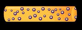
5 Size Shell Gas DeMaria. Clin Cardiol. 1997;20(suppl I):I-3. Microbubble µm RBC 6 8 µm Cheng et al. Am J Cardiol. 1998;81:41G. 5
6 Shell Composition Proteins Biocompatible polymers Phospholipids Shell Properties Elasticity Fragility Biodistribution Elimination Burns PN. In: Rumack CM et al, eds. Diagnostic Ultrasound. Vol. 1. 2nd ed. St. Louis, MO: Mosby; 1998:57. Air Highly soluble Low persistence and stability Rapid diffusion after disruption Heavy Gases High molecular weight Low solubility High persistence and stability Inert & safe Villarraga et al. Tex Heart Inst J. 1996;23:90. 6

7 Duration of clinically useful contrast effect (min) P < P < Albunex Optison 0 Albunex EchoGen Adapted from Cohen et al. J Am Coll Cardiol. 1998;32:746. Adapted from Grayburn et al. J Am Coll Cardiol. 1998;32:230. MI Bubble Behavior < 0.1 Linear Oscillation Acoustic Behavior Backscatter Enhancement Clinical Application Fundamental LVO Spectral Doppler Nonlinear Oscillation Harmonic Backscatter Harmonic LVO Real time perfusion >1.0 Disruption Transient Harmonic Echos Doppler LVO Triggered perfusion 7

8 P. Burns & H. Becher. Handbook of Contrast Echocardiography: LV Function and Myocardial Perfusion. Springer;


9 Very strong nonlinear backscatter of extremely short duration Wei et al. J Am Coll Cardiol. 1997;29:1081. Linear resonance Nonlinear resonance Transient scattering POWER POWER POWER Fundamental enhancement Harmonic enhancement Bubble disruption Burns PN. Echocardiography. 2002;19:

10 Tissue and blood reflect at the fundamental frequency 2.5 MHz 2.5 MHz Microbubbles reflect at both the fundamental and the harmonic frequencies 2.5 MHz + 5 MHz Backscattered signal Imaged signal Tissue Filter Bubbles Burns. In Rumack et al, eds. Diagnostic Ultrasound. Vol. 1. 2nd ed. St. Louis: Mosby; 1998:57. 10


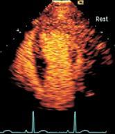
11 1. Define ultrasound contrast? 2. Recognize the interaction of the bubbles with ultrasound 3. Describe how contrast maximizes value Incremental value for LVO Incremental value for spectral Doppler Structural abnormalities Tissue characterization Differentiate real from Memorex : Artifacts 4. Explain how to set up the pictures/pitfalls 5. Perfusion 6. Safety Most common use of diagnostic echocardiography Global ventricular function Regional wall motion Rest Stress 11


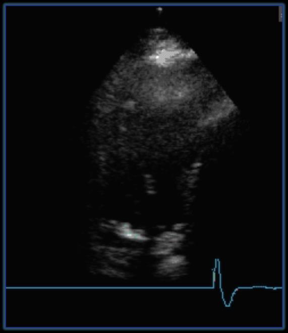
12 Modified Biplane Simpson s Method Biplane volume 20 i=l a i b i =. 4 L 20 12


13 Increased sensitivity Heightened diagnostic confidence Improved accuracy and reproducibility Enhanced clinical utility Echo Modified Biplane Simpson s Method vs. RNA Harmonic Imaging with Contrast Better correlation with reference standards! Nahar T et al: Am J Cardiol 86:1358,


14 Myocardial Border Detection vs Angiography Angio outline Echo outline No contrast Columnae carnae bases enclosed by angiographic dye vs apices imaged by ultrasound Schnittger I et al: Am J Cardiol 50:512, 1982 Take Home Points 1. Defines the endocardial border better than unenhanced echocardiography. 2. The underestimation of cardiac volumes by echocardiography is nearly resolved when contrast agents are used. 3. Reduced intra and interobserver variability in measures of LV volumes and EF with better correlation with reference standards. 4. Recommended for use when > 2 LV segments are not well visualized. 14


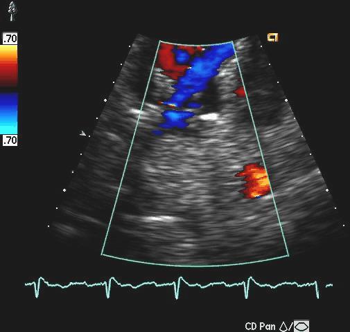
15 Spectral Doppler 15
:919-23 Structural Definition 1.")
16 Percent (%) Spectral Doppler 1= Excellent 2= Fair 3= Poor Spectral Doppler Score = 1 Pulmonary Vein Routine Mitral Inflow Discretionary Tricuspid Regurgitation Lester SJ et al. J Am Soc Echocardiogr 2006 Jul; 19(7): Structural Definition 1. LV Structural Abnormalities - Apical hypertrophy - Aneurysm / pseudoaneurysm - Thrombus - Noncompaction - Myocardial rupture 16

17 17


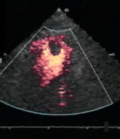
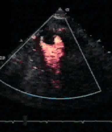
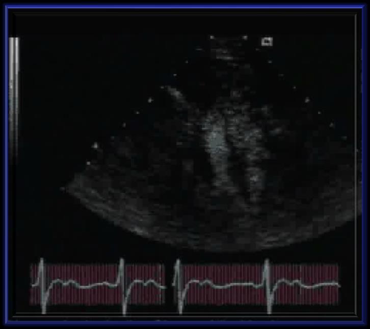
18 LV Aneurysm LV Aneurysm & More 18

19 LV Aneurysm LV Aneurysm & More 19

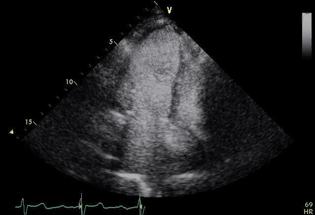
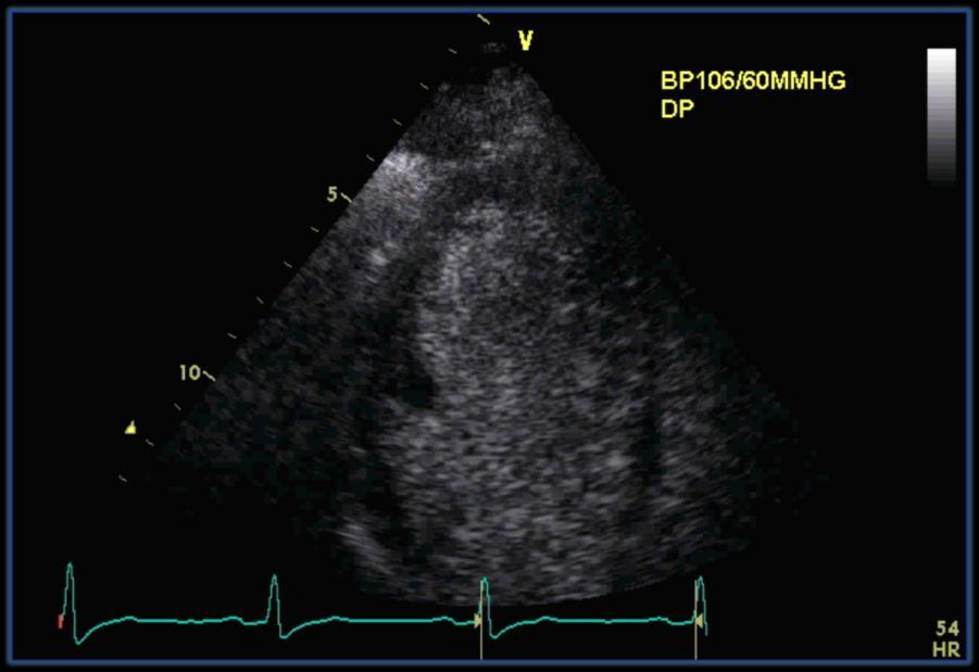
 LV apical thrombus in patient post MI, no enhancement")
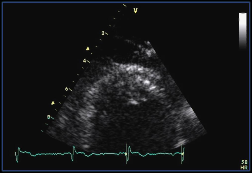
20 Structural Definition 1. LV Structural Abnormalities - Apical hypertrophy - Aneurysm / pseudoaneurysm - False chord - Trabeculation: Noncompaction - Myocardial rupture 2. Characterize intracardiac masses (tissue characterization) LV apical thrombus in patient post MI, no enhancement Secondary cardiac tumor (renal sarcoma) located in RA, complete enhancement LA myxoma, partial enhancement Mansencal et al. Archives of Cardiovascular Disease (2009) 102,


21 Structural Definition 1. LV Structural Abnormalities - Apical hypertrophy - Aneurysm / pseudoaneurysm - False chord - Trabeculation:Noncompaction - Myocardial rupture 2. Characterize intracardiac masses (tissue characterization) 3. Is it real or Memorex? Artifacts 21
22 22


23 1. Define ultrasound contrast? 2. Recognize the interaction of the bubbles with ultrasound 3. Describe how contrast maximizes value Incremental value for LVO Incremental value for spectral Doppler Structural abnormalities Tissue characterization Differentiate real from Memorex : Artifacts 4. Explain how to set up the pictures/pitfalls 5. Perfusion 6. Safety Machine and administration frequency adjusted to provide the best image 23


24 Focal Area Focal Area Single Focus Single Focus Dual Focus 24


25 Mechanical Index Measure of output acoustic power High MI increases bubble destruction MI 0.24 increased to 0.6 MI = 1.4 CP

26 MI =


 Dosing")

 Clinician")
27 POTENTIAL CAUSES System settings (focal zone misplacement) Dosing and administration (low concentration) POTENTIAL CAUSES Dosing (high concentration) Administration (infusion rate too fast) Clinician (obtain off-axis windows) 27


28 28

 Dosing (low")

 Poor LV")
29 POTENTIAL CAUSES System settings (high MI) Dosing (low concentration) Administration (low infusion rate) Poor LV function 29
30 30


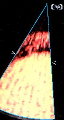
31 Perfusion Right Ventricle LVO Phase Myocardial Phase J Am Soc Echocardiogr

32 Perfusion Low MI, Nondestructive Real Time High MI, Destructive Triggered Continuous Imaging Intermittent Imaging Low MI, Nondestructive Real Time Perfusion Continuous Imaging 32

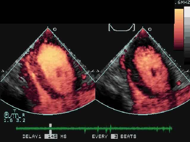

33 High MI, Destructive Triggered Perfusion Intermittent Imaging 1 beat 5 beats 32 beats 44 beats 33

34 Intensity Plateau intensity ( ) Appearance rate ( ) Intensity = (1-exp 1 ) Baseline intensity Bolus Time (t) Feigenbaum s Echocardiography 6 th edition Baseline Curves A B Vasodilator Curves A: normal reference area B: area of coronary obstruction Both normal Infarct territory Both normal Infarct territory 75% stenosis 75% stenosis Feigenbaum s Echocardiography 6 th edition 34


35 Perfusion Low MI, Nondestructive Real Time High MI, Destructive Triggered Continuous Imaging Intermittent Imaging When I look back on all the worries I remember the story of the old man who said on his deathbed that he had a lot of trouble in his life, most of which never happened Sir Winston Leonard Spencer Churchill ( ) 35

 - Back pain n=75 - Headache n=9 - Urticaria n=17 Urticaria/urticarial-like reaction 0.")
36 1. Suspected hypersensitivity to the microsphere components. Most serious reactions occur within 30 minutes of administration No 30 minute monitoring period! Mayo Clinic: Doses Any Adverse Reaction 0.6% (1 in 154) - Back pain n=75 - Headache n=9 - Urticaria n=17 Urticaria/urticarial-like reaction 0.1% (1 in 915) - Without Respiratory Symptoms n=12 - With Respiratory Symptoms n=5 Lester SJ et al J Am Soc Echocardiogr 2008:15;417 36


37 CARPA Complement Activation Related Pseudo Allergy Features similar IgE-mediated Type 1 reactions. Angioedema, bronchospasm, hypoxemia, hypotension, low back pain, and urticaria Can occur without prior exposure, decrease in severity with subsequent exposure, resolve spontaneously. Tool Used to Build Excellence in ECHO 37
38 1. The clinical utility of a bubble depends on its size, shell and type of gas 2. The backscattered signal radiating from a bubble that oscillates in a nonlinear fashion will contain a harmonic component. 3. Perfusion: Low MI, nondestructive, real time imaging High MI, destruction, triggered imaging 4. The only contraindications is a hypersensitivity to the microsphere components 38
39 39
40 40


41 Positive Impact : Makes a Difference Avoid the Pitfalls: Big 5 Innovation Workflow For any innovation in echocardiography to be widely adopted it must be equaled or paralleled by an innovation in workflow 41


42 Minutes Imaging Protocol (a) : Workflow Efficiency The 60 Second Echo Impact of the 60 second Echo P = NS P < Total Procedure Time Sonographer Scan Time Routine Discretionary Lester SJ et al. J Am Soc Echocardiogr 2006 Jul; 19(7):


43 Percent We Don t Use Enough Contrast Visualization 1= Excellent or adequate full endocardial visualization 2= Incomplete endocardial visualization 3= only epicardium visualized 4= segment not visualized Visualization Score < 1.1 Routine 88% Discretionary 50% Discretionary No Contrast 33% Lester SJ et al. J Am Soc Echocardiogr 2006 Jul; 19(7):


44 Without Contrast With Contrast Avoid The Pitfalls 1. Contraindications (safety) 2. Protocol Development (The 60 second Echo) 3. Use It 4. Spectral Doppler 5. Instrument Settings 44
Basics and Perfusion Imaging. Steven J. Lester, MD, FACC, FRCP(C), FASE Mayo Clinic, Arizona
, FASE Mayo Clinic, Arizona") Basics and Perfusion Imaging Steven J. Lester, MD, FACC, FRCP(C), FASE Mayo Clinic, Arizona Relevant Financial Relationship(s) None Off Label Usage a. Produce only a harmonic backscatter signal. b. Produce
Basics and Perfusion Imaging Steven J. Lester, MD, FACC, FRCP(C), FASE Mayo Clinic, Arizona Relevant Financial Relationship(s) None Off Label Usage a. Produce only a harmonic backscatter signal. b. Produce
CONTRAST ECHOCARDIOGRAPHY
 CONTRAST ECHOCARDIOGRAPHY How Should it Be Administered and How Do I Optimize My Machine Settings? Keith Collins, MS RDCS FASE Monday, Feb. 15, 2016 State of the Art Tscc.exe Contrast Is Needed When Poor
Where Contrast Administration Makes a Difference Contrast 2017 State of the Art
 Where Contrast Administration Makes a Difference Contrast 2017 State of the Art Linda D. Gillam, MD, MPH, FASE Chair, Cardiovascular Medicine Medical Director, CV Service Line Morristown Medical Center/Atlantic
Basics of Contrast Echocardiography Echo Hawaii 2017
 Basics of Contrast Echocardiography Echo Hawaii 2017 Maryellen Orsinelli, RN, RDCS, FASE Lead Cardiac Sonographer The Ohio State University The Ross Heart Hospital 1 DISCLOSURES NONE 1 OBJECTIVES Indications
Contrast Echocardiography. What is the critical need? Meets a critical need! Cavity Opacification. What contrast is approved for: chamber assessment?
 Contrast Echocardiography for LV Opacification Natesa G. Pandian What contrast is approved for: Cavity Opacification Meets a critical need! Disclosure: Speakers Bureau, Lantheus Inc What is the critical
Qualitative and Quantitative Assessment of Perfusion
 APCDE 2011 Qualitative and Quantitative Assessment of Perfusion Hyun Ju Yoon Chonnam National University Hospital Gwangju, Korea ISCHEMIC CASCADE Blood flow mismatch Perfusion defects on nuclear imaging,
Myocardial Contrast Echo
 Myocardial Contrast Echo Anthony DeMaria Myocardial Contrast Echocardiography: Problems and Potential Anthony DeMaria MD Judith and Jack White Chair Founding Director, Sulpizio Cardiovascular Center University
ROLE OF CONTRAST AGENTS TO ENHANCE WALL MOTION AND DOPPLER SIGNALS
 ROLE OF CONTRAST AGENTS TO ENHANCE WALL MOTION AND DOPPLER SIGNALS Adj Assoc Prof. Tong Khim Leng MBBS MRCP FAMS FACC FASE FRCP Chief, and Sn Consultant Department of Cardiology, Changi General Hospital
Cardiac Chamber Quantification by Echocardiography
 Cardiac Chamber Quantification by Echocardiography Maryam Bokhamseen, RCS, RCDS, EACVI Echotechnologist ǁ, Non invasive Cardiac Laboratory King Abdulaziz Cardiac Center. Outline: Introduction. Background
Mechanisms and role of contrast echocardiography
 Mechanisms and role of contrast echocardiography Seol Sang-Hoon Inje University College of Medicine, Haeundae Paik Hospital, Busan, Korea Physical Principles of Contrast Ultrasound Contrast echocardiography
Adult Echocardiography Examination Content Outline
 Adult Echocardiography Examination Content Outline (Outline Summary) # Domain Subdomain Percentage 1 2 3 4 5 Anatomy and Physiology Pathology Clinical Care and Safety Measurement Techniques, Maneuvers,
CONTRAST ENHANCED IMAGING HOW TO GET STARTED: A Tale From A Facility Like Yours
 The presentation will begin in a few moments. CONTRAST ENHANCED IMAGING HOW TO GET STARTED: A Tale From A Facility Like Yours For technical support, call 800-679-3646 CONTRAST ENHANCED IMAGING HOW TO GET
Disclosures. Stress Echocardiography 2010 Appropriate Use & Further Applications. Stress Echo Clinical Utility 9/8/10
 2010 & Further Applications Disclosures None Geoffrey A. Rose, MD FACC FASE Director, Cardiac Ultrasound Laboratory Sanger Heart & Vascular Institute Clinical Utility and Event-free Survival Cortigiani,
Top 10 Facts in Contrast Echocardiography. Pamela R. Burgess, BS, RDCS, RDMS, RVT, FASE
 Top 10 Facts in Contrast Echocardiography Pamela R. Burgess, BS, RDCS, RDMS, RVT, FASE Presenter Disclosure The following relationship exist related to this presentation: Pamela R. Burgess, BS, RDCS, RDMS,
Case Report. Case Report. Ana Lúcia Martins Arruda, Altamiro Ozório, Eloisa Mattos, José Lázaro de Andrade, Thomas Porter, Wilson Mathias Jr
 Case Report Hypoperfusion of the Left Ventricle in the Absence of Changes in Segmental Contractility as Observed through Echocardiography by Using Microbubbles During Dobutamine Infusion Ana Lúcia Martins
Chapter 1. General introduction and outline of the thesis. Jeroen Slikkerveer Otto Kamp
 Chapter 1 General introduction and outline of the thesis Jeroen Slikkerveer Otto Kamp Chapter 1 Introduction The introduction of microbubbles and the technical development in ultrasound facilitates the
The Normal Echocardiogram
 The Normal Echocardiogram Pravin V. Patil, MD FACC Lewis Katz School of Medicine at Temple University Acknowledgments Dr. Susan Wiegers Dr. Martin Keane Temple Cardiac Sonographers Disclosures No relevant
Power Doppler Myocardial Contrast Echocardiography Using an Improved Multiple Frame Triggered Harmonic Angio Technique
 Reprinted with permission from ECHOCARDIOGRAPHY, Volume 18, No. 3, April 2001 Copyright 2001 by Futura Publishing Company, Inc., Armonk, NY 10504-0418 Power Doppler Myocardial Contrast Echocardiography
10/7/2013. Systolic Function How to Measure, How Accurate is Echo, Role of Contrast. Thanks to our Course Director: Neil J.
 Systolic Function How to Measure, How Accurate is Echo, Role of Contrast Neil J. Weissman, MD MedStar Health Research Institute & Professor of Medicine Georgetown University Washington, D.C. No Disclosures
The last decade has seen a revolution in the technique of contrast echocardiography, with
 342 * Imaging techniques CONTRAST ECHOCARDIOGRAPHY c CONTRAST Correspondence to: Dr Michael J Stewart, Cardiothoracic Unit, The James Cook University Hospital, Marton Road, Middlesbrough, TS4 3BW, UK;
Contrast Enhanced Voiding Urosonography (cevus): How we do it
: How we do it") Contrast Enhanced Voiding Urosonography (cevus): How we do it Susan J. Back, MD Department of Radiology, The Children s Hospital of Philadelphia No Disclosures cevus What it is What to do What not to do
Contrast Enhanced Voiding Urosonography (cevus): How we do it Susan J. Back, MD Department of Radiology, The Children s Hospital of Philadelphia No Disclosures cevus What it is What to do What not to do
Echocardiography has been widely used as a. Contrast Microbubbles Improve Diagnostic Yield in ICU Patients With Poor Echocardiographic Windows*
 Contrast Microbubbles Improve Diagnostic Yield in ICU Patients With Poor Echocardiographic Windows* Thanh T. Nguyen, DO; Milind R. Dhond, MD; Raju Sabapathy, MD; and William J. Bommer, MD Objective: To
Concepts of Imaging and Knobology
 Concepts of Imaging and Knobology Pravin Patil, MD FACC FASE Associate Professor of Medicine Director, Cardiovascular Disease Training Program Lewis Katz School of Medicine at Temple University Disclosures
DOWNLOAD PDF MYOCARDIAL CONTRAST TWO DIMENSIONAL ECHOCARDIOGRAPHY (DEVELOPMENTS IN CARDIOVASCULAR MEDICINE)
") Chapter 1 : Imaging Cardiovascular Medicine Stanford Medicine contrast two-dimensional echocardiography (MC-2DE), a new and exciting diagnostic methodology for assessment of myocardial perfusion, which
Chapter 1 : Imaging Cardiovascular Medicine Stanford Medicine contrast two-dimensional echocardiography (MC-2DE), a new and exciting diagnostic methodology for assessment of myocardial perfusion, which
Stress, strain and contrast. UK available agents. Safety 13/06/2018. Which enhancing agent do you use? Ultrasound enhancing agents.
 Stress, strain and contrast Stephen Glen Ultrasound enhancing agents Safety Effectiveness during stress Perfusion / myocardial contrast UK available agents Which enhancing agent do you use? Name Bubble
Ultrasound Contrast Agents
 Ultrasound Contrast Agents Recent Safety Studies, Quality Assurance Documents & Consensus Documents March 27, 2009 1 2 FDA Revised Product Labeling May 2008 "Boxed Warning" remains for Definity and Optison
Tissue Doppler and Strain Imaging
 Tissue Doppler and Strain Imaging Steven J. Lester MD, FRCP(C), FACC, FASE Relevant Financial Relationship(s) None Off Label Usage None 1 Objective way with which to quantify the minor amplitude and temporal
Tissue Doppler and Strain Imaging
 Tissue Doppler and Strain Imaging Steven J. Lester MD, FRCP(C), FACC, FASE Relevant Financial Relationship(s) None Off Label Usage None 1 Objective way with which to quantify the minor amplitude and temporal
3D-stress echocardiography Bernard Cosyns, MD, PhD
 3D-stress echocardiography Bernard Cosyns, MD, PhD No Disclosure The Pro-Technology bias Sicari et al. Cardiovascular Ultrasound 2006, 4:11 Overview 2D stress echocardiography: main limitations 3D echocardiography:
Cardiac Masses. Cardiac Masses: Considerations. Dennis A. Tighe, MD, FASE. University of Massachusetts Medical School Worcester, MA 4/16/2018
 Cardiac Masses Dennis A. Tighe, MD, FASE University of Massachusetts Medical School Worcester, MA Cardiac Masses: Considerations Definition of the mass Nature Location Benign or malignant Presentation
Cardiac Masses. Dennis A. Tighe, MD, FASE. University of Massachusetts Medical School Worcester, MA
 Cardiac Masses Dennis A. Tighe, MD, FASE University of Massachusetts Medical School Worcester, MA Cardiac Masses: Considerations Definition of the mass Nature Location Benign or malignant Presentation
Contrast-enhanced echocardiography improves agreement on the assessment of ejection fraction and left ventricular function. A multicentre study
 Eur J Echocardiography 7 Suppl. 2 (2006) S16 S21 Contrast-enhanced echocardiography improves agreement on the assessment of ejection fraction and left ventricular function. A multicentre study Rainer Hoffmann*
Clinical Applications of Ultrasonic Contrast Agents in Echocardiography
 Author: Sherif F. Nagueh, MD,FACC, FAHA, FASE TABLE OF CONTENTS Faculty Disclosure page 1 Accreditation page 2 Biography page 3 Learning Objectives page 4 Introduction page 5 Conclusions page 11 Figures
imagine 2018 Echocardiography Today - Do, Learn, Do More, Learn More November 3-4, 2018 Georgia Technology Hotel and Conference Center
 Piedmont Heart presents imagine 2018 Echocardiography Today - Do, Learn, Do More, Learn More November 3-4, 2018 Georgia Technology Hotel and Conference Center Program Co-Directors Mani A. Vannan MBBS FASE
Multiple Gated Acquisition (MUGA) Scanning
 Scanning") Multiple Gated Acquisition (MUGA) Scanning Dmitry Beyder MPA, CNMT Nuclear Medicine, Radiology Barnes-Jewish Hospital / Washington University St. Louis, MO Disclaimers/Relationships Standard of care research
Multiple Gated Acquisition (MUGA) Scanning Dmitry Beyder MPA, CNMT Nuclear Medicine, Radiology Barnes-Jewish Hospital / Washington University St. Louis, MO Disclaimers/Relationships Standard of care research
Pregnancy and Heart Disease Sharon L. Roble, MD Echo Hawaii 2016
 1 Pregnancy and Heart Disease Sharon L. Roble, MD Echo Hawaii 2016 DISCLOSURES I have no disclosures relevant to today s talk 2 Cardiovascular Effects of Pregnancy Anatomic Ventricular muscle mass increases
Echocardiography as a diagnostic and management tool in medical emergencies
 Echocardiography as a diagnostic and management tool in medical emergencies Frank van der Heusen MD Department of Anesthesia and perioperative Care UCSF Medical Center Objective of this presentation Indications
Imaging of Coronary Artery Disease: II
 Acta Radiológica Portuguesa, Vol.XIX, nº 74, pág. 45-51, Abr.-Jun., 2007 Imaging of Coronary Artery Disease: II Jean Jeudy University of Maryland School of Medicine Department of Diagnostic Radiology Armed
좌심실수축기능평가 Cardiac Function
 Basic Echo Review Course 좌심실수축기능평가 Cardiac Function Seonghoon Choi Cardiology Hallym university LV systolic function Systolic function 좌심실수축기능 - 심근의수축으로심실에서혈액을대동맥으로박출하는기능 실제임상에서 LV function 의의미 1Diagnosis
Review of Cardiac Imaging Modalities in the Renal Patient. George Youssef
 Review of Cardiac Imaging Modalities in the Renal Patient George Youssef ECHO Left ventricular hypertrophy (LVH) assessment Diastolic dysfunction Stress ECHO Cardiac CT angiography Echocardiography - positives
Correlation Between Regional Wall Motion Abnormalities via 2-Dimensional Echocardiography, and Coronary Angiographic Findings
 THE ECHOCARDIOGRAPHY, IRAQI POSTGRADUATE MEDICAL AND CORONARY JOURNAL ANGIOGRAPHIC FINDINGS VOL.11, SUPPLEMENT,2012 Correlation Between Regional Wall Motion Abnormalities via 2-Dimensional Echocardiography,
Value of echocardiography in chronic dyspnea
 Value of echocardiography in chronic dyspnea Jahrestagung Schweizerische Gesellschaft für /Schweizerische Gesellschaft für Pneumologie B. Kaufmann 16.06.2016 Chronic dyspnea Shortness of breath lasting
Improvement in Endocardia1 Border Delineation Using Tissue Harmonic Imaging
 Improvement in Endocardia1 Border Delineation Using Tissue Harmonic Imaging HARALD BECHER, M.D.," KLAUS TIEMA", M.D.," THOMAS SCHLOSSER," CHRISTOPH POHL, M.D.," NAVIN C. NANDA, M.D.,? MICHALAKIS A. AVERKIOU,
THE LEFT ATRIUM HOW CAN ECHO HELP US?
 THE LEFT ATRIUM HOW CAN ECHO HELP US? Dr. Dragos COZMA BACKGROUND Left atrium (LA) dilation can occur in a broad spectrum of cardiovascular diseases including hypertension, left ventricular dysfunction,
Echo in CAD: Wall Motion Assessment
 Echo in CAD: Wall Motion Assessment Joe M. Moody, Jr, MD UTHSCSA and STVHCS October 2007 Relevant References ACC/AHA/ASE 2003 Guideline Update for the Clinical Application of Echocardiography Bayes de
Cardiac MRI in ACHD What We. ACHD Patients
 Cardiac MRI in ACHD What We Have Learned to Apply to ACHD Patients Faris Al Mousily, MBChB, FAAC, FACC Consultant, Pediatric Cardiology, KFSH&RC/Jeddah Adjunct Faculty, Division of Pediatric Cardiology
A New Method to Rapidly Evaluate LVEF from a Contractility Polar Map. Lebeau et al.
 A New Method to Rapidly Evaluate LVEF from a Contractility Polar Map Lebeau et al. Good afternoon It is my pleasure to present to you a new method to rapidly evaluate LVEF from a contractility polar map
Tissue Doppler and Strain Imaging. Steven J. Lester MD, FRCP(C), FACC, FASE
, FACC, FASE") Tissue Doppler and Strain Imaging Steven J. Lester MD, FRCP(C), FACC, FASE Relevant Financial Relationship(s) None Off Label Usage None a. Turn the wall filters on and turn down the receiver gain. b. Turn
Tissue Doppler and Strain Imaging Steven J. Lester MD, FRCP(C), FACC, FASE Relevant Financial Relationship(s) None Off Label Usage None a. Turn the wall filters on and turn down the receiver gain. b. Turn
Left atrial function. Aliakbar Arvandi MD
 In the clinic Left atrial function Abstract The left atrium (LA) is a left posterior cardiac chamber which is located adjacent to the esophagus. It is separated from the right atrium by the inter-atrial
ΚΑΡΔΙΟΛΟΓΟΣ EUROPEAN ACCREDITATION IN TRANSTHORACIC AND TRANSESOPHAGEAL ECHOCARDIOGRAPHY
 1 ΚΑΡΔΙΟΛΟΓΟΣ EUROPEAN ACCREDITATION IN TRANSTHORACIC AND TRANSESOPHAGEAL ECHOCARDIOGRAPHY 2 Constrictive pericarditis (CP) is characterized by impaired ventricular filling due to a stiffened or noncompliant
Noncoronary Cardiac MDCT
 Noncoronary Cardiac MDCT David A. Bluemke, M.D., Ph.D. Professor, of Radiology and Medicine Johns Hopkins University School of Medicine Baltimore, Maryland Toshiba Disclosures Grant support Noncoronary
You Won t Believe What I Saw on. Disclosures. Goals. Dimensions 2013 October 18 th Michael Pfeiffer, MD. No Financial Disclosures
 You Won t Believe What I Saw on that ECHO! Dimensions 2013 October 18 th Michael Pfeiffer, MD Disclosures No Financial Disclosures Goals Review unusual and unique echocardiographic images. Briefly present
Cardiac Computed Tomography
 Cardiac Computed Tomography Authored and approved by Koen Nieman Stephan Achenbach Francesca Pugliese Bernard Cosyns Patrizio Lancellotti Anastasia Kitsiou Contents CARDIAC COMPUTED TOMOGRAPHY Page 1.
Complications of Myocardial Infarction
 Complications of Myocardial Infarction Sunil Mankad, MD, FACC, FCCP, FASE Associate Professor of Medicine Mayo Clinic College of Medicine Director, Transesophageal Echocardiography Associate Director,
Handbook of Contrast Echocardiography Left ventricular function and myocardial perfusion
 Harald Becher. Peter N Burns Handbook of Contrast Echocardiography Left ventricular function and myocardial perfusion Peter N Burns Professor of Medical Biophysics and Radiology University of Toronto Imaging
Hemodynamic Assessment. Assessment of Systolic Function Doppler Hemodynamics
 Hemodynamic Assessment Matt M. Umland, RDCS, FASE Aurora Medical Group Milwaukee, WI Assessment of Systolic Function Doppler Hemodynamics Stroke Volume Cardiac Output Cardiac Index Tei Index/Index of myocardial
Abdomen Sonography Examination Content Outline
 Abdomen Sonography Examination Content Outline (Outline Summary) # Domain Subdomain Percentage 1 2 3 Anatomy, Perfusion, and Function Pathology, Vascular Abnormalities, Trauma, and Postoperative Anatomy
ASCeXAM / ReASCE. Practice Board Exam Questions Monday Morning
 ASCeXAM / ReASCE Practice Board Exam Questions Monday Morning Ultrasound Physics Artifacts Doppler Physics Imaging, Knobology, and Artifacts Echocardiographic Evaluation of the RV Tricuspid and Pulmonary
Prof. JL Zamorano Hospital Universitario Ramón y Cajal
 Prof. JL Zamorano Hospital Universitario Ramón y Cajal Fully Automated Quantification Software Adaptive analytical algorithm consists in knowledge-based identification of global shape and specific adaptation
ARTIFACTS: THEORY AND ILLUSTRATIVE EXAMPLES
 ARTIFACTS: THEORY AND ILLUSTRATIVE EXAMPLES Robert A. Levine, M.D. Marielle Scherrer-Crosbie, M.D. Eric M. Isselbacher, M.D. No conflicts of interest Philippe Bertrand, Pieter Vendervoort, Hasselt and
PROSTHETIC VALVE BOARD REVIEW
 PROSTHETIC VALVE BOARD REVIEW The correct answer D This two chamber view shows a porcine mitral prosthesis with the typical appearance of the struts although the leaflets are not well seen. The valve
Perflutren lipid microsphere injectable suspension for cardiac ultrasound
 CONTRAST AGENT EVALUATION Perflutren lipid microsphere injectable suspension for cardiac ultrasound Echocardiography is a widely available noninvasive procedure for diagnosing cardiovascular disease. With
Diagnostic approach to heart disease
 Diagnostic approach to heart disease Initial work up History Physical exam Chest radiographs ECG Special studies Echocardiography Cardiac catheterization Echocardiography principles Technique of producing
NAKAO, RDCS 2. J Cardiol 2002 Dec; 40 6 :
 J Cardiol 2002 Dec; 40 6 : 259 265 T Usefulness of Left Ventricular Opacification With Intravenous Contrast Echocardiography in Patients With Asymptomatic Negative T Waves on Electrocardiography 1 2 2
Detection of Resting Myocardial Perfusion
 Detection of Resting Myocardial Perfusion Defects by SonoVue R Myocardial Contrast Echocardiography Tamanna Nahar, M.D., Peng Li, M.D., Ph.D., Bettina Kuersten, M.D., Sanjay Batra, Ph.D., and Mani A. Vannan,
Right Ventricle Steven J. Lester MD, FACC, FRCP(C), FASE Mayo Clinic, Arizona
, FASE Mayo Clinic, Arizona") Right Ventricle Steven J. Lester MD, FACC, FRCP(C), FASE Mayo Clinic, Arizona 1. In which scenario will applying the simplified Bernoulli equation to the peak tricuspid regurgitation velocity and adding
Right Ventricle Steven J. Lester MD, FACC, FRCP(C), FASE Mayo Clinic, Arizona 1. In which scenario will applying the simplified Bernoulli equation to the peak tricuspid regurgitation velocity and adding
Echocardiography Conference
 Echocardiography Conference David Stultz, MD Cardiology Fellow, PGY-6 September 20, 2005 Atrial Septal Aneurysm Bulging of Fossa Ovalis Associated commonly with Atrial septal defect or small perforations
Imaging of the Heart Todd Tessendorf MD FACC
 Imaging of the Heart Todd Tessendorf MD FACC Outline Imaging Modalities for Structural Heart Disease ECHO, MRI Imaging Modalities for Ischemic Heart Disease SPECT, PET, CCTA Show lots of pretty pictures
PISA Evaluation of Mitral Regurgitation. Raymond Graber, MD Cardiac Anesthesia Group University Hospitals Case Medical Center 4/07/2011
 PISA Evaluation of Mitral Regurgitation Raymond Graber, MD Cardiac Anesthesia Group University Hospitals Case Medical Center 4/07/2011 Introduction Evaluation of MR. What is PISA? Physiologic basis Issues
가천의대길병원소아심장과최덕영 PA C IVS THE EVALUATION AND PRINCIPLES OF TREATMENT STRATEGY
 가천의대길병원소아심장과최덕영 PA C IVS THE EVALUATION AND PRINCIPLES OF TREATMENT STRATEGY PA c IVS (not only pulmonary valve disease) Edwards JE. Pathologic Alteration of the right heart. In: Konstam MA, Isner M, eds.
DECLARATION OF CONFLICT OF INTEREST
 DECLARATION OF CONFLICT OF INTEREST The additive prognostic value of myocardial perfusion defects, coronary flow reserve and wall motion abnormalities during dipyridamole contrast stress-echo: a prospective
Pediatric Echocardiography Examination Content Outline
 Pediatric Echocardiography Examination Content Outline (Outline Summary) # Domain Subdomain Percentage 1 Anatomy and Physiology Normal Anatomy and Physiology 10% 2 Abnormal Pathology and Pathophysiology
MAYON VOLCANO: FAST FACTS
 MAYON VOLCANO: FAST FACTS Type of Volcano: Stratovolcano Elevation: 2.46 km Base Diameter: 20 km Base Circumference: 62.8 km Area: 314.1 km 2 Reference: http://www.phivolcs.dost.gov.ph/html/update_vmepd/volcano/volcanolist/mayon.htm
Appropriateness of Stress Echocardiography and Nuclear Stress Thallium/Sesta Mibi Testing Methods
 Appropriateness of Stress Echocardiography and Nuclear Stress Thallium/Sesta Mibi Testing Methods Review by Michael Devenport, MS, RDCS Objectives: A basic description and review of appropriate use of
Heart Failure in Women: Dr Goh Ping Ping Cardiologist Asian Heart & Vascular Centre
 Heart Failure in Women: More than EF? Dr Goh Ping Ping Cardiologist Asian Heart & Vascular Centre Overview Review pathophysiology as it relates to diagnosis and management Rational approach to workup:
April 16, 09:00-09:15 중앙대학교 윤신원
 April 16, 09:00-09:15 중앙대학교 윤신원 When to perform Echocardiography in IE? Vegetations?(pathologic Whatever the level hallmark) of suspicion Intracardiac abscess? Confirm or R/O at the Earliest opportunity.
Getting the Most Out of Stress Echo
 Getting the Most Out of Stress Echo Vera H. Rigolin, MD, FASE, FACC, FAHA Professor of Medicine Northwestern University Feinberg School of Medicine Medical Director, Echocardiography Laboratory Northwestern
Safety of Dobutamine Stress Real-Time Myocardial Contrast Echocardiography
 Journal of the American College of Cardiology Vol. 45, No. 8, 2005 2005 by the American College of Cardiology Foundation ISSN 0735-1097/05/$30.00 Published by Elsevier Inc. doi:10.1016/j.jacc.2005.01.024
Reproducibility and Accuracy of Echocardiographic Measurements of Left Ventricular Parameters Using Real-Time Three-Dimensional Echocardiography
 Journal of the American College of Cardiology Vol. 44, No. 4, 2004 2004 by the American College of Cardiology Foundation ISSN 0735-1097/04/$30.00 Published by Elsevier Inc. doi:10.1016/j.jacc.2004.05.050
The Fontan circulation. Folkert Meijboom
 The Fontan circulation Folkert Meijboom What to expect? Why a Fontan-circulation Indications How does it work Types of Fontan circulation Historical overview Role of echocardiography What to expect? Why
Contrast echocardiography in critical care: echoes of the future? A review of the role of microsphere contrast echocardiography
 Contrast echocardiography in critical care: echoes of the future? A review of the role of microsphere contrast echocardiography David G Platts and John F Fraser Transthoracic echocardiography (TTE) is
Quantitation of right ventricular dimensions and function
 SCCS Basics of cardiac assessment Quantitation of right ventricular dimensions and function Tomasz Kukulski, MD PhD Dept of Cardiology, Congenital Heart Disease and Electrotherapy Silesian Medical University
How NOT to miss Hypertrophic Cardiomyopathy? Adaya Weissler-Snir, MD University Health Network, University of Toronto
 How NOT to miss Hypertrophic Cardiomyopathy? Adaya Weissler-Snir, MD University Health Network, University of Toronto Introduction Hypertrophic cardiomyopathy is the most common genetic cardiomyopathy,
Index. Note: Page numbers of article titles are in boldface type.
 Index Note: Page numbers of article titles are in boldface type. A Acute coronary syndrome(s), anticoagulant therapy in, 706, 707 antiplatelet therapy in, 702 ß-blockers in, 703 cardiac biomarkers in,
Cardiovascular Imaging Stress Echo
 Cardiovascular Imaging Stress Echo Theodora A Zaglavara, MD, PhD Cardiac Imaging Department INTERBALKAN MEDICAL CENTER Thessaloniki GREECE Evolution of Stress Echo: From Innovation to a Widely Established
My Patient Needs a Stress Test
 My Patient Needs a Stress Test Amy S. Burhanna,, MD, FACC Coastal Cardiology Cape May Court House, New Jersey Absolute and relative contraindications to exercise testing Absolute Acute myocardial infarction
Pitfalls and how to avoid them
 Pitfalls and how to avoid them I.M. Coman MD, PhD, FESC Pitfalls victims? WHO : all of us WHEN : - Learning curve! - Don t hurry! WHICH conditions : Poor windows Complex diseases/procedures Arrhythmias
Basic Assessment of Left Ventricular Systolic Function
 WINFOCUS BASIC ECHO (WBE) Basic Assessment of Left Ventricular Systolic Function Ritesh Dhar, MD Director, Echocardiography Lab and Staff Cardiologist Intermountain Medical Center Murray, Utah Outline
Cardiac MRI: Cardiomyopathy
 Cardiac MRI: Cardiomyopathy Laura E. Heyneman, MD I do not have any relevant financial relationships with any commercial interests Cardiac MRI: Cardiomyopathy Laura E. Heyneman, MD Duke University Medical
EDITOR S PICK CURRENT STATUS OF FULLY AUTOMATED SOFTWARE WITH THREE-DIMENSIONAL ECHOCARDIOGRAPHY FOR THE QUANTIFICATION OF LEFT VENTRICULAR FUNCTION
 ITOR S PICK This paper, courtesy of Yang and Takeuchi, provides a timely and well-considered update on the current status of fully-automated software with three-dimensional echocardiography for quantifying
What effects will proximal or distal disease have on a waveform?
 Spectral Doppler Interpretation Director of Ultrasound Education & Quality Assurance Baylor College of Medicine Division of Maternal-Fetal Medicine Maternal Fetal Center Imaging Manager Texas Children
Access to the published version may require journal subscription. Published with permission from: Blackwell Synergy
 This is an author produced version of a paper published in Clin Physiol Funct Imaging. This paper has been peer-reviewed but does not include the final publisher proof-corrections or journal pagination.
Index. radiologic.theclinics.com. Note: Page numbers of article titles are in boldface type.
 Index Note: Page numbers of article titles are in boldface type. A ALCAPA. See Anomalous left coronary artery from the pulmonary artery. Angiosarcoma computed tomographic assessment of, 809 811 Anomalous
Martin G. Keane, MD, FASE Temple University School of Medicine
 Martin G. Keane, MD, FASE Temple University School of Medicine Measurement of end-diastolic LV internal diameter (LVIDd) made by properly-oriented M-Mode techniques in the Parasternal Long Axis View (PLAX):
A Practical Approach to Prosthetic Valves
 A Practical Approach to Prosthetic Valves Bonita Anderson DMU (Cardiac), MApplSc (Med Ultrasound), ACS, AMS, FASE https://doi.org/10.1161/circulationaha.108.778886 Disclosures None 1 Know the Product Know
Department of Pediatrics and Child Health, Kurume University School of Medicine, Kurume, 830 Japan. Received for publication October 26, 1992
 THE KURUME MEDICAL JOURNAL Vol.39, p.291-296, 1992 Jon-Invasive Evaluation of Pulmonary Arterial and Right Ventricular Pressures with Contrast Enhanced Doppler Signals of Tricuspid Regurgitation Flow Using
Brief View of Calculation and Measurement of Cardiac Hemodynamics
 Cronicon OPEN ACCESS EC CARDIOLOGY Review Article Brief View of Calculation and Measurement of Cardiac Hemodynamics Samah Alasrawi* Pediatric Cardiologist, Al Jalila Children Heart Center, Dubai, UAE *
Νεότερα ςτην Υπερηχοκαρδιογραφία. Βαςίλειοσ Καμπερίδησ Clinical research fellow in Cardiology
 Νεότερα ςτην Υπερηχοκαρδιογραφία Βαςίλειοσ Καμπερίδησ Clinical research fellow in Cardiology Disclosures ESC training grant EACVI research grant HCS training grant ELIKAR research grant Evolution of Echocardiography
Index. K Knobology, TTE artifact, image resolution, ultrasound, 14
 A Acute aortic regurgitation (AR), 124 128 Acute aortic syndrome (AAS) classic aortic dissection diagnosis, 251 263 evolutive patterns, 253 255 pathology, 250 251 classifications, 247 248 incomplete aortic
Myocardial performance index, Tissue Doppler echocardiography
 Value of Measuring Myocardial Performance Index by Tissue Doppler Echocardiography in Normal and Diseased Heart Tarkan TEKTEN, 1 MD, Alper O. ONBASILI, 1 MD, Ceyhun CEYHAN, 1 MD, Selim ÜNAL, 1 MD, and