OBJECTIVES. To emphasize the important components of a detailed obstetrical scan versus a routine 2 nd or 3 rd trimester scan
|
|
|
- Diana Logan
- 6 years ago
- Views:
Transcription
1
2 OBJECTIVES To discuss the different types of OB ultrasound examinations with emphasis on what is done here at CHOP at the time of the initial evaluation To emphasize the important components of a detailed obstetrical scan versus a routine 2 nd or 3 rd trimester scan To illustrate with interesting cases how we perform these scans and to show how additional findings can significantly influence patient counseling, management and ultimately outcome S L I D E : 2
3 OBSTETRICAL US EXAMINATIONS DIFFERENT TYPES OF OB US SCANS CODE DESCRIPTION Standard US<14 wks Standard US>14 wks Detailed Anatomy Scan Limited Scan= Quickie Follow Up Scan Umbilical Artery Doppler Middle Cerebral Artery Doppler Uterine Artery Doppler
4 STANDARD ANATOMY SCAN WHAT FETAL ANATOMY DOES THIS SCAN INCLUDE? The Five C s in the Fetal Head CP, Cerebral ventricles, CSP, Cerebellum & Cisterna Magna 4CH with Inflow and Outflow Tracts Stomach Abdominal Cord Insertion Kidneys Bladder Spine Longitudinal & Transverse Three Vessel Umbilical Cord
5 DETAILED ANATOMY SCAN WHAT FETAL ANATOMY DOES THIS SCAN INCLUDE? Consensus report on the Detailed Fetal Anatomic Ultrasound Examination: Indications, Components, and Qualifications. J Ultrasound Med 33(2): , 2014 Wax J, Minkoff H, Johnson A, Coleman B, Levine D, Helfgoff A, O Keefe D, Henningsen C, Benson, C. Participants included representatives from the American Institute of Ultrasound in Medicine (AIUM), Society of Maternal Fetal Medicine (SMFM), American College of Obstetricians and Gynecologists (ACOG), American College of Osteopathic Obstetricians and Gynecologists (ACOOG), American College of Radiology (ACR), Society of Diagnostic Medical Sonography (SDMS) and Society of Radiologists in Ultrasound(SRU)
6 HEAD & NECK FACE THORAX DETAILED ANATOMY SCAN TOP to BOTTOM STRUCTURES 3 rd & 4 th Ventricles CB Lobes, Vermis & Folia Brain Parenchyma Corpus Callosum Cranial Vault Neck and Pharynx Profile & Coronal Nose/Lips Views Maxilla, Mandible & Tongue Orbits Ear Position, Shape & Size Aortic & Ductal Arches 3VV/3 Vessel & Trachea Views Superior & Inferior Vena Cava Lungs & Thymus Esophagus Diaphragm Integrity Ribs
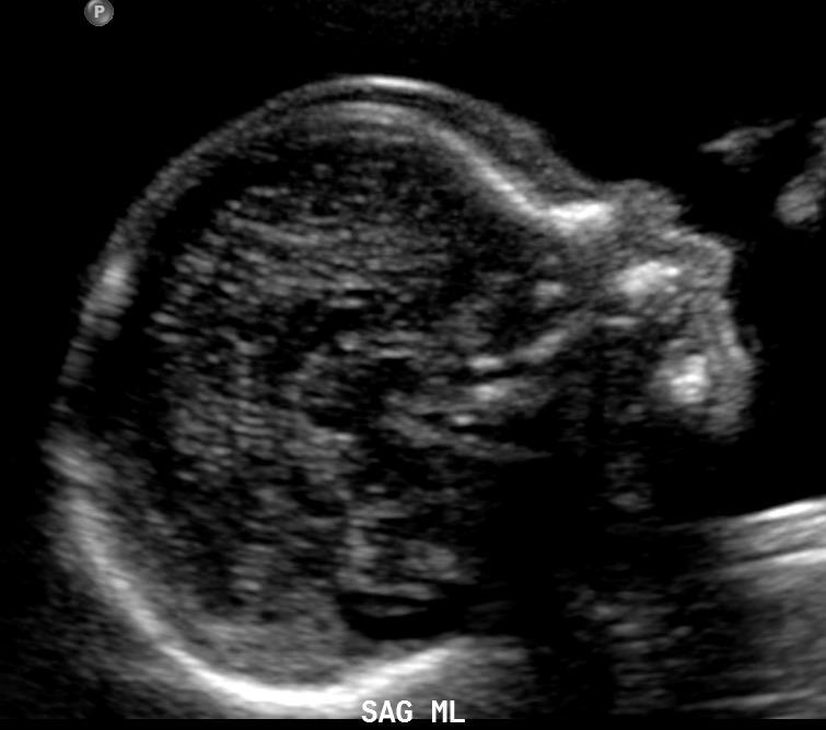
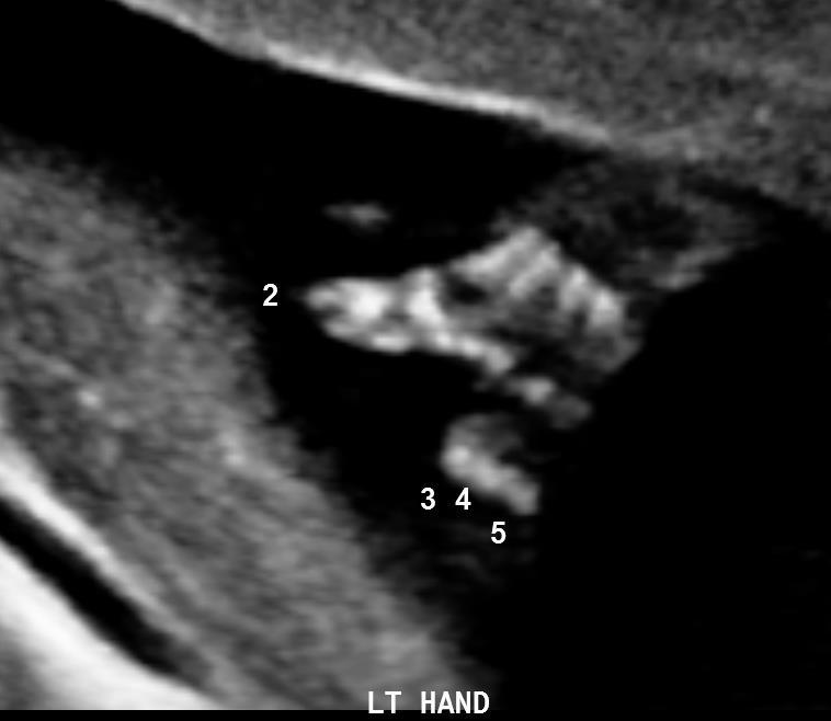
7 DETAILED ANATOMY SCAN ABDOMEN SPINE EXTREMITIES GENITALIA STRUCTURES Liver-Location, Size &Texture Gallbladder Spleen Small & Large Bowel Renal Vessels Adrenal Glands Perineum, Rectum & Anus Integrity, Alignment, Soft Tissues Spinal Cord & Conus Medullaris Number, Architecture & Position Hands & Feet Digits-Number, Position, Motion Gender Endometrium & Uterus Penile Shaft, Scrotum & Testes
8 COMMON CHOP CLINICAL SCENARIOS Patients with known or suspected CHD to assess for other findings Patients ref for possible or suspected CHD with normal CHOP Echo and detailed scan with other findings unrelated to the cardiovascular system Patients ref for other diagnoses but found to have CHD on detailed scan and CHOP Echo Patients with suspected complications of multiple gestations for cardiovascular analysis and treatment such as laser procedures, RFA, etc. S L I D E : 8
9 DIAGNOSIS COMMON CHD ABBREVIATIONS ABBREVIATION Ventricular Septal Defect VSD Truncus Arteriosus TA Transposition of Great Arteries TGA Tetralogy of Fallot TOF Single Ventricle SV Pulmonic Stenosis/Atresia PS/PA Patent Ductus Arteriosus PDA Interrupted Aortic Arch IAA Hypoplastic Left Heart Syndrome HLHS Double Outlet Right Ventricle DORV Coarctation of the Aorta CoA Atrioventricular Septal Defect AVSD Atrioventricular Canal AVC Atrial Septal Defect ASD
10 THE BALTIMORE WASHINGTON INFANT STUDY DIAGNOSIS CHILDREN with CHD (%) Ventricular Septal Defect 26 Tetralogy of Fallot 9 Atrioventricular Septal Defect 9 Atrial Septal Defect 8 Pulmonary Valvular Stenosis 7 Coarctation of the Aorta 7 Hypoplastic Left Heart Syndrome 6 D-Transposition of the Great Arteries 5 Other 23 S L I D E : 10
11 DISORDERS ASSOCIATED WITH CHD Cardiosplenic Heterotaxy Syndromes Chromosomal Abnormalities Acronym Syndromes Disorders with Unique CV Pathology Other Syndromes and Disorders
12 CARDIOSPLENIC HETEROTAXY SYNDROMES LA Isomerism Polysplenia ML, R or L Liver Absent, Small or ML GB RA Isomerism Asplenia Central Liver ML GB Bilobed Lungs Malrotated Bowel Biliary Atresia Trilobed Lungs Malrotated Bowel Microgastria


13 TOO MUCH LEFT SIDEDNESS Polysplenia Left IVC


14 TOO MUCH RIGHT SIDEDNESS Duplicated SVC
15 CARDIOSPLENIC HETEROTAXY SYNDROMES Left Atrial Isomerism Right Atrial Isomerism Dextrocardia 30-40% Dextrocardia 30-40% Bilateral SVC 40% Bilateral SVC 50-70% PAPVR (partial) 20-40% TAPVR 50-70% Common Atrium ASD 80% ASD 90% Atrioventricular Canal 20-40% AVC 85% Single Ventricle 10% SV >50% Conotruncal Defects 15-30% Conotruncal Defects 80% Interrupted IVC >70% LVOT Obstruction 40%


16 CHD TYPE CAN BE A TIP! Dextrocardia, DORV, AVSD ML Liver and Left GB 32 yo ref at 30 wks for cx CHD
17 SPECIFIC CHROMOSOMAL ABNORMALITIES Chromosome Disorder CHD T21 AVSD, VSD, TOF, etc T18 VSD, TOF, DORV, AVSD, etc T13 HLHS, DORV, AVSD, etc XO Turner s CoA, HLHS, etc 22q11 DiGeorge TA, TOF, IAA, etc Triploidy VSD, ASD, TA, etc


18 UNBALANCED AV CANAL DEFECT (CAVC/AVSD) Tethered Cord 37 yo ref at 24 wks for CHD with no other T21 markers Polyhydramnios
19 TRISOMY 21 MAJOR MARKERS Atrioventricular Canal (AVC or AVSD) Duodenal Atresia Esophageal Atresia Ventricular Septal Defect (VSD)
20 T21 MINOR MARKERS Marker LR for T21 (Isolated Sign) General Pop Incidence Incidence in T21 Thick NF % 40-75% Short Femur % 24% Short Humerus % 24% Echogenic Bowel % 15% Mild RP % 18% IEF/EICF % ethnic 20% Abnormal NB % ethnic 10-60% Mild VM 0.15% 1.5%


21 Thickened Nuchal Fold Mild UTD Clinodactyly


22 EICF AND SMALL VSD FL within normal range T21 NB within normal range

23 COMMON FEATURES OF T21
24 ULTRASOUND FEATURES OF T18 CPC (50%) is the hallmark!! Cardiac Defects (90%) Clenched Hands (50%) IUGR (50%) often early onset wks Single Umbilical Artery (50%) Brain Anomalies (30%) 1) Posterior Fossa Anomalies 2) ACC, Ventriculomegaly Facial Anomalies (20%) GI Anomalies (20%) Spina Bifida (12%)


25 LARGE BILATERAL CPC S VSD T18 2VC


26 MULTIPLE CNS FINDINGS Large CSP Microtia T18 Partial ACC Clenched Hands

27 COMMON FEATURES OF TRISOMY 18


28 CB Hypoplasia T18 Long Philtrum


29 T18 Macrodactyly Sacral Vertebrae Long Bones
30 ULTRASOUND FEATURES OF T13 CNS Anomalies (70%) 1) Holoprosencephaly is the Hallmark Cardiac Defects (80%) Postaxial Polydactyly (75%) Facial Anomalies (50%) 1) The face predicts the brain!! Renal Anomalies (50%) IUGR (50%)


31 SEVERE SEMILOBAR HSP Fused Thalami Micropthalmia


32 Ectrodactyly T13 Rockerbottom Foot

33 COMMON FEATURES OF T13


34 FACE PREDICTS THE BRAIN Partial ACC Hypoplastic CB Vermian Hypoplasia


35 GIANT OMPHALOCELE & TRUNCUS ARTERIOSUS Pulsative UV Reversed Flow UA
36 DIGEORGE SYNDROME MICRODELETION 22Q11.2 CATCH 22 Cardiac Anomalies (74%) 1) Conotruncal malformations most common Abnormal Characteristic Facies Thymic Hypoplasia/Aplasia Cleft Palate (69%) Hypocalcemia (50%)


37 DIGEORGE SYNDROME Hypolastic LVOT Large VSD


38 DIGEORGE SYNDROME FL within FL within

39 DISORDERS WITH UNIQUE CV PATHOLOGY Tuberous Sclerosis Pentalogy of Cantrell Scimitar Syndrome S L I D E : 39


40 TUBEROUS SCLEROSIS


41 TUBEROUS SCLEROSIS Rhabdomyomas Subependymal Nodules


42 JUST GIANT OMPHALOCELE? Where s the Pericardium?


43 PENTALOGY OF CANTRELL Tethered Cord Loose Cord Knot Velamentous PCI


44 SCIMITAR SYNDROME Large Anomalous Pulmonary Vein


45 SCIMITAR SYNDROME THC 4 weeks Right Lung<< Left Lung
46 ACRONYM SYNDROMES CHARGE VACTERL PHACES S L I D E : 46
47 CHARGE C=Colobomas H=Heart Malformations A=Choanal Atresia R=Growth Delay IUGR G=Abnormal Genitalia E=Eye Anomalies S L I D E : 47


48 CHARGE SYNDROME Microtia Coloboma


49 CHARGE SYNDROME Undescended Testes Adducted Flexed Thumbs
50 VACTERL ASSOCIATION V=Vertebral Anomalies A=Anal Atresia C=Cardiac Anomalies T=Tracheoesophageal Fistula E=Eophageal Atresia R=Renal Abnormalities L=Limb Defects S L I D E : 50


51 VACTERL Segmentation Anomalies Plantar Flexed 5 th Digit


52 VACTERL Decreased Renal Length & Volume
53 PHACES P=Posterior Fossa Anomalies H=Hemangiomas A=Arterial Abnormalities C=Cardiac Defects E=Eye Anomalies S=Sternal Defects S L I D E : 53


54 PHACES SYNDROME IVH TOF


55 PHACES SYNDROME Coloboma Microtia
56 OTHER SYNDROMES & DISORDERS Carpenter Syndrome Cornelia de Lange Diabetic Embryopathy Fryn s Syndrome Holt-Oram Syndrome Meckel Gruber Smith Lemli Opitz Valproate Embryopathy


57 DIABETIC EMBRYOPATHY AVSD UNBALANCED TO RT


58 Malaligned Iliac Bones & EB Segmentation Anomalies


59 Abnormal Ribs & Toes Talipes & RRA
60 CONCLUSIONS A detailed evaluation of the fetal thorax differs from the routine 4CH and outflow tract views, including scans of the aortic & ductal arches, 3 vessel & trachea view, superior & inferior vena cava, lungs, thymus, esophagus, diaphragm integrity and ribs. Congenital heart disease is associated with many conditions including heterotaxy, aneuploidy and various syndromes which have structural anomalies that can be accurately diagnosed with high resolution 2d/3d scans. Prenatal evaluation at a tertiary center such as CHOP can optimize diagnosis, parental counseling and the overall management of patients. S L I D E : 6 0
Heart and Soul Evaluation of the Fetal Heart
 Heart and Soul Evaluation of the Fetal Heart Ivana M. Vettraino, M.D., M.B.A. Clinical Associate Professor, Michigan State University College of Human Medicine Objectives Review the embryology of the formation
Ultrasound Anomaly Details
 Appendix 2. Association of Copy Number Variants With Specific Ultrasonographically Detected Fetal Anomalies Ultrasound Anomaly Details Abdominal wall Bladder exstrophy Body-stalk anomaly Cloacal exstrophy
Basic Training. ISUOG Basic Training The 20 Planes Approach to the Routine Mid Trimester Scan
 ISUOG The 20 Planes Approach to the Routine Mid Trimester Scan Learning objective At the end of the lecture you will be able to: Explain how to perform a structured routine examination, including measurements,
Basic Training. ISUOG Basic Training Examining the Upper Lip, Face & Profile
 ISUOG Examining the Upper Lip, Face & Profile Learning objectives At the end of the lecture you will be able to: Describe how to obtain the 3 planes required to assess the anatomy of the fetal face Recognise
Supplemental Information
 ARTICLE Supplemental Information SUPPLEMENTAL TABLE 6 Mosaic and Partial Trisomies Thirty-eight VLBW infants were identified with T13, of whom 2 had mosaic T13. T18 was reported for 128 infants, of whom
ISUOG Basic Training. Assessing the Neck & Chest Gihad Chalouhi, Lebanon
 ISUOG Basic Training Assessing the Neck & Chest Gihad Chalouhi, Lebanon Learning objectives 9 & 10 At the end of the lecture you will be able to: recognise the differences between the normal & most common
Heart and Lungs. LUNG Coronal section demonstrates relationship of pulmonary parenchyma to heart and chest wall.
 Heart and Lungs Normal Sonographic Anatomy THORAX Axial and coronal sections demonstrate integrity of thorax, fetal breathing movements, and overall size and shape. LUNG Coronal section demonstrates relationship
ULTRASOUND OF THE FETAL HEART
 ULTRASOUND OF THE FETAL HEART Cameron A. Manbeian, MD Disclosure Statement Today s faculty: Cameron Manbeian, MD does not have any relevant financial relationships with commercial interests or affiliations
MEDICAL MANAGEMENT WITH CAVEATS 1. In one study of 50 CHARGE patients with CHD, 75% required surgery. 2. Children with CHARGE may be resistant to chlo
 CARDIOLOGY IN CHARGE SYNDROME: FOR THE PHYSICIAN Angela E. Lin, M.D. Teratology Program/Active Malformation Surveillance, Brigham and Women's Hospital, Old PBBH-B501, 75 Francis St., Boston, MA 02115 alin@partners.org
Before we are Born: Fetal Diagnosis of Congenital Heart Disease
 Before we are Born: Fetal Diagnosis of Congenital Heart Disease Mohamed Sulaiman, MD Pediatric cardiologist Kidsheart: American Fetal & Children's Heart Center Dubai Healthcare City, Dubai-UAE First Pediatric
Screening for Critical Congenital Heart Disease
 Screening for Critical Congenital Heart Disease Caroline K. Lee, MD Pediatric Cardiology Disclosures I have no relevant financial relationships or conflicts of interest 1 Most Common Birth Defect Most
3/14/2011 MANAGEMENT OF NEWBORNS CARDIAC INTENSIVE CARE CONFERENCE FOR HEALTH PROFESSIONALS IRVINE, CA. MARCH 7, 2011 WITH HEART DEFECTS
 CONFERENCE FOR HEALTH PROFESSIONALS IRVINE, CA. MARCH 7, 2011 MANAGEMENT OF NEWBORNS WITH HEART DEFECTS A NTHONY C. CHANG, MD, MBA, MPH M E D I C AL D I RE C T OR, HEART I N S T I T U T E C H I LDRE N
Making Sense of Cardiac Views and Imaging Characteristics for 13 Congenital Heart Defects (CHDs)
") Making Sense of Cardiac Views and Imaging Characteristics for 13 Congenital Heart Defects (CHDs) Manny Gaziano, MD, FACOG obimages.net obimages.net@gmail.com Acknowledgements: Krista Wald, RDMS, sonographer,
Making Sense of Cardiac Views and Imaging Characteristics for 13 Congenital Heart Defects (CHDs) Manny Gaziano, MD, FACOG obimages.net obimages.net@gmail.com Acknowledgements: Krista Wald, RDMS, sonographer,
Cardiopulmonary Syndromes: Conditions With Concomitant Cardiac and Pulmonary Abnormalities
 Cardiopulmonary Syndromes: Conditions With Concomitant Cardiac and Pulmonary Abnormalities Carlos S. Restrepo M.D. Professor of Radiology The University of Texas HSC at San Antonio Cardiopulmonary Syndromes
Congenital Heart Defects
 Normal Heart Congenital Heart Defects 1. Patent Ductus Arteriosus The ductus arteriosus connects the main pulmonary artery to the aorta. In utero, it allows the blood leaving the right ventricle to bypass
September 28-30, 2018
 September 28-30, 2018 Course Director Optimizing Detection of Congenital Heart Disease: Important Anatomic Cardiac Regions The Top 5 Critical Anatomic Regions in Fetal Cardiac Imaging Alfred Abuhamad,
ISUOG Basic Training Distinguishing Between Normal and Abnormal Appearances of the Fetal Anatomy
 ISUOG Basic Training Distinguishing Between Normal and Abnormal Appearances of the Fetal Anatomy Reem S. Abu-Rustum, Lebanon Learning Objective At the end of the lecture you will be able to: Compare the
ISUOG Basic Training Distinguishing Between Normal and Abnormal Appearances of the Fetal Anatomy. Basic Training
 ISUOG Distinguishing Between Normal and Abnormal Appearances of the Fetal Anatomy Learning Objective At the end of the lecture you will be able to: Compare the differences between the ultrasound appearances
Outflow Tracts Anomalies
 Diagnosis of Outflow Tract Anomalies in the Fetus General Framing D.Paladini Fetal Medicine & Surgery Unit Gasllini Children s Hospital - Genoa dariopaladini@ospedale-gaslini.ge.it Outflow Tracts Anomalies
Systematic approach to Fetal Echocardiography. Objectives. Introduction 11/2/2015
 Systematic approach to Fetal Echocardiography. Pediatric Echocardiography Conference, JCMCH November 7, 2015 Rajani Anand Objectives Fetal cardiology pre-test Introduction Embryology and Physiology of
COMPREHENSIVE EVALUATION OF FETAL HEART R. GOWDAMARAJAN MD
 COMPREHENSIVE EVALUATION OF FETAL HEART R. GOWDAMARAJAN MD Disclosure No Relevant Financial Relationships with Commercial Interests Fetal Echo: How to do it? Timing of Study -optimally between 22-24 weeks
List by Region - Visceral Anomalies
 1 List by Region - Visceral Anomalies General Terms 10127 Situs inversus 80,00 10125 Aneurysm 68,42 10126Fluid-filled abdomen -35,00 Brain 10131 Hydrocephaly 10128 Dilated cerebral ventricle 20,00 10132
Pediatric Echocardiography Examination Content Outline
 Pediatric Echocardiography Examination Content Outline (Outline Summary) # Domain Subdomain Percentage 1 Anatomy and Physiology Normal Anatomy and Physiology 10% 2 Abnormal Pathology and Pathophysiology
Cardiac Catheterization Cases Primary Cardiac Diagnoses Facility 12 month period from to PRIMARY DIAGNOSES (one per patient)
") PRIMARY DIAGNOSES (one per patient) Septal Defects ASD (Atrial Septal Defect) PFO (Patent Foramen Ovale) ASD, Secundum ASD, Sinus venosus ASD, Coronary sinus ASD, Common atrium (single atrium) VSD (Ventricular
PRIMARY DIAGNOSES (one per patient) Septal Defects ASD (Atrial Septal Defect) PFO (Patent Foramen Ovale) ASD, Secundum ASD, Sinus venosus ASD, Coronary sinus ASD, Common atrium (single atrium) VSD (Ventricular
Why did this happen? Genetic and teratologic considerations in CHD Jennifer Kussmann, MS,CGC. The Children's Mercy Hospital, 2014.
 Why did this happen? Genetic and teratologic considerations in CHD Jennifer Kussmann, MS,CGC The Children's Mercy Hospital, 2014. 03/14 Risk factors Family history Maternal medical condition/ illness Monochorionic
List by Terms Visceral anomalies
 1 List by Terms Visceral anomalies Dilated 10128 Dilated cerebral ventricle 11 7 2 0 20,00 10201 Dilated aorta 9 8 2 1 5,26 10207 Dilated aortic arch 9 8 3 0 5,00 10213 Dilated carotid 3 12 4 1-47,37 10218
Most common fetal cardiac anomalies
 Most common fetal cardiac anomalies Common congenital heart defects CHD % of cardiac defects Chromosomal Infants Fetuses anomaly (%) 22q11 deletion (%) VSD 30 5~10 20~40 10 PS 9 5 (PA w/ VSD) HLHS 7~9
Accuracy of prenatal diagnosis of fetal congenital heart disease by different
 Accuracy of prenatal diagnosis of fetal congenital heart disease by different methods with echocardiography Ying Zhang 1* * Corresponding author Email: baogoubei@hotmail.com Ai-Lu Cai 1 Email: caial_us@hotmail.com
ISUOG Basic Training Distinguishing between Normal & Abnormal Appearances of the Long Bones & Extremities. Basic Training
 ISUOG Basic Training Distinguishing between Normal & Abnormal Appearances of the Long Bones & Extremities Basic Training Learning objectives At the end of the lecture you will be able to: Describe how
Data Collected: June 17, Reported: June 30, Survey Dates 05/24/ /07/2010
 Job Task Analysis for ARDMS Pediatric Echocardiography Data Collected: June 17, 2010 Reported: Analysis Summary For: Pediatric Echocardiography Exam Survey Dates 05/24/2010-06/07/2010 Invited Respondents
Chapter 2 Cardiac Interpretation of Pediatric Chest X-Ray
 Chapter 2 Cardiac Interpretation of Pediatric Chest X-Ray Ra-id Abdulla and Douglas M. Luxenberg Key Facts The cardiac silhouette occupies 50 55% of the chest width on an anterior posterior chest X-ray
ISUOG Basic Training. Obtaining & Interpreting Heart Views Correctly Alfred Abuhamad, USA. Basic training. Editable text here
 ISUOG Basic Training Obtaining & Interpreting Heart Views Correctly Alfred Abuhamad, USA Learning Objectives 6, 7 & 8 At the end of the lecture you will be able to: describe how to assess cardiac situs
9/8/2009 < 1 1,2 3,4 5,6 7,8 9,10 11,12 13,14 15,16 17,18 > 18. Tetralogy of Fallot. Complex Congenital Heart Disease.
 Current Indications for Pediatric CTA S Bruce Greenberg Professor of Radiology Arkansas Children s Hospital University of Arkansas for Medical Sciences greenbergsbruce@uams.edu 45 40 35 30 25 20 15 10
NASCI 2012 Segmental Analysis
 NASCI 2012 Segmental Analysis Frandics Chan, M.D., Ph.D. Stanford University Medical Center Lucile Packard Department Children s of Radiology Hospital Menagerie of Congenital Cardiac Lesions 1. Absent
The Fetal Cardiology Program
 The Fetal Cardiology Program at Texas Children s Fetal Center About the program Since the 1980s, Texas Children s Fetal Cardiology Program has provided comprehensive fetal cardiac care to expecting families
Congenital Anomalies
 Congenital Anomalies Down Syndrome 7580 7580 DOWN''S SYNDROME Q900 Q90.0 : Trisomy 21, meiotic nondisjunction 7580 7580 DOWN''S SYNDROME Q901 Q90.1 : Trisomy 21, mosaicism (mitotic nondisjunction) 7580
Basic Fetal Cardiac Evaluation
 Basic Fetal Cardiac Evaluation Mert Ozan Bahtiyar, MD Director, Fetal Care Center Division of Maternal Fetal Medicine Department of Obstetrics, Gynecology and Reproductive Sciences S L I D E 1 Background
Slide 1. Slide 2. Slide 3 CONGENITAL HEART DISEASE. Papworth Hospital NHS Trust INTRODUCTION. Jakub Kadlec/Catherine Sudarshan INTRODUCTION
 Slide 1 CONGENITAL HEART DISEASE Jakub Kadlec/Catherine Sudarshan NHS Trust Slide 2 INTRODUCTION Most common congenital illness in the newborn Affects about 4 9 / 1000 full-term live births in the UK 1.5
Disclosures. Outline. Learning Objectives. Introduction. Introduction. Sonographic Screening Examination of the Fetal Heart
 Sonographic Screening Examination of the Fetal Heart Lami Yeo, MD Director of Fetal Cardiology Perinatology Research Branch of NICHD / NIH / DHHS Bethesda, MD and Detroit, Michigan, USA Professor, Division
ECHOCARDIOGRAPHIC APPROACH TO CONGENITAL HEART DISEASE: THE UNOPERATED ADULT
 ECHOCARDIOGRAPHIC APPROACH TO CONGENITAL HEART DISEASE: THE UNOPERATED ADULT Karen Stout, MD, FACC Divisions of Cardiology University of Washington Medical Center Seattle Children s Hospital NO DISCLOSURES
FETAL ICD-10 CODES QUICK REFERENCE GUIDE
 FETAL ICD-10 CODES QUICK REFERENCE GUIDE Page CONTENTS 1 Cardiac Anomalies 3 Chromosome Abnormalities 4 Central Nervous System Anomalies 5 Extremity Anomalies 6 Face / Neck Anomalies 7 Gastrointestinal
Auswertung visceraler Anomalien:
 Auswertung visceraler Anomalien: General 10125 Aneurysm 15 2 2 1 20 68,42 10126 Fluid-filled abdomen 4 11 5 0 20-35,00 10127 Situs inversus 18 2 0 0 20 80,00 Brain 10128 Dilated cerebral ventricle 11 7
Diagnosis of Congenital Cardiac Defects Between 11 and 14 Weeks Gestation in High-Risk Patients
 Article Diagnosis of Congenital Cardiac Defects Between 11 and 14 Weeks Gestation in High-Risk Patients Zeev Weiner, MD, Abraham Lorber, MD, Eliezer Shalev, MD Objective. To examine the feasibility of
"Lecture Index. 1) Heart Progenitors. 2) Cardiac Tube Formation. 3) Valvulogenesis and Chamber Formation. 4) Epicardium Development.
 Heart Progenitors. 2) Cardiac Tube Formation. 3) Valvulogenesis and Chamber Formation. 4) Epicardium Development.") "Lecture Index 1) Heart Progenitors. 2) Cardiac Tube Formation. 3) Valvulogenesis and Chamber Formation. 4) Epicardium Development. 5) Septation and Maturation. 6) Changes in Blood Flow during Development.
"Lecture Index 1) Heart Progenitors. 2) Cardiac Tube Formation. 3) Valvulogenesis and Chamber Formation. 4) Epicardium Development. 5) Septation and Maturation. 6) Changes in Blood Flow during Development.
UPDATE FETAL ECHO REVIEW
 UPDATE 1 FETAL ECHO REVIEW Study Alert for RDCS Candidates D A V I E S P U B L I S H I N G I N C. Fetal Echo Review Study Alert U P D A T E D A U G U S T 1, 2 0 1 2 Nikki Stahl, RT(R)(M)(CT), RDMS, RVT
Fetal Tetralogy of Fallot
 36 Fetal Tetralogy of Fallot E.D. Bespalova, R.M. Gasanova, O.A.Pitirimova National Scientific and Practical Center of Cardiovascular Surgery, Moscow Elena D. Bespalova, MD Professor, Director Rena M,
Appendix A.1: Tier 1 Surgical Procedure Terms and Definitions
 Appendix A.1: Tier 1 Surgical Procedure Terms and Definitions Tier 1 surgeries AV Canal Atrioventricular Septal Repair, Complete Repair of complete AV canal (AVSD) using one- or two-patch or other technique,
All You Need to Know About Situs and Looping Disorders: Embryology, Anatomy, and Echocardiography
 All You Need to Know About Situs and Looping Disorders: Embryology, Anatomy, and Echocardiography Helena Gardiner Co-Director of Fetal Cardiology, The Fetal Center, University of Texas at Houston Situs
Anatomy. Contents Brain (Questions)
") Anatomy 12 Contents 12.1 Brain (Questions).................................................... 683 12.2 Head and Neck (Questions)............................................. 685 12.3 Thorax (Questions)...................................................
Anatomy 12 Contents 12.1 Brain (Questions).................................................... 683 12.2 Head and Neck (Questions)............................................. 685 12.3 Thorax (Questions)...................................................
5/22/2013. Alan Zuckerman 1, Swapna Abhyankar 1, Tiffany Colarusso 2, Richard Olney 2, Kristin Burns 3, Marci Sontag 4
 Alan Zuckerman 1, Swapna Abhyankar 1, Tiffany Colarusso 2, Richard Olney 2, Kristin Burns 3, Marci Sontag 4 1 National Library of Medicine, NIH, Bethesda, MD, USA, 2 Centers for Disease Control and Prevention,
Congenital Heart Disease An Approach for Simple and Complex Anomalies
 Congenital Heart Disease An Approach for Simple and Complex Anomalies Michael D. Pettersen, MD Director, Echocardiography Rocky Mountain Hospital for Children Denver, CO None Disclosures 1 ASCeXAM Contains
Bits and Bobs secondary causes of heart problems. Dr Angela McBrien 9 th September 2017
 Bits and Bobs secondary causes of heart problems Dr Angela McBrien 9 th September 2017 Not the heart Dextroposition Heart in the right chest with the apex to the left Often caused by left sided chest mass
Bench to Bassinet Pediatric Cardiac Genomics Consortium: CHD GENES Form 105: Congenital Extracardiac Findings Version: B - 11/01/2010
 Bench to Bassinet Pediatric Cardiac Genomics Consortium: CHD GENES Form 105: Congenital Extracardiac Findings Version: B - 11/01/2010 SECTION A: ADMINISTRATIVE INFORMATION A1. Study Identification Number:
Segmental approach to normal and abnormal situs arrangement - Echocardiography -
 Segmental approach to normal and abnormal situs arrangement - Echocardiography - Jan Marek Great Ormond Street Hospital & Institute of Cardiovascular Sciences, University College London No disclosures
Anomalous Systemic Venous Connection Systemic venous anomaly
 World Database for Pediatric and Congenital Heart Surgery Appendix B: Diagnosis (International Paediatric and Congenital Cardiac Codes (IPCCC) and definitions) Anomalous Systemic Venous Connection Systemic
Dear Parent/Guardian,
 Dear Parent/Guardian, You have indicated on school records that your child has an ongoing health problem that may require medication and/or treatment during the school day with rescue medication. Attached
From Head to Toe Use of Advanced Dynamic Flow in prenatal ultrasound
 From Head to Toe Use of Advanced Dynamic Flow in prenatal ultrasound Without doubt, the B- Schwerdtfeger, R. tant diagnostic instrument. Furthermore, we use colour in feto- mode imaging is the most important
Common Defects With Expected Adult Survival:
 Common Defects With Expected Adult Survival: Bicuspid aortic valve :Acyanotic Mitral valve prolapse Coarctation of aorta Pulmonary valve stenosis Atrial septal defect Patent ductus arteriosus (V.S.D.)
Summary. HVRA s Cardio Vascular Genetic Detailed L2 Obstetrical Ultrasound. CPT 76811, 76825, _ 90% CHD detection. _ 90% DS detection.
 What is the role of fetal echocardiography (2D 76825, cardiovascular color flow mapping 93325) as performed in conjunction with detailed fetal anatomy scan (CPT 76811) now that AIUM requires limited outflow
Children with Single Ventricle Physiology: The Possibilities
 Children with Single Ventricle Physiology: The Possibilities William I. Douglas, M.D. Pediatric Cardiovascular Surgery Children s Memorial Hermann Hospital The University of Texas Health Science Center
Heart Development and Congenital Heart Disease
 Heart Development and Congenital Heart Disease Sally Dunwoodie s.dunwoodie@victorchang.edu.au Developmental and Stem Cell Biology Division Victor Chang Cardiac Research Institute for the heart of Australia...
Fetal Pig Visual Dissection Guide
 Fetal Pig Visual Dissection Guide WARD470156-776 Orientation Cranial Anterior Sagittal plane Frontal plane Ventral Dorsal Transverse plane Caudal Posterior 1 Incisions 1 Gender Key Male Female Both 4 3
Foetal Cardiology: How to predict perinatal problems. Prof. I.Witters Prof.M.Gewillig UZ Leuven
 Foetal Cardiology: How to predict perinatal problems Prof. I.Witters Prof.M.Gewillig UZ Leuven Cardiopathies Incidence : 8-12 / 1000 births ( 1% ) Most frequent - Ventricle Septum Defect 20% - Atrium Septum
Comparing levocardia and dextrocardia in fetuses with heterotaxy syndrome: prenatal features, clinical significance and outcomes
 Wang et al. BMC Pregnancy and Childbirth (217) 17:393 DOI 1.1186/s12884-17-1579-y RESEARCH ARTICLE Open Access Comparing levocardia and dextrocardia in fetuses with heterotaxy syndrome: prenatal features,
Adult Congenital Heart Disease: What All Echocardiographers Should Know Sharon L. Roble, MD, FACC Echo Hawaii 2016
 1 Adult Congenital Heart Disease: What All Echocardiographers Should Know Sharon L. Roble, MD, FACC Echo Hawaii 2016 DISCLOSURES I have no disclosures relevant to today s talk 2 Why should all echocardiographers
Accuracy of the Fetal Echocardiogram in Double-outlet Right Ventricle
 Blackwell Publishing IncMalden, USACHDCongenital Heart Disease 2006 The Authors; Journal compilation 2006 Blackwell Publishing, Inc.? 200723237Original ArticleFetal Echocardiogram in Double-outlet Right
CNS Embryology 5th Menstrual Week (Dorsal View)
") Imaging of the Fetal Brain; Normal & Abnormal Alfred Abuhamad, M.D. Eastern Virginia Medical School CNS Embryology 5th Menstrual Week (Dorsal View) Day 20 from fertilization Neural plate formed in ectoderm
Imaging of the Fetal Brain; Normal & Abnormal Alfred Abuhamad, M.D. Eastern Virginia Medical School CNS Embryology 5th Menstrual Week (Dorsal View) Day 20 from fertilization Neural plate formed in ectoderm
List of Videos. Video 1.1
 Video 1.1 Video 1.2 Video 1.3 Video 1.4 Video 1.5 Video 1.6 Video 1.7 Video 1.8 The parasternal long-axis view of the left ventricle shows the left ventricular inflow and outflow tract. The left atrium
Research article. Primary detection of congenital heart diseases in the Kyrgyz Republic
 Research article Primary detection of congenital heart diseases in the Kyrgyz Republic Irina A. Akhmedova, Gulzada A. Imanalieva, Damir A.Abibillaev, Taalaibek Z. Kudaiberdiev Scientific Research Institute
Congenital Heart Disease Systematic Interpretation of CT Suhny Abbara, MD
 Congenital Heart Disease Systematic Interpretation of CT Suhny Abbara, MD Chief, Cardiothoracic Imaging Division Professor of Radiology UT Southwestern Medical Center, Dallas, TX Suhny.Abbara@UTSouthwestern.edu
The Brain: Prenatal and Postnatal Effects of Congenital Heart Disease. Dianna M. E. Bardo, M D Swedish Cherry Hill Radia, Inc.
 The Brain: Prenatal and Postnatal Effects of Congenital Heart Disease Dianna M. E. Bardo, M D Swedish Cherry Hill Radia, Inc. Seattle, WA embryology We recognize the VACTERL association and frequency of
Anatomy & Physiology
 1 Anatomy & Physiology Heart is divided into four chambers, two atrias & two ventricles. Atrioventricular valves (tricuspid & mitral) separate the atria from ventricles. they open & close to control flow
МОРФОЛОГИЯ ВРОЖДЕННЫХ ПОРОКОВ СЕРДЦА Бабий Леся Николаевна, Сосамма Йоханнан Сану Харьковский национальный медицинский университет Украина, Харьков
 МОРФОЛОГИЯ ВРОЖДЕННЫХ ПОРОКОВ СЕРДЦА Бабий Леся Николаевна, Сосамма Йоханнан Сану Харьковский национальный медицинский университет Украина, Харьков.MORPHOLOGY OF CONGENITAL HEART DEFECT Babiy L.,Sanu.S.Yohannan
The Chest X-ray for Cardiologists
 Mayo Clinic & British Cardiovascular Society at the Royal College of Physicians, London : 21-23-October 2013 Cases-Controversies-Updates 2013 The Chest X-ray for Cardiologists Michael Rubens Royal Brompton
DEVELOPMENT OF THE CIRCULATORY SYSTEM L E C T U R E 5
 DEVELOPMENT OF THE CIRCULATORY SYSTEM L E C T U R E 5 REVIEW OF CARDIAC ANATOMY Heart 4 chambers Base and apex Valves Pericardial sac 3 layers: epi, myo, endo cardium Major blood vessels Aorta and its
A. Incorrect! Think of a therapy that reduces prostaglandin synthesis. B. Incorrect! Think of a therapy that reduces prostaglandin synthesis.
 USMLE Step 1 - Problem Drill 02: Embryology Question No. 1 of 10 1. A premature infant is born with a patent ductus arteriosis. Which of the following treatments may be used as part of the treatment regimen?
Leitlinien. Hypoplastisches Linksherzsyndrom. Hypoplastic left heart syndrome (HLHS)
") 1.Title Hypoplastic left heart syndrome (HLHS) N.A. Haas, Bad Oeynhausen Ch. Jux, Giessen J. Photiadis, Berlin H.-H. Kramer, Kiel Typical forms: Mitral atresia/aortic atresia (MA/AoA) Mitral stenosis/aortic
1.Title Hypoplastic left heart syndrome (HLHS) N.A. Haas, Bad Oeynhausen Ch. Jux, Giessen J. Photiadis, Berlin H.-H. Kramer, Kiel Typical forms: Mitral atresia/aortic atresia (MA/AoA) Mitral stenosis/aortic
How to Recognize a Suspected Cardiac Defect in the Neonate
 Neonatal Nursing Education Brief: How to Recognize a Suspected Cardiac Defect in the Neonate https://www.seattlechildrens.org/healthcareprofessionals/education/continuing-medical-nursing-education/neonatalnursing-education-briefs/
ISUOG Basic Training Distinguishing between Normal & Abnormal Appearances of the Long Bones & Extremities
 ISUOG Distinguishing between Normal & Abnormal Appearances of the Long Bones & Extremities Learning objectives At the end of the lecture you will be able to: Describe how to obtain the planes required
CONGENITAL HEART DISEASE (CHD)
") CONGENITAL HEART DISEASE (CHD) DEFINITION It is the result of a structural or functional abnormality of the cardiovascular system at birth GENERAL FEATURES OF CHD Structural defects due to specific disturbance
CONGENITAL HEART DISEASE (CHD) DEFINITION It is the result of a structural or functional abnormality of the cardiovascular system at birth GENERAL FEATURES OF CHD Structural defects due to specific disturbance
Disclosures 5/2/17. None
 Joshua A. Copel, MD Professor, Ob-Gyn & Pediatrics Yale University School of Medicine New Haven, CT None Disclosures 1 Infant mortality, USA, 2006 # Rate* % Congenital anomalies 5,769 133.3 19.7 Premat,
5.8 Congenital Heart Disease
 5.8 Congenital Heart Disease Congenital heart diseases (CHD) refer to structural or functional heart diseases, which are present at birth. Some of these lesions may be discovered later. prevalence of Chd
Hypoplastic Left Heart Syndrome: Echocardiographic Assessment
 Hypoplastic Left Heart Syndrome: Echocardiographic Assessment Craig E Fleishman, MD, FACC, FASE Director, Non-invasive Cardiac Imaging The Hear Center at Arnold Palmer Hospital for Children, Orlando SCAI
Paediatric Cardiology. Acyanotic CHD. Prof F F Takawira
 Paediatric Cardiology Acyanotic CHD Prof F F Takawira Aetiology Chromosomal Down syndrome, T13, T18 Genetic syndromes (gene defects) Velo-Cardio-facial (22 del) Genetic syndromes (undefined aetiology)
PRACTICAL GUIDE TO FETAL ECHOCARDIOGRAPHY IC Huggon and LD Allan
 PRACTICAL GUIDE TO FETAL ECHOCARDIOGRAPHY IC Huggon and LD Allan Fetal Cardiology Unit, Harris Birthright Research Centre for Fetal Medicine, King's College Hospital, London, UK IMPORTANCE OF PRENATAL
Fetal Echocardiography and the Routine Obstetric Sonogram
 JDMS 23:143 149 May/June 2007 143 Fetal Echocardiography and the Routine Obstetric Sonogram SHELLY ZIMBELMAN, RT(R)(CT), RDMS, RDCS ASAD SHEIKH, MD, RDCS Congenital heart disease (CHD) is the most common
Segmental Analysis. Gautam K. Singh, M.D. Washington University School of Medicine St. Louis
 Segmental Analysis Gautam K. Singh, M.D. Washington University School of Medicine St. Louis Segmental Analysis Segmental Analysis: From Veins to Ventricles Segmental Approach to Evaluation of Congenital
SLOS? (Smith-Lemli-Opitz Syndrome) Dr E. P. Frohlich Sunninghill Hospital, Private Practice
 Dr E. P. Frohlich Sunninghill Hospital, Private Practice") SLOS? (Smith-Lemli-Opitz Syndrome) Dr E. P. Frohlich Sunninghill Hospital, Private Practice Baby D Introduction SLOS is part of a heterogenic group of monogenically (AR, AD, XD) determined syndrome of
SLOS? (Smith-Lemli-Opitz Syndrome) Dr E. P. Frohlich Sunninghill Hospital, Private Practice Baby D Introduction SLOS is part of a heterogenic group of monogenically (AR, AD, XD) determined syndrome of
Congenital Heart Disease
 Congenital Heart Disease Mohammed Alghamdi, MD, FRCPC, FAAP, FACC Associate Professor and Consultant Pediatric Cardiology, Cardiac Science King Fahad Cardiac Centre King Saud University INTRODUCTION CHD
Cardiovascular Pathophysiology: Right to Left Shunts aka Cyanotic Lesions
 Cardiovascular Pathophysiology: Right to Left Shunts aka Cyanotic Lesions Ismee A. Williams, MD, MS iib6@columbia.edu Pediatric Cardiology Learning Objectives To discuss the hemodynamic significance of
Cardiovascular Pathophysiology: Right to Left Shunts aka Cyanotic Lesions Ismee A. Williams, MD, MS Pediatric Cardiology
 Cardiovascular Pathophysiology: Right to Left Shunts aka Cyanotic Lesions Ismee A. Williams, MD, MS iib6@columbia.edu Pediatric Cardiology Learning Objectives To discuss the hemodynamic significance of
Multimodality Imaging of Septal Defects
 Multimodality Imaging of Septal Defects Ohio-ACC 2018 Annual Meeting October 27, 2018 Kan N. Hor, MD Director, Cardiac Magnetic Resonance Imaging Associate Professor of Pediatrics The Heart Center, Nationwide
intracranial anomalies
 Chapter 5: Fetal Central Nervous System 84 intracranial anomalies Hydrocephaly Dilatation of ventricular system secondary to an increase in the amount of CSF. Effects of hydrocephalus include flattening
By Dickens ATURWANAHO & ORIBA DAN LANGOYA MAKchs, MBchB CONGENTAL HEART DISEASE
 By Dickens ATURWANAHO & ORIBA DAN LANGOYA MAKchs, MBchB CONGENTAL HEART DISEASE Introduction CHDs are abnormalities of the heart or great vessels that are present at birth. Common type of heart disease
The Fetal Care Center at NewYork-Presbyterian/ Weill Cornell Medicine
 The Fetal Care Center at NewYork-Presbyterian/ Weill Cornell Medicine Prompt and Personalized Care for Women with Complex Pregnancies A Team of Experts additional training in maternal and fetal complications
Potential Data Sources
 Potential Data Sources Addressing Congenital Heart Defects: Occurrence, Populations and Risk Factors Centers for Disease Control and Prevention (CDC) Birth Defects Data and Statistics http://www.cdc.gov/ncbddd/birthdefects/data.html
When is Risky to Apply Oxygen for Congenital Heart Disease 부천세종병원 소아청소년과최은영
 When is Risky to Apply Oxygen for Congenital Heart Disease 부천세종병원 소아청소년과최은영 The Korean Society of Cardiology COI Disclosure Eun-Young Choi The author have no financial conflicts of interest to disclose
Congenital Heart Disease: Physiology and Common Defects
 Congenital Heart Disease: Physiology and Common Defects Jamie S. Sutherell, M.D, M.Ed. Associate Professor, Pediatrics Division of Cardiology Director, Medical Student Education in Pediatrics Director,
Surgical Procedures. Direct suture of small ASDs Patch repair Transcatheter closure with a prosthetic device called occluder
 PEDIATRIC Review Surgical Procedures Atrial Septal Defect repair: Direct suture of small ASDs Patch repair Transcatheter closure with a prosthetic device called occluder Balloon atrial septostomy (Rashkind)
ORIGINAL RESEARCH PAPER
 ORIGINAL RESEARCH PAPER ROLE OF CT PULMONARY ANGIOGRAPHY IN CONGENITAL HEART DISEASES IN PAEDIATRIC POPULATION Radiology KEY WORDS: Congenital heart disease, CT pulmonary angiography, pediatric heart disease,