A 5' Dystrophin Duplication Mutation Causes Membrane Deficiency of a-dystroglycan in a Family with X-linked Cardiomyopathy
|
|
|
- Mervin Harrison
- 5 years ago
- Views:
Transcription

1 J Mol Cell Cardiol 29, (1997) Feature Article A 5' Dystrophin Duplication Mutation Causes Membrane Deficiency of a-dystroglycan in a Family with X-linked Cardiomyopathy Roger D. Biesl, Masato ~aeda', Steven L. Roberds2, Emma ~older', Teresa ~ohlmeyer', James B. Young3 and Kevin P. Campbell2 'Division of Cardiology, VA Medical Center and University of Colorado Health Sciences Center, Denver, CO 80262; 2Howard Hughes Medical Institute and Department of Physiology and Biophysics, University of Iowa College of Medicine, Iowa City, IA 52242; and 3Heart Failure and Transplant Division, Cleveland Clinic, Cleveland, OH , USA (Received 25 July 1997, accepted in revised form 28 August 1997) R. D. BIES, M. MAEDA, S. L. ROBERDS, E. HOLDER, T. BOHLMEYER, J. B. YOUNG AND K. P. CAMPBELL. A 5' Dystrophin Duplication Mutation Causes Membrane Deficiency of a-dystroglycan in a Family with X-linked Cardiomyopathy. lourr~nl of Molecular and Cellular Cardiology (1997) 29, '-mutations in the dystrophin gene can result in cardiomyopathy without clinically-apparent skeletal myopathy. The effect of dystrophin mutations on the assembly and stability of the dystrophin associated protein (DAP) complex in human heart are not fully understood. The molecular defect in the dystrophin complex was explored in a family with an X-linked pedigree and severe dilated cardiomyopathy. Dystrophin gene analysis demonstrated a 5' duplication involving exons 2-7, which encodes the N-terminal actin binding domain of dystrophin. Ribonuclease protection and PCR assays demonstrated a reduction in muscle promoter transcribed dystrophin mrna in the heart compared to skeletal muscle. A deficiency of cardiac dystrophin protein was observed by Western blot and lack of membrane localization by immunocytochemistry. The cardiac expression of the dystrophin related protein utrophin was increased, and the 43 kda (p-dystroglycan), 50 kda (a-sarcoglycan) and 59 kda (syntrophin) dystrophin associated proteins (DAPs) were co-isolated and present in nearly normal amounts in the membrane. However, cardiac dystrophin deficiency and increased utrophin expression were associated with loss of extracellular 156 kda dystrophin associated glycoprotein (a-dystroglycan) binding to the cardiomyocyte membrane, a-dystroglycan is responsible for linkage of the dystrophin complex to the extracellular matrix protein laminin. Therefore, 5' dystrophin mutations can reduce cardiac dystrophin mrna, protein expression, and dystrophin function in X-linked cardiomyopathy (XLCM). The presence of membrane-associated p-dystroglycan, a-sarcoglycan, syntrophin, and utrophin are insufficient to maintain cardiac function. This XLCM family has a 5' dystrophin gene mutation resulting in cardiac dystrophin deficiency and a loss of a-dystroglycan membrane binding. O 1997 Academic Press Limited KEY WORDS: Dystrophin; Utrophin: a-dystroglycan; Cardiomyopathy: Muscular dystrophy. Introduction Familial cardiomyopathy and idiopathic dilated cardiomyopathy (IDC) are conditions associated with a high mortality (Goldstein and Brown, 1988), and in some families appear to have a genetic etiology (Michels et al., 1992). Duchenne and Becker muscular dystrophies (DMDIBMD) are examples where a mutation in the gene encoding the cytoskeletal protein dystrophin can cause muscular Please address all corrrespondence to: Roger D. Bies, Cardiology Division. Department of Veterans Affairs Medical Center Clermont Street, Denver, CO 80220, USA /97/ $25.00/0 mc O 1997 Academic Press Limited
.")
2 3176 R. D. Bies et al. dystrophy and severe heart failure (Perloff et al., 1966; Palmucci et al., 1992; Rees et al., 1993). Patients with BMD and cardiomyopathy have been shown to have decreased localization of dystrophin protein at the cardiomyocyte membrane (Maeda et al a). The degree of cardiac and skeletal involvement in BMD may differ, and severe cardiomyopathy may be present with either mild or clinically inapparent skeletal muscle disease. Patients in such families may be misdiagnosed with IDC. Families with heart failure and mutations in the dystrophin gene have been designated as BMD with dilated cardiomyopathy or X-linked cardiomyopathy (XLCM) (Muntoni et d., 1993: Towbin et d., 1993; Yoshida et d., 1993; Franz et d., 1995). Several reports now suggest that most patients with severe heart failure and mild or inapparent skeletal myopathy have mutations in the 5' region of the dystrophin gene. The proposed mechanisms which may explain the XLCM phenotype in these families include either mutations which cause diminished expression of cardiac dystrophin (Muntoni et al., 1995, Milasin et al., 19961, or cardiac sensitive changes in N-terminal amino acid sequence (~rtiz-~opez et al., 1997). What remains unclear, is whether some common molecular or biochemical feature could explain the similar phenotype in these different families. Are there other types of mutations which can result in XLCM? What role does the dystrophin related protein, utrophin, play in modulating disease phenotype? How is the stability and organization of the dystrophin associated protein complex affected in XLCM? Dystrophin is found at the inner membrane surface of cardiac and skeletal muscle fibers and is bound to a dystrophin-associated protein (DAP) complex. This complex forms a structural network which stabilizes the muscle membrane and links intracellular actin microfilaments to the extracellular matrix protein laminin (Ervasti and Campbell, 1991 j. Loss of dystrophin can cause destabilization and loss of the DAP complex at the cell membrane (Ohlendieck et al., 1991a, 1993). The importance of this complex has been demonstrated by the fact that mutations in either dystrophin or the dystrophin associated glycoproteins [SO kda DAG (a-sarcoglycanj, 43 kda DAG (Psarcoglycan), and 35 kda DAG (y-sarcoglycan)] can affect skeletal muscle causing muscular dystrophies (Hoffman et al ; Roberds et al ; Bonneman et al., 1995; Lim et al ; Noguchi et al., 1995). A review by Towbin (1995) suggested that a reduction in the abundance of cardiac 156 kda a-dystroglycan may occur in some cases of XLCM. Thus. the organization of the DAP complex may also affect cardiac muscle and play an important role in XLCM. The autosomal homologue of dystrophin, dystrophin related protein (DRP) or utrophin, has also been shown to associate with the DAP complex in the membrane (Matsumura et al., 1992). Data in small-caliber skeletal muscle in the mdx mouse has led to the hypothesis that utrophin may be substituted for dystrophin as an approach to therapy in DMDIBMD (Matsumura and Campbell, 1994). The organization of DAPs and utrophin may therefore influence the phenotypic presentation of dystrophin mutations in diseases like XLCM. In this report, we have analysed cardiac and skeletal muscle dystrophin, utrophin, and DAP expression and membrane affinity in a family with X-linked cardiomyopathy. Our results suggest that a mutation in the 5' region of the dystrophin gene can cause muscle-promoter-derived mrna and protein to be reduced in heart compared with skeletal muscle. This abnormality is associated with an increase in cardiac utrophin, and a loss of a-dystroglycan binding with the cardiomyocyte membrane. Materials and Methods Case descriptions The family's pedigree is shown in Figure 1. The Propositus (W.W.) was a 20-year-01d clerk suffering from dyspnea~ fatigue and weight loss. Physical examination showed bibasilar rales LW Figure 1 Pedigree of a family with X-linked (Becker) dilated cardiomyopathy. Darkened squares indicate affected males and half darkened circles indicate carrier females. The open circles and square are unaffected females and male respectively. Clinical data from subjects L.W., C.W., J.W. and W.W. are described in Materials and Methods. Arrow indicates proband who underwent heart transplantation. Diagonal slash indicates death from heart failure.


3
 which spans exons 1-9 (cdna bp 1-1538), detected the mutation, and the other probes showed no abnormality.")
4 3178 R. D. Bies et al. as previously described (Bies et al., 1992a). Dystrophin cdna probe 9.7 (ATCC, Rockville, MD, USA) which spans exons 1-9 (cdna bp ), detected the mutation, and the other probes showed no abnormality. Purified DNA was digested with Hind 111, separated by 1% agarose gel electrophoresis, and transferred to nitrocellulose membrane (Genescreen, Dupont, Boston, MA, USA). Probes used in this study were labeled with 32PdCTP (New England Nuclear, Boston, MA, USA) using a random hexamer priming kit (Pharmacia Biotech Inc., Piscataway, NJ, USA). Exon numbering of Hind I11 fragments detected on Southern blot follows the convention established by Koenig et al., (1989). Analysis of band intensity was performed using a scanning densitometer (Helena Labs, Beumont, TX, USA) on the autoradiograms. Tissue sources Fresh heart muscle was obtained at the time of cardiac transplantation and immediately frozen in liquid nitrogen-colled isopentane. Biopsy specimens from left biceps muscle were prepared in a similar fashion. Control skeletal muscle samples were obtained from gastrocnemius muscle from two diabetic subjects post-leg-amputation. Control cardiac tissues were obtained from three explanted hearts at the time of transplantation from non-dystrophic (ischemic) cardiomyopathy. All tissues were pathological specimens obtained at the time of surgery with informed consent per institutional guidelines. PCR analysis of dystrophin cdna Evaluation of dystrophin mrna transcribed from the genomic mutation (exon 2-7 duplication) was performed by reverse transcription of heart and skeletal muscle RNA into cdna with a dystrophinspecific primer, followed by nested PCR amplification. Primers flanking the duplication (in exons 1 and 10) were selected for an initial round of reverse transcription PCR. Total RNA was prepared as described by Chirgwin (1979). Five pg of RNA was incubated with 50 ng of reverse primer (bases ) in exon 10 for reverse transcription into cdna (Bies et al., 1992). The cdna product was diluted to 5 pl and PCR performed using 500 ng exon 1 forward primer (cdna sequence 36-61) and 450 ng exon 10 reverse primer (cdna sequence ) for 30 cycles. This reaction was diluted 1 to 50 and a 1 p1 aliquot was used as a template for subsequent PCR reactions. Figure 4 shows the location of primers used in PCR reactions 1-4. Reaction 1 utilized primers in exon 1 (cdna sequence ) and exon 8 (cdna sequence ), spanning the duplication, to synthesize a large 1387 base pair fragment. Reaction 2 utilized a forward primer in exon 1 (cdna sequence ) and a reverse primer in duplicated exon 4 (cdna sequence ) to synthesize both 852 bp (2a) and 235 bp (2b) fragments from each of the exon 4 annealing sites. Reaction 3 utilized a reverse primer in exon 8 (cdna sequence ) and a forward primer in duplicated exon 7 (cdna sequence ) to synthesize both 821 bp (3a) and 203 bp (3b) fragments from each of the exon 7 annealing sites. Reaction 4 utilized a forward exon 7 primer (cdna sequence ) and a reverse exon 4 primer (cdna sequence ) predicted to synthesize a 286 bp fragment from the center of the duplication. Dystrophin gene transcription is regulated in a tissue and developmentally specific manner by multiple promoterlenhancer elements in the 5' region. The majority of cardiac and skeletal muscle dystrophin expression is driven by the "muscle" promoter, though expression from "brain" promoters has been described (Feener et al., 1989; Nude1 et al., 1989; Bies et al., 1992b). To test for transcripts derived from muscle and non-muscle dystrophin promoters, PCR amplification was performed with promoter-specific exon 1 forward primers for the muscle, cortical brain, and Purkinje transcripts according to previous reports (Bies et al., 1992b; Holder et al., 1996). Ten p1 of all PCR reactions were analysed on 3% NuSieve agarose gels (FMC Bioproducts, Rockland, ME, USA) containing 0.2 mglml of ethidium bromide prior to photography. Subcloning and sequencing The PCR products were subcloned directly into the TA cloning vector kit (Invitrogen Cor., San Diego, CA, USA). Plasmid DNA containing dystrophin cdna inserts were purified (Maxiprep, Promega Cor., Madison, WI, USA) and sequenced using T7 and SP6 primers with a Sequenase version 2.0 sequencing kit (United States Biochemical Cor., Cleveland, OH, USA).
. The probe starts with the last 22 bp from the muscle promoter exon 1, and ends with the first 58 bp from exon 4.")
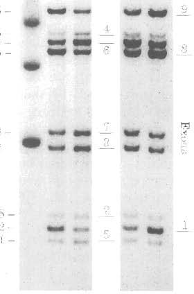
5 a-dystroglycan in X-linked Cardiomyopathy 3179 Ribonuclease protection assay A nt riboprobe spanning dystrophin exons 1-4 (cdna bases ) was selected to determine the abundance of dystrophin mrna in affected heart and skeletal muscle (Holder et al., 1996). The probe starts with the last 22 bp from the muscle promoter exon 1, and ends with the first 58 bp from exon 4. Hybridization to the duplicated dystrophin transcript should protect two equal fragments; one normal sized (235 nt) fragment corresponding to hybridization to the 5'-end of the mutant transcript containing exon 1-4 sequence, and one smaller 2 13 nt fragment of approximately equal intensity hybridizing to the downstream duplication sequence junction containing exons 2, 3 and 4. The 22 nt of sequence in the riboprobe from exon 1 should not hybridize to the exon 7 sequence which preceeds exon 2 at the central portion of the duplicated transcript. Hybridization to dystrophin transcript in control tissues should detect a 235 nt fragment. The low level transcription from the brain (Bies et al., 1992b) and Purkinje promoter (Holder et al., 1996) in control muscle tissues should also detect a small amount of the 2 13 nt (exon 2-4) fragment due to mismatch from brain exon 1 and Purkinje exon 1 (Holder et al., 1996). Ribonuclease protection assay (RPA I1 kit, Promega Corp.) of dystrophin transcripts was performed using 5 pg of total RNA as previously described (Holder et al., 1996). To assess equal RNA loading, 5 pg of total RNA from affected and control heart and skeletal muscle was examined by Northern blots using a GAPDH 32P-labeled cdna probe (ATCC, Rockville, MD, USA). RPA samples were analysed by electrophoresis in an 8% polyacrylamide gel and exposed to x-ray film for 4 days. Western blot analysis Whole tissue heart and skeletal muscle tissue homogenates (Bies et al., 1992c), and microsomal membranes from human cardiac tissue were prepared (Ohlendieck et al., 1991b). Proteins were resolved electrophorectically in 3-12% polyacrylamide gradient gels which were then electrophoretically transferred to nitrocellulose membrane (0.2 pm, Bio-Rad Laboratories Inc.. Hercules, CA, USA) (Bies et al., 1992~). The antibodies used in this study are as follows: rabbit polyclonal anti-dystrophin antibody (Bies et al., 1992c) at 1:500 dilution in Tris (10 mm; ph 8.0) buffered saline, 0.05% Tween 20 (TBST), anti-utrophin (Ohlendieck et al., 1991a) rabbit polyclonal antibody at 1: 100, anti-1 56 DAG (Ibraghimov-Beskrovnaya et al., 1992) sheep polyclonal antibody at 1:50, anti-59 DAP (Ohlendieck et al., 1991b) sheep polyclonal antibody at 1:50, anti-50 DAG (Roberds et al., 1993) sheep polyclonal antibody at 1:500, and anti-43 DAG (Ohlendieck et al., 199lb) sheep polyclonal antibody at 1:50 dilution. Immunoreactive bands were detected with secondary antibodies coupled to alkaline phosphatase (Sigma Chemical Co.) (Bies et al., 1992c) or to horseradish peroxodase using the enhanced chemiluminescent method (Amersham Corp.). Protein concentrations of tissue homogenates were determined by BCA protein assay (Pierce, Rockford, IL, USA) and 24 pg was loaded in each lane. Equal sample loading was assessed by staining the gel with Coomassie blue and insuring a consistent density of the myosin band in each lane as previously described (Maeda et al., 1996). A scanning densitometer (Bio-Rad Laboratories Inc.) was used to analyse dystrophin, utrophin, and DAP band intensity relative to controls. lmmunocytochemistry and histopathology Unfixed frozen samples of heart and skeletal muscle were cut into 6 pm cryosections and placed on Superfrost microscope slides (Fischer Scientific, Pittsburgh, PA, USA). The slides were blocked with 3% BSA in phosphate buffered saline (PBS, ph 7.4) for 20 min at room temperature and then incubated with anti-dystrophin antibody (Bies et al., 1992c) or anti-vinculin antibody (Maeda et al., 1997) for 2 h. Slides were incubated with secondary antibody for 1 h; tetramethylrhodamine isothiocyanate (TRITC) conjugated anti-rabbit IgG antibody (1:80, Sigma Chemical Co.) The slides were washed with PBS and mounted with 90% glycerol in PBS. Staining with secondary antibodies alone was used as a control for non-specific fluorescence. Stained sections were photographed under uv light with a Nikon microscope. Light microscopy of hemotoxylin and eosin stained sections were performed on 6- pm sections of 10% buffered neutral formalin fixed paraffin blocks of affected tissue. DNA analysis Southern blot analysis using dystrophin cdna probes showed a 5' gene duplication. Probe 9.7 demonstrated the presence of nine Hind I11

6


7


8


9


10


11

12 3186 R. D. Hies et al. the heart. This abnormality was associated with a loss of a-dystroglycan association with the cardiomyocyte membrane. Skeletal muscle dystrophin mrna, protein abundance, and localization to the sarcolemma were not as severely afffected. Sufficient dystrophin expression in skeletal muscle accompanied by continuous immunolocalization at the sarcolemma in all myocytes was probably capable of maintaining partial integrity of the skeletal muscle DAP complex. The concept that some el---l-la e -P d-.-t-,,h:,,,tt,,..,t, phenotype in mdx mice (Wells et al., 1995). Therefore, differences in dystrophin abundance and membrane association probably account for most of the differences in disease severity in heart v skeletal muscle. Utrophin can also bind and maintain membrane association of the DAP complex in skeletal muscle (Tinsley et al., 1996). Skeletal muscle utrophin was mildly increased in the family studied, and therefore could potentially stabilize the skeletal muscle DAP membrane complex and attenuate the development of skeletal myopathy. However, we found cardiac utrophin expression was significantly increased and co-isolated in cardiac microsomal membranes with some components of the DAP complex. The cardiac utrophinidap complex appeared unable to maintain the membrane binding affinity of a-dystroglycan, or prevent the development of cardiomyopathy. This observation could be due to possible differences in utrophin expression in cardiac and skeletal myocytes. a-dystroglycan is localized to the cardiac membrane, and is believed to link the dystrophin cytoskeletal complex to the extracellular matrix protein laminin (Ervasti and Campbell, 1991). Utrophin has been localized to the intercalated disc and T-tubules of bovine and mouse heart (Klietsch et al., 1993: Pons et al., 1994), and laminin is absent from the intercalated disc (Klietsch et al., 1993). Therefore, the interaction of DAPs with other cytoskeletal elements like utrophin at the intercalated disc may lack the appropriate structural organization or subcellular localization to maintain DAP (a-dystroglycan) binding in the heart. The recent observation that a truncated utrophin transgene is capable of maintaining skeletal muscle membrane DAPs, and reduce the dystrophic pheneotype in mdx mouse, has not been proven to be effective in cardiac tissues (Tinsley et al., 1996). There is growing evidence that XLCM is commonly associated with 5' dystrophin gene mut- ations. The 5' mutation in this study is located in the same general region of the dystrophin gene described in other XLCM reports (Muntoni et al., 1993; Milasin et al., 1995; Ortiz-Lopez et al., 1997), and had likely occurred via a cross-over event with breakpoints in intron 1 and intron 7. As in our report, the other families studied had some elevation of serum CK and subclinical muscle degeneration1 regeneration was probably present. However, the lack of severe skeletal muscle symptoms suggests th-t th- A..~t..-..h:,,,,,l,., :,,l,,l,t,l,..,,1, function in the heart has been reported as one possible mechanism (Ortiz-Lopez et al., 199 7). In two other families, complete loss of muscle promoter activity may explain the phenotype. In these cases, selective activation of the brain or Purkinje promoters appeared to provide adequate dystrophin transcription in skeletal muscle, but not in the heart (Milasin et al., 1995; Mutoni et al., 1995b). The current study expands the possible mechanisms involved in phenotypic heart and skeletal muscle disease expression. The same mutant dystrophin transcript was present in both tissues, yet dystrophin immunolocalized at the membrane in skeletal muscle, but was almost absent in heart. The mutation may have caused a cardiac-sensitive change in dystyropin function. However, part of the difference was also likely due to an alteration of dystrophin abundance in the heart. In other XLCM reports, cardiac dystrophin expression was selectively reduced (and skeletal muscle expression maintained) due to differential activity of nonmuscle promoters in the absence of normal muscle promoter activity. This mechanism was probably not the cause of low cardiac dystrophin mrna in our family. In this study, the amount of musclepromoter-derived dystrophin mrna in heart and skeletal muscle was different. Cardiac dystrophin mrna was almost undetectable, and skeletal muscle dystrophin mrna was present in near normal amounts. Differential activity of non-muscle promoters did not appear to play a major role in dystrophin expression. This observation suggests that the mutation breakpoints could have involved unidentified regulatory elements for dystrophin muscle promoter activity or dystrophin mrna stability in the heart. Therefore, several potential mechanisms have been identified which could contribute to the unusual XLCM phenotype. These include a reduction in cardiac dystrophin mrna expression or stability,

13 cc-dystroglycan in X-linked Cardiomyopathy 3187 abnormal dystrophin protein stabilitylfunction, and the potential salutary effect of increased skeletal muscle utrophin. The current literature for XLCM does not allow us to conclude that any one of these mechanisms predominate. However, when dystrophin is abnormally expressed in the heart, the final common pathway may be a loss of membrane bound a-dystroglycan and development of cardiomyopathy. Studies on other families will be required to determine whether a defect in cardiac membrane a-dystroglycan binding is a unique feature of XLCM. Acknowledgements This work was supported in part by the Temple Hoyne Buell Foundation and a grant from the VA Research Advisory Group to RDB. SLR was a Paul Cohen Fellow of the Muscular Dystrophy Association. KPC is an investigator for the Howard Hughes Medical Institute. References BIES RD, CASKEY CT, FENWICK R, 1992a. An intact cysteine-rich domain is required for dystrophin function. J Clin Invest 90: BIES RD, PHELPS S, CORTEZ M, ROBERTS R, CASKEY CT, CHAMBERLAIN JS, 1992b. Human and murine dystrophin mrna transcripts are differentially expressed during skeletal muscle, heart and brain development. Nucl Acids Res 20: BIES RD, FREIDMAN D. ROBERTS R, PERRYMAN MB. CASKEY CT, 1992c. Expression and localization of dystrophin in human cardiac Purkinje fibers. Circulation 86: BONNEMAN C, MODI R, NOGIJCHI S, MIZUNO Y, YOSHIDA M, GUSSONI E, MCNALLY E. DUGGAN D, ANGELINI C, HOFF- MAN E, OZAWA E, KUNKE L, /I-sarcoglycan (A3b) mutations cause autosomal recessive muscular dystrophy with loss of the sarcoglycan complex. Nature Genet 11: CHIRGWIN JM, PRZYBUKLA AE, MACDONALD RJ, RUTTER WJ, Isolation of biologically active ribonucleic acid from sources rich in ribonucleases. Biochemistry 13: ERVASTI JM, CAMPBELL KP, Membrane organization of the dystrophin-glycoprotein complex. Cell 66: FEENER CA, KOENIG M. KUNKELM, Alternative splicing of human dystrophin mrna generates isoforms at the carboxy terminus. Nature 338: FRANZ WM, CREMER M, HERRMANN R, GRUNIG E, FOGEL W, SCHEFFOLD R, GOEBEL H. KIRCHEISEN R, KUBLER W, VOIT T, KATUS H X-linked dilated cardiomyopathy. Ann NY Acad Sci 752: GOLDSTEIN J, BROWN MS, Genetics and cardiovascular disease. In: Braunwald E (ed.). Heart Disease. 3rd ed. Philadelphia, PA; W,B. Saunders company, GORECKI DC, MONACO AP, DERRY JMJ, WALKER AP. BAR- NARD EA, BARNARD PJ, Expression of four alternative dystrophin transcripts in brain regions regulated by different promoters. Hum Mol Genet 1: HOFFMAN E. BROWN R. KUNKEL Dystrophin: the protein product of the Duchenne muscular dystrophy locus. Cell 51: HOLDER E. MAEDA M, BIES RD, Expression and regulation of the dystrophin Purkinje promoter in human skeletal muscle, heart, and brain. Hurn Genet 97: IBRAGHIMOV-BESKROVNAYA 0, ERVASTI JM, LEVEILLE CJ, SLAUGHTER CA, SERNETT SW, CAMPBELL KP, Primary structure of dystrophin-associated glycoproteins linking dystrophin to the extracellular matrix. Nature 355: KLIETSCH R, ERVASTI JM, ARNOLD W, CAMPBELL KP, JOR- GENSEN AO, Dystrophin-glycoprotein complex and laminin colocalize to the sarcolemma and transverse tubules of cardiac muscle. Circ Res 72: KOENIG M, BEGGS AH, MOYER M, et al., The molecular basis for Duchenne verses Becker muscular dystrophy: correlation of severity with type of deletion. Am J Hum Genet 45: LIM L, DUCLOS, F, BROUX 0, BOURG N, SUNADA Y, ALLAMAND V, MEYER J, RICHARD I, MOOMAW C, SLAUGHTER C, TOME E, FARDEAU M, JACKSON C, BECKMANN J, CAMPBELL K P-sarcoglycan: characterization and role in limbgirdle muscular dystrophy. Nature Genet 11: MAEDA M, NAKAO S, MIYAZATO H, SETOGUCHI M, ARIMA S, HIGUCHI I, OSAME M, TAIRA A, NOMOTO K. TODA H, TAHARA M, ATSUCHI Y, TANAKA H, 1995a. Cardiac dystrophin abnormalities in Becker muscular dystrophy assessed by endomyocardial biopsy. Am Heart J 129: MAEDA M, TAFT C, BUSH E, HOLDER E. NEVILLE H, BAILEY W, PERRYMAN MB, BIES RD, 1995b. Identification tissue specific expression, and subcellular localization of the 71- and 80-kDa forms of myotonic dystrophy kinase protein. J Biol Chem 270: MAEDA M, HOLDER E. LOWES B, BIES RD, Dilated cardiomyopathy associated with deficiency of the cytoskeletel protein metavinculin. Circulation 95: MATSUMUR~ K, CAMPBELL KP, Dystrophinglycoprotein complex: its role in the molecular pathogenesis of muscular dystrophies. Muscle Nerve 17: MATSUMURA K, ERVASTI J, OHLENDIEK K, KAHL S, CAMPBELL K, Association of dystrophin-related protein with dystrophin-associated proteins in mdx mouse muscle. Nature 360: MICHELS V, MOLL P, MILLER F, TAJAK A, CHU J, DRISKOLL D. BURNETT J, ROCLEHEFFER RJ, TAJIK JA, BEGGS AH, KUNKELM. THIBODEAU SN, The frequency of familial dilated cardiomyopathy in a series of patients with idiopathic dilated cardiomyopathy. N Engl J Med 326: MILASIN J, FRANCESCO M, SEVERINI GM. BARTOLONI L, VATTA M, KRAJINOVIC M, MATEDDU A, ANGELINI, C, CAMERINI F, FALASCHI A, MESTRONI L, GIACCA M, A point mutation in the 5' splice site of the dystrophin gene first intron responsible for X-linked dilated cardiomyopathy. Hum Mol Genet 5: MUNTONI F, CAU M, GANAU A, CONGIU R, ARVEDI G, MATEDDU A, MARROSU M, CIANCHETTI C, REALDI G, CAO

14 3188 R. D. Bies et al. A. MELIS M Deletion of the muscle-promoter region associated with X-linked dilated cardiomyopathy. N Engl 1 Med 329: MUNTONI P, WII.SON L. MARROSIJ, MARROSIJ MG, CI- ANCHETTI C, MESTRONI K. GANAU A. DOBOWITZ V. SEWRY C. 1995a. A Mutation in the dystrophin gene selectively affecting dystrophin expression in the heart. 1 Clin Invest 96: MUNTONI F, MEI.IS MA, GANAU A. DIJBOWITZ V, 1995b. Transcription of the dystrophin gene in normal tissues and in skeletal muscle of a family with X-linked dilated cardiomyopathy. Aiil 1 Hum Genet 56: NOGUCHI S, MCNALLY E. OTHMANE K, HAGIWARA Y, MIZUNO Y, YOSHIDA M, YAMAMOTO H, BONNEMANN CG. GUSSONI E, DENTON PH, KYRIAKIDES T, MIDDLETON, HENTATI F, HAMIDA MB, NONAKA I, VANCE JM, KUNKEL LM. OZAWA E, Mutations in the dystrophin-associated protein y-sarcoglycan in chromosome 13 muscular dystrophy. Science 240: NIJDEL U, ZUK D. ZEELON E. LEVY Z. NEUMAN S, YAFFE D, Duchenne muscular dystrophy gene product is not identical in muscle and brain. Nature 337: OHLENDIECK K, ERVASTI JM, MATSUMURA K, KAHL SD, LEVEILLE CJ, CAMPBELL KP, 1991a. Dystrophin-related protein is localized to neuromuscular junctions of adult skeletal muscle. Neuron 7: OHLEND~ECK K. CAMPBELI. KP, 1991b. Dystrophin-associated proteins are greatly reduced in skeletal muscle from mdx mice. 7 Cell Biol 115: OHLENDIECK K, MATSITMURA K, IONASESCU VV, TOWBIN JA, BOSCH EP, WEINSTEIN SL, SERNETT SW. CAMPBELL KP, Duchenne muscular dystrophy: deficiency of dystrophin-associated proteins in the sarcolemma. Neurology 43: ORTIZ-LOPEZ R, LI H, SIT J. GOYTIA V, TOWBIN J, Evidence for a dystrophin missense mutation as a cause of X-linked dilated cardiomyopathy. Circulation 95: PALMUCCI L. DORIGUZZI L, MONGINI T, CHIADO-PIAT L. RESTAGANO G. CARBONARA A, PAOLILLO V, Dilating cardiomyopathy as the expression of Xp21 Becker type muscular dystrophy. 1 Neurol Sci 111: PERLOFF J, DEI.EON A. O'DOHERTY D, The cardiomyopathy of progressive muscular dystrophy. Circulation 33: PONS F, ROBERT A, FABBRIZIO E, HUGON G, CALIFANO JC, FEHRENTZ JA, MARTINEZ J, MORNET D, Utrophin localization in normal and dystrophin-deficient heart. Circulation 90: REES W, SCHULER S, HUMMEL M. HETZER, Heart transplantation in patients with muscular dystrophy associated with end-stage cardiomyopathy. 1 Heart Lung Dansplant 12: ROBERDS S, ERVASTI J. ANDERSON R, OHLENDIECK K, KAHL S. ZOLOTO D. CAMPBELL K Disruption of the dystrophin-glycoprotein complex in the cardiomyopathic hamster. 1 Biol Chem 268: ROBERDS S, LETURCQ F, ALLAMAND V, PICCOLO F. JEANPIERRE M. ANDERSON R, LIM L. LEE JC, TOME FMS. R~MERO NB, FARDEA[I M, BECKMANN JS. KAPLAN J-C, CAMPBEI,~, KP, Missense Mutations in the Adhalin Gene Linked to Autosomal Recessive Muscular Dystrophy. Cell 78: TINSLEY JM, POTTER AC. PHELP SR, FISHE R, TRICRETT JI, DAVIES KE, Amelioration of the dystrophic phenotype of mdx mice using a truncated utrophin transgene. Nature 384: TOWBIN J. HEJTMANCIK J. BRINK P, GELB B. ZHU X, CHAM- BERLAIN J, MCCABE, SWIFT M X-linked dilated cardiornyopathy: molecular genetic evidence of linkage to the Duchenne muscular dystrophy (dystrophin) gene at the Xp21 locus. Circ~ilation 87: TOWBIN JA, Biochemical and molecular characterization of X-linked dilated cardiomyopathy (XLCM). In: Clark EB, Markwald RR and Talcao A (eds). Developmental Mechanisms of Heart Disease. New York: Futura Publishing Co. Inc., WELLS DJ, WEELS KE, ASANTEA, TURNER G. SUNADA Y, CAMPBELL KP, WALSH FS. DICKSON G, Expression of full-length and minidystrophin in transgenic rildx mice: implications for gene therapy of Duchenne muscular dystrophy. Hurt1 Mol Genet 4: WINNARD AV, MENDELL JR, PRIOR TW, FLORENCE J. BURGHES AHM, Frameshift deletions of exons 3-7 and revertant fibers in Duchenne muscular dystrophy: mechanisms of dystrophin production. Am 1 Hum Genet 56: YOSHIDA K. IKEDA S, NAKAMURE A. KAGOSHIMA M, TAKEDA S. SHOJI S, YANAGISAWA N Molecular analysis of the Duchenne muscular dystrophy gene in patients with Becker muscular dystrophy presenting with dilated cardiomyopathy. Muscle Nerve 16:
Three Muscular Dystrophies: Loss of Cytoskeleton-Extracellular Matrix Linkage
 Cell, Vol. 80, 675-679, March 10, 1995, Copyright 1995 by Cell Press Three Muscular Dystrophies: Loss of Cytoskeleton-Extracellular Matrix Linkage Review Kevin P. Campbell Howard Hughes Medical Institute
Muscular Dystrophy. Biol 405 Molecular Medicine
 Muscular Dystrophy Biol 405 Molecular Medicine Duchenne muscular dystrophy Duchenne muscular dystrophy is a neuromuscular disease that occurs in ~ 1/3,500 male births. The disease causes developmental
Dystrophy Patients Lacking COOH-terminal Domains of Dystrophin
 Deficiency of Dystrophin-associated Proteins in Duchenne Muscular Dystrophy Patients Lacking COOH-terminal Domains of Dystrophin Kiichiro Matsumura, * Fernando M. S. Tome,t Victor lonasescu, James M. Ervasti,
CARDIAC muscle is commonly affected in muscular
 362 THE NEW ENGLAND JOURNAL OF MEDICINE Feb. 8, 1996 BRIEF REPORT: DEFICIENCY OF A DYSTROPHIN-ASSOCIATED GLYCOPROTEIN (ADHALIN) IN A PATIENT WITH MUSCULAR DYSTROPHY AND CARDIOMYOPATHY RICARDO FADIC, M.D.,
The New England Journal of Medicine MUTATIONS IN THE SARCOGLYCAN GENES IN PATIENTS WITH MYOPATHY
 MUTATIONS IN THE SARCOGLYCAN GENES IN PATIENTS WITH MYOPATHY DAVID J. DUGGAN, B.S., J. RAFAEL GOROSPE, M.D., PH.D., MARINA FANIN, M.S., ERIC P. HOFFMAN, PH.D., A CORRADO ANGELINI, M.D. ABSTRACT Background
DMD Genetics: complicated, complex and critical to understand
 DMD Genetics: complicated, complex and critical to understand Stanley Nelson, MD Professor of Human Genetics, Pathology and Laboratory Medicine, and Psychiatry Co Director, Center for Duchenne Muscular
Peripheral nerve dystroglycan: its function and potential role in the molecular pathogenesis of neuromuscular diseases
 Y. Fukuyama, M. Osawa and K. Saito (Eds.), Congenital Muscular Dyrfrophies O 1997 Elsevier Science B.V. All rights reserved CHAPTER 22 Peripheral nerve dystroglycan: its function and potential role in
Functional significance of dystrophin positive fibres
 632 Muscular Dystrophy Group Research Laboratories, Regional Neurosciences Centre, Newcastle General Hospital, Newcastle upon Tyne L V B Nicholson M A Johnson K M D Bushby D Gardner-Medwin Correspondence
Dystrophin-glycoprotein complex: molecular organization and critical roles in skeletal muscle
 Dystrophin-glycoprotein complex: molecular organization and critical roles in skeletal muscle Yoshihide Sunada and Kevin P. Campbell Howard Hughes Medical Institute, Department of Physiology and Biophysics,
READ ORPHA.NET WEBSITE ABOUT BETA-SARCOGLYOCANOPATHY LIMB-GIRDLE MUSCULAR DYSTROPHIES
 READ ORPHA.NET WEBSITE ABOUT BETA-SARCOGLYOCANOPATHY LIMB-GIRDLE MUSCULAR DYSTROPHIES (LGMD) Limb-girdle muscular dystrophies (LGMD) are a heterogeneous group of genetically determined disorders with a
Disruption of heart sarcoglycan complex and severe cardiomyopathy caused by β sarcoglycan mutations
 102 Department of Neuromuscular Diseases, Istituto Nazionale Neurologico C Besta, Via Celoria 11, 20133 Milano, Italy R Barresi C Di Blasi T Negri R Brugnoni S Daniel F Cornelio L Morandi M Mora Department
Familial DilatedCardiomyopathy Georgios K Efthimiadis, MD
 Familial DilatedCardiomyopathy Georgios K Efthimiadis, MD Dilated Cardiomyopathy Dilated LV/RV, reduced EF, in the absence of CAD valvulopathy pericardial disease Prevalence:40/100.000 persons Natural
The dystrophin glycoprotein complex (DGC)1 consists
1 consists") Molecular Organization of Sarcoglycan Complex in Mouse Myotubes in Culture Yiu-mo Chan,* Carsten G. Bönnemann,* Hart G.W. Lidov, and Louis M. Kunkel* *Howard Hughes Medical Institute, Division of Genetics,
Molecular Organization of Sarcoglycan Complex in Mouse Myotubes in Culture Yiu-mo Chan,* Carsten G. Bönnemann,* Hart G.W. Lidov, and Louis M. Kunkel* *Howard Hughes Medical Institute, Division of Genetics,
Dystrophin gene abnormalities in two patients with idiopathic dilated cardiomyopathy
 608 Heart 1997;78:608 612 CASE STUDY Dystrophin gene abnormalities in two patients with idiopathic dilated cardiomyopathy Francesco Muntoni, Andrea Di Lenarda, Maurizio Porcu, Gianfranco Sinagra, Anna
Understanding genetics, mutation and other details. Stanley F. Nelson, MD 6/29/18
 Understanding genetics, mutation and other details Stanley F. Nelson, MD 6/29/18 1 6 11 16 21 Duchenne muscular dystrophy 26 31 36 41 46 51 56 61 66 71 76 81 86 91 96 600 500 400 300 200 100 0 Duchenne/Becker
(A) PCR primers (arrows) designed to distinguish wild type (P1+P2), targeted (P1+P2) and excised (P1+P3)14-
 PCR primers (arrows) designed to distinguish wild type (P1+P2), targeted (P1+P2) and excised (P1+P3)14-") 1 Supplemental Figure Legends Figure S1. Mammary tumors of ErbB2 KI mice with 14-3-3σ ablation have elevated ErbB2 transcript levels and cell proliferation (A) PCR primers (arrows) designed to distinguish
1 Supplemental Figure Legends Figure S1. Mammary tumors of ErbB2 KI mice with 14-3-3σ ablation have elevated ErbB2 transcript levels and cell proliferation (A) PCR primers (arrows) designed to distinguish
BRIEF REPORT: DEFICIENCY OF A DYSTROPHIN-ASSOCIATED GLYCOPROTEIN (ADHALIN) IN A PATIENT WITH MUSCULAR DYSTROPHY AND CARDIOMYOPATHY
 IN A PATIENT WITH MUSCULAR DYSTROPHY AND CARDIOMYOPATHY") BRIEF REPORT: DEFICIENCY OF A DYSTROPHIN-ASSOCIATED GLYCOPROTEIN (ADHALIN) IN A PATIENT WITH MUSCULAR DYSTROPHY AND CARDIOMYOPATHY RICARDO FADIC, M.D., YOSHIHIDA SUNADA, PH.D., ANDREW J. WACLAWIK, M.D.,
BRIEF REPORT: DEFICIENCY OF A DYSTROPHIN-ASSOCIATED GLYCOPROTEIN (ADHALIN) IN A PATIENT WITH MUSCULAR DYSTROPHY AND CARDIOMYOPATHY RICARDO FADIC, M.D., YOSHIHIDA SUNADA, PH.D., ANDREW J. WACLAWIK, M.D.,
K. P. Loss of sarcolemma nnos in sarcoglycandeficient
 Loss of sarcolemma nnos in sarcoglycan-deficient muscle RACHELLE H. CROSBIE 1,2, RITA BARRESI 1, AND KEVIN P. CAMPBELL 1 Howard Hughes Medical Institute, Department of Physiology and Biophysics, Department
Progress in human genetics has identified a number. -Sarcoglycan Deficiency Leads to Muscle Membrane Defects and Apoptosis Independent of Dystrophin
 -Sarcoglycan Deficiency Leads to Muscle Membrane Defects and Apoptosis Independent of Dystrophin Andrew A. Hack,* Chantal T. Ly, Fang Jiang, Cynthia J. Clendenin, Kirsten S. Sigrist, Robert L. Wollmann,
Online Data Supplement. Anti-aging Gene Klotho Enhances Glucose-induced Insulin Secretion by Upregulating Plasma Membrane Retention of TRPV2
 Online Data Supplement Anti-aging Gene Klotho Enhances Glucose-induced Insulin Secretion by Upregulating Plasma Membrane Retention of TRPV2 Yi Lin and Zhongjie Sun Department of physiology, college of
Islet viability assay and Glucose Stimulated Insulin Secretion assay RT-PCR and Western Blot
 Islet viability assay and Glucose Stimulated Insulin Secretion assay Islet cell viability was determined by colorimetric (3-(4,5-dimethylthiazol-2-yl)-2,5- diphenyltetrazolium bromide assay using CellTiter
MicroRNA sponges: competitive inhibitors of small RNAs in mammalian cells
 MicroRNA sponges: competitive inhibitors of small RNAs in mammalian cells Margaret S Ebert, Joel R Neilson & Phillip A Sharp Supplementary figures and text: Supplementary Figure 1. Effect of sponges on
Exercise induced cramps and myoglobinuria in dystrophinopathy a report of three Malaysian patients
 Neurology Asia 2010; 15(2) : 125 131 Exercise induced cramps and myoglobinuria in dystrophinopathy a report of three Malaysian patients 1 Azlina Ahmad Annuar, 2 Kum Thong Wong, 1 Ai Sze Ching, 3 Meow Keong
Differentiation-induced Changes of Mediterranean Fever Gene (MEFV) Expression in HL-60 Cell
 Expression in HL-60 Cell") Differentiation-induced Changes of Mediterranean Fever Gene (MEFV) Expression in HL-60 Cell Wenxin Li Department of Biological Sciences Fordham University Abstract MEFV is a human gene that codes for an
Differentiation-induced Changes of Mediterranean Fever Gene (MEFV) Expression in HL-60 Cell Wenxin Li Department of Biological Sciences Fordham University Abstract MEFV is a human gene that codes for an
Supplementary methods:
 Supplementary methods: Primers sequences used in real-time PCR analyses: β-actin F: GACCTCTATGCCAACACAGT β-actin [11] R: AGTACTTGCGCTCAGGAGGA MMP13 F: TTCTGGTCTTCTGGCACACGCTTT MMP13 R: CCAAGCTCATGGGCAGCAACAATA
Supplementary Figure 1. AdipoR1 silencing and overexpression controls. (a) Representative blots (upper and lower panels) showing the AdipoR1 protein
 Representative blots (upper and lower panels) showing the AdipoR1 protein") Supplementary Figure 1. AdipoR1 silencing and overexpression controls. (a) Representative blots (upper and lower panels) showing the AdipoR1 protein content relative to GAPDH in two independent experiments.
Supplementary Figure 1. AdipoR1 silencing and overexpression controls. (a) Representative blots (upper and lower panels) showing the AdipoR1 protein content relative to GAPDH in two independent experiments.
SUPPLEMENTARY INFORMATION
 SUPPLEMENTARY INFORMATION FOR Liver X Receptor α mediates hepatic triglyceride accumulation through upregulation of G0/G1 Switch Gene 2 (G0S2) expression I: SUPPLEMENTARY METHODS II: SUPPLEMENTARY FIGURES
Naturally Protected Muscle Phenotypes: Development of Novel Treatment Strategies for Duchenne Muscular Dystrophy
 Naturally Protected Muscle Phenotypes: Development of Novel Treatment Strategies for Duchenne Muscular Dystrophy Paul Dowling, Philip Doran, James Lohan, Kevin Culligan and Kay Ohlendieck Department of
The Childhood Muscular Dystrophies: Diseases Sharing a Common Pathogenesis of Membrane Instability
 Carrell-Krusen Symposium Invited Lecture The Childhood Muscular Dystrophies: Diseases Sharing a Common Pathogenesis of Membrane Instability Jerry R. Mendell, MD; Zarife Sahenk, MD; Thomas W. Prior, PhD
Dystrophin analysis using a panel of anti-dystrophin antibodies in Duchenne and Becker muscular dystrophy
 26 6Journal of Neurology, Neurosurgery, and Psychiatry 1993;56:26-31 Institute of Child Neurology and Psychiatry, Cagliari, Italy F Muntoni A Mateddu C Cianchetti M Marrosu Jerry Lewis Muscle Centre, Hammersmith
MUSCLE DISEASE ANTIBODIES NOVOCASTRA ADVANCING MUSCLE DISEASE DIAGNOSIS, MANAGEMENT AND RESEARCH RESULTS YOU CAN RELY ON
 MUSCLE DISEASE ANTIBODIES ADVANCING MUSCLE DISEASE DIAGNOSIS, MANAGEMENT AND RESEARCH NOVOCASTRA RESULTS YOU CAN RELY ON Novocastra Muscle Disease Antibodies The Novocastra muscle disease portfolio comprises
Anti-Lamin B1/LMNB1 Picoband Antibody
 Anti-Lamin B1/LMNB1 Picoband Antibody Catalog Number:PB9611 About LMNB1 Lamin-B1 is a protein that in humans is encoded by the LMNB1 gene. The nuclear lamina consists of a two-dimensional matrix of proteins
18 (2), DOI: /bjmg
, DOI: /bjmg") 18 (2), 2015 71-76 DOI: 10.1515/bjmg-2015-0088 CASE REPORT SARCOLEMMAL DEFICIENCY OF SARCOGLYCAN COMPLEX IN AN 18-MONTH-OLD TURKISH BOY WITH A LARGE DELETION IN THE BETA SARCOGLYCAN GENE Diniz G 1,*, Tekgul
18 (2), 2015 71-76 DOI: 10.1515/bjmg-2015-0088 CASE REPORT SARCOLEMMAL DEFICIENCY OF SARCOGLYCAN COMPLEX IN AN 18-MONTH-OLD TURKISH BOY WITH A LARGE DELETION IN THE BETA SARCOGLYCAN GENE Diniz G 1,*, Tekgul
SALSA MLPA KIT P060-B2 SMA
 SALSA MLPA KIT P6-B2 SMA Lot 111, 511: As compared to the previous version B1 (lot 11), the 88 and 96 nt DNA Denaturation control fragments have been replaced (QDX2). Please note that, in contrast to the
Research Article: Histology and Cell Biology Preliminary study on sarcoglycan sub-complex in rat cerebral and cerebellar cortex
 IJAE Vol. 117, n. 1: 54-64, 2012 Italian Journal of Anatomy and Embryology Research Article: Histology and Cell Biology Preliminary study on sarcoglycan sub-complex in rat cerebral and cerebellar cortex
In cardiac muscle, the dystrophin glycoprotein complex includes
 Dissociation of Sarcoglycans and the Dystrophin Carboxyl Terminus From the Sarcolemma in Enteroviral Cardiomyopathy Gil-Hwan Lee,* Cornel Badorff,* Kirk U. Knowlton Abstract Enteroviral infection can cause
SMA IS A SEVERE NEUROLOGICAL DISORDER [1]
![SMA IS A SEVERE NEUROLOGICAL DISORDER [1]](/thumbs/72/67086153.jpg "SMA IS A SEVERE NEUROLOGICAL DISORDER [1]") SMA OVERVIEW SMA IS A SEVERE NEUROLOGICAL DISORDER [1] Autosomal recessive genetic inheritance 1 in 50 people (approximately 6 million Americans) are carriers [2] 1 in 6,000 to 1 in 10,000 children born
SMA OVERVIEW SMA IS A SEVERE NEUROLOGICAL DISORDER [1] Autosomal recessive genetic inheritance 1 in 50 people (approximately 6 million Americans) are carriers [2] 1 in 6,000 to 1 in 10,000 children born
Supplementary data Supplementary Figure 1 Supplementary Figure 2
 Supplementary data Supplementary Figure 1 SPHK1 sirna increases RANKL-induced osteoclastogenesis in RAW264.7 cell culture. (A) RAW264.7 cells were transfected with oligocassettes containing SPHK1 sirna
Immunohistochemical Analysis of Dystrophin-associated Proteins in Becker/Duchenne Muscular Dystrophy with Huge In-frame
 Immunohistochemical Analysis of Dystrophin-associated Proteins in Becker/Duchenne Muscular Dystrophy with Huge In-frame Deletions in the NH2-Terminal and Rod Domains of Dystrophin Kiichiro Matsumura,*
Identification and characterization of multiple splice variants of Cdc2-like kinase 4 (Clk4)
") Identification and characterization of multiple splice variants of Cdc2-like kinase 4 (Clk4) Vahagn Stepanyan Department of Biological Sciences, Fordham University Abstract: Alternative splicing is an
Identification and characterization of multiple splice variants of Cdc2-like kinase 4 (Clk4) Vahagn Stepanyan Department of Biological Sciences, Fordham University Abstract: Alternative splicing is an
Treatment of Duchenne Muscular Dystrophy with Oligonucleotides
 Treatment of Duchenne Muscular Dystrophy with Oligonucleotides against an Exonic Splicing Enhancer Sequence Masafumi Matsuo, Mariko Yagi and Yasuhiro Takeshima Department of Pediatrics, Kobe University
Journal of the American College of Cardiology Vol. 43, No. 5, by the American College of Cardiology Foundation ISSN /04/$30.
 Journal of the American College of Cardiology Vol. 43, No. 5, 2004 2004 by the American College of Cardiology Foundation ISSN 0735-1097/04/$30.00 Published by Elsevier Inc. doi:10.1016/j.jacc.2003.09.052
Duchenne muscular dystrophy quantification of muscular parameters and prednisone therapy Beenakker, Ernesto Alexander Christiaan
 University of Groningen Duchenne muscular dystrophy quantification of muscular parameters and prednisone therapy Beenakker, Ernesto Alexander Christiaan IMPORTANT NOTE: You are advised to consult the publisher's
Gene therapy and genome editing technologies for the study and potential treatment of :
 WORKSHOP ON GENOME EDITING Gene therapy and genome editing technologies for the study and potential treatment of : Duchenne Muscular Dystrophy by Dr France Piétri-Rouxel, Institut de Myologie Centre de
Expression of acid base transporters in the kidney collecting duct in Slc2a7 -/-
 Supplemental Material Results. Expression of acid base transporters in the kidney collecting duct in Slc2a7 -/- and Slc2a7 -/- mice. The expression of AE1 in the kidney was examined in Slc26a7 KO mice.
The functional role of dystrophin in the heart: implications for inherited and non-inherited heart disease. Matthew Scott Barnabei
 The functional role of dystrophin in the heart: implications for inherited and non-inherited heart disease by Matthew Scott Barnabei A dissertation submitted in partial fulfillment of the requirements
MRC-Holland MLPA. Description version 19;
 SALSA MLPA probemix P6-B2 SMA Lot B2-712, B2-312, B2-111, B2-511: As compared to the previous version B1 (lot B1-11), the 88 and 96 nt DNA Denaturation control fragments have been replaced (QDX2). SPINAL
Protection against doxorubicin-induced myocardial dysfunction in mice by cardiac-specific expression of carboxyl terminus of hsp70-interacting protein
 Protection against doxorubicin-induced myocardial dysfunction in mice by cardiac-specific expression of carboxyl terminus of hsp70-interacting protein Lei Wang 1, Tian-Peng Zhang 1, Yuan Zhang 2, Hai-Lian
Supplementary Information Titles Journal: Nature Medicine
 Supplementary Information Titles Journal: Nature Medicine Article Title: Corresponding Author: Supplementary Item & Number Supplementary Fig.1 Fig.2 Fig.3 Fig.4 Fig.5 Fig.6 Fig.7 Fig.8 Fig.9 Fig. Fig.11
CHAPTER 4 RESULTS. showed that all three replicates had similar growth trends (Figure 4.1) (p<0.05; p=0.0000)
 (p<0.05; p=0.0000)") CHAPTER 4 RESULTS 4.1 Growth Characterization of C. vulgaris 4.1.1 Optical Density Growth study of Chlorella vulgaris based on optical density at 620 nm (OD 620 ) showed that all three replicates had similar
CHAPTER 4 RESULTS 4.1 Growth Characterization of C. vulgaris 4.1.1 Optical Density Growth study of Chlorella vulgaris based on optical density at 620 nm (OD 620 ) showed that all three replicates had similar
Predicted and observed sizes of dystrophin in some patients with gene deletions that disrupt the open reading frame
 892 8 Med Genet 1992; 29: 892-896 Muscular Dystrophy Group Research Laboratories, Regional Neurosciences Centre, Newcastle General Hospital, Newcastle upon Tyne NE4 6BE. L V B Nicholson K M D Bushby M
1. Identify and characterize interesting phenomena! 2. Characterization should stimulate some questions/models! 3. Combine biochemistry and genetics
 1. Identify and characterize interesting phenomena! 2. Characterization should stimulate some questions/models! 3. Combine biochemistry and genetics to gain mechanistic insight! 4. Return to step 2, as
Supplementary Figure 1. Generation of knockin mice expressing L-selectinN138G. (a) Schematics of the Sellg allele (top), the targeting vector, the
 Schematics of the Sellg allele (top), the targeting vector, the") Supplementary Figure 1. Generation of knockin mice expressing L-selectinN138G. (a) Schematics of the Sellg allele (top), the targeting vector, the targeted allele in ES cells, and the mutant allele in
Supplementary Figure 1. Generation of knockin mice expressing L-selectinN138G. (a) Schematics of the Sellg allele (top), the targeting vector, the targeted allele in ES cells, and the mutant allele in
Aminoglycoside antibiotics restore dystrophin function to skeletal muscles of mdx mice
 Aminoglycoside antibiotics restore dystrophin function to skeletal muscles of mdx mice Elisabeth R. Barton-Davis,, Stuart E. Leland, H. Lee Sweeney J Clin Invest. 1999;104(4):375-381. https://doi.org/10.1172/jci7866.
Supplementary Appendix
 Supplementary Appendix This appendix has been provided by the authors to give readers additional information about their work. Supplement to: Choi YL, Soda M, Yamashita Y, et al. EML4-ALK mutations in
In vitro DNase I foot printing. In vitro DNase I footprinting was performed as described
 Supplemental Methods In vitro DNase I foot printing. In vitro DNase I footprinting was performed as described previously 1 2 using 32P-labeled 211 bp fragment from 3 HS1. Footprinting reaction mixes contained
Protocol for Gene Transfection & Western Blotting
 The schedule and the manual of basic techniques for cell culture Advanced Protocol for Gene Transfection & Western Blotting Schedule Day 1 26/07/2008 Transfection Day 3 28/07/2008 Cell lysis Immunoprecipitation
Construction of a hepatocellular carcinoma cell line that stably expresses stathmin with a Ser25 phosphorylation site mutation
 Construction of a hepatocellular carcinoma cell line that stably expresses stathmin with a Ser25 phosphorylation site mutation J. Du 1, Z.H. Tao 2, J. Li 2, Y.K. Liu 3 and L. Gan 2 1 Department of Chemistry,
Mutations in several components of the dystrophin glycoprotein. Animal Models for Muscular Dystrophy Show Different Patterns of Sarcolemmal Disruption
 Animal Models for Muscular Dystrophy Show Different Patterns of Sarcolemmal Disruption Volker Straub,* Jill A. Rafael, Jeffrey S. Chamberlain, and Kevin P. Campbell* Department of *Physiology and Biophysics
Cover Page. The handle holds various files of this Leiden University dissertation.
 Cover Page The handle http://hdl.handle.net/1887/29354 holds various files of this Leiden University dissertation. Author: Straathof, Chiara Title: dystrophinopathies : heterogeneous clinical aspects of
Screening of dystrophin gene deletions in Egyptian patients with DMD/BMD muscular dystrophies
 125 Screening of dystrophin gene deletions in Egyptian patients with DMD/BMD muscular dystrophies Laila K. Effat a, Ashraf A. El-Harouni a, Khalda S. Amr a, Tarik I. El-Minisi b, Nagwa Abdel Meguid a and
Supplementary Information
 Supplementary Information HBV maintains electrostatic homeostasis by modulating negative charges from phosphoserine and encapsidated nucleic acids Authors: Pei-Yi Su 1,2,3, Ching-Jen Yang 2, Tien-Hua Chu
TSH Receptor Monoclonal Antibody (49) Catalog Number MA3-218 Product data sheet
 Catalog Number MA3-218 Product data sheet") Website: thermofisher.com Customer Service (US): 1 800 955 6288 ext. 1 Technical Support (US): 1 800 955 6288 ext. 441 TSH Receptor Monoclonal Antibody (49) Catalog Number MA3-218 Product data sheet Details
Website: thermofisher.com Customer Service (US): 1 800 955 6288 ext. 1 Technical Support (US): 1 800 955 6288 ext. 441 TSH Receptor Monoclonal Antibody (49) Catalog Number MA3-218 Product data sheet Details
Distribution of Dystroglycan in Normal Adult Mouse Tissues
 Volume 46(4): 449 457, 1998 The Journal of Histochemistry & Cytochemistry http://www.jhc.org ARTICLE Distribution of Dystroglycan in Normal Adult Mouse Tissues Madeleine Durbeej, Michael D. Henry, Maria
HCC1937 is the HCC1937-pcDNA3 cell line, which was derived from a breast cancer with a mutation
 SUPPLEMENTARY INFORMATION Materials and Methods Human cell lines and culture conditions HCC1937 is the HCC1937-pcDNA3 cell line, which was derived from a breast cancer with a mutation in exon 20 of BRCA1
CURRICULUM VITAE. Phone: (614) Lane Road Department of Pathology MR5 Building, Room Charlottesville, VA 22903
 Lane Road Department of Pathology MR5 Building, Room Charlottesville, VA 22903") CURRICULUM VITAE NAME: AFFILIATION: CITIZENSHIP: HOME ADDRESS: OFFICE ADDRESS: Jordan T. Gladman Postdoctoral Research Associate The University of Virginia Charlottesville, Virginia 22908 Citizen of U.S.A.
CURRICULUM VITAE NAME: AFFILIATION: CITIZENSHIP: HOME ADDRESS: OFFICE ADDRESS: Jordan T. Gladman Postdoctoral Research Associate The University of Virginia Charlottesville, Virginia 22908 Citizen of U.S.A.
Target Protein Antibody name Product number Manufacturer Species Epitope Dilution Aggrecan Anti-aggrecan AB1031 EMD Millipore Corp Rabbit
 Family history Hypertension/ Maximum Degree of aortic Bicuspid Disease Age/Sex Diagnosed CTD of aortic disease Treated aortic insufficiency/stenosis aortic Aneurysm 63/M No Yes Yes/Yes diameter 59mm 1-2+/None
Supporting Online Material for
 www.sciencemag.org/cgi/content/full/1171320/dc1 Supporting Online Material for A Frazzled/DCC-Dependent Transcriptional Switch Regulates Midline Axon Guidance Long Yang, David S. Garbe, Greg J. Bashaw*
TITLE: The Role of hcdc4 as a Tumor Suppressor Gene in Genomic Instability Underlying Prostate Cancer
 AD Award Number: TITLE: The Role of hcdc4 as a Tumor Suppressor Gene in Genomic Instability Underlying Prostate Cancer PRINCIPAL INVESTIGATOR: Audrey van Drogen, Ph.D. CONTRACTING ORGANIZATION: Sidney
Materials and Methods , The two-hybrid principle.
 The enzymatic activity of an unknown protein which cleaves the phosphodiester bond between the tyrosine residue of a viral protein and the 5 terminus of the picornavirus RNA Introduction Every day there
Genetic diagnosis of limb girdle muscular dystrophy type 2A, A Case Report
 Genetic diagnosis of limb girdle muscular dystrophy type 2A, A Case Report Roshanak Jazayeri, MD, PhD Assistant Professor of Medical Genetics Faculty of Medicine, Alborz University of Medical Sciences
Sarcoglycan Subcomplex Expression in Normal Human Smooth Muscle
 Volume 55(8): 831 843, 2007 Journal of Histochemistry & Cytochemistry http://www.jhc.org ARTICLE Sarcoglycan Subcomplex Expression in Normal Human Smooth Muscle Giuseppe Anastasi, Giuseppina Cutroneo,
Rare Monogenic Disorders. Function. Pathophysiology
 Rare Monogenic Disorders Function Pathophysiology Protein Gene Episodic Nervous System Diseases Migraine Epilepsy Periodic Paralysis LQTS Episodic Ataxia Paroxysmal Dyskinesias Phenotypes Muscle diseases
Corporate Medical Policy
 Corporate Medical Policy Genetic Testing for Duchenne and Becker Muscular Dystrophy File Name: Origination: Last CAP Review: Next CAP Review: Last Review: genetic_testing_for_duchenne_and_becker_muscular_dystrophy
Myopathies of Unknown Etiology (Western Blot)
") Myopathies of Unknown Etiology (Western Blot) April 2013 DISCLAIMER: This document was originally drafted in French by the Institut national d'excellence en santé et en services sociaux (INESSS), and that
Myopathies of Unknown Etiology (Western Blot) April 2013 DISCLAIMER: This document was originally drafted in French by the Institut national d'excellence en santé et en services sociaux (INESSS), and that
BMP6 treatment compensates for the molecular defect and ameliorates hemochromatosis in Hfe knockout mice
 SUPPLEMENTARY MATERIALS BMP6 treatment compensates for the molecular defect and ameliorates hemochromatosis in Hfe knockout mice Elena Corradini, Paul J. Schmidt, Delphine Meynard, Cinzia Garuti, Giuliana
MTC-TT and TPC-1 cell lines were cultured in RPMI medium (Gibco, Breda, The Netherlands)
") Supplemental data Materials and Methods Cell culture MTC-TT and TPC-1 cell lines were cultured in RPMI medium (Gibco, Breda, The Netherlands) supplemented with 15% or 10% (for TPC-1) fetal bovine serum
Supplemental data Materials and Methods Cell culture MTC-TT and TPC-1 cell lines were cultured in RPMI medium (Gibco, Breda, The Netherlands) supplemented with 15% or 10% (for TPC-1) fetal bovine serum
Original Articles. Analysis of Dystrophin Deletion Mutations Predicts Age of Cardiomyopathy Onset in Becker Muscular Dystrophy
 Original Articles Analysis of Dystrophin Deletion Mutations Predicts Age of Cardiomyopathy Onset in Becker Muscular Dystrophy Rita Wen Kaspar, PhD, RN; Hugh D. Allen, MD; Will C. Ray, PhD; Carlos E. Alvarez,
T he limb-girdle muscular dystrophies (LGMDs) are genetically
 are genetically") 1of8 ELECTRONIC LETTER Genotype-phenotype correlations in 35 Brazilian families with sarcoglycanopathies including the description of three novel mutations E S Moreira, M Vainzof, O T Suzuki, RCMPavanello,
1of8 ELECTRONIC LETTER Genotype-phenotype correlations in 35 Brazilian families with sarcoglycanopathies including the description of three novel mutations E S Moreira, M Vainzof, O T Suzuki, RCMPavanello,
Skeletal Muscle and the Molecular Basis of Contraction. Lanny Shulman, O.D., Ph.D. University of Houston College of Optometry
 Skeletal Muscle and the Molecular Basis of Contraction Lanny Shulman, O.D., Ph.D. University of Houston College of Optometry Like neurons, all muscle cells can be excited chemically, electrically, and
R J M E Romanian Journal of Morphology & Embryology
 Rom J Morphol Embryol 2011, 52(1):111 115 ORIGINAL PAPER R J M E Romanian Journal of Morphology & Embryology http://www.rjme.ro/ Value of immunohistochemical investigation in the diagnosis of neuromuscular
Probe. Hind III Q,!?R'!! /0!!!!D1"?R'! vector. Homologous recombination
 Supple-Zhang Page 1 Wild-type locus Targeting construct Targeted allele Exon Exon3 Exon Probe P1 P P3 FRT FRT loxp loxp neo vector amh I Homologous recombination neo P1 P P3 FLPe recombination Q,!?R'!!
The Human Major Histocompatibility Complex
 The Human Major Histocompatibility Complex 1 Location and Organization of the HLA Complex on Chromosome 6 NEJM 343(10):702-9 2 Inheritance of the HLA Complex Haplotype Inheritance (Family Study) 3 Structure
Supplemental Materials and Methods Plasmids and viruses Quantitative Reverse Transcription PCR Generation of molecular standard for quantitative PCR
 Supplemental Materials and Methods Plasmids and viruses To generate pseudotyped viruses, the previously described recombinant plasmids pnl4-3-δnef-gfp or pnl4-3-δ6-drgfp and a vector expressing HIV-1 X4
MRC-Holland MLPA. Description version 30; 06 June 2017
 SALSA MLPA probemix P081-C1/P082-C1 NF1 P081 Lot C1-0517, C1-0114. As compared to the previous B2 version (lot B2-0813, B2-0912), 11 target probes are replaced or added, and 10 new reference probes are
Replication Defective Enterovirus Infections: Implications for Type I Diabetes
 Replication Defective Enterovirus Infections: Implications for Type I Diabetes N. M. Chapman Department of Pathology & Microbiology University of Nebraska Medical Center Enterovirus Genome and 2 Capsid
Gene therapy of monogenic diseases
 Gene therapy of monogenic diseases Hemophilia Cystic fibrosis Duchenne muscular dystrophy Lecture 12 7th January 2013 1 Disease targets for gene therapy Disease Cystic fibrosis Gaucher disease Hemophilia
Electron microscopy in the investigation and diagnosis of muscle disease
 Electron microscopy in the investigation and diagnosis of muscle disease Roy Weller Clinical Neurosciences University of Southampton School of Medicine Normal Muscle Normal Muscle The Sarcomere The names
SHORT COMMUNICATION. Human Papillomavirus Type 11 E1 Ú E4 and L1 Proteins Colocalize in the Mouse Xenograft System at Multiple Time Points
 VIROLOGY 214, 259 263 (1995) SHORT COMMUNICATION Human Papillomavirus Type 11 E1 Ú E4 and L1 Proteins Colocalize in the Mouse Xenograft System at Multiple Time Points DARRON R. BROWN,*,,1 JANINE T. BRYAN,
Clinical Genetics in Cardiomyopathies
 Clinical Genetics in Cardiomyopathies Γεώργιος Κ Ευθυμιάδης Αναπληρωτής Καθηγητής Καρδιολογίας ΑΠΘ No conflict of interest Genetic terms Proband: The first individual diagnosed in a family Mutation: A
Supplementary Figure 1. Spitzoid Melanoma with PPFIBP1-MET fusion. (a) Histopathology (4x) shows a domed papule with melanocytes extending into the
 Histopathology (4x) shows a domed papule with melanocytes extending into the") Supplementary Figure 1. Spitzoid Melanoma with PPFIBP1-MET fusion. (a) Histopathology (4x) shows a domed papule with melanocytes extending into the deep dermis. (b) The melanocytes demonstrate abundant
Supplementary Figure 1. Spitzoid Melanoma with PPFIBP1-MET fusion. (a) Histopathology (4x) shows a domed papule with melanocytes extending into the deep dermis. (b) The melanocytes demonstrate abundant
Chapter 4. Estrogen receptor expression in human macrophages
 Chapter 4 Estrogen receptor expression in human macrophages 4.1. Introduction Macrophages respond to estrogen present in their microenvironment and hence should express functional estrogen receptors unless
MRC-Holland MLPA. Description version 29; 31 July 2015
 SALSA MLPA probemix P081-C1/P082-C1 NF1 P081 Lot C1-0114. As compared to the previous B2 version (lot 0813 and 0912), 11 target probes are replaced or added, and 10 new reference probes are included. P082
Immunohistochemical Study of Dystrophin Associated Glycoproteins in Limb-girdle Muscular Dystrophies
 Dystrophin Immunohistochemical Study of Dystrophin Associated Glycoproteins in Limb-girdle Muscular Dystrophies NSC 89-2314-B-002-111 88 8 1 89 7 31 ( Peroxidase -AntiPeroxidase Immnofluorescence) Abstract
Bio 111 Study Guide Chapter 17 From Gene to Protein
 Bio 111 Study Guide Chapter 17 From Gene to Protein BEFORE CLASS: Reading: Read the introduction on p. 333, skip the beginning of Concept 17.1 from p. 334 to the bottom of the first column on p. 336, and
T4-BINDING globulin (TBG) is a 54-kDa glycoprotein
 is a 54-kDa glycoprotein") 0021-972X/98/$03.00/0 Vol. 83, No. 10 Journal of Clinical Endocrinology and Metabolism Printed in U.S.A. Copyright 1998 by The Endocrine Society Complete Thyroxine-Binding Globulin (TBG) Deficiency Produced
0021-972X/98/$03.00/0 Vol. 83, No. 10 Journal of Clinical Endocrinology and Metabolism Printed in U.S.A. Copyright 1998 by The Endocrine Society Complete Thyroxine-Binding Globulin (TBG) Deficiency Produced
Protein MultiColor Stable, Low Range
 Product Name: DynaMarker Protein MultiColor Stable, Low Range Code No: DM670L Lot No: ******* Size: 200 μl x 3 (DM670 x 3) (120 mini-gel lanes) Storage: 4 C Stability: 12 months at 4 C Storage Buffer:
DSS-1. No financial disclosures
 DSS-1 No financial disclosures Clinical History 9 year old boy with past medical history significant for cerebral palsy, in-turning right foot, left clubfoot that was surgically corrected at 3 years of
III./10.4. Diagnosis. Introduction. A.) Laboratory tests. Laboratory tests, electrophysiology, muscle biopsy, genetic testing, imaging techniques
 Laboratory tests. Laboratory tests, electrophysiology, muscle biopsy, genetic testing, imaging techniques") III./10.4. Diagnosis Laboratory tests, electrophysiology, muscle biopsy, genetic testing, imaging techniques After studying this chapter, you will become familiar with the most commonly used diagnostic
III./10.4. Diagnosis Laboratory tests, electrophysiology, muscle biopsy, genetic testing, imaging techniques After studying this chapter, you will become familiar with the most commonly used diagnostic
BIOL2005 WORKSHEET 2008
 BIOL2005 WORKSHEET 2008 Answer all 6 questions in the space provided using additional sheets where necessary. Hand your completed answers in to the Biology office by 3 p.m. Friday 8th February. 1. Your