Radiological changes of renal papillary necrosis
|
|
|
- Harriet Harrington
- 5 years ago
- Views:
Transcription
1 Kidney International, Vol. 13 (1978), pp Radiological changes of renal papillary necrosis NILs LINDVALL Department of Diagnostic Radiology, Karolinska Sjukhuset, Stockholm, Sweden Necrosis of the renal papilae was first described by von Friedreich [1] in 1877, and the radiologic appearances were first described by Praetorius [2] in Further radiologic details were published by GUnther [3, 4], Lagergren and Lindvall [5], Lindvall [6], and Hare and Poynter [7]. Necrosis of a papilla will first result in a local disturbance in function and a swelling of the papilla, which later shrinks irregularly. The fornices of the calyx become widened. The necrotic papila may then either be detached, and a cavity is formed, or the papilla may remain attached (in situ). In the latter case, calcium deposits may occur in the necrotic portion. When detached, the necrotic tissue may either remain in the cavity, shrink and be absorbed, or it may be passed in the urine, whole or in fragments. Sometimes the necrotic tissue may form the nucleus of a calculus. The necrotic changes never encroach upon the cortex or the columns of Bertin. GUnther [4] differentiated between two types of necrosis with detachment: the papillary form, where the whole papilla or a greater part of the medulla was necrotized, and the medullary form, where only the central part of the papila was necrotized. As these names indicate different degrees of necrosis, and are confusing, Hare and Poynter [7] suggested a more appropriate nomenclature: total papillary necrosis (TPN) instead of papillary necrosis (Fig. 1), and partial papillary necrosis (PPN) instead of medullary necrosis (Fig. 2). They also suggested the name necrosis in situ (NIS) for those instances where there is no detachment of the necrotic papilla (Fig. 3). As this nomenclature is more adequate than that of GUnther [4], I will use it here. Radiologic changes Total papillary necrosis (TPN) and partial papillary necrosis (PPN). The radiologic changes, possibly apart from NIS, will be independent of the etiology (Fig. 2). In TPN and PPN, the whole course of events will be reflected radiologically if urography (i.v.) is used instead of pyelography (retrograde). The first stage will be a reduced contrast density or no contrast filling at all in the actual minor calyx (Fig. 4) because of the disturbed function. The swelling of the papila and subsequent shrinkage with irregular contours may be revealed, and also the widening of the fomices (Fig. 5). The incipient detachment is manifested by contrast penetrating into the parenchyma, in TPN from the fornix (Fig. 6) and in PPN from the tip of the papila (Fig. 7). After the detachment of the necrotic papilla, the typical ring-shadow, caused by the detached papilla lying in the contrast-filled cavity, is seen in TPN (Fig. 8). This ring-shadow has most often a triangular appearance (Fig. 6), but it is sometimes more irregular because of more than one pyramid forming the papillary tip (Fig. 9). The ring-shadow is seldom seen in PPN. After the sloughed necrotic tissue has disappeared, a typical empty cavity is formed, both in TPN (Fig. 1) and PPN (Fig. 2). In PPN, there will be only one cavity in each pyramid; but as two or more pyramids often unite to form one papilla, two or even three PPN cavities may be found associated with a minor calyx (Fig. 10). As the necrotizing process may proceed, especially if the etiologic factors are not removed, the cavities may enlarge, and a typical PPN cavity may change to a typical TPN cavity (Fig. 4), and a rather small TPN cavity may become larger. As described above, there is no basic difference between TPN and PPN, but the radiologist should recognize the different appearances of the cavities. As the kidney shrinks, the cavities can also shrink. In TPN and also in PPN, the detached necrotic papillary tissue may form the nucleus of a calculus. Such calculi will have, especially in TPN, a very characteristic appearance, showing a radiopaque shell around a radiolucent center (Figs. 8 and 9). Small calculi seen in PPN are often atypical, even if /78/ $ , by the International Society ot Nephrology. 93

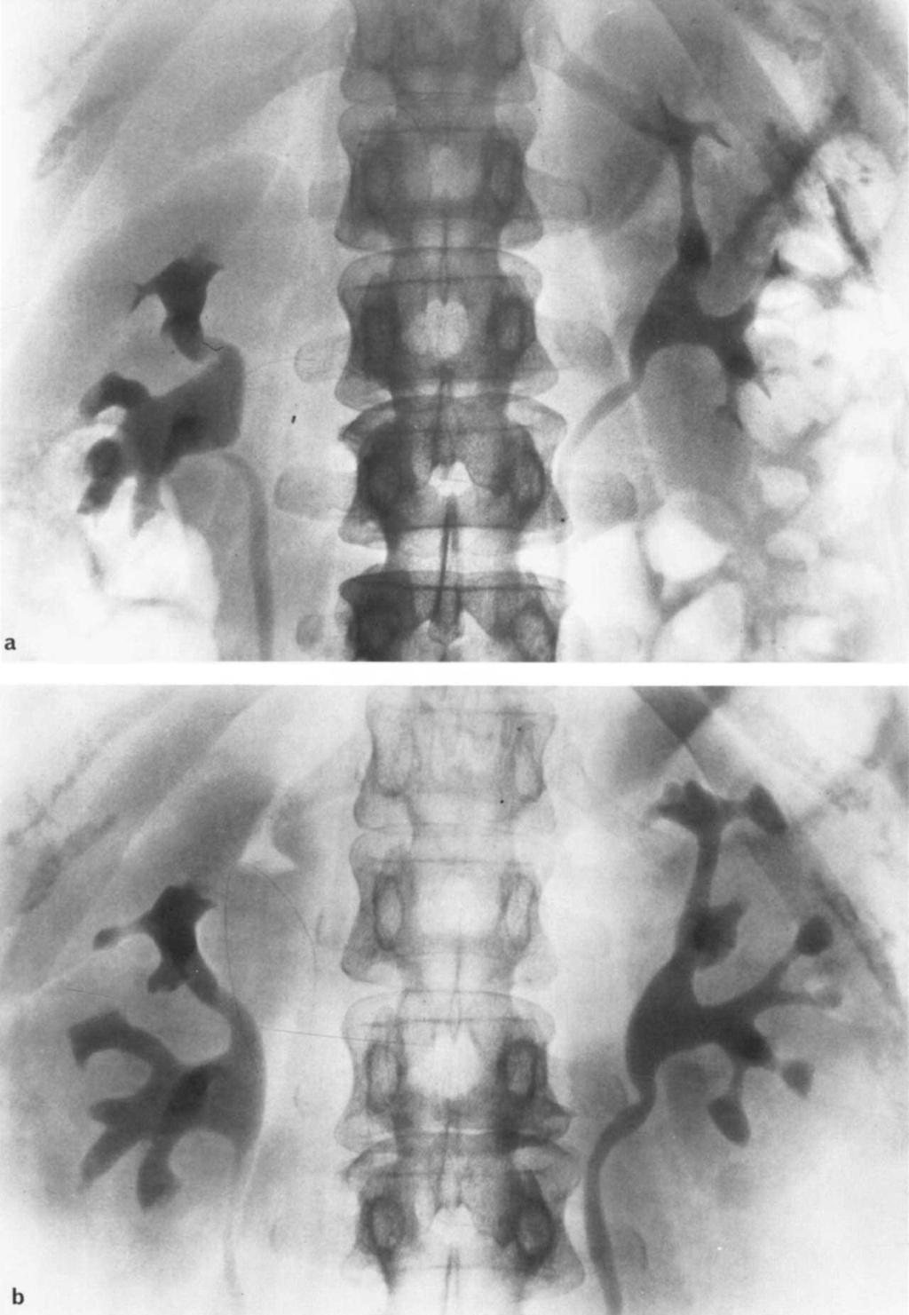
 in all papilae contrast density is slightly")
2 94 Lindvall I Fig. 1. Urographic findings in woman with migraine, analgesic abuse, and urinary tract infection: a) Normal findings initially. b) Three years later. There is total papillary necrosis (TPN) in all papilae contrast density is slightly reduced, and the kidney size is unchanged. (Reprinted with permission from Acta Radiologica [61.)
 in most papillae: a) Woman with analgesic abuse but without symptoms from the urinary tract.")
.")
3 Renal papillary necrosis: Radiologic changes 95 a, I b Fig. 2. Urography showing partial papillary necrosis (PPN) in most papillae: a) Woman with analgesic abuse but without symptoms from the urinary tract. b) Woman with diabetes and recurrent urinary tract infection. (Reprinted with permission from Acta Radiologica [61.) they have a stroma of necrotic papillary tissue. Calculi built up on a necrotic nondetached papilla do not have a typical radiologic appearance (Fig. 11). Sometimes, a typical calculus in papillary necrosis may grow and form a coral calculus; then the typical appearance will be lost. Formation of calculi in TPN and PPN is most common in connection with infection with Proteus organisms. That the necrotic tissue does constitute the nucleus of these calculi has been verified by microradiographic examinations [5] (Figs. 9 and 11). In the course of a renal colic in the presence of papillary necrosis, it is sometimes possible to find contrast accumulation in the kidney at urography. It may occur when a sloughed papilla obstructs the ureter, but it is said also to happen when all the papillae become necrotic simultaneously. Most often this contrast accumulation is rather slight and not as dense as in ureteric colic, due to other forms of obstruction, Necrosis in situ (NIS). The radiologic appearance in NIS, which differs somewhat from those described in TPN and PPN, is described in detail by Hare and Poynter [71. In this type, there will be no detachment of the necrotic tissue, which remains "in situ." The first stages described in TPN and PPN, as reduced contrast density, swelling, and shrinkage of the papilla, and widening of the fornices, are also found in the NIS. In the necrotic papillae, there will often be calcification, and even ossification is noted [81. The only typical features of this type of renal papillary necrosis are the calcifications (Fig. 3). If none of the papillae in the kidney have TPN or PPN, the radiologic diagnosis in NIS without calcification
 with calcifications. The kidney has shrunk and the contrast density is reduced.")
4 96 Lindvall Fig. 3. Urographic findings in analgesic abuse: a) Woman with analgesic abuse. To the left, findings are normal. To the right, eight years later, there are small PPN cavities, some with atypical calculi, and necrosis in situ (NIS) with calcifications. The kidney has shrunk and the contrast density is reduced. (Reprinted with permission from Acta Radiologica [6].) b) Man with analgesic abuse. Small kidneys with heavily reduced contrast density are shown. There is NIS with calcifications.
 One year later. There is one small PPN in lowermost, lateral papila and TPN in the earlier not contrast-filled medial minor calyx.")
 Normalfindings. b) Ten months later.")
5 Renal papillary necrosis: Radiologic changes 97 C Fig. 4. Urographic findings in woman with analgesic abuse: a) Ring-shadows in uppermost papillae. Lowermost, the medial minor calyx is not contrast-filled. b) One year later. There is one small PPN in lowermost, lateral papila and TPN in the earlier not contrast-filled medial minor calyx. c) One and a half year later. The small PPN has changed to TPN. There is TPN in the other papillae too. (Reprinted with permission from Acta Radiologica [61.) C Fig. S. Urographic findings in woman with analgesic abuse: a) Normalfindings. b) Ten months later. The uppermost papilla is somewhat irregular at its tip and the fomices around the papila are wider than the other in the same kidney. c) Small PPN in uppermost papilla. TPN is in lower pole. (Reprinted with permission from Ada Radiologica [6].)
 Two months later.")
 Normal findings initially. b) Two months later. There is an incipient detachment of PPM type. c) Further 2 months later.")
6 98 Lindvail b C Fig. 6. Urographic findings in woman with analgesic abuse: a) Normalfindings. b) Eight months later. There is an incipient detachment of the uppermost papilla starting from the calyceal fornix. c) Two months later. There is complete detachment of uppermost papilla, showing typical triangular ring-shadow. (Reprinted with permission from Acta Radiologica [61.) Fig. 7. Urographic findings in woman with analgesic abuse: a) Normal findings initially. b) Two months later. There is an incipient detachment of PPM type. c) Further 2 months later. Large PPN cavity has formed. (Reprinted with permission from Ada Radiologica [61.) may be difficult or impossible. There may be TPN and PPN and NIS in the same kidney (Figs. 5 and 13) Ṡize of kidney in papillary necrosis. The size of the kidney will change in different phases of renal papillary necrosis. In renal colic, there may be a swelling of the kidney, which will retain its normal size after the acute attack is gone. Mostly, the kidney will shrink (Fig. 12), but it might keep an almost normal size for many years (Fig. 13). There will be a slight thinning of the cortex, but if there is no complicating pyelonephritis the shrinkage will not be as great as in chronic pyelonephritis. The outline of the kidney in such cases is not more than slightly irregular; the outline is rather distinct (Fig. 13). Hare and Poynter [71 have described that there is sometimes a swelling of the columns of Bertin; however, sometimes there is a shrinkage of this part of the kidney too. Kidney function. Kidney function as judged by urography varies. In renal colic the function will deteriorate, but after the attack is over the urographic function may return to normal again. If the etiologic factors are removed and the complicating pyelonephritis is treated, the kidney can retain an almost normal urographic function for many years, even if all the papillae are detached (Figs. 1 and 13). In other cases, the function in the later stages may be so poor that pyelography, instead of urography, is necessary to demonstrate the anatomic changes. Changes in the ureter and the bladder. Those seen
 Plain film. The detached papilae have been calcified and form typical TPNcalculi. (Reprinted with permission from Acta Radiologica [6].) I C 4 Fig.")
 Plain film. Calcification of detached papilae occurred when the E. coli infection was replaced by Proteus infection. d) Microradiography of two of these calculi. [Shown on next page.")
7 Renal papillary necrosis: Radiologic changes 99 b Fig. 8. Urographic findings in man with analgesic abuse: a) Normal findings initially. b) Contrast density slightly reduced. Kidney size is normal. TPN with ring-shadows is present in all papillae. c) Plain film. The detached papilae have been calcified and form typical TPNcalculi. (Reprinted with permission from Acta Radiologica [6].) I C 4 Fig. 9. Urographic findings in woman with analgesic abase: a) Incipient detachment in central papilla. b) One year later. Note the typical irregular TPN ring-shadows and one empty TPN cavity. c) Plain film. Calcification of detached papilae occurred when the E. coli infection was replaced by Proteus infection. d) Microradiography of two of these calculi. [Shown on next page.] This shows them to be formed around detached necrotic papillae. (Reprinted with permission from Acta Radiologica [6].)
 Normalfindings initially. b) Two and a half months later. Shown are two PPN cavities at the uppermost minor calyx.")

8 100 Lindvall Fig. 94 (Cont'd from preceding page). This shows calculi to be formed around detached necrotic papilae. Fig. 10. Urographic findings in woman with analgesic abuse: a) Normalfindings initially. b) Two and a half months later. Shown are two PPN cavities at the uppermost minor calyx. (Reprinted with permission from Ada Radiologica [61.) are due to the complicating infection and the analgesic abuse. The infection of the urinary tract may, rarely, cause ureteritis, and in such cases strictures b may occur. The changes in the bladder are the same as those seen in ordinary urinary tract infection. During a typical attack of renal colic in renal papillary necrosis, it is common to find a contraction of the bladder on the same side as the renal colic. The complications caused by the abuse of analgesics containing phenacetin are transitional cell carcinomas in the renal pelvis or the bladder [9, 101. The radiologic appearance of these carcinomas are the same as in patients without analgesic abuse. The renal pelvis carcinoma may easily be missed, as in these cases the kidney function often has deteriorated too much for using urography. Differential diagnosis When the typical cavities of TPN and PPN have formed, there will be no diagnostic problems. The appearance of the cavities is unlike those seen in
 Calculi built up on NIS papilla.")
![d)microradiography showing the concretion to have been built on a not detached necrotic papilla with calcf1cations. (Reprinted with permission from Acta Radiologica [6].](/docs-images/81/82787313/images/9-2.jpg ") tuberculosis or medullary sponge kidney. Pyelogenic cysts nearly always communicate with the minor calyx at the fornix, but it can communicate with the minor calyx near the tip of the papila.")
9 Renal papillary necrosis: Radiologic changes 101 Fig. 11. Urographic findings in woman with analgesic abuse: a) Normal findings initially. b) 11 months later. Contrast density is appreciably reduced. There are PPN cavities and NIS in uppermost papilla. c) Calculi built up on NIS papilla. d)microradiography showing the concretion to have been built on a not detached necrotic papilla with calcf1cations. (Reprinted with permission from Acta Radiologica [6].) tuberculosis or medullary sponge kidney. Pyelogenic cysts nearly always communicate with the minor calyx at the fornix, but it can communicate with the minor calyx near the tip of the papila. In such a case, it can be hard to differentiate it from PPN with only one cavity; however, the further course of the disease will reveal its true nature. The so-called back pressure atrophy may, especially if the kidney is shrunken, simulate TPN. Von Friedreich [1] pointed out that the back pressure atrophy probably sometimes was no real atrophy but a healing stage after papillary necrosis. The only type of calculi that can simulate those in TPN are the "coagulate" calculi. The nephrocalcinosis seen in NIS is nonspecific, and it is not possible to radiologically differentiate it from other types of nephrocalcinosis, for example, in hyperparathyroidism. In short: the radiologic appearances in TPN and PPN are virtually always highly characteristic, and difficulties in differential diagnosis are rare unless marked shrinkage or hydronephrosis is present.

![) In NIS, the differential diagnostic problems are greater and, as pointed out by Hare and Poynter [7], unless there is calcification, the radiologic diagnosis may be impossible.](/docs-images/81/82787313/images/10-1.jpg "Discussion Urography (i.v.) and not pyelography should be used, if possible, to study the kidney in suspected renal papillary necrosis.")
10 102 Lindvall b Fig. 12. Urographic findings in woman with analgesic abase: a) Contrast density moderately reduced. There are no cavities, but there is probably shrinkage of uppermost papila. 14 One year later. There is marked contraction and PPN-cavities in all papillae. (Reprinted with pemlission from Acta Radialogica [61.) In NIS, the differential diagnostic problems are greater and, as pointed out by Hare and Poynter [7], unless there is calcification, the radiologic diagnosis may be impossible. Discussion Urography (i.v.) and not pyelography should be used, if possible, to study the kidney in suspected renal papillary necrosis. With urography, it is possible to study even the early changes, such as reduced function and incipient detachment. In the acute phase or in the later stages with shrinkage of the kidney and markedly depressed function, pyelography may be necessary. In the rare cases with ureteral strictures, kidney puncture and anterograde pyelography may be necessary. When using urography, it is important to thoroughly study the films taken before and after the application of the ureteral compression, as small PPN cavities can be difficult to detect owing to the dilatation of the calyceal fornices. Even a small detached papilla may be obscured by the dense contrast material after application of the ureteral compression. Serial examinations and comparison with earlier examinations are of great value to detect the early radiologic changes in renal papillary necrosis (Fig. 14). The period during which the necrosis of type TPN or PPN develops may vary widely, from exceptional instances in which all the papillae are involved simultaneously (Fig. 1) to a slowly progressive process involving one papilla after another over a very long Fig. 13. Urographic findings in woman with analgesic abase: a) Normal findings initially. b) len and PEN in most papillae. There is probably NIS in the others. The uppermost papilla left kidney is shrunken. c) (see p. 104) After 11 years withont analgesics. There is moderate shrinkage of the kidneys. Outlines are still distinct; otherwise, the appearance is the same.
11

12 104 Lindvall -4 a b Fig. 14. Urographicfindings in woman with analgesic abuse: a) Norma/findings initially. b) Three years later. One PPN has appeared in the uppermost papilla. The diagnosis of renal papillary necrosis would be difficult without comparison with the earlier normal urogram. (Reprinted with permission from Acta Radiologica [61.)
21 years later. One large PPN cavity has developed latemlly in the upper calyceal group and one medially in the lower group. (Reprinted with permission from Acta Radiologica [6].) period (Fig. 15).")
![If the etiology is analgesic abuse, all papillae should be affected at the same time, according to Hare and Poynter [7].](/docs-images/81/82787313/images/13-1.jpg "I have seen cases, however, with radiologically quite normal papillae together with typical TPN or PPN changes, which were followed for more than 10 years.")
13 Renal papillary necrosis: Radiologic changes 105 b Fig. 15. Urographic findings in woman with recurrent urinary tract infection, analgesic abuse denied: a)ppn in uppermost and undermost papilla. b)21 years later. One large PPN cavity has developed latemlly in the upper calyceal group and one medially in the lower group. (Reprinted with permission from Acta Radiologica [6].) period (Fig. 15). If the etiology is analgesic abuse, all papillae should be affected at the same time, according to Hare and Poynter [7]. I have seen cases, however, with radiologically quite normal papillae together with typical TPN or PPN changes, which were followed for more than 10 years. Perhaps the necrotic changes in those papillae are so small that they do not show up at radiography. In analgesic abuse, although there is bilateral renal papillary necrosis, the radiologically demonstrable changes may not always appear in both kidneys simultaneously. In exceptional cases, the typical changes may appear in the kidneys with a time-lag of two to three years. Unilateral renal papillary necrosis in analgesic abuse may occur only if it is favored by local predisposing conditions, such as unilateral urinary obstruction or unilateral medullary sponge kidney. One thing to keep in mind concerning the radiologic appearances is that the clinical signs and symptoms from the urinary tract do not correlate with the type of radiologic changes, and that the clinical course never indicates the distribution of the lesions in renal papillary necrosis, nor does it afford information frequently as to their presence or absence (Figs. 2, 12, 13, 15). Summary The radiological changes of renal papillary necrosis are independent of its etiology. If total papillary necrosis (TPN) or partial papillary necrosis (PPN) is present, radiological findings are diagnostic. Whereas, if the necrotic papillae remain in situ (NIS) none of the typical radiologic features of papillary necrosis are seen. Serial radiologic studies are useful in renal papillary necrosis. Extension of papillary or medullary cavities, shrinkage of the kidney, and calcification thereby may be noted. Radiologic changes involving the ureter and bladder are those of complications such as ureteritis or development of a transitional cell carcinoma. The latter most often appears in the renal pelvis. Reprint requests to Dr. N. Lindvall, Department of Diagnostic Radiology, Karolinska Sjukhuset, Stockholm, Sweden. References 1. VON FRIEDREICH N: Ueber Nekrose der Nierenpapillen bei Hydronephrose. Virchows Arch [PatholAnat] 69:308, PRAETORIUS G: Papillitis necroticans bei schwerer chronischer Pyelonephritis. Z Urol Nephrol 31:298, GUNTHER GW: Die Papilennekrosen der Niere bei Diabetes. Munch Med Wochenschr 84:1695, 1937
14 GUNTHER GW: Die Mark- und Papillennekrosen der Niere, Pyelonephritis und Diabetes. Z Uro! Nephrol 41:310, LAGEROREN C, LINOVALL N: Renal papillary necrosis: Roentgenologic diagnosis and formation of calculi. Acta Radio! (Stockh) 49:249, LINDVALL N: Renal papillary necrosis, Ada Radio! [Suppi] (Stockh) 192, H.&a WSC, POYNTER JD: The radiology of renal papillary necrosis as seen in analgesic nephropathy. C!in Radio! 25:423, MURPHY KJ: Calcification of the renal papillae as a sign of analgesic nephropathy. C!in Radio! 19: HULTENOREN N, LAGEROREN C, LJUNGQVI5T A: Carcinoma of the renal pelvis in renal papillary necrosis. Acta Chir Scand 130:314, BENOT5SON U, ANOERVALL L. REMAN H. LEHMANN L: Transitional cell tumours of the renal pelvis in analgesic abusers. Scand Uro! Nephro! 2:145, 1968
Excretory urography (EU) or IVP US CT & radionuclide imaging
 or IVP US CT & radionuclide imaging") Excretory urography (EU) or IVP US CT & radionuclide imaging MRI arteriography studies requiring catherization or direct puncture of collecting system EU & to a lesser extent CT provide both functional
Excretory urography (EU) or IVP US CT & radionuclide imaging MRI arteriography studies requiring catherization or direct puncture of collecting system EU & to a lesser extent CT provide both functional
Sonographic Features of Necrosed Renal Papillae Causing Hydronephrosis
 Case Series Sonographic Features of Necrosed Renal Papillae Causing Hydronephrosis S. Boopathy Vijayaraghavan, MD, DMRD, Sangampalayam Vedhanayagam Kandasamy, MS, MCh, Mylsamy Arul, MS, DNB (Uro), Muniappan
Chapter IV. Angionephrography in Simple Renal Cysts
 Acta Radiologica ISSN: 0001-6926 (Print) (Online) Journal homepage: http://www.tandfonline.com/loi/iaro20 Chapter IV. Angionephrography in Simple Renal Cysts To cite this article: (1957) Chapter IV. Angionephrography
US in non-traumatic acute abdomen. Lalita, M.D. Radiologist Department of radiology Faculty of Medicine ChiangMai university
 US in non-traumatic acute abdomen Lalita, M.D. Radiologist Department of radiology Faculty of Medicine ChiangMai university Sagittal Orientation Transverse (Axial) Orientation Coronal Orientation Intercostal
PROFESSIONAL SKILLS 1 3RD YEAR SEMESTER 6 RADIOGRAPHY. THE URINARY SYSTEM Uz. Fatema shmus aldeen Tel
 PROFESSIONAL SKILLS 1 3RD YEAR SEMESTER 6 RADIOGRAPHY THE URINARY SYSTEM Uz. Fatema shmus aldeen Tel. 0925111552 Professional skills-2 THE URINARY SYSTEM The urinary system (review anatomy and physiology)
Outline. Introduction to imaging modalities of the urinary system. Case base learning of common diseases in urinary tract
 Outline Introduction to imaging modalities of the urinary system Case base learning of common diseases in urinary tract Outline Introduction to imaging modalities of the urinary system Case base learning
Outline. Introduction to imaging modalities of the urinary system. Case base learning of common diseases in urinary tract
 Outline Introduction to imaging modalities of the urinary system Case base learning of common diseases in urinary tract Diagnostic Investigations in Urinary System PLAIN KUB EXCRETORY UROGRAPHY RETROGRADE
Roentgenologic diagnosis of medullary sponge kidney
 Acta Radiologica ISSN: 0001-6926 (Print) (Online) Journal homepage: http://www.tandfonline.com/loi/iaro20 Roentgenologic diagnosis of medullary sponge kidney Nils Lindvall To cite this article: Nils Lindvall
I N A previous paper3 extensive forms of renal papillary necrosis from analgesic
 VoL. No. 2 EARLY FORMS OF RENAL PAPILLARY NECROSIS* By BENEDICT R. HARROW, M.D. MIAMI, I N A previous paper3 extensive forms of renal papillary necrosis from analgesic abuse and from diabetes were described
Functions of the kidney:
 Diseases of renal system : Normal anatomy of renal system : Each human adult kidney weighs about 150 gm, the ureter enters the kidney at the hilum, it dilates into a funnel-shaped cavity, the pelvis, from
Analgesic and NSAID-induced Kidney Disease
 Analgesic and NSAID-induced Kidney Disease Edited by J.H.STEWART Associate Dean, Western Clinical School University of Sydney, Australia Oxford New York Tokyo Melbourne OXFORD UNIVERSITY PRESS 1993 CONTENTS
Acute renal colic Radiological investigation in patients with renal colic
 Acute renal colic Radiological investigation in patients with renal colic Mikael Hellström Professor Department of Radiology Sahlgrenska University Hospital Göteborg University 0.9-1.8/1.000 inhabitants
R adio logical investigations of urinary system
 R adio logical investigations of urinary system There are 4 main radiological Ix: 1 IVU: Intravenous urography. 2- U/S 3-CT scan 4-Radioisotope scan. Others (not frequently used): MRI, arteriography, antegrade
A Case of Calcified Ureteritis Cystica: An Indiscernible Condition from Ureterolithiasis
 Prague Medical Report / Vol. 110 (2009) No. 3, p. 245 249 245) A Case of Calcified Ureteritis Cystica: An Indiscernible Condition from Ureterolithiasis Alicioglu B. 1, Kaplan M. 2, Aktoz T. 3, Atakan I.
Separating and Distorted Nephroliths Signs of Renal Squamous Cell Carcinoma
 Chin J Radiol 2003; 28: 203-208 203 Separating and Distorted Nephroliths Signs of Renal Squamous Cell Carcinoma TZE-YU LEE SHEUNG-FAT KO CHUNG-CHENG HUANG YU-FENG CHENG Department of Radiology, Chang Gung
CYSTIC DISEASES of THE KIDNEY. Dr. Nisreen Abu Shahin
 CYSTIC DISEASES of THE KIDNEY Dr. Nisreen Abu Shahin 1 Types of cysts 1-Simple Cysts 2-Dialysis-associated acquired cysts 3-Autosomal Dominant (Adult) Polycystic Kidney Disease 4-Autosomal Recessive (Childhood)
IMAGING OF THE UROGENITAL TRACT
 IMAGING OF THE UROGENITAL TRACT 1 A) URINARY TRACT There are many methods of imaging the urinary tract but plain abdominal X-ray and ultrasound scan are usually done first in most cases, especially in
Intrarenal reflux and the scarred kidney
 Archives of Disease in Childhood, 1974, 49, 531. Intrarenal reflux and the scarred kidney G. L. ROLLESTON, T. M. J. MALING, and C. J. HODSON* From the Department of Radiology, Christchurch Hospital and
URINARY SYSTEM I. Kidneys II. Nephron Unit and Urine Formation
 URINARY SYSTEM I. Kidneys A. Location and Structure 1. Retroperitoneal 2. Between T12 and L3 3. Rt. kidney slightly lower 4. Two bean shaped organs 5. Adrenal gland 6. Internal construction a. Renal cortex
THE NATURAL HISTORY OF THE UPPER RENAL TRACTS IN ADULTS FOLLOWING URETERO- ILEAL DIVERSION (BRICKER PROCEDURE)*
*") DECEMBER, 1975 THE NATURAL HISTORY OF THE UPPER RENAL TRACTS IN ADULTS FOLLOWING URETERO- ILEAL DIVERSION (BRICKER PROCEDURE)* By PHILIP STANLEY, M.R.C.P., F.R.C.R.,f J. DUNCAN CRAVEN, M.R.C.P., F.R.C.R.,
DECEMBER, 1975 THE NATURAL HISTORY OF THE UPPER RENAL TRACTS IN ADULTS FOLLOWING URETERO- ILEAL DIVERSION (BRICKER PROCEDURE)* By PHILIP STANLEY, M.R.C.P., F.R.C.R.,f J. DUNCAN CRAVEN, M.R.C.P., F.R.C.R.,
Hydronephrosis. What is hydronephrosis?
 What is hydronephrosis? Hydronephrosis Hydronephrosis describes the situation where the urine collecting system of the kidney is dilated. This may be a normal variant or it may be due to an underlying
By GEORGE E. NELIGAN, M.C., M.A., B.M,, B.Ch. (Oxon.), F.R.C.S. (Swrgeon with charge of Out-patients and Surgeon in charge of the Genito-Urinary
, F.R.C.S. (Swrgeon with charge of Out-patients and Surgeon in charge of the Genito-Urinary") 426 POST-GRADUATE MEDICAL JOURNAL November, 1935 RENAL TUMOURS. By GEORGE E. NELIGAN, M.C., M.A., B.M,, B.Ch. (Oxon.), F.R.C.S. (Swrgeon with charge of Out-patients and Surgeon in charge of the Genito-Urinary
426 POST-GRADUATE MEDICAL JOURNAL November, 1935 RENAL TUMOURS. By GEORGE E. NELIGAN, M.C., M.A., B.M,, B.Ch. (Oxon.), F.R.C.S. (Swrgeon with charge of Out-patients and Surgeon in charge of the Genito-Urinary
Uroradiology Tutorial For Medical Students
 Uroradiology Tutorial For Medical Students Lesson 3: Cystography & Urethrography Part 1 American Urological Association Introduction Conventional radiography of the urinary tract includes several diagnostic
Proceedings of the 34th World Small Animal Veterinary Congress WSAVA 2009
 www.ivis.org Proceedings of the 34th World Small Animal Veterinary Congress WSAVA 2009 São Paulo, Brazil - 2009 Next WSAVA Congress : Reprinted in IVIS with the permission of the Congress Organizers IMAGING
Urinary system Ultrasound (Renal & Urinary bladder)
") Urinary system Ultrasound (Renal & Urinary bladder) Edited & Presented by ; Hussien A.B ALI DINAR. Msc.Phd ISRRT Associate Member Lecturer (National university) Reporting Sonographer (PHC) Objective By
Urinary system Ultrasound (Renal & Urinary bladder) Edited & Presented by ; Hussien A.B ALI DINAR. Msc.Phd ISRRT Associate Member Lecturer (National university) Reporting Sonographer (PHC) Objective By
Treatment of Staghorn Calculi by Pole Resection
 International Urology and Nephrology 3 (4), pp. 353--358 (1971) Treatment of Staghorn Calculi by Pole Resection M. FRYCZKOWSKI Department of Urology, Surgical Clinic No. I, Silesian Academy of Medicine,
CASE REPORT RENAL TUBERCULOSIS CAUSE OF RENAL REPLACEMENT LIPOMATOSIS : A RARE ASSOCIATION
 CASE REPORT RENAL TUBERCULOSIS CAUSE OF RENAL REPLACEMENT LIPOMATOSIS : A RARE ASSOCIATION DR ANAND AARTI 1, DR CHANDAK PRIYA 2,DR SURESH PARVATHY 3 1. PROF AND HOD, DEPARTMENT OF RADIODIAGNOSIS, GOVERNMENT
Radiological Assessment of the Kidney in Patients with Hematuria
 March 2005 Radiological Assessment of the Kidney in Patients with Hematuria Jeremy L. McKay, Harvard Medical School Year III Hematuria Signs and Symptoms Microscopic or gross hematuria Abdominal pain Fever
Primary Squamous Cell Carcinoma Of Kidney - A Case Report And Review Of Literature.
 ISPUB.COM The Internet Journal of Nephrology Volume 6 Number 1 Primary Squamous Cell Carcinoma Of Kidney - A Case Report And Review Of Literature. P Kaur, A Chauhan, G Singh, S Kataria, R Kalra Citation
Ultrasonographic diagnosis and typing of renal tuberculosis
 International Journal of Urology (2008) 15, 135 139 doi: 10.1111/j.1442-2042.2007.01962.x, Original Article: Clinical Investigation Ultrasonographic diagnosis and typing of renal tuberculosis Xuefang Rui,
The Kidney Dissection (photos curtosy of Murray Jensen at UMN)
") CJ Shuster AP2 Lab Addenum Kidney Dissection 1 The Kidney Dissection (photos curtosy of Murray Jensen at UMN) BACKGROUND INFORMATION The human urinary system consists of two kidneys, two ureters, one urinary
CJ Shuster AP2 Lab Addenum Kidney Dissection 1 The Kidney Dissection (photos curtosy of Murray Jensen at UMN) BACKGROUND INFORMATION The human urinary system consists of two kidneys, two ureters, one urinary
Vascular Pattern in Tumours
 Acta Radiologica ISSN: 0001-6926 (Print) (Online) Journal homepage: https://www.tandfonline.com/loi/iaro20 Vascular Pattern in Tumours To cite this article: (1957) Vascular Pattern in Tumours, Acta Radiologica,
Fetal Renal Malformations: The Role of Ultrasound in Diagnosis & Management
 Fetal Renal Malformations: The Role of Ultrasound in Diagnosis & Management 12 weeks Alfred Abuhamad, M.D. Eastern Virginia Medical School 13 weeks 2nd trimester Medullary pyramids Renal Sinus Cortex 2nd
Urinary System Laboratory
 Urinary System Laboratory 1 Adrenal gland Organs of The Urinary System Renal artery and vein Kidney Ureter Urinary bladder Figure 26.1 2 Urethra Functions of the urinary system organs: Urethra expels urine
AP2, Lab 7 - THE URINARY SYSTEM
 AP2, Lab 7 - THE URINARY SYSTEM I. SYSTEM COMPONENTS (Figs. 25.1 25.4) KIDNEYS Each kidney contains approx. 1,000,000 tubular NEPHRONS which produce FILTRATE from the plasma and then add to or take from
DISEASES AFFECTING TUBULES AND INTERSTITIUM
 DISEASES AFFECTING TUBULES AND INTERSTITIUM Acute tubular injury (ATI) Pyelonephritis Drug-induced tubulointerstitial nephritis (TIN) Myeloma cast NP Renal stones Urinary outflow obstruction: hydronephrosis
THE DYSFUNCTIONAL 'LAZY' BLADDER SYNDROME IN CHILDREN*
 THE DYSFUNCTIONAL 'LAZY' BLADDER SYNDROME IN CHILDREN* BY FRANK G. DELUCA, ORVAR SWENSONt, JOHN H. FISHER and ADEL H. LOUTFI From the Boston Floating Hospital for Infants and Children, Boston, Massachusetts
Primary Renal Candidiasis
 Case Series Primary Renal Candidiasis Importance of Imaging and Clinical History in Diagnosis and Management Barry J. Sadegi, MD, Bhargavi K. Patel, MD, Andrew C. Wilbur, MD, Anil Khosla, MD, Ejaz Shamim,
Pediatric Ure-Radiology*
 Pediatric Ure-Radiology* HERMAN GROSSMAN, M.D. Professor of Radiology and Pediatrics, Duke University Medical Center, Durham, North Carolina "Routine" radiologic studies do not, often enough, concentrate
Matsunaga, Naofumi; Saito, Yutaka. Citation Acta medica Nagasakiensia. 1991, 36
 NAOSITE: Nagasaki University's Ac Title Author(s) A Large Periureteral Lipoma Associa Hydronephrosis. Hayashi, Tomayoshi; Imamura, Atushi Matsunaga, Naofumi; Saito, Yutaka Citation Acta medica Nagasakiensia.
Urologic Stone Disease. Urologic Stone Disease. Urologic Stone Disease. Urologic Stone Disease. Urologic Stone Disease 5/7/2010
 Diagnosis and Treatment Stephen E. Strup MD William Farish Professor and Chief of Urology Director of Minimally Invasive Urologic Surgery University of Kentucky I will not cut, even for the stone, but
XANTHOGRANULOMATOUS PYELONEPHRITIS: radiologic review.
 XANTHOGRANULOMATOUS PYELONEPHRITIS: radiologic review. Poster No.: C-0557 Congress: ECR 2014 Type: Educational Exhibit Authors: M. Barral, J. M. Sánchez Crespo, J. C. Pérez Herrera, J. L. 1 2 3 1 1 1 Ortega
Imaging spectrum of genitourinary tuberculosis: Our experience at a tertiary care centre of a third world country
 Imaging spectrum of genitourinary tuberculosis: Our experience at a tertiary care centre of a third world country Poster No.: C-361 Congress: ECR 2009 Type: Educational Exhibit Topic: Genitourinary Authors:
Lec-8 جراحة بولية د.نعمان
 4th stage Lec-8 جراحة بولية د.نعمان 11/10/2015 بسم هللا الرحمن الرحيم Ureteric, Vesical, & urethral stones Ureteric Calculus Epidemiology like renal stones Etiology like renal stones Risk factors like
Urinary Anatomy. Lab 40. Kidneys. Nephrons. Renal Corpuscle
 Urinary Anatomy Lab 40. Urinary Anatomy and Kidney Dissection Kidneys: filters blood, produces urine Ureters: convey urine to bladder Bladder: holding tank Urethra: carries urine to the outside for elimination
54 year-old Female with Recurrent Bronchopneumonia and Tumor of the Left Kidney
 Original Report TheScientificWorldJOURNAL (2004) 4 (S1), 353 356 ISSN 1537-744X; DOI 10.1100/tsw.2004.89 54 year-old Female with Recurrent Bronchopneumonia and Tumor of the Left Kidney Burkhard Ubrig,
General Anatomy of Urinary System
 General Anatomy of Urinary System URINARY SYSTEM ORGANS Kidneys (2) Ureters (2) Urinary bladder Urethra KIDNEY FUNCTIONS Control blood volume and composition KIDNEY FUNCTIONS Filter blood plasma, eliminate
Five Views of Transitional Cell Carcinoma: One Man s Journey
 September 2006 Five Views of Transitional Cell Carcinoma: One Man s Journey Amsalu Dabela, Harvard Medical School III Outline Overview: Renal Anatomy Our Patient s Story Diagnostic Imaging Studies Appearance
Kidney & Urinary Tract Ultrasound. Fatina Fadel Hafez Bazaraa
 Kidney & Urinary Tract Ultrasound Fatina Fadel Hafez Bazaraa Ultrasonography Ultrasound Available Rapid Inexpensive Painless & no sedation needed No adverse effects/ complications Can be repeated Useful
Abdominal Ultrasound : Aorta, Kidneys, Bladder
 Abdominal Ultrasound : Aorta, Kidneys, Bladder Nilam J. Soni, MD, MSc Associate Professor of Medicine Divisions of Hospital Medicine and Pulmonary/Critical Care Medicine Department of Medicine University
Obstructive Uropathy. PATHOPHYSIOLOGIC CHANGES UUO vs BUO. Arry Rodjani Urology Department Ciptomangunkusumo Hospital Jakarta
 Obstructive Uropathy PATHOPHYSIOLOGIC CHANGES UUO vs BUO Arry Rodjani Urology Department Ciptomangunkusumo Hospital Jakarta INTRODUCTION Obstructive uropathy refers to the functional or anatomic obstruction
Nonurographic evaluation of renal calculous disease 1
 Contributions Nonurographic evaluation of renal calculous disease 1 Gregory P. Borkowski, M.D. Craig R. George, M.D. Peter B. O'Donovan, M.D. While excretory urography has been useful in the evaluation
PICTORIAL ESSAY. Experiences of using a single post-contrast CT scan of the urinary tract after triphasic contrast injection
 Experiences of using a single post-contrast CT scan of the urinary tract after triphasic contrast injection P C Pretorius, FCRad (Diag) SA Drs Visser, Erasmus, Vawda & Partners, Port Elizabeth Corresponding
Imaging the Urinary Tract
 Imaging the Urinary Tract Laura Armbrust, DVM, DACVR Gregory F. Grauer, DVM, MS, DACVIM Kansas State University Radiographic and ultrasound imaging in addition to history, physical examination, and clinicopathologic
1. Urinary System, General
 S T U D Y G U I D E 16 1. Urinary System, General a. Label the figure by placing the numbers of the structures in the spaces by the correct labels. 7 Aorta 6 Kidney 8 Ureter 2 Inferior vena cava 4 Renal
90% of bladder tumours are transitional cell carcinoma (TCC), the remaining 10% of cases are squamous cell carcinoma, adenocarcinoma and sarcoma.
, the remaining 10% of cases are squamous cell carcinoma, adenocarcinoma and sarcoma.") The Role of the Interventional Radiologist in Management of Post-Radical Cystectomy Ureteral Obstruction : A Case Review of Retrograde Transileal Conduit Ureteric Stents. Poster No.: C-2288 Congress: ECR
The Role of the Interventional Radiologist in Management of Post-Radical Cystectomy Ureteral Obstruction : A Case Review of Retrograde Transileal Conduit Ureteric Stents. Poster No.: C-2288 Congress: ECR
Basic Urinary Tract Anatomy and Histology
 Basic Urinary Tract Anatomy and Histology The two kidneys are located in the retroperitoneum on either side of the vertebral bladder and the contraction of the detrusor muscle. Any mechanical barrier,
Audit of split-bolus CT urography for the investigation of haematuria over a 12 month period at two district general hospitals
 Audit of split-bolus CT urography for the investigation of haematuria over a 12 month period at two district general hospitals Poster No.: C-1349 Congress: ECR 2010 Type: Educational Exhibit Topic: Genitourinary
Part III: Extrarenal Arteries Arising from the Renal Artery
 Acta Radiologica ISSN: 0001-6926 (Print) (Online) Journal homepage: https://www.tandfonline.com/loi/iaro20 Part III: Extrarenal Arteries Arising from the Renal Artery To cite this article: (1959) Part
URINARY TRACT IMAGING - BASIC PRINCIPLES
 URINARY TRACT IMAGING - BASIC PRINCIPLES Clinical Radiology Every physician needs a basic understanding of diagnostic imaging to understand how to order the appropriate studies and to understand the resulting
Obstetrics Content Outline Obstetrics - Fetal Abnormalities
 Obstetrics Content Outline Obstetrics - Fetal Abnormalities Effective February 2007 10 16% renal agenesis complete absence of the kidneys occurs when ureteric buds fail to develop Or degenerate before
The Kidney in Diabetics: Other than Nephropathy
 The Kidney in Diabetics: Other than Nephropathy Abeer Al-Saweer, MD* The kidney is thoroughly studied in diabetics since it is one of the target organs for major complication caused by diabetes. Diabetes
Secondary Gout Associated with Myeloproliferative Diseases* imt. Sinai Hospital
 Chapter VI Secondary Gout Associated with Myeloproliferative Diseases* By TS'AI-FAN Yu, M.D. imt. Sinai Hospital In a variety of disorders of hemopoiesis, the turnover of nucleic acids is greatly augmented,
E XTRAVASATIONS from the pelvicalyceal
 VOL. 112, No. 3 PELVICALYCEAL EXTRAVASATIONS* By FILEMON LOPEZ, M.D. BRONX, NEW YORK E XTRAVASATIONS from the pelvicalyceal system, previously considered rare, are being demonstrated with increasing frequency.
Physiologic Anatomy and Nervous Connections of the Bladder
 Micturition Objectives: 1. Review the anatomical organization of the urinary system from a physiological point of view. 2. Describe the micturition reflex. 3. Predict the lines of treatment of renal failure.
Uroradiology For Medical Students
 Uroradiology For Medical Students Lesson 4: Cystography & Urethrography - Part 2 American Urological Association Review Cystography is useful in evaluating the bladder, the urethra and the competence of
Role of imaging in RCC. Ultrasonography. Solid lesion. Cystic RCC. Solid RCC 31/08/60. From Diagnosis to Treatment: the Radiologist Perspective
 Role of imaging in RCC From Diagnosis to Treatment: the Radiologist Perspective Diagnosis Staging Follow up Imaging modalities Limitations and pitfalls Duangkamon Prapruttam, MD Department of Therapeutic
The urinary system consists of:
 Urinary system The urinary system consists of: - Two kidneys: this organ extracts wastes from the blood, balance body fluids and form urine. - Two ureters: this tube conducts urine from the kidneys to
Contents. Review anatomy of the urinary tract Imaging modalities
 Contents Review anatomy of the urinary tract Imaging modalities The Urinary Tract Kidneys ตาแหน งไต (position) อย ใน retroperitoneum ระด บ T12-L3 โดยไต ขวาจะม ระด บตากว าไตซ ายเล กน อย ร ปร าง (shape)
Congenital Pediatric Anomalies: A Collection of Abdominal Scintigraphy Findings: An Imaging Atlas
 ISPUB.COM The Internet Journal of Nuclear Medicine Volume 5 Number 1 Congenital Pediatric Anomalies: A Collection of Abdominal Scintigraphy Findings: An Imaging Atlas V Vijayakumar, T Nishino Citation
URINARY SYSTEM. Lecturer Dr.Firdous M.Jaafar Department of anatomy/histology section Lecture 3
 URINARY SYSTEM Lecturer Dr.Firdous M.Jaafar Department of anatomy/histology section Lecture 3 Objectives 1- Describe the structure of the urinary bladder, 2- Describe the structure of the ureters, bladder,
이학종분당서울대학교병원. Ultrasound in Urinary Colic
 이학종분당서울대학교병원 Ultrasound in Urinary Colic U l t r a s o u n d i n U r i n a US: Normal Kidney r y C o l i c Contents 1. 1. Definition and clinical consideration 2. 2. Pathophysiology 3. 3. US in in obstructive
Nephrographic and Pyelographic Analysis of CT Urography: Principles, Patterns, and Pathophysiology
 Genitourinary Imaging Review Wolin et al. CT Urography Principles, Patterns, and Genitourinary Imaging Review FOCUS ON: Ely A. Wolin 1 David S. Hartman J. Ryan Olson Wolin EA, Hartman DS, Olson JR Keywords:
Find Medical Solutions to Your Problems HYDRONEPHROSIS. (Distension of Renal Calyces & Pelvis)
") HYDRONEPHROSIS (Distension of Renal Calyces & Pelvis) Hydronephrosis is the distension of the renal calyces and pelvis due to accumulation of the urine as a result of the obstruction to the outflow of
HYDRONEPHROSIS (Distension of Renal Calyces & Pelvis) Hydronephrosis is the distension of the renal calyces and pelvis due to accumulation of the urine as a result of the obstruction to the outflow of
Long-Term Follow-Up of Reflux Nephropathy in Adults with Vesicoureteral Reflux - Radiological and Pathoanatomical Analysis
 Acta Radiologica ISSN: 0284-1851 (Print) 1600-0455 (Online) Journal homepage: http://www.tandfonline.com/loi/iard20 Long-Term Follow-Up of Reflux Nephropathy in Adults with Vesicoureteral Reflux - Radiological
Urinary System. Genitourinary System (Part A-1) Module 10 -Chapter 11. Overview 1/4/2013
 Module 10 -Chapter 11. Overview 1/4/2013") Genitourinary System (Part A-1) Module 10 -Chapter 11 Overview Urinary system Kidneys Ureter Bladder Nephron Male reproductive system Testes Accessory Structures Susie Turner, M.D. 71/3/13 Major homeostatic
Genitourinary System (Part A-1) Module 10 -Chapter 11 Overview Urinary system Kidneys Ureter Bladder Nephron Male reproductive system Testes Accessory Structures Susie Turner, M.D. 71/3/13 Major homeostatic
P RIMARY carcinoma of the ureter, once considered rare, makes up about I per
 JANUARY, 1973 TUMORS OF THE URETER PROBLEMS IN DIAGNOSIS By J. J. CANCELMO, JR., M.D., R. C. UHLMAN, M.D., J. L. ESHLEMAN, M.D., andn. F. V1EK, M.D. WAYNE, P RIMARY carcinoma of the ureter, once considered
In Canada, there was a 25% reduction in incidence of genitourinary TB in the period compared with An interesting speculation
 Renal T B EPIDEMIOLOGY Young to middle age usually affected, rare in children Male : female ratio = 2:1 True prevalence and incidence not known as patients are usually asymptomatic With HIV pandemic, there
Renal size in healthy Malaysian adults by ultrasonography
 Med. J. Malaysia Vol. 44 No. 1 March 1989 Renal size in healthy Malaysian adults by ultrasonography F. Wang, FRCPEd, FRACP Professor and Head Department ofmedicine Faculty ofmedicine, University ofmalaya,
RATIONALE: The organs making up the urinary system consist of the kidneys, bladder, urethra, and ureters.
 Chapter 12 Section Review 12.1 1. A. Kidneys RATIONALE: The renal pelvis receives urine from the kidney, travels through the ureters on the way to the bladder, but urine is formed in the kidney. 2. C.
IVU ((INTRAVENOUSUROGRAM))
)") IVU ((INTRAVENOUSUROGRAM)) Anatomy The urinary system consists of : 2 kidneys, 2 ureters,1 bladder, 1 urethra Renal pelvis Minor calyx Major calyx Renal parenchyma Proximal ureter Pelvi-uretric junction
IVU ((INTRAVENOUSUROGRAM)) Anatomy The urinary system consists of : 2 kidneys, 2 ureters,1 bladder, 1 urethra Renal pelvis Minor calyx Major calyx Renal parenchyma Proximal ureter Pelvi-uretric junction
Caveat sonologist Mistakes to avoid in Kidney Ultrasound
 Caveat sonologist Mistakes to avoid in Kidney Ultrasound Simon Freeman Derriford Hospital, Plymouth simonfreeman@nhs.net Bear trap 1 Report: There is a 4cm solid mass arising from the left kidney likely
27-Apr-15 1 UAF ANOMALIES OF DEVELOPMENT RENAL SYSTEM - 1 DR. MUHAMMAD TARIQ JAVED UAF UAF
 RENAL SYSTEM - 1 DR. MUHAMMAD TARIQ JAVED Professor, Department of Pathology, Faculty of Veterinary Science, University of Agriculture, Faisalabad, Pakistan. Email: mtjaved@uaf.edu.pk RENAL AGENESIS Renal
ENDOSCOPIC MANAGEMENT OF A URETERAL OBSTRUCTION CAUSED BY ENDOMETRIOSIS: A CASE REPORT
 ENDOSCOPIC MANAGEMENT OF A URETERAL OBSTRUCTION CAUSED BY ENDOMETRIOSIS: A CASE REPORT Hsu-Cheng Juan, 1 Hsin-Chih Yeh, 1 Hsi-Lin Hsiao, 1 Shean-Fang Yang, 2 and Wen-Jeng Wu 1,3 Departments of 1 Urology
Imaging findings in renal infections
 Imaging findings in renal infections Poster No.: C-0221 Congress: ECR 2013 Type: Educational Exhibit Authors: I. lópez blasco, D. Soriano Mena, R. Pastor Toledo, S. Paz Maya, A. M. Julve Parreño, J. Palmero
 1 2 Infertile women are seven to ten times more likely to have endometriosis than their fertile 3 The mechanism by which endometriosis develops is unknown Theories for the histogenesis of endometriosis
Chapter 23. The Nephron. (functional unit of the kidney
 Chapter 23 The Nephron (functional unit of the kidney Renal capsule The Nephron Renal cortex Nephron Collecting duct Efferent arteriole Afferent arteriole (a) Renal corpuscle: Glomerular capsule Glomerulus
ISSN East Cent. Afr. J. surg. (Online)
") 87 Ureteroscopy in a Resource Limited Setting: The Tikur Anbessa General Specialized Hospital Experience in Addis Ababa, Ethiopia. D. Andualem, L. Be-ede, T. Mulat, L. Samodi Addis Ababa University-School
87 Ureteroscopy in a Resource Limited Setting: The Tikur Anbessa General Specialized Hospital Experience in Addis Ababa, Ethiopia. D. Andualem, L. Be-ede, T. Mulat, L. Samodi Addis Ababa University-School
Gross Anatomy of the Urinary System
 Gross Anatomy of the Urinary System Lecture Objectives Overview of the urinary system. Describe the external and internal anatomical structure of the kidney. Describe the anatomical structure of the ureter
RENAL SCINTIGRAPHY IN THE 21 st CENTURY
 RENAL SCINTIGRAPHY IN THE 21 st CENTURY 99m Tc- MAG 3 with zero time injection of Furosemide (MAG 3 -F 0 ) : A Fast and Easy Protocol, One for All Indications Clinical Experience Congenital Disorders PROTOCOL
Title with transitional cell carcinoma: a. Citation 泌尿器科紀要 (1992), 38(6):
, 38(6):") Title Primary localized amyloidosis of th with transitional cell carcinoma: a Author(s) Shiramizu, Miki; Nakamura, Kaoru; B Yoji; Kinoshita, Hidechika Citation 泌尿器科紀要 (1992), 38(6): 699-702 Issue Date
Title Primary localized amyloidosis of th with transitional cell carcinoma: a Author(s) Shiramizu, Miki; Nakamura, Kaoru; B Yoji; Kinoshita, Hidechika Citation 泌尿器科紀要 (1992), 38(6): 699-702 Issue Date
Urinary Schistosomiasis: Urological manifestations &complications. Dr MJ Engelbrecht Department of Urology
 Urinary Schistosomiasis: Urological manifestations &complications Dr MJ Engelbrecht Department of Urology Clinical presentation Three clinical stages Swimmer s itch Acute schistosomiasis Chronic schistosomiasis
Percutaneous Nephrolithotomy in a Patient with Mainz Pouch II Urinary Diversion: A Case Report
 198) Prague Medical Report / Vol. 117 (2016) No. 4, p. 198 203 Percutaneous Nephrolithotomy in a Patient with Mainz Pouch II Urinary Diversion: A Case Report Stavros Sfoungaristos 1, Ioannis Mykoniatis
RADIOLOGY OF THE URINARY TRACT CHAPTER 9 239
 RADIOLOGY OF THE URINARY TRACT CHAPTER 9 239 in length. They lie cephalad to the kidneys, with the right just posterior to the inferior vena cava (IVC) and the left anteromedial to the upper pole of the
URINARY SYSTEM. These organs lie posterior or inferior to the. (membrane).
.") URINARY SYSTEM I. INTRODUCTION Each kidney is made up of about a million tiny tubules called nephrons. Each nephron individually filters the blood and makes urine and it does the job completely, from start
URINARY SYSTEM I. INTRODUCTION Each kidney is made up of about a million tiny tubules called nephrons. Each nephron individually filters the blood and makes urine and it does the job completely, from start
An Unexpected Cause Of Spontaneous Perinephric Urinoma: A Case Report. L Chandrasekharan, T Abdl Ghaffar, M Venkatramana, K Mammigatty
 ISPUB.COM The Internet Journal of Radiology Volume 4 Number 1 An Unexpected Cause Of Spontaneous Perinephric Urinoma: A Case Report L Chandrasekharan, T Abdl Ghaffar, M Venkatramana, K Mammigatty Citation
Randall plaque versus renal stone?
 Commentary Randall plaque versus renal stone? Thomas Chi, Joe Miller, Marshall L. Stoller Department of Urology, University of California, San Francisco, USA Correspondence to: Thomas Chi, MD. Department
ULTRASONOGRAPHY RADIOLOGY Simple abdominal X-ray Intravenous urography Retrograde/anterograde pieloureterography Renal angiography CT
 ULTRASONOGRAPHY RADIOLOGY Simple abdominal X-ray Intravenous urography Retrograde/anterograde pieloureterography Renal angiography CT NUCLEAR MEDICINE Static studies: static renal scintigraphy Dynamic
My Patient Has Abdominal Pain PoCUS of the Biliary Tract and the Urinary Tract
 My Patient Has Abdominal Pain PoCUS of the Biliary Tract and the Urinary Tract Objectives PoCUS for Biliary Disease PoCUS for Renal Colic PoCUS for Urinary Retention Biliary Disease A patient presents