FCC2 5CY7 FCC1 5CY8. Actinonin 5CVQ
|
|
|
- Morgan Sullivan
- 5 years ago
- Views:
Transcription
1 Table S1. Data collection and refinement statistics Data collection Apo 5E5D MA 5CPD MAS 5CP0 Actinonin 5CVQ FCC1 5CY8 FCC2 5CY7 FCC 5CWY FCC4 5CVK FCC5 5CWX FCC6 5CVP X-ray source 17A-KEK PAL4A PAL4A PAL4A PAL4A PAL4A PAL4A PAL4A PAL4A PAL4A Space group P6 122 P6 122 P6 122 P6 122 P6 122 P6 122 P6 122 P6 122 P6 122 P6 122 Unit-cell parameters a(b), c (Å ) 58.7, , , , , , , , , , Resolution range (Å ) ( ) ( ) ( ) ( ) ( ) ( ) ( ) ( ) ( ) ( ) No. of observed 86,56 159,50 204,72 91,979 11, , , ,756 71, ,999 reflections (9,011) (16,778) (20,724) (22,857) (11,77) (11,015) (20,246) (,248) (25,491) (18,979) (unique) Completeness (%) 96.9 (97.4) 98.5(99.6) 98.6 (95.7) 87.6 (8.1) 98.8 (99.1) 95.2 (88.0) 97.9 (96,4) 95.2 (82.7) 98.9 (97.9) 97.1 (84.7) R sym (%) 15.5 (44.) 17.7 (59.) 11.0 (65.2) 18.6 (64.8) 9.1 (42.5) 6.8 (28.9) 9.7 (51.0) 11.1 (48.0) 9.8 (48.4) 8.2 (7.8) Average I/ (I) 22.5 (6.6) 9.8 (1.7) 6.2 (4.6) 7.2 (1.6) 25 (4.5) 5. (7.4) 18.8 (.2) 16.9 (2.2) 12.4 (.2) 1.6 (4.0) Refinement Resolution (Å ) 0.4 (2.6) 0.4 (2.2) 29.4 (2.0) 27.8 (2.5) (2.8) 0.4 (2.8) 0.4 (2.4) 27.0 (2.1) 0.4 (2.2) 0.4 (2.0) Number of reflections Working set 8,502 1,851 18,225 9,226 4,800 10,401 10,91 15,907 1,777 17,907 Free R set R/R free (%) c 18.6 (24.) 19.1 (24.7) 18.1 (21.7) 17.2 (21.8) 18.7 (22.7) 18.1 (21.2) 18.4 (22.7) 17.6 (22.4) 20.7 (25.9) 17.4 (21.6) Protein atoms number Substrate MA MAS Inhibitor 1,5 1, ,0 20 1, ,5 1 1, , , ,57 1 1,14 1 Water molecules
2 Cadmium ion Sodium ion Acetate Glycerol RMS bond lengths (Å ) RMS bond angles( o ) Mean B factors (Å 2 ) Main-chain atoms Side-chain atoms Substrate MA MAS Inhibitor Water atoms Cadmium Sodium ion Acetate Glycerol Ramachandran plot (%) Favored regions Allowed regions Outlier regions ,881 2, ,96 2,148 1,919 2,157 2,042 2,146 1, Values in parentheses are for the highest resolution shell.
3 Rmerge = hkl i (I i (hkl)) I(hkl) / hkl i I i (hkl), where I i (hkl) is the mean intensity of ith observation of symmetry-related reflections hkl. R free = hkl F obs F calc / hkl F obs, where F calc is the calculated protein structure factor from the atomic model (Rfree was calculated from a randomly selected 5% sample of all reflections).
4 Fig. S1


5 Fig. S2 A B C C99 C H H141 H H141

6 Fig. S A B C D E F
7 Fig. S4

8 Fig. S5 * * *


9 Fig. S6 A B C F107 R149 G95 R147 D G95 R129 R147 W146 F14 F87 L126 F14

10 Fig. S7 E97 H141 V45

11 Fig. S8 A B F14 F14 Y69

12 Fig. S9 F
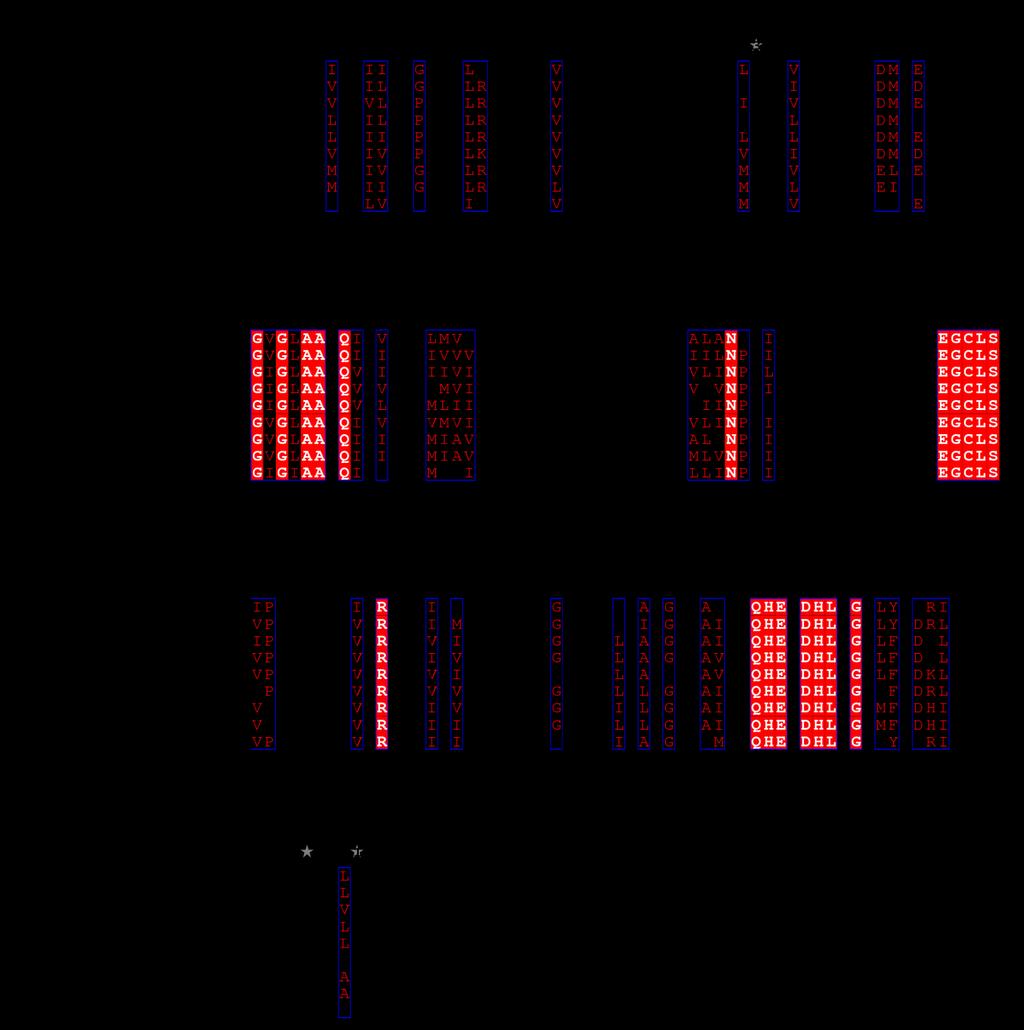
13 SUPPLEMENTARY FIGURE LEGENDS Figure S1. Sequence alignment of bacterial PDFs. The amino acid sequences were aligned with the secondary structure information of XoPDF; XoPDF (Xoo1075; Def) from Xanthomonas oryzae pv. oryzae, PDF from Leptospira interrogans, PDF from E. coli, PDF (Xoo0585) from Xanthomonas oryzae pv. oryzae, PDF from Helicobacter pylori, PDF from Aquifex aeolicus, PDF from Bacillus subtilis 184, PDF from Staphylococccus aureus and PDF from Mycoplasma pneumonia. Residues coordinating the metal ion in the active site are indicated with red inverted triangles. Signature motif 1 is shown in blue, motif 2 in cyan, motif in purple and CD loop in orange. Figure S2. Metal binding and coordination of XoPDF. (a) Cd 2+ ion in the metal site of MAS-bound XoPDF structure with 2Fofc and FoFc maps. (b) Zn 2+ ion in the metal site of MAS-bound XoPDF structure with 2Fofc and FoFc maps. 2Fofc map (blue mesh) is contoured at 1.0 σ and Fofc map (green mesh) at 5.0 σ. (c) Superimposed metal sites of Cd 2+ -bound XoPDF (purple), Co 2+ -bound EcPDF (PDB ID: 4AZ4; cyan), Ni 2+ -bound EcPDF (PDB ID: 4AL2; yellow), Zn 2+ -bound EcPDF (PDB ID: 1XEM; green) and Fe 2+ -bound EcPDF (PDB ID: 1XEN; orange). Figure S. Structures of PDF CD-loops. (a) XoPDF. (b) PDF from Leptospira interrogans (PDB ID: 1SV2). (c) PDF from Ehrlichia chaffeensis (PDB ID: OCA). (d) PDF from human (PDB ID: G5P). (e) PDF from Bacillus cereus (PDB ID: 1WS0). (f) PDF from Staphylococcus aureus (PDB ID: 2AI9). CD-loops are shown with purple (closed) and orange (opened) shades. Figure S4. Time-resolved transcriptional gene expression of XoPDF (Xoo1075, def) and Xoo0585 genes Transcriptional gene expression levels of XoPDF and Xoo0585 were measured in a time-resolved manner via RNA-Seq, after the activation of Xoo pathogenicity. Xoo1075_RLX and Xoo1075_con exhibit transcriptional expression of the XoPDF gene within 1 hour in the pathogenicity-activated Xoo cell and the control Xoo cell, 1
14 respectively. Xoo0585_RLX and Xoo0585_con exhibit transcriptional expression of the Xoo0585 gene within 1 hour in the pathogenicity-activated Xoo cell and the control Xoo cell, respectively. Figure S5. Sequence alignment of XoPDF and eukaryotic PDFs: XoPDF (Xoo1075; Def) from Xanthomonas oryzae pv. oryzae, OsPDF1B from rice, AtPDF1B from Arabidopsis thaliana, PfPDF from Plasmodium falciparum, OsPDF1A from rice and AtPDF1A from Arabidopsis thaliana. Residues in the upper layer of the hydrophobic pocket for substrate methionine side chain are marked with asterisk. Figure S6. Structure comparison between XoPDF and AtPDFs. (a) Superimposed structures of actinonin-bound XoPDF (green) and MAS-bound AtPDF1A (brown). Actinonin in XoPDF is shown in yellow. CD-loop of AtPDF1A is shaded in purple. (b) Superimposed structures of actinonin-bound XoPDF (green) and actinoninbound AtPDF1B (pale-blue). Actinonin in XoPDF is shown in yellow. CD-loop of AtPDF1B is shaded in salmon. (c) Superimposed substrate-binding site of actinonin-bound XoPDF (green) and MAS-bound AtPDF1A (brown) with the upper layer residues of the hydrophobic pocket for substrate methionine side chain. (d) Superimposed substrate-binding site of actinonin-bound XoPDF (green) and actinonin-bound AtPDF1B (pale-blue) with the upper layer residues of the hydrophobic pocket for substrate methionine side chain. Figure S7. Bottom view of substrate methionine-binding site. The fragment chemical compounds (FCCs)-bound structures were superimposed to the MA-bound structure and only FCCs and substrate methionine-binding site are shown in the same bottom view of Fig. 4b. Figure S8. Exposure of Phe14 residue in XoPDF with the flexible open conformation of CD loop. (a) The structure of closed CD loop. (b) The structure of opened CD loop. 2
15 Figure S9. Actinonin structure in XoPDF. The hydrophobic van der Waals interactions between the pentyl group and the other chain structure with the pyrrolidine ring are shown as black dashed lines. The hydrophobic van der Waals interactions between Phe14 and the 5-member pyrrolidine ring is shown as blue arcs.
Supplementary Materials for
 www.sciencemag.org/cgi/content/full/science.aal4326/dc1 Supplementary Materials for Structure of a eukaryotic voltage-gated sodium channel at near-atomic resolution Huaizong Shen, Qiang Zhou, Xiaojing
Table S1. X-ray data collection and refinement statistics
 Table S1. X-ray data collection and refinement statistics Data collection H7.167 Fab-Sh2/H7 complex Beamline SSRL 12-2 Wavelength (Å) 0.97950 Space group I2 1 3 Unit cell parameters (Å, º) a = b = c=207.3,
Supplementary Table 1. Data collection and refinement statistics (molecular replacement).
.") Supplementary Table 1. Data collection and refinement statistics (molecular replacement). Data set statistics HLA A*0201- ALWGPDPAAA PPI TCR PPI TCR/A2- ALWGPDPAAA PPI TCR/A2- ALWGPDPAAA Space Group P2
Supplementary Table 1. Data collection and refinement statistics (molecular replacement). Data set statistics HLA A*0201- ALWGPDPAAA PPI TCR PPI TCR/A2- ALWGPDPAAA PPI TCR/A2- ALWGPDPAAA Space Group P2
Detergent solubilised 5 TMD binds pregnanolone at the Q245 neurosteroid potentiation site.
 Supplementary Figure 1 Detergent solubilised 5 TMD binds pregnanolone at the Q245 neurosteroid potentiation site. (a) Gel filtration profiles of purified 5 TMD samples at 100 nm, heated beforehand for
(B D) Three views of the final refined 2Fo-Fc electron density map of the Vpr (red)-ung2 (green) interacting region, contoured at 1.4σ.
 Three views of the final refined 2Fo-Fc electron density map of the Vpr (red)-ung2 (green) interacting region, contoured at 1.4σ.") Supplementary Figure 1 Overall structure of the DDB1 DCAF1 Vpr UNG2 complex. (A) The final refined 2Fo-Fc electron density map, contoured at 1.4σ of Vpr, illustrating well-defined side chains. (B D) Three
Supplementary Figure 1 Overall structure of the DDB1 DCAF1 Vpr UNG2 complex. (A) The final refined 2Fo-Fc electron density map, contoured at 1.4σ of Vpr, illustrating well-defined side chains. (B D) Three
Supplementary Materials for
 advances.sciencemag.org/cgi/content/full/2/9/e1600292/dc1 Supplementary Materials for Native phasing of x-ray free-electron laser data for a G protein coupled receptor Alexander Batyuk, Lorenzo Galli,
Supplementary Figure 1 (previous page). EM analysis of full-length GCGR. (a) Exemplary tilt pair images of the GCGR mab23 complex acquired for Random
. EM analysis of full-length GCGR. (a) Exemplary tilt pair images of the GCGR mab23 complex acquired for Random") S1 Supplementary Figure 1 (previous page). EM analysis of full-length GCGR. (a) Exemplary tilt pair images of the GCGR mab23 complex acquired for Random Conical Tilt (RCT) reconstruction (left: -50,right:
S1 Supplementary Figure 1 (previous page). EM analysis of full-length GCGR. (a) Exemplary tilt pair images of the GCGR mab23 complex acquired for Random Conical Tilt (RCT) reconstruction (left: -50,right:
Chapter 6. X-ray structure analysis of D30N tethered HIV-1 protease. dimer/saquinavir complex
 Chapter 6 X-ray structure analysis of D30N tethered HIV-1 protease dimer/saquinavir complex 6.1 Introduction: The arrival of HIV protease inhibitors (PIs) in late 1995 marked the beginning of an important
SUPPLEMENTARY INFORMATION
 doi: 10.1038/nature07422 SUPPLEMENTARY INFRMATIN K S(P) R S I M Q(L4) R M 7 6 Sp Q I K R 5 4 3 2 1 L(L0) L L S E 0 +1 +2 +3 Figure S1a Difference electron density (mfo DFc) for the peptide (Qpeptide),
SUPPLEMENTARY INFORMATION
 doi:10.1038/nature10913 Supplementary Figure 1 2F o -F c electron density maps of cognate and near-cognate trna Leu 2 in the A site of the 70S ribosome. The maps are contoured at 1.2 sigma and some of
Atypical Natural Killer T-cell receptor recognition of CD1d-lipid antigens supplementary Information.
 Atypical Natural Killer T-cell receptor recognition of CD1d-lipid antigens supplementary Information. Supplementary Figure 1. Phenotypic analysis of TRBV25-1 + and TRBV25-1 - CD1d-α-GalCerreactive cells.
Supporting Information
 Supporting Information Mechanism of inactivation of -aminobutyric acid aminotransferase by (1S,3S)-3-amino-4-difluoromethylenyl-1- cyclopentanoic acid (CPP-115) Hyunbeom Lee, 1, Emma H. Doud, 1,2 Rui Wu,
SUPPLEMENTARY INFORMATION FOR. (R)-Profens Are Substrate-Selective Inhibitors of Endocannabinoid Oxygenation. by COX-2
-Profens Are Substrate-Selective Inhibitors of Endocannabinoid Oxygenation. by COX-2") SUPPLEMENTARY INFORMATION FOR (R)-Profens Are Substrate-Selective Inhibitors of Endocannabinoid Oxygenation by COX-2 Kelsey C. Duggan, Daniel J. Hermanson, Joel Musee, Jeffery J. Prusakiewicz, Jami L.
SUPPLEMENTARY INFORMATION FOR (R)-Profens Are Substrate-Selective Inhibitors of Endocannabinoid Oxygenation by COX-2 Kelsey C. Duggan, Daniel J. Hermanson, Joel Musee, Jeffery J. Prusakiewicz, Jami L.
Amino Acids. Review I: Protein Structure. Amino Acids: Structures. Amino Acids (contd.) Rajan Munshi
 Rajan Munshi") Review I: Protein Structure Rajan Munshi BBSI @ Pitt 2005 Department of Computational Biology University of Pittsburgh School of Medicine May 24, 2005 Amino Acids Building blocks of proteins 20 amino acids
Review I: Protein Structure Rajan Munshi BBSI @ Pitt 2005 Department of Computational Biology University of Pittsburgh School of Medicine May 24, 2005 Amino Acids Building blocks of proteins 20 amino acids
172R 172K TAM-2/172R TAM-2/172K. AZT concentration [nm] AZT concentration [nm] MgCl 2 2.5K 2.5K 5K 2.5K 5K 2.5K K 5K 2.5K 5K 2.5K 50 2.
![172R 172K TAM-2/172R TAM-2/172K. AZT concentration [nm] AZT concentration [nm] MgCl 2 2.5K 2.5K 5K 2.5K 5K 2.5K K 5K 2.5K 5K 2.5K 50 2.](/thumbs/82/85138523.jpg "172R 172K TAM-2/172R TAM-2/172K. AZT concentration [nm] AZT concentration [nm] MgCl 2 2.5K 2.5K 5K 2.5K 5K 2.5K K 5K 2.5K 5K 2.5K 50 2.") 5 5 5 5 A MgCl 2 172R 172K TAM-2/172R TAM-2/172K AZT concentration [nm] B 172R 172K TAM-2/172R TAM-2/172K AZT concentration [nm] ATP + ATP - Supplemental Figure 1. Primer extension of HIV-1 RT polymorphisms
5 5 5 5 A MgCl 2 172R 172K TAM-2/172R TAM-2/172K AZT concentration [nm] B 172R 172K TAM-2/172R TAM-2/172K AZT concentration [nm] ATP + ATP - Supplemental Figure 1. Primer extension of HIV-1 RT polymorphisms
Supplementary Materials for
 advances.sciencemag.org/cgi/content/full/2/4/e1500980/dc1 Supplementary Materials for The crystal structure of human dopamine -hydroxylase at 2.9 Å resolution Trine V. Vendelboe, Pernille Harris, Yuguang
Nature Structural & Molecular Biology: doi: /nsmb.1933
 The structural basis of open channel block in a prokaryotic pentameric ligand-gated ion channel Ricarda J. C. Hilf, Carlo Bertozzi, Iwan Zimmermann, Alwin Reiter, Dirk Trauner and Raimund Dutzler a GLIC
Structure of the measles virus hemagglutinin bound to the CD46 receptor. César Santiago, María L. Celma, Thilo Stehle and José M.
 Supporting Figures and Table for Structure of the measles virus hemagglutinin bound to the CD46 receptor César Santiago, María L. Celma, Thilo Stehle and José M. Casasnovas This PDF file includes: Supplementary
Supplementary Information
 Supplementary Information Two common structural motifs for TCR recognition by staphylococcal enterotoxins Karin Erica Johanna Rödström 1, Paulina Regenthal 1, Christopher Bahl 2, Alex Ford 2, David Baker
SUPPLEMENTARY INFORMATION
 Supplementary Discussion The HGGG sequence motif forms an oxyanion hole in HLSs. In the case of GID1, the third Gly residue of the sequence motif is replaced with Ser (Ser123 in OsGID1), i.e., the sequence
Supplementary Information
 Supplementary Information Using the pimeloyl-coa synthetase adenylation fold to synthesise fatty acid thioesters Menglu Wang 1a ; Lucile Moynié 2a, Peter J. Harrison 1, Van Kelly 1, Andrew Piper 1, James
Supplementary Material
 Supplementary Material Materials and methods Enzyme assay The enzymatic activity of -glucosidase toward salicin was measured with the Miller method (Miller, 1959) using glucose as the standard. A total
Insights into the Giardia intestinalis Enolase and Human Plasminogen interaction
 Electronic Supplementary Material (ESI) for Molecular BioSystems. This journal is The Royal Society of Chemistry 2017 Supplementary Information Insights into the Giardia intestinalis Enolase and Human
Transient β-hairpin Formation in α-synuclein Monomer Revealed by Coarse-grained Molecular Dynamics Simulation
 Transient β-hairpin Formation in α-synuclein Monomer Revealed by Coarse-grained Molecular Dynamics Simulation Hang Yu, 1, 2, a) Wei Han, 1, 3, b) Wen Ma, 1, 2 1, 2, 3, c) and Klaus Schulten 1) Beckman
Supplementary Materials for
 advances.sciencemag.org/cgi/content/full/4/3/eaaq0762/dc1 Supplementary Materials for Structures of monomeric and oligomeric forms of the Toxoplasma gondii perforin-like protein 1 Tao Ni, Sophie I. Williams,
Introduction to proteins and protein structure
 Introduction to proteins and protein structure The questions and answers below constitute an introduction to the fundamental principles of protein structure. They are all available at [link]. What are
Molecular Dynamics Simulation Study Explaining Inhibitor Selectivity in Different Class of Histone Deacetylases
 Journal of Biomolecular Structure & Dynamics, ISSN 0739-1102 Volume 29, Issue Number 4, (2012) Adenine Press (2012) Abstract Molecular Dynamics Simulation Study Explaining Inhibitor Selectivity in Different
SUPPLEMENTARY INFORMATION (SI) FIGURES AND TABLES
 FIGURES AND TABLES") SUPPLEMENTARY INFORMATION (SI) FIGURES AND TABLES 1 Title: Discovery of a junctional epitope antibody that stabilizes IL-6 and gp80 protein:protein interaction and modulates its downstream signaling Authors:
SUPPLEMENTARY INFORMATION (SI) FIGURES AND TABLES 1 Title: Discovery of a junctional epitope antibody that stabilizes IL-6 and gp80 protein:protein interaction and modulates its downstream signaling Authors:
CS612 - Algorithms in Bioinformatics
 Spring 2016 Protein Structure February 7, 2016 Introduction to Protein Structure A protein is a linear chain of organic molecular building blocks called amino acids. Introduction to Protein Structure Amine
Structural biology of viruses
 Structural biology of viruses Biophysical Chemistry 1, Fall 2010 Coat proteins DNA/RNA packaging Reading assignment: Chap. 15 Virus particles self-assemble from coat monomers Virus Structure and Function
The mechanism of patellamide macrocyclization revealed by study of the Prochloron sp PatG macrocyclase domain
 The mechanism of patellamide macrocyclization revealed by study of the Prochloron sp PatG macrocyclase domain Jesko Koehnke 1,2, Andrew Bent 1,2, Wael E. Houssen 2,3,4, David Zollman 1, Falk Morawitz 1,
HOMEWORK II and Swiss-PDB Viewer Tutorial DUE 9/26/03 62 points total. The ph at which a peptide has no net charge is its isoelectric point.
 BIOCHEMISTRY I HOMEWORK II and Swiss-PDB Viewer Tutorial DUE 9/26/03 62 points total 1). 8 points total T or F (2 points each; if false, briefly state why it is false) The ph at which a peptide has no
This exam consists of two parts. Part I is multiple choice. Each of these 25 questions is worth 2 points.
 MBB 407/511 Molecular Biology and Biochemistry First Examination - October 1, 2002 Name Social Security Number This exam consists of two parts. Part I is multiple choice. Each of these 25 questions is
Nature Structural & Molecular Biology: doi: /nsmb Supplementary Figure 1
 Supplementary Figure 1 The UBL and RING1 interface remains associated in the complex structures of Parkin and pub. a) Asymmetric Unit of crystal structure of UBLR0RBR and pub complex showing UBL (green),
Supporting Information
 Supporting Information McCullough et al. 10.1073/pnas.0801567105 A α10 α8 α9 N α7 α6 α5 C β2 β1 α4 α3 α2 α1 C B N C Fig. S1. ALIX Bro1 in complex with the C-terminal CHMP4A helix. (A) Ribbon diagram showing
SUPPLEMENTARY INFORMATION
 SUPPLEMENTARY INFORMATION doi:10.1038/nature22394 Supplementary Table 1 Observed intermolecular interactions within the GLP-1:GLP-1R TMD interface. Superscripts refer to the Wootten residue numbering system
An esterase from anaerobic Clostridium hathewayi can hydrolyze aliphatic-aromatic polyesters
 Supporting Information An esterase from anaerobic Clostridium hathewayi can hydrolyze aliphatic-aromatic polyesters Veronika Perz a, Altijana Hromic b,c, Armin Baumschlager b, Georg Steinkellner b, Tea
Supplemental Information. An Atlas of b-glucuronidases. in the Human Intestinal Microbiome
 Structure, Volume 2 Supplemental Information An Atlas of b-glucuronidases in the Human Intestinal Microbiome Rebecca M. Pollet, Emma H. D'Agostino, William G. Walton, Yongmei Xu, Michael S. Little, Kristen
Supplementary Figure 1 Preparation, crystallization and structure determination of EpEX. (a), Purified EpEX and EpEX analyzed on homogenous 12.
, Purified EpEX and EpEX analyzed on homogenous 12.") Supplementary Figure 1 Preparation, crystallization and structure determination of EpEX. (a), Purified EpEX and EpEX analyzed on homogenous 12.5 % SDS-PAGE gel under reducing and non-reducing conditions.
Supplementary Figure 1 Preparation, crystallization and structure determination of EpEX. (a), Purified EpEX and EpEX analyzed on homogenous 12.5 % SDS-PAGE gel under reducing and non-reducing conditions.
Thermal shift binding experiments were carried out using Thermofluor 384 ELS system. Protein
 Supplementary Methods Thermal shift assays Thermal shift binding experiments were carried out using Thermofluor 384 ELS system. Protein unfolding was examined by monitoring the fluorescence of ANS (1-anilinonaphthalene-8-
Main Functions maintain homeostasis
 The Cell Membrane Main Functions The main goal is to maintain homeostasis. Regulates materials moving in and out of the cell. Provides a large surface area on which specific chemical reactions can occur.
Supporting Information
 Supporting Information Guan et al. 10.1073/pnas.1217609110 Fig. S1. Three patterns of reactivity for CD4-induced (CD4i) mabs. The following representative ELISAs show three patterns of reactivity for CD4i
Cbl ubiquitin ligase: Lord of the RINGs
 Cbl ubiquitin ligase: Lord of the RINGs Not just quite interesting - really interesting! A cell must be able to degrade proteins when their activity is no longer required. Many eukaryotic proteins are
BIRKBECK COLLEGE (University of London)
") BIRKBECK COLLEGE (University of London) SCHOOL OF BIOLOGICAL SCIENCES M.Sc. EXAMINATION FOR INTERNAL STUDENTS ON: Postgraduate Certificate in Principles of Protein Structure MSc Structural Molecular Biology
BIRKBECK COLLEGE (University of London) SCHOOL OF BIOLOGICAL SCIENCES M.Sc. EXAMINATION FOR INTERNAL STUDENTS ON: Postgraduate Certificate in Principles of Protein Structure MSc Structural Molecular Biology
BBSI Lab Assignment for 6/15/2005. ** Turn in the answers to the questions on a separate piece of paper.
 BBSI Lab Assignment for 6/15/2005 ** Turn in the answers to the questions on a separate piece of paper. 1. Ramachandran Plot and Molecular Dynamics of Alanine Dipeptide In this Molecular Dynamics section,
Bioinformatics for molecular biology
 Bioinformatics for molecular biology Structural bioinformatics tools, predictors, and 3D modeling Structural Biology Review Dr Research Scientist Department of Microbiology, Oslo University Hospital -
Supplementary Figure 1. Heavy chain sequences of 2G1 and 8M2 aligned with V H 1-69
 Supplementary Figure 1. Heavy chain sequences of 2G1 and 8M2 aligned with V H 1-69 germline gene sequence. 2G1 and 8M2 acquired 14 and 18 mutations from the germline gene sequence, respectively. In 2G1,
Phenylketonuria (PKU) Structure of Phenylalanine Hydroxylase. Biol 405 Molecular Medicine
 Structure of Phenylalanine Hydroxylase. Biol 405 Molecular Medicine") Phenylketonuria (PKU) Structure of Phenylalanine Hydroxylase Biol 405 Molecular Medicine 1998 Crystal structure of phenylalanine hydroxylase solved. The polypeptide consists of three regions: Regulatory
Phenylketonuria (PKU) Structure of Phenylalanine Hydroxylase Biol 405 Molecular Medicine 1998 Crystal structure of phenylalanine hydroxylase solved. The polypeptide consists of three regions: Regulatory
Data are contained in multiple tabs in Excel spreadsheets and in CSV files.
 Contents Overview Curves Methods Measuring enzymatic activity (figure 2) Enzyme characterisation (Figure S1, S2) Enzyme kinetics (Table 3) Effect of ph on activity (figure 3B) Effect of metals and inhibitors
Insulin mrna to Protein Kit
 Insulin mrna to Protein Kit A 3DMD Paper BioInformatics and Mini-Toober Folding Activity Student Handout www.3dmoleculardesigns.com Insulin mrna to Protein Kit Contents Becoming Familiar with the Data...
SUPPLEMENTAL MATERIAL. UNC119 is required for G protein trafficking in sensory neurons
 1 SUPPLEMENTAL MATERIAL UNC119 is required for G protein trafficking in sensory neurons Houbin Zhang, Ryan N. Constantine, Sergey Vorobiev, Yang Chen, Jayaraman Seetharaman, Yuanpeng Janet Huang, Rong
KDM2A. Reactions. containing. Reactions
 Supplementary Figure 1 KDM2A catalyses only lysine demethylation.. MALDI-TOF MS of demethylation of the shown variant histone peptides as catalysed by recombinant KDM2A. Reactions containing enzyme are
Porphyrins: Chemistry and Biology
 Porphyrins: Chemistry and Biology 20.109 Lecture 6 24 February, 2011 Goals Explore some essential roles of heme in biology Appreciate how ature has used the same cofactor to achieve diverse functions Gain
SUPPLEMENTARY INFORMATION. Computational Assay of H7N9 Influenza Neuraminidase Reveals R292K Mutation Reduces Drug Binding Affinity
 SUPPLEMENTARY INFORMATION Computational Assay of H7N9 Influenza Neuraminidase Reveals R292K Mutation Reduces Drug Binding Affinity Christopher Woods 1, Maturos Malaisree 1, Ben Long 2, Simon McIntosh-Smith
obtained for the simulations of the E2 conformation of SERCA in a pure POPC lipid bilayer (blue) and in a
 and in a") Supplementary Figure S1. Distribution of atoms along the bilayer normal. Normalized density profiles obtained for the simulations of the E2 conformation of SERCA in a pure POPC lipid bilayer (blue) and
Supplementary Figure S1. Distribution of atoms along the bilayer normal. Normalized density profiles obtained for the simulations of the E2 conformation of SERCA in a pure POPC lipid bilayer (blue) and
Supplementary Material
 10.1071/CH15728_AC The Authors 2016 Australian Journal of Chemistry 2016, 69(8), 846-855 Supplementary Material Structural Diversity and Properties of Six Zn II /Cd II Coordination Polymers Based on a
Excerpt from J. Mol. Biol. (2002) 320, :
 320, :") Excerpt from J. Mol. Biol. (2002) 320, 1095 1108: Crystal Structure of the Ternary Complex of the Catalytic Domain of Human Phenylalanine Hydroxylase with Tetrahydrobiopterin and 3-(2-Thienyl)-L-alanine,
Excerpt from J. Mol. Biol. (2002) 320, 1095 1108: Crystal Structure of the Ternary Complex of the Catalytic Domain of Human Phenylalanine Hydroxylase with Tetrahydrobiopterin and 3-(2-Thienyl)-L-alanine,
Protein Secondary Structure
 Protein Secondary Structure Reading: Berg, Tymoczko & Stryer, 6th ed., Chapter 2, pp. 37-45 Problems in textbook: chapter 2, pp. 63-64, #1,5,9 Directory of Jmol structures of proteins: http://www.biochem.arizona.edu/classes/bioc462/462a/jmol/routines/routines.html
Structural analysis of fungus-derived FAD glucose dehydrogenase
 Structural analysis of fungus-derived FAD glucose dehydrogenase Hiromi Yoshida 1, Genki Sakai 2, Kazushige Mori 3, Katsuhiro Kojima 3, Shigehiro Kamitori 1, and Koji Sode 2,3,* 1 Life Science Research
Catalysis & specificity: Proteins at work
 Catalysis & specificity: Proteins at work Introduction Having spent some time looking at the elements of structure of proteins and DNA, as well as their ability to form intermolecular interactions, it
Supporting Information Identification of Amino Acids with Sensitive Nanoporous MoS 2 : Towards Machine Learning-Based Prediction
 Supporting Information Identification of Amino Acids with Sensitive Nanoporous MoS 2 : Towards Machine Learning-Based Prediction Amir Barati Farimani, Mohammad Heiranian, Narayana R. Aluru 1 Department
Introduction to Protein Structure Collection
 Introduction to Protein Structure Collection Teaching Points This collection is designed to introduce students to the concepts of protein structure and biochemistry. Different activities guide students
CHAPTER 9: CATALYTIC STRATEGIES. Chess vs Enzymes King vs Substrate
 CHAPTER 9: CATALYTIC STRATEGIES Chess vs Enzymes King vs Substrate INTRODUCTION CHAPTER 9 What are the sources of the catalytic power and specificity of enzymes? Problems in reactions in cells Neutral
Review II: The Molecules of Life
 Review II: The Molecules of Life Judy Wieber BBSI @ Pitt 2007 Department of Computational Biology University of Pittsburgh School of Medicine May 24, 2007 Outline Introduction Proteins Carbohydrates Lipids
Arginine side chain interactions and the role of arginine as a mobile charge carrier in voltage sensitive ion channels. Supplementary Information
 Arginine side chain interactions and the role of arginine as a mobile charge carrier in voltage sensitive ion channels Craig T. Armstrong, Philip E. Mason, J. L. Ross Anderson and Christopher E. Dempsey
Objective: You will be able to explain how the subcomponents of
 Objective: You will be able to explain how the subcomponents of nucleic acids determine the properties of that polymer. Do Now: Read the first two paragraphs from enduring understanding 4.A Essential knowledge:
Simulate enzymatic actions. Explain enzymatic specificity. Investigate two types of enzyme inhibitors used in regulating enzymatic activity.
 Name: Enzymes in Action Objectives: You will use the model pieces in the kit to: Simulate enzymatic actions. Explain enzymatic specificity. Investigate two types of enzyme inhibitors used in regulating
2. Which of the following amino acids is most likely to be found on the outer surface of a properly folded protein?
 Name: WHITE Student Number: Answer the following questions on the computer scoring sheet. 1 mark each 1. Which of the following amino acids would have the highest relative mobility R f in normal thin layer
SDS-Assisted Protein Transport Through Solid-State Nanopores
 Supplementary Information for: SDS-Assisted Protein Transport Through Solid-State Nanopores Laura Restrepo-Pérez 1, Shalini John 2, Aleksei Aksimentiev 2 *, Chirlmin Joo 1 *, Cees Dekker 1 * 1 Department
Figure S1. HP1α localizes to centromeres in mitosis and interacts with INCENP. (A&B) HeLa
 HeLa") SUPPLEMENTARY FIGURES Figure S1. HP1α localizes to centromeres in mitosis and interacts with INCENP. (A&B) HeLa tet-on cells that stably express HP1α-CFP, HP1β-CFP, or HP1γ-CFP were monitored with livecell
SUPPLEMENTARY FIGURES Figure S1. HP1α localizes to centromeres in mitosis and interacts with INCENP. (A&B) HeLa tet-on cells that stably express HP1α-CFP, HP1β-CFP, or HP1γ-CFP were monitored with livecell
SUPPORTING INFORMATION FOR. A Computational Approach to Enzyme Design: Using Docking and MM- GBSA Scoring
 SUPPRTING INFRMATIN FR A Computational Approach to Enzyme Design: Predicting ω- Aminotransferase Catalytic Activity Using Docking and MM- GBSA Scoring Sarah Sirin, 1 Rajesh Kumar, 2 Carlos Martinez, 2
Interactions of Polyethylenimines with Zwitterionic and. Anionic Lipid Membranes
 Interactions of Polyethylenimines with Zwitterionic and Anionic Lipid Membranes Urszula Kwolek, Dorota Jamróz, Małgorzata Janiczek, Maria Nowakowska, Paweł Wydro, Mariusz Kepczynski Faculty of Chemistry,
Proteins. (b) Protein Structure and Conformational Change
 Protein Structure and Conformational Change") Proteins (b) Protein Structure and Conformational Change Protein Structure and Conformational Change Proteins contain the elements carbon (C), hydrogen (H), oxygen (O2) and nitrogen (N2) Some may also
Proteins (b) Protein Structure and Conformational Change Protein Structure and Conformational Change Proteins contain the elements carbon (C), hydrogen (H), oxygen (O2) and nitrogen (N2) Some may also
Supplementary materials
 Supplementary materials Chemical library from ChemBridge 50,240 structurally diverse small molecule compounds dissolved in DMSO Hits Controls: No virus added μ Primary screening at 20 g/ml of compounds
Lecture 10 More about proteins
 Lecture 10 More about proteins Today we're going to extend our discussion of protein structure. This may seem far-removed from gene cloning, but it is the path to understanding the genes that we are cloning.
HDL surface lipids mediate CETP binding as revealed by electron microscopy and molecular dynamics simulation
 HDL surface lipids mediate CETP binding as revealed by electron microscopy and molecular dynamics simulation Meng Zhang 1, River Charles 1, Huimin Tong 1, Lei Zhang 1, Mili Patel 2, Francis Wang 1, Matthew
List of Figures. List of Tables
 Supporting Information for: Signaling Domain of Sonic Hedgehog as Cannibalistic Calcium-Regulated Zinc-Peptidase Rocio Rebollido-Rios 1, Shyam Bandari 3, Christoph Wilms 1, Stanislav Jakuschev 1, Andrea
biochem480 [Spring 2018] Enzyme Bio-informatics project
![biochem480 [Spring 2018] Enzyme Bio-informatics project](/thumbs/95/122795322.jpg "biochem480 [Spring 2018] Enzyme Bio-informatics project") biochem480 [Spring 2018] Enzyme Bio-informatics project Student Name: Alissa Burbridge Enzyme Name: Fructose-bisphosphate aldolase # of PDB entries for this enzyme: 129 PDB code: 4ALD E.C. # 4.1.2.13 Authors
biochem480 [Spring 2018] Enzyme Bio-informatics project Student Name: Alissa Burbridge Enzyme Name: Fructose-bisphosphate aldolase # of PDB entries for this enzyme: 129 PDB code: 4ALD E.C. # 4.1.2.13 Authors
Biological systems interact, and these systems and their interactions possess complex properties. STOP at enduring understanding 4A
 Biological systems interact, and these systems and their interactions possess complex properties. STOP at enduring understanding 4A Homework Watch the Bozeman video called, Biological Molecules Objective:
Amprenavir complexes with HIV-1 protease and its drug-resistant mutants altering hydrophobic clusters
 Amprenavir complexes with HIV-1 protease and its drug-resistant mutants altering hydrophobic clusters Chen-Hsiang Shen 1, Yuan-Fang Wang 1, Andrey Y. Kovalevsky 1, *, Robert W. Harrison 1,2 and Irene T.
Determination of the Structure of the Zn 2+ -Oxytocin Complex: Implications for Oxytocin-Receptor Binding
 Determination of the Structure of the Zn 2+ -Oxytocin Complex: Implications for Oxytocin-Receptor Binding Alexandra Seuthe, Dengfeng Liu, Oli Th. Ehrler, Xiaohua Zhang, Thomas Wyttenbach and Michael T.
Molecular and Cellular Biology. 2. Bio-Chemical Foundations & Key Molecules of a Cell
 Molecular and Cellular Biology 2. Bio-Chemical Foundations & Key Molecules of a Cell Prof. Dr. Klaus Heese Cell Function & Chemistry Interaction 1 Molecular Bonds Define Cellular Functions Interactions
Supplementary Figure-1. SDS PAGE analysis of purified designed carbonic anhydrase enzymes. M1-M4 shown in lanes 1-4, respectively, with molecular
 Supplementary Figure-1. SDS PAGE analysis of purified designed carbonic anhydrase enzymes. M1-M4 shown in lanes 1-4, respectively, with molecular weight markers (M). Supplementary Figure-2. Overlay of
MBB 694:407, 115:511. Please use BLOCK CAPITAL letters like this --- A, B, C, D, E. Not lowercase!
 MBB 694:407, 115:511 First Test Severinov/Deis Tue. Sep. 30, 2003 Name Index number (not SSN) Row Letter Seat Number This exam consists of two parts. Part I is multiple choice. Each of these 25 questions
Chymotrypsin Lecture. Aims: to understand (1) the catalytic strategies used by enzymes and (2) the mechanism of chymotrypsin
 the catalytic strategies used by enzymes and (2) the mechanism of chymotrypsin") Chymotrypsin Lecture Aims: to understand (1) the catalytic strategies used by enzymes and (2) the mechanism of chymotrypsin What s so great about enzymes? They accomplish large rate accelerations (10 10-10
Chymotrypsin Lecture Aims: to understand (1) the catalytic strategies used by enzymes and (2) the mechanism of chymotrypsin What s so great about enzymes? They accomplish large rate accelerations (10 10-10
BCH Graduate Survey of Biochemistry
 BCH 5045 Graduate Survey of Biochemistry Instructor: Charles Guy Producer: Ron Thomas Director: Glen Graham Lecture 10 Slide sets available at: http://hort.ifas.ufl.edu/teach/guyweb/bch5045/index.html
Structure-Function Relationship of Autotaxin and the Importance of its Inhibitors. Lexi Tatem
 Structure-Function Relationship of Autotaxin and the Importance of its Inhibitors Lexi Tatem Department of Chemistry and Biochemistry, The University of Arizona, Tucson, Arizona 85721 Abstract Word Count:
Structural Analysis of TCRpMHC Complexes Using Computational Tools. Feroze Mohideen Briarcliff High School
 Structural Analysis of TCRpMHC Complexes Using Computational Tools Feroze Mohideen Briarcliff High School TCR-pMHC Complexes Peptide Structure of TCR-pMHC complex PDB AO7 Crossreactivity is the ability
Supplementary Materials for
 advances.sciencemag.org/cgi/content/full/3/11/e1701208/dc1 Supplementary Materials for Competitive chiral induction in a 2D molecular assembly: Intrinsic chirality versus coadsorber-induced chirality This
Supplementary Information
 Supplementary Information Structural basis of improved second generation 3-nitro-tyrosine trna synthetases Richard B. Cooley, Jessica L. Feldman, Camden M. Driggers, Taylor Bundy, Audrey L. Stokes, P.
Nature Structural & Molecular Biology: doi: /nsmb Supplementary Figure 1
 Supplementary Figure 1 Design of isolated protein and RNC constructs, and homogeneity of purified RNCs. (a) Schematic depicting the design and nomenclature used for all the isolated proteins and RNCs used
!"#$%&' (#%) /&'(2+"( /&3&4,, ! " #$% - &'()!% *-sheet -(!-Helix - &'(&') +,(-. - &'()&+) /&%.(0&+(! - &'(1&2%( Basic amino acids
 /&'(2+( /&3&4,, ! #$% - &'()!% *-sheet -(!-Helix - &'(&') +,(-. - &'()&+) /&%.(0&+(! - &'(1&2%( Basic amino acids") Basic amino acids pk ~ 10.5 pk ~ 12.5 pk ~ 6.0 Polar 25!"#$%&' (#%)! " #$% - &'()!% *-sheet -(!-Helix - &'(&') +,(-. - &'()&+) /&%.(0&+(! - &'(1&2%( /&'(2+"( /&3&4,, :++55 ('&.! 6($.(" 40 > 3&4,, ('&.!
Basic amino acids pk ~ 10.5 pk ~ 12.5 pk ~ 6.0 Polar 25!"#$%&' (#%)! " #$% - &'()!% *-sheet -(!-Helix - &'(&') +,(-. - &'()&+) /&%.(0&+(! - &'(1&2%( /&'(2+"( /&3&4,, :++55 ('&.! 6($.(" 40 > 3&4,, ('&.!
Supplementary Information Janssen et al.
 Supplementary Information Janssen et al. Insights into complement convertase formation based on the structure of the factor B CVF complex Bert J.C. Janssen 1, Lucio Gomes 1, Roman I. Koning 2, Dmitri I.
Supporting information: Evidence for a C14 Frank-Kasper phase in. One-Size Gold Nanoparticle Superlattices.
 Supporting information: Evidence for a C14 Frank-Kasper phase in One-Size Gold Nanoparticle Superlattices. Stéphanie Hajiw, Brigitte Pansu, and Jean-François Sadoc Laboratoire de Physique des Solides,
Macromolecules Cut & Paste
 Macromolecules Cut & Paste Adapted from http://mrswords.weebly.com/uploads/1/5/2/4/15244382/ch_6-3_life_molecules_cut-out_lab.pdf INTRODUCTION Many of the molecules in living cells are so large that they
SUPPLEMENTARY INFORMATION
 SUPPLEMENTARY INFORMATION TITLE: Structural Basis of Signal Sequence Surveillance and Selection by the SRP-SR Complex AUTHORS and AFFILIATIONS Ottilie von Loeffelholz 1,2, Kèvin Knoops 1,2,6, Aileen Ariosa
Stereochemistry: biological significance of isomerism
 S Stereochemistry: biological significance of isomerism Craig Wheelock October 17 th, 2008 craig.wheelock@ki.se http://www.metabolomics.se/ (copies of slides can be downloaded from my homepage) Learning
More powerful virus inhibitors from structure-based analysis of
 More powerful virus inhibitors from structure-based analysis of HEV71 capsid-binding molecules Luigi De Colibus, Xiangxi Wang, John A. B. Spyrou, James Kelly, Jingshan Ren, Jonathan Grimes, Gerhard Puerstinger,
Supplementary information: Binding of N-methylscopolamine to the extracellular domain of muscarinic acetylcholine receptors
 Supplementary information: Binding of N-methylscopolamine to the extracellular domain of muscarinic acetylcholine receptors Jan Jakubík, Alena Randáková, Pavel Zimčík, Esam E. El-Fakahany, and Vladimír
Supplemental Figure 1. Small RNA size distribution from different soybean tissues.
 Supplemental Figure 1. Small RNA size distribution from different soybean tissues. The size of small RNAs was plotted versus frequency (percentage) among total sequences (A, C, E and G) or distinct sequences