Effectiveness of a Non-penetrating Captive Bolt for Euthanasia of Suckling and Weaned Piglets
|
|
|
- Griselda Summers
- 5 years ago
- Views:
Transcription
1 Effectiveness of a Non-penetrating Captive Bolt for Euthanasia of Suckling and Weaned Piglets by Teresa Casey-Trott A Thesis Presented to The University of Guelph In partial fulfillment of requirements for the degree of Master of Science in Animal and Poultry Science Guelph, Ontario, Canada Teresa Marie Casey-Trott, August, 2012
2 ABSTRACT EFFECTIVENESS OF A NON-PENETRATING CAPTIVE BOLT FOR EUTHANASIA OF SUCKLING AND WEANED PIGLETS Teresa Marie Casey-Trott University of Guelph, 2012 Advisor: Professor Tina M. Widowski There has been minimal research into the most humane, practical method for on-farm euthanasia of suckling and weaned piglets. The goal of the research presented in this thesis was to test the effectiveness of a non-penetrating captive bolt (Zephyr-E) for euthanasia of piglets 9 kg. Brainstem and spinal reflexes and heartbeat were used to determine the time to insensibility and death. Post-mortem damage was scored to assess the degree of traumatic brain injury (TBI) induced by the Zephyr-E. The Zephyr-E consistently resulted in immediate, sustained insensibility until death in piglets 9 kg. Skull fractures and subdural and parenchymal hemorrhage were present in all piglets. Neonatal piglets had longer durations of convulsions and heartbeat and more severe damage than weaned piglets, suggesting age and weight effect TBI. Overall, the Zephyr-E was a highly effective, single step method of euthanasia for suckling and weaned piglets up to 9 kg.
3 iii ACKNOWLEDGEMENTS First and foremost, I would like to thank my advisor, Dr. Tina Widowski, for her support, patience, and guidance as I learned what it takes to earn a Masters degree. Her confidence in me encouraged me to push my boundaries and get the most out of this experience. I cannot imagine a better advisory match for me. Thank you to my committee members Drs. Suzanne Millman, Pat Turner, and Leah Bent as well as Dr. Stephanie Nykamp for offering their time, resources, and expertise. Thank you to Dr. Margaret Quinton and Michelle Edwards, who provided me with the tools and confidence to understand statistics and SAS coding. This work would not have been possible without the help of a long list of co-workers, volunteers, and research teams in Ontario, Iowa, and Saskatchewan. Their positive attitudes and willingness to learn helped ease the stress of a sometimes emotionally challenging task. Thank you to all the producers and stock personnel who opened their doors to me and my research team to spend the morning focusing on a difficult topic. Their unrestricted feedback and first-hand experience helped me to gain a better understanding of the swine industry and reminded me that my research topic affected the welfare of humans as well as animals. Lastly, I would like to thank my family and friends who convinced me that I could take on this challenging topic with the appropriate balance of science and empathy. Thank you to my animals who gave me simple chores to accomplish at the end of the day when it felt like the writing stage was futile. Thank you to my son James who helped me to forget that I had work to do, and remember that there is so much more to life. Finally, I would like to thank my dedicated husband, David, who took on countless extra responsibilities to allow me to pursue this path. With his unfailing love and support, I look forward to our next adventure.
4 TABLE OF CONTENTS Acknowledgements... iii Table of Contents... iv List of Tables... vi List of Figures... viii iv Chapter INTRODUCTION Swine Industry Overview...2 Causes of Pre-weaning Piglet Mortality...3 Weaned and Nursery Piglet Morality...4 Chapter LITERATURE REVIEW Welfare Concerns Associated with Euthanasia...6 Consciousness in the Mammalian Brain...7 Assessing Insensibility and Time of Death...8 Euthanasia Methods and Modes of Action...11 Acceptable Euthanasia Techniques for Piglets...12 Non-Inhalant Pharmaceuticals...13 Anesthetic Overdose...13 Gaseous Inhalants...13 Carbon Dioxide...13 Physical Methods...15 Electrocution...16 Manual Blunt Force Trauma...17 Gunshot...18 Captive Bolt Guns...19 Traumatic Brain Injury from BFT and NPCB...21 Research Objectives...24
5 Chapter Effectiveness of a Non-Penetrating Captive Bolt for Euthanasia of Piglets < 3 Days of Age 3.1 Abstract Introduction Methods Results Discussion...37 Chapter Effectiveness of a Non-penetrating Captive Bolt for Inducing Cardiac Arrest in Anesthetized Piglets Ranging from 3 to 9 kg 3.1 Abstract Introduction Methods Results Discussion...61 Chapter Effectiveness of a Non-Penetrating Captive Bolt for Euthanasia of Piglets Ranging from 3 to 9 kg 3.1 Abstract Introduction Methods Results Discussion...84 Chapter GENERAL DISCUSSION Literature Cited Appendix A Training Materials for Use of the Zephyr-E Appendix B Stock Person Survey from Chapter 3 v
6 vi LIST OF TABLES Table Macroscopic scoring system (Adapted from Erasmus et al 2010b). Table Microscopic scoring system. (Adapted from Erasmus et al., 2010b). Table Distribution of macroscopic scores for subcutaneous (SC), subdural-dorsal (SDD), and subdural-ventral (SDV) hemorrhage and skull fracture (SK). For SC, SDD, and SK n=100. Table Distribution of microscopic scores for subdural (SDH) and parenchymal (PH) hemorrhage within each brain section. Table Distribution of microscopic scores for the highest subdural (SDH) and parenchymal (PH) hemorrhage scores reported for each entire piglet brain (n=10). Table Mean macroscopic scores (± SE) for each weight class. Table Distribution of microscopic scores among weight classes and brain sections for subdural (SDH) and parenchymal (PH) hemorrhage. Table Mean scores (±SE) for subdural (SDH) hemorrhage across different weight classes and brain sections. Table Mean scores (±SE) for parenchymal (PH) hemorrhage across different weight classes and brain sections. Table Mean scores (±SE) for highest subdural (SDH) and parenchymal (PH) hemorrhage scores across weight classes for each piglet brain. Table Mean macroscopic scores (±SE) for each weight class.
7 Table Distribution of microscopic scores among weight classes and brain sections for subdural (SDH) and parenchymal (PH) hemorrhage scores. Table Mean scores (±SE) for subdural (SDH) hemorrhage across different weight classes and brain sections. Table Mean scores (±SE) for parenchymal (PH) hemorrhage across different weight classes and brain sections. Table Mean scores (±SE) for highest subdural (SDH) and parenchymal (PH) hemorrhage scores across weight classes for each piglet brain. vii
8 viii LIST OF FIGURES Figure Cumulative percentage of piglets ceasing clonic convulsions (CC) across time. Figure Cumulative percentage of piglets achieving brain death across time. Figure Cumulative percentage of piglets achieving cardiac arrest across time. Figure Stock person (SP) on time to cardiac arrest. Figure BMI as a covariate had a negative effect on the duration of clonic convulsions (CC). Figure Computed tomography scan image of piglet euthanized with the Zephyr-E. Example of typical fracture displacement associated with application of the Zephyr-E in suckling piglets. Figure Photomicrographs of subdural hemorrhage (20x magnification) and parenchymal hemorrhage (100x magnification) of piglets euthanized with the Zephyr-E. Figure Cumulative percentage of piglets ceasing clonic convulsions (CC) across time. Figure Cumulative percentage of piglets achieving brain death across time. Figure Cumulative percentage of piglets achieving cardiac arrest across time. Figure Positive linear relationship between the duration of clonic convulsions (CC) and body weight. Figure Positive linear relationship between fracture displacement (FD) and body weight (BW). Figure Positive linear relationship between fracture displacement (FD) and BMI.
9 Figure Positive linear relationship between the skull thickness and body weight (BW). Figure Positive linear relationship between skull thickness and BMI. Figure Cumulative percentage of piglets ceasing clonic convulsions (CC) across time. Figure Cumulative percentage of piglets achieving brain death across time. Figure Cumulative percentage of piglets achieving cardiac arrest across time. Figure Stock person (SP) effect on the duration of total convulsions (TC). Figure Age as a covariate had a positive effect on the duration of clonic convulsions (CC). Figure Age as a covariate had a positive effect on the duration of total convulsions (TC). Figure Age as a covariate had a positive effect on the duration of heartbeat (HB). Figure Stock person (SP) effect on time to cardiac arrest. Figure BMI as a covariate had a negative effect on the duration of clonic convulsions (CC). Figure Computed tomography scan image of piglet euthanized with the Zephyr-E. Example of typical fracture displacement associated with application of the Zephyr-E in weaned piglets. Figure Skull thickness had a positive linear relationship with BMI. Figure Photomicrographs of subdural hemorrhage (20x magnification) and parenchymal hemorrhage (100x magnification) of piglets euthanized with the Zephyr-E. ix
10 CHAPTER 1 Introduction Euthanasia of sick, injured or compromised animals is a challenging task that every livestock producer has to consider. When an animal is in pain or suffering and is unlikely to survive, euthanasia is often times the most humane option. When an appropriate euthanasia technique is chosen and carried out humanely, the welfare of both the animal and the stock person can be greatly improved. For piglets, there are three main reasons to euthanize: 1. Individual pigs with obvious disease, deformation or injury 2. Individual pigs with low birth weight or that are unthrifty 3. Populations of pigs during disease eradication or market collapse (mass euthanasia) Each of these reasons for euthanasia involves a varying degree of emotional and physical difficulty to perform for the stockperson. In most cases, the first situation is the easiest to address due to the outward suffering of the animal that can be eliminated by a timely euthanasia. The second situation, dealing with the low birth weight or unthrifty piglet, is often overlooked as the piglet outwardly appears to be functional yet may struggle to survive in a competitive system ultimately negatively affecting the producer financially. The third situation is the least common but most extensive. Humane mass depopulation is not only physically exhausting to carry out due to the shear enormity of the task, but it is emotionally draining since otherwise normal, healthy animals must be killed (Mort et al., 2008; Whiting and Marion, 2011) affecting people directly involved the task as well as the public. Regardless of the situation, on-farm euthanasia is 1
11 an issue that has major implications for hundreds of thousands of piglets around the world and is a task that deserves thorough consideration not only for the sake of the piglets but also for the well-being of the personnel who must carry out this difficult task on a daily basis. SWINE INDUSTRY OVERVIEW For the swine industry, piglet morbidity and mortality is a prevalent issue. Current preweaning mortality rates are 12.6% in the United States and 12.1% in Canada (PigCHAMP Benchmarking, 2010). The average values for total number of piglets born per litter (TB), total live born per litter (LB), and total weaned (W) per litter for the US are 12.8 (TB), 11.5 (LB), 10.2 (W), respectively, with similar averages for Canada (PigCHAMP Benchmarking, 2010). To put this in perspective, according to the 2010 Annual Summary Benchmarking Report of the US swine industry (which surveyed 388 stock personnel from 47 swine operations), approximately 40,600 piglets are born annually in the average US sow farrowing barn. That would amount to 780 piglets born each week with 111 piglets born daily. With an annual pre-weaning mortality rate of 12.6%, this amounts to a loss of 100 piglets per week in an average barn and up to 300 piglets per week for larger operations. Thus, approximately 5,000-15,000 piglets die or are euthanized prior to weaning within each individual swine operation every year. While some of these piglets may be found dead during routine daily inspection, others would benefit from more timely intervention. Canadian farms are generally smaller, producing just over 21,000 live born piglets per year with 2,500 piglets lost annually prior to weaning in an average barn (PigCHAMP Benchmarking, 2010). These numbers do not account for losses from disease outbreaks or situations requiring humane mass depopulation, such as that which occurred in 2008 and
12 when the collapse of the North American swine market led to the mass culling of thousands of nursery piglets in Manitoba, Canada (Whiting et al., 2011). Because of the large numbers of animals involved and the ever increasing size of swine operations, it is essential to research and implement acceptable methods for euthanasia of piglets in production settings. Causes of pre-weaning piglet mortality Numerous factors contribute to pre-weaning mortality. By definition, this includes any piglet born alive that dies or is euthanized prior to weaning (18-21 days). Neonatal mortality, which is defined to occur within the first 7 days of life, accounts for a large number of preweaning loses, with the majority of deaths occurring within the first hours (Mellor and Stafford, 2004). Hypothermia, starvation, mis-mothering, disease, injury and low birth weights are some of the factors associated with piglet morbidity. Often times it is the interaction of two or more of these underlying factors that ultimately leads to death (Lay et al., 2001; Mellor and Stafford, 2004). Of all the causes of piglet mortality, accidental crushing by the sow and starvation account for 71.7% of deaths (USDA, 2007) with low birth weight piglets at the greatest risk (English, 1998). In one study, piglets weighing less than 0.8 kg had a 32% survival rate whereas 2 kg piglets had a 97% survival rate (Gardner et al., 1989). Small, weak or injured piglets are naturally out-competed by their larger, stronger siblings for access to prime teats preventing them from accessing essential nutrients, hormones and maternal antibodies from the sow s milk (Fraser, 1979). These compromised piglets continue to grow weaker from malnutrition, dehydration and hypothermia, significantly reducing their welfare and leaving them more 3
13 vulnerable to die from crushing and starvation (Milligan et al., 2002). Cross fostering can help increase piglet survival, but it requires the movement of at least 5% of piglets to balance the piglet to functional teat ratio, plus the movement of another 15-20% of piglets to create litters of equal-sized piglets (Straw et al., 1997). Few stock persons fully utilize cross fostering (Straw et al., 1997) and for some operations it is likely not practical. Weaned and nursery piglet mortality The second stage of piglet rearing in commercial swine production is the nursery phase. In this phase the piglets are weaned from the sow and moved into a group pen with piglets of a similar age either at a separate location within the barn or at a different production site all together. Although the majority of piglet mortality occurs prior to weaning, an additional 2% to 3% of piglets die during the nursery stage (USDA, 2007). The transition into the nursery exposes the piglets to social stressors and disease as they are removed from the sow and mixed with new pen mates (Hameister et al., 2010). The majority of these deaths are caused by respiratory problems (44%), with approximately 10-15% dying from starvation or injury (USDA, 2007). Low birth weight piglets have been reported to have very low nursery survival rates (Smith et al., 2007), because low body weight at birth commonly translates into low weaning weights which reduces nursery survival (Larriestra et al., 2006). Additionally, piglets classified as weak, lame, or having hernias at the time of movement into the nursery are reported to have high nursery mortality rates (Morrow et al., 2006). Not only do the small or compromised piglets have a greater risk of poor welfare and mortality, they also can be costly to a producer in the form of increased feed costs and poor market quality (Fix et al., 2010). An aggressive euthanasia program recommending euthanasia of compromised piglets upon arrival into the nursery 4
14 provides an immediate improvement of group welfare scores at the lowest cost to the producer (Morrow et al., 2006). Even with an aggressive euthanasia approach during weaning, there is still potential for new injuries or disease among the remaining group with the greatest losses typically within the first 4 weeks in the nursery (Larriestra et al., 2006). Regardless of the production stage, timely euthanasia generally provides an economic benefit to the producer, as well as improved animal welfare by eliminating unnecessary suffering of the compromised piglet and improving the overall health and welfare of the remaining group. 5
15 CHAPTER 2 Literature Review WELFARE CONCERNS ASSOCIATED WITH EUTHANASIA The term euthanasia stems from the greek term eu meaning good, and thanos meaning death. By definition, euthanasia is the act of inducing a humane death for a hopelessly diseased or injured individual (Merriam-Webster, 2012). When euthanasia is deemed to be necessary, it is essential that the entire process be carried out in a manner that minimizes pain and distress (AVMA, 2007). According to the International Association for the Study of Pain (IASP, 1994), pain is defined as an unpleasant sensory and emotional experience associated with actual or potential tissue damage, or described in terms of such damage. Both pain initiated in the form of an unpleasant sensory experience, as well as pain caused by activation of nociceptors, can significantly reduce animal welfare. Distress is a secondary effect of acute or chronic pain and (eu)stress occurs when the animal is no longer biologically able to cope with the situation, thereby reducing its welfare (Carstens and Moberg, 2000). Unfortunately, since animals are unable to offer verbal feedback, the presence of pain and distress is often overlooked (Rutherford, 2002). In order to achieve the humane end that euthanasia is meant to provide, the process of death should begin with rapid loss of consciousness (insensibility) in conjunction with or followed shortly by full loss of respiratory and cardiac functions (AVMA, 2007). This can be achieved in a single step or as a two-stage process requiring first that the animal be quickly rendered unconscious by stunning, followed by a secondary step to confirm death, such as exsanguination (AVMA, 2007). Regardless of the method, the chosen technique should seek to 6
16 reduce handling stress and ensure a pain-free experience for the animal, in addition to minimizing distress experienced by the operator (Blackmore, 1993). CONSCIOUSNESS IN THE MAMMALIAN BRAIN Although consciousness is difficult to define, it is generally accepted as the subjective awareness of sensory perceptions, emotions and thoughts. Without consciousness, no subjective awareness or feelings (pleasant, unpleasant or neutral) can be felt (Kirkwood and Hubrecht, 2004). With the goal in mind of inducing rapid loss of consciousness, also known as a state of insensibility, it is important to understand the neurological components responsible for consciousness. Within the mammalian brain, the mechanism of consciousness is believed to be dispersed (Turner and Knapp, 1995) with several neuroanatomical features responsible for its existence. The mammalian brain is comprised of the telencephalon (cerebrum), metenecephalon (cerebellum), diencephalon, and brainstem (Walker, 1988). In humans, the cerebrum, the largest most frontal region of the brain, is responsible for voluntary motor function and sensory processing. Specifically, the cerebral cortex, the outermost layer of the cerebrum, plays a crucial role in memory, attention, thought, language, awareness, and consciousness. The cerebellum helps to coordinate gross motor movements, locomotion and balance. The diencephalon, located in the central core of the brain, enveloped by the cerebral hemispheres, is comprised of the epithalamus, thalamus, and hypothalamus. The thalamus plays a significant role in consciousness and serves as the main relay centre connecting sensory input form the cerebral cortex to the cerebellum, brainstem, and spinal cord. The brainstem comprises the mesencephalon (midbrain), medulla oblongata, and pons. All three regions have a role in consciousness. The mesencephalon 7
17 processes input from the environment, the medulla oblongata controls autonomic function, and the pons integrates information from the cerebrum and cerebellum. Together the medulla oblongata and pons house the reticular formation, which is an extensive network of nerve fibres that play a crucial role in a variety of neurologic functions including sensory processing for autonomic regulation (Orrison 2008). In order to induce unconsciousness, damage must occur in at least one of the key regions responsible for consciousness. Specifically, lesions in the brainstem (Parvizi, 2003), reticular formation (Shaw, 2002), or cerebral cortex (Brierly, 1971), or general disruption of signalling pathways of the reticular formation (Foltz and Schmidt, 1956) are believed to play a role in loss of consciousness. By understanding the function and location of these structures, euthanasia techniques can be refined to directly target these areas. Loss of consciousness can be achieved by concussing the brain, effectively inducing diffuse and direct or indirect damage to these regions. Fatal injuries frequently possess numerous small hemorrhages dispersed throughout the brain with the subjects dying within minutes of initial injury (Finnie, 2001). Skull fractures, subdural and subarachnoid hemorrhage, and hypotension are also considered reliable indicators of a fatal response to traumatic brain injury (TBI: Fabbri et al., 2008; Tseng et al., 2011). ASSESSING INSENSIBILITY AND TIME OF DEATH When determining whether or not a technique proposed for euthanasia is humane, it is essential to test for signs of sensibility (consciousness) to ensure that the technique causes rapid loss of consciousness that is sustained until the animal dies. In a laboratory setting, an electroencephalogram (EEG) is considered the gold standard for assessing sensibility (Erasmus 8
18 et al., 2010c). By monitoring wave forms, the level of arousal and cortical function can be determined (Molony, 1986). Several types of brain electrical patterns are considered to be incompatible with sensibility, including epileptiform activity, characterized by high frequency bipolar high amplitude spikes (Anil et al., 2000). The absence of Visual Evoked Potentials (VEP) and Somatosensory Evoked Potential (SEP) can also be used as indications of insensibility (Gregory, 2008). EEGs have also been used to monitor the time of brain death, at which point an isoelectric (flat-line) reading is observed (Turner et al., 2012) in combination with the absence of reflexes and respiration (Brierley et al., 1971; Ropper, 1984). Although an EEG provides highly reliable evidence of unconsciousness and brain death, it is typically not practical for use in on farm situations, nor is it compatible with physical methods of euthanasia, due to the risk of damaging the electrodes. Instead, brainstem and spinal reflexes and behavioural measures associated with loss of brain function can be used to monitor loss of consciousness in the field (Erasmus et al., 2010c). Observation of brainstem and spinal (nociceptive) reflexes, similar to those used to determine effective stunning prior to slaughter or depth of anesthesia during surgery, are the most practical measures for determining insensibility during euthanasia of animals on farms (Erasmus et al. 2010c). Brainstem reflexes provide insight into the degree and location of injury through the link between the reflex itself and its location of innervation on the brainstem. Key brainstem reflexes used for humans and animals include the corneal, palpebral, and pupillary light reflexes. The palpebral and corneal reflexes test for a blink in response when either the eyelid or cornea is touched, respectively. The pupillary light reflex tests for pupil constriction in the presence of bright light. The palpebral and corneal reflexes are regulated by cranial nerves V and VII in the pons and medulla oblongata; whereas the pupillary light reflex is related to cranial 9
19 nerves II and III in the mesencephalon. Damage to the structures or pathways associated with these brain regions indicates sufficient damage to impair function and thus implies insensibility (Wijdicks, 2001). Spinal reflexes, such as the pedal reflex or nose prick response, are commonly used to determine anesthetic depth (Kaiser et al., 2006) and can be used to further confirm loss of sensibility during euthanasia. Spinal reflexes indicate a lack of activation of nociceptors responsible for relaying pain information from the spinal cord (Kaiser et al., 2006). These reflexes involve a withdrawal in response to a pinch of the toe or nose. Although the spinal reflexes are useful indicators of impaired motor response (Wijdicks, 1995) they cannot be used as the sole source of insensibility testing, as motor responses will also be absent in conscious, but paralyzed individuals (Blackmore and Delany, 1987). In addition to sensory reflexes, several behavioural observations are important for assessing effectiveness of a euthanasia technique. These include absence of rhythmic breathing, absence of vocalizations, and loss of muscle tone (Gregory, 2008). Although respiration can be observed in unconscious animals under anesthesia, certain stunning methods such as electrocution, report that respiration is typically absent during clonic and tonic convulsion phases (Wotton, 1995). If the animal is not progressing to death by the end of convulsions, return of rhythmic breathing may occur as one of the earliest signs of return to consciousness (Anil, 1991). Voluntary vocalizations are a sign of pain or distress and should not be present at any time during the euthanasia process (Warriss et al., 1994). Loss of posture and muscle tone occur with the onset of unconsciousness and a limp jaw or tongue is a reliable indicator of insensibility in pigs (Anil, 1991). Clonic muscle spasms, characterized by kicking or paddling movements, and tonic spasms, characterized by rigid extension of the limbs, are associated with epileptiform 10
20 brain activity and are considered to be signs of insensibility following stunning in pigs (McKinstry and Anil, 2004). These are involuntary neuromuscular spasms and should not be confused with voluntary movements or deliberate escape attempts (Shaw, 2002; Grandin, 2010). Brainstem and spinal reflexes are also consistently used to determine brain death in humans. According to the American Academy of Neurology (1995), brain death is described as the irreversible loss of all brain function, including that of the brainstem as determined by loss of consciousness by a known cause, absence of all brainstem and spinal reflexes, and apnea (AAN, 1995; Wijdicks, 1995, 2001; Hills, 2010). Research has also been done to determine outward signs of brain death in poultry (Dawson, 2007, 2009; Turner et al., 2012). In both studies, brain death was achieved at approximately the same time as cessation of tonic-clonic movements, as confirmed by an EEG. The final stage in determining successful euthanasia is confirming cardiac arrest. The medulla oblongata and pons are responsible to autonomic function and continued heart function (Orrison, 2008). Although the presence of a heartbeat can be detected for prolonged periods after brain death, Turner et al. (2012) reported that these beats were considered arrhythmias inconsistent with functional blood circulation, and confirmed by irregular electrocardiogram (ECG) wave forms. By destroying the ability of the hindbrain to regulate respiratory and cardiac activity, vital function cannot be maintained. EUTHANASIA METHODS AND MODES OF ACTION According the AVMA (2007), there are three general categories of euthanasia methods: inhalant, non-inhalant pharmaceutical agents, and physical. Each category has a different mode of action for inducing death. Gaseous inhalants such as carbon dioxide (CO2), argon, nitrogen, or 11
21 a mixture of gases are used to induce hypoxia. Hypoxic conditions in the brain ultimately to tissue death, shutting down vital centers for heart and lung function. Non-inhalant pharmaceutical agents are injectable compounds, typically barbiturates, which induce depression of the central nervous system (CNS). Direct depression of the CNS by anesthetic overdose initiates unconsciousness by induction into a deep state of anesthesia. If prolonged, cardiac and respiratory failure occurs. Physical methods include manual blunt force trauma, penetrating and non-penetrating captive bolt guns, and gunshot, all of which cause physical destruction of neuronal tissue essential for life. Electrocution is a physical method that does not physically destroy or displace the brain tissue, but instead directly depolarizes neurons responsible for vital function (AVMA, 2007). Physical destruction of the brain, whether by concussion, depolarization by electrocution or direct injury, targets the cerebral cortex and brainstem damaging critical pathways and regions of the brain responsible for consciousness and vital function. Acceptable euthanasia technique for piglets To date there has been little research as to the most appropriate methods for euthanasia of piglets. Currently, the recommended methods for euthanasia of piglets < 5.5 kg include manual blunt force trauma (BFT), anesthetic overdose, CO 2, electrocution (piglets > 4.5 kg), and nonpenetrating captive bolt (NPCB). For piglets > 5.5 kg, anesthetic overdose, CO 2, electrocution, gunshot, and both penetrating and non-penetrating captive bolt guns are considered acceptable (NPB, 2009). Although there are several methods available to producers, when taking into account worker safety, costs, and practicality, the producer is left with few viable alternatives. Not only 12
22 this, but few studies have been conducted to verify the humaneness of each recommended technique, especially for young pigs (Irwin, 2010). As a result, the National Pork Board issued a call for proposals to verify and refine euthanasia techniques that are both effective and humane (National Hog Farmer, 2009). Currently, research is underway on a variety of techniques to determine the most humane, cost-effective, and esthetically acceptable method for euthanasia of piglets. Non-Inhalant Pharmaceuticals Anesthetic overdose Anesthetic overdose is a humane and esthetically acceptable method; however it is not practical for use on the farm. The method involves the use of controlled drugs, which can only be administered under veterinary supervision. Additionally, the method of administration can influence the effectiveness. Whiting et al (2011) found that IP injection of pentobarbital resulted in some piglets regaining consciousness and some failing to die during a mass euthanasia of segregated early weaned (SEW) piglets. Use of anesthetics also restricts carcass disposal due to the presence of chemical residues. Thus, anesthetic overdose is more appropriate for use in a laboratory or veterinary setting. Gaseous Inhalants Carbon Dioxide Euthanasia by CO 2 inhalation offers producers an esthetically acceptable alternative; however, whether the method is humane in piglets is a subject of current debate. With this technique there is no visible damage or bleeding and the operator has a more indirect role in the 13
23 death of the animal in comparison to a very direct role of inflicting the physical damage of BFT. CO 2 is inexpensive, non-flammable, and non-explosive, but may pose significant risks to the operator if adequate ventilation is not available. Its use requires minimal restraint and handling, and animals can be euthanized in groups, all of which reduce stress for the animal (EFSA, 2004). Carbon dioxide induces unconsciousness by both hypercapnia and hypoxia. CO 2 displaces erythrocyte oxygen (O 2 ), reducing O 2 delivery to the brain. As a result the ph of cerebrospinal fluid decreases and ultimately causes death by cessation of respiratory and cardiac function (Rodriguez et al 2008). CO 2 euthanasia requires constant exposure to 80-90% CO 2 for at least five minutes in either a pre-charged or gradual fill system. With a gradual fill system, the necessary amount of time to achieve this high exposure is dependent on the flow rate of the gas (Sadler et al., 2011a). Although CO 2 effectively causes death, this method remains controversial since, loss of consciousness is not immediate (Chevillon et al., 2004; Blackmore, 1993), and vocalizations, signs of breathlessness and active avoidance have been observed during the inhalation phase while the pigs remained conscious (Raj and Gregory, 1996; Rodriguez et al., 2008; Sutherland, 2010; Sadler et al., 2011a). At high concentrations, CO 2 is absorbed by the nasal and tracheal mucosa and converted to carbonic acid which is believed to cause a painful, burning sensation distressful to pigs (Gregory et al., 1987). Studies have shown that pigs have an awareness of the presence of CO 2 and will avoid contact with the gas even when highly motivated by food rewards (Raj and Gregory, 1995). Since CO 2 gas is denser than oxygen, CO 2 sinks to the bottom of the container. Some species have been recorded as actively avoiding the rising CO 2 level (AVMA, 2007). It has been suggested that a pre-filled system may be more humane for swine since the onset of unconsciousness is shortened and the duration of breathlessness is reduced 14
24 (EFSA, 2004; Bryer et al., 2010, Sadler et al., 2011a). However, Velarde et al. (2007) found that pigs exposed to higher levels of CO 2 showed severe aversion and were more reluctant to re-enter the filled chamber after recovery, suggesting that a longer time of breathlessness may be less aversive than the more intense but shorter period of high CO 2 levels. Based on the research, it is clear that pigs are capable of detecting CO 2 to some degree, and find exposure to the gas aversive. Although it has been proposed that neonatal animals may be more resistant to hypoxia (AVMA, 2007) and thus CO 2 euthanasia, both Sadler et al. (2011b) and Sutherland (2011) found that younger piglets were as susceptible to CO 2 euthanasia as older piglets. Some possible solutions for improving gas euthanasia methods are currently being researched. Alternative gases such as argon and nitrogen are being tested for effectiveness, and the optimum flow rate is also under investigation (Sadler et al., 2010; Sutherland, 2011). Until the major welfare concerns associated with CO 2 euthanasia are resolved, a search for improvements or alternatives is essential. Physical Methods Physical methods have been in practice for centuries, with documentation dating back to early 1900s with the use a poll-axe to strike the head of the animal to crush the seat of sensation and cause death (Youatt, 1839). Physical methods cause direct damage to the CNS either by neuronal depolarization or direct destruction of neuronal tissue (AVMA, 2007). For swine producers, the practicality, low cost, and effectiveness of physical methods make them the preferred method especially for nursing and weaned piglets (Matthis, 2004). Unlike other 15
25 techniques, physical methods are capable of inducing immediate insensibility when carried out correctly; however, the nature and degree of the injury also poses the risk of painful, non-fatal injury if the technique is not carried out properly (Blackmore, 1993). Electrocution Currently, electrocution is considered an acceptable method of euthanasia for piglets > 4.5 kg (NPB, 2009), although concerns for human safety make it a less favourable option in some circumstances (AVMA, 2007; Woods et al, 2010a). Only commercially available electric stunners should be used. It is important to note that both head-to-heart and head-only methods are acceptable. However, head-only electrocution only causes loss of consciousness; therefore, it is reversible and must be followed by a secondary step within 15 seconds (Blackmore and Newhook, 1981; Anil, 1991). A voltage has been shown to be effective at 3 to 5 second intervals for piglets > 3 days of age up to 15 lbs (Probst-Miller, 2010). Tonic and clonic neuromuscular spasms should be present with the head-only method (McKinstry and Anil, 2004), but these are not seen in association with head-to-heart electrocution (Wotton and Gregory, 1986; Gregory, 2008). Head-to-heart electrocution induces unconsciousness via a current through the brain that globally depolarizes neurons causing loss of consciousness (AVMA, 2007). An electrode is subsequently placed on the chest to deliver a current through the body inducing cardiac fibrillation. When using this method, the head electrode must be used first or simultaneously with the heart electrode. The heart electrode should never be used first, as the animal will remain conscious without current delivery through the brain (Grandin, 2010). Vogel et al., (2011) reported a clear advantage of head-to-heart stunning over head-only, as it significantly reduced return to sensibility in comparison to head-only stunning. 16
26 Electrocution has been shown to be effective in weaned piglets from 5-15 kg (Van Beusekom et al., 2010); however, for piglets less than 4.5 kg, the electrical current more readily passes through the skin rather than across the body; therefore, electrocution for piglets < 4.5 kg is not considered effective, nor is it acceptable (NPB, 2009). Recent results from Probst-Miller (2010) suggest a novel, effective electrocution device for piglets < 4.5 kg; however the device was not effective for piglets < 3 days of age. While this device slightly expands the recommended weight range for electrocution in piglets, it also confirms that electrocution for newly born piglets is not acceptable as the reduced resistance in the skin of neonatal piglets causes the current to travel more readily across the skin instead of through the body. Manual Blunt Force Trauma Manual blunt force trauma (BFT) is the most commonly used method of euthanasia for neonatal piglets, and although BFT is not recommended for use on piglets > 5.5 kg, it is also the most commonly used method for weaned and early stage nursery piglets (Matthis, 2004). To be effective, BFT requires sufficient force directed at the central skull bones to cause destruction of the skull and brain tissue and disrupt CNS function (AVMA, 2007). BFT can be delivered as a blow to the head with a heavy instrument or by striking the cranium against a flat hard surface. Although BFT has been shown to be effective for young piglets (Chevillon et al., 2004; Widowksi et al., 2008), it is the only physical method that can be carried out without mechanized equipment; therefore, all the force required for this technique is entirely dependent on the strength of the operator. While this makes BFT an extremely cost-effective and a low maintenance technique, it is also cause for concern in terms of repeatability and worker fatigue. Furthermore, for piglets > 5.5 kg, BFT is not recommended as the failure rate and necessity of 17
27 repeated blows to the head (Whiting et al., 2011) do not conform to the guidelines of euthanasia (AVMA, 2007). Not only this, but the particularly grotesque nature of this technique, which involves a blow to the head by a blunt object (club or bat) or by forcefully impacting the skull on the floor, has led to debate over the psychological impact on stock personnel or observers. While the humaneness of the technique takes precedence, esthetic effect still needs to be taken into account (Blackmore, 1993). Other physical methods that effectively mechanize the delivery of trauma maintain the benefit of immediate insensibility seen with physical methods while reducing the chance of worker variability and insufficient force application. These methods include gunshot, penetrating captive bolt, and non-penetrating captive bolt. Gunshot A gunshot to the head can be effective for euthanasia of piglets greater than 5.5 kg (12lbs) (AVMA, 2007; NPB, 2009; OIE, 2011). The trajectory of the bullet should follow the angle of the spine ideally passing through the brain and lodging in the brainstem (AVMA, 2007; OIE, 2011). The penetration of the bullet concusses the brain as well as causes destruction of vital brain tissue (NPB, 2009). Although when properly applied, gunshot is an effective, humane method of euthanasia for weaned piglets, it is not without error. In a mass culling of SEW piglets, Whiting et al. (2011) reported a 5% to18% application failure rate, resulting in non-lethal injuries. Gunshot is more commonly used for the euthanasia of larger animals on the farm. Although effective, gunshot may be perceived as excessive for use on small piglets. With this method, safety of the operator and all others present (including other piglets) must be considered as complete pass through of the bullet is not uncommon (Whiting et al., 2011). The shooter must 18
28 be trained to operate a firearm and should be highly skilled in its use (AVMA, 2007). In most cases, gunshot is not preferred of small piglets (5-15 kg). Captive Bolt Guns The second type of mechanical method of euthanasia is a captive bolt. Captive bolt guns are commonly used in abattoirs for stunning livestock. Captive bolts may be powered by cartridge, compressed air or by internal combustion. There is considerable variation in the design of captive bolts that affect the amount of force and damage they deliver (Woods, 2010a) The effectiveness relies on sufficient transfer of energy from the device to the brain tissue with an emphasis on the importance of both the diameter and velocity of the bolt (Gregory, 1998). For both penetrating and non-penetrating captive bolt guns, the piglet should be restrained as the shot is precisely directed at the midline of the forehead, with the gun directed perpendicular to and flush with the forehead (Chevillon, 2005), although different designs may require adjustments to the placement on the skull (Woods et al., 2010b). Maintenance of the captive bolt device is crucial to proper performance. The equipment must be routinely cleaned and serviced as described by the manufacturer (Grandin, 2003). A penetrating captive bolt is currently considered to be acceptable without a secondary step for nursery piglets < 5 kg (NPB, 2009). This method requires sufficient energy to allow for penetration of the skull and proper placement to ensure adequate destruction of vital brain tissue. The barrel of the gun should be firmly pressed flush against the skull aimed in the direction of passing through the brain toward the tail (NPB, 2009). Neuromuscular leg spasms should be present immediately following effective application (Grandin, 2010). In a survey of over 300 stock people regarding their perceptions about euthanasia, Matthis (2004) reported that although 19
29 penetrating captive bolt gun was the second most commonly used method for nursery piglets, over 20% of respondents felt that safety was an issue. Additionally, over 40 % of respondents reported that the open wound left on the head of the piglet by the penetrating captive bolt was unsightly or upsetting (Matthis, 2004). A non-penetrating captive bolt gun (NPCB) follows the same principles as the penetrating captive bolt, but without penetrating the skin. The bolt extends to impact the skull, without breaking the skin, causing concussion and brain damage, then retracts back to the original position. For this method, the shape of the percussive bolt head, depth of depression of the bolt head into the cranium and force of the gun are all important factors. Research into the success of NPCB guns as a single-step method of euthanasia has led to variable results. Widowski et al. (2008) found that some piglets showed signs of return of sensibility and variable time to cardiac arrest when a round-head, pneumatic-driven percussive bolt was used for neonatal piglets. When the bolt head was modified to a conical shape for a pilot study involving one day-old piglets, the efficacy of the method was greatly improved. Finnie and colleagues (2000) reported successful stunning of lambs with a NPCB, whereas NPCB caused minimal damage in nursery piglets (Finnie et al., 2003). In both of these studies the NPCB was applied in the temporal position. Similarly, Woods and colleagues (2011a; 2011b) found cartridge-powered NPCB to be effective for piglets up to 10 lbs without a secondary step. Currently, a non-penetrating captive bolt is considered acceptable as a single-step euthanasia method for piglets up to 12 lbs, and can be used with a secondary step for piglets exceeding this weight limit (NPB, 2009). The barrel should be positioned on the frontal bone between the eyes and pressed firmly against the skull in the direction of the tail (Widowski et al., 2008). Proper maintenance and positioning are critical to the success of this method. The blow to 20
30 the head should inflict sufficient physical damage to both the cerebral cortex and brainstem to causing immediate and irreversible brain damage leading to death. Clonic and tonic convulsions can be expected following application of the percussive bolt. In general, to ensure the effectiveness of physical methods, proper placement as well as sufficient voltage or force is crucial to achieve unconsciousness and death. All the mechanical methods recommended for swine target the brain directly, with an emphasis on causing direct damage to the cerebral cortex and brainstem (NPB, 2009). TRAUMATIC BRAIN INJURY FROM BFT AND NPCB There are two mechanisms involved in the neuropathology of TBI. The first is related to the direct impact of the brain by an object or surface commonly associated with skull fracture and contusions. The second mechanism results from strain of the brain tissue from acceleration/deceleration motion (Finnie, 2001; Gaetz, 2004). TBI can develop from primary damage occurring at the instant of injury, or secondary injury resulting from complications from the initial event. Further classification designates contusions, intracranial hematoma, and secondary damage related to ischemic-hypoxia as a focal injury, whereas a diffuse axonal injury with microvascular damage without obvious hematoma is considered a diffuse injury (Finnie, 2001). Impact injuries, such as those caused by physical methods of euthanasia, are typically considered a focal TBI, and are typically associated with a poor prognosis in human infants (Squier and Mack, 2009). Gennarelli and Graham (1998) describe several forms of hemorrhage usually present following an impact head injury: intracranial (associated with direct blood vessel rupture), extradural (associated with skull fracture), and subdural (associated with rupture of bridging 21
31 veins of the dura or cortical arteries). Severe hemorrhage in any of these regions may result in blood extending into the surrounding cortex increasing the likelihood of secondary necrosis of neurons due to ischemia. Increased pressure induced by hemorrhage within the brain tissue reduces regional blood flow, leading to hypoxia and ischemia, and is correlated with a poor outcome following TBI (Fritz et al., 2005). Severe edema, whether from seepage of fluid from damaged vessels, hypertension or a cytotoxic influx of ions, may also cause irreversible neuronal damage (Gaetz, 2004). Although the pathophysiology of TBI is known, individual differences may still cause variation in the physiologic response to a particular brain injury. In particular, neonatal humans and animals are known to have differing responses to TBI because of a maturation-dependent response (Duhaime et al., 2000; Finnie, 2001; Durham and Duhaime, 2007). Specially, young animals have increased plasticity in regards to hematoma and intracranial pressure, allowing neonates to withstand higher levels of injury without as severe an outcome when compared to that seen in older individuals of the same species. Older piglets also have been shown to have larger tissue deformation strains resulting from TBI than neonates (Ibrahim et al., 2010). Sex differences are also thought to play a role in susceptibility to TBI with circulating sex steroids (namely progesterone and estrogen) thought to provide some resistance to neurologic damage (Hurn et al., 2005), although these effects are age dependent in pigs (Missios et al., 2009). It is possible that some of these differences in responses to TBI with age in piglets are due to a postnatal spurt in brain growth that occurs at 4 weeks of age (Pond et al., 2000). Differences in brain mass and myelination are likely related to the variability as the piglet brain is still immature at 4 weeks of age and physical differences in brains continue into the adolescent stage of piglets (Duhaime et al., 2000). It is also possible that the relative neuroprotection of neonates following 22
32 TBI is due to the lack of rigidity of the skull and resistance to subdural hematoma, both characteristics that ensure survival following potential compression during vaginal birth (Squier and Mack, 2009). With the well documented effect of age on resistance to TBI, it is also possible that body weight or body mass index (BMI) have an impact on the success of physical methods. Baxter et al. (2008), reported body weight and BMI influenced the number of stillbirths and postnatal deaths in piglets. These increases in mortality were hypothesized to be related to physiological differences in the piglets. Overall, it is clear that by directly targeting the CNS, physical methods are capable of reliably inducing unconsciousness and death in piglets. Although individual variability is inevitable due to the diversity of physiological function, by understanding the general age and weight categories that a euthanasia technique is recommended for, a humane death can be provided, improving the welfare of the animal euthanized. 23
33 RESEARCH OBJECTIVES 1. To test the ability of a non-penetrating captive bolt, Zephyr-E, to induce rapid and sustained unconsciousness in piglets up to 9 kg 2. To test the ability of a non-penetrating captive bolt, Zephyr-E, to cause cardiac arrest in a timely manner in piglets up to 9 kg The study took place in three stages, with each experiment dependent on the effectiveness of the Zephyr-E at each stage. Experiment 1 used signs of sensibility and death to test the effectiveness of the Zephyr-E on conscious, neonatal piglets (n=100) less than 3 days of age (1.04 kg ±SE). Post-mortem examinations were completed to determine the degree of skull and brain damage associated with the technique. Experiment 2 tested the ability of the Zephyr-E to cause death in larger suckling and weaned piglets from 3 to 9 kg. Since the Zephyr-E was a novel technique as a single step method for piglets > 5.5 kg, all piglets (n=20) were anesthetized prior to Zephyr-E application. Signs of sensibility could not be observed for this experiment because the piglets were anesthetized. Postmortem examinations were conducted to determine the degree of skull and brain damage resulting from the Zephyr-E. Time to cardiac arrest and post-mortem data results were then compared to Experiment 1 results in an attempt to determine whether the technique would be effective and humane for use in the final stage of the study. Experiment 3 tested the effectiveness of the Zephyr-E on conscious suckling and weaned piglets ranging from 3 to 9 kg. Signs of sensibility and death were observed followed by postmortem damage assessment. Due to the size and weight of the piglets in this experiment, a restraint sling was used in conjunction with the Zephyr-E to ensure worker safety. 24
34 CHAPTER 3 Effectiveness of Non-Penetrating Captive Bolt for Euthanasia of Piglets < 3 Days of Age 3.1 Abstract The objective of this study was to determine the effectiveness of a non-penetrating captive bolt, the Zephyr-E, for euthanasia of neonatal piglets < 72 hours of age using signs of insensibility and death as well as post mortem assessment of traumatic brain injury. The Zephyr- E was used by 10 stock people to euthanize 100 low viability suckling piglets from 3 commercial swine farms and 1 research farm. Brainstem and spinal reflexes, convulsions, and heartbeat were used to assess loss of consciousness, time of brain death and cardiac arrest following Zephyr-E application. The degree of hemorrhage severity (HS) and skull fracture displacement (FD) was quantified from computed tomography (CT) scans, macroscopic scoring was used to assess brain hemorrhage and skull fracture severity, and microscopic scoring was used to assess subdural (SDH) and parenchymal (PH) hemorrhage within specific brain regions that are responsible for consciousness and vital function. Mixed model analyses of variance were used to determine differences among stock persons with either body weight (BW) or body mass index (BMI) as a covariate. A linear regression was used to test for relationships between FD and BW or BMI. Microscopic hemorrhage data was ranked and two-way analysis of variance was run on the ranked data to test the effects of brain sections on hemorrhage scores. All 100 piglets were rendered immediately insensible without any returning to consciousness. Cardiac arrest occurred within 15 minutes in 94% of piglets. On average clonic convulsions ceased in 101 sec (± 7.4 SE), brain death was achieved in 229 sec (± 9.18 SE) and cardiac arrest was achieved in 420 sec (± SE). The duration of piglet heartbeat differed significantly among stock people when 25
35 either BW (p=0.0053) or BMI (p=0.0059) was used as a covariate. BMI had a negative effect on the duration of clonic convulsions (p=0.0227). Moderate to severe HS was reported in all but one of 10 piglets from CT scans. There was no linear relationship between FD and BW (p=0.8408) or BMI (p=0.6439). Macroscopic analysis indicated moderate to severe hemorrhage and skull fracture in all piglets. No differences were found among brain sections for SDH (p=0.3454) or PH (0.1874) hemorrhage. By reliably causing immediate, sustained insensibility and death, the Zephyr-E provided a humane death for piglets < 72 hours of age. Post mortem assessment confirmed that the Zephyr-E causes widespread, irreversible brain damage and provided insight into the damage associated with loss of consciousness and death in suckling piglets. 26
36 3.2 Introduction As a polytoccus species, swine have evolved to survive despite relatively high losses of their offspring (Stanton and Carroll, 1974). Although many piglets are born healthy, there remains a consistent percentage that will maintain physiological and functional impairment after birth and still others within the litter who will simply not survive (Mellor and Stafford, 2004). Timely euthanasia of low viability piglets prevents prolonged suffering of the compromised piglet and provides an economic benefit to the producer by improving the overall quality of the piglets remaining in the group (Fix et al., 2010). As a result, piglet euthanasia is common practice within the swine industry. Although few studies have been conducted to evaluate their welfare impact, a variety of techniques are considered to be acceptable or conditionally acceptable for on-farm euthanasia (NPB /09; AVMA, 2007) and culling for disease control purposes (OIE, 2011). For piglets < 5 kg these methods include: manual blunt force trauma (BFT), non-penetrating captive bolt (NPCB), carbon dioxide (CO 2 ), nitrogen (N 2 ), or a mixture of inert gases, electrocution, and anesthetic overdose. Unconsciousness (insensibility) and death can be caused by direct destruction of the brain tissue, hypoxia-induced loss of vital brain function, or chemical depression of respiratory or cardiac function (AVMA, 2007). Physical methods, such as BFT, are the most practical and preferred techniques for euthanasia of piglets on farms (Matthis, 2004). Physical methods cause frank damage to the central nervous system disrupting sensory processing and preventing an adequate amount of oxygen from reaching regions of the brain responsible for vital function (Gaetz, 2004; Fritz et al., 2005). The main targets of physical methods should be the cerebral cortex, as this is the region 27
37 primarily involved in maintenance of consciousness, and the brainstem, since this region is also involved in consciousness, as well as control of respiratory and cardiac function (Gaetz, 2004; Shaw, 2002). The absence of brainstem and spinal reflexes can be used to determine insensibility in pigs (Anil, 1991; Vogel et al., 2011). Furthermore, the absence of these reflexes in combination with sustained apnea is used to confirm brain death in humans (Wijdicks, 2001; Hills, 2010). Recent work comparing a NPCB (Zephyr- Rabbit Stunner (RS), round head, 120 PSI) to BFT found that both methods rendered piglets immediately insensible; however, some piglets showed signs of returning to consciousness when the Zephyr-RS was used (Widowski et al., 2008). Subsequently, the design of the nylon bolt head attachment of the Zephyr-RS was modified to a conical shape to increase the depth of depression without causing penetration when used at 120 PSI. This newly modified gun, Zephyr-Euthanasia (Zephyr-E), was designed specifically for euthanasia purposes whereas the Zephyr-RS was designed for stunning. The objective of the study reported here was to determine the effectiveness of the Zephyr-E as a euthanasia technique for neonatal piglets. Time to loss of consciousness and cardiac arrest were used to determine whether the technique was effective and humane. Postmortem assessments were then completed to determine the nature and degree of traumatic brain injury (TBI) associated with the use of the Zephyr-E. 3.3 Methods All procedures were approved by the Animal Care Committee at the University of Guelph and the Institutional Animal Care and Use Committee at Iowa State University. 28
38 3.3.1 Euthanasia Device The Zephyr-E is a pneumatic nail gun (NS 100A ¼ Narrow Crown Stapler, Porter Cable, USA) that was modified to hold a conical nylon bolt head (diameter: 2.5 cm, length: 3.8 cm) attached to a cylindrical bolt (diameter: 0.8 cm) (Erasmus et al., 2010a). The nylon bolt head recesses 3.3 cm into a metal barrel of the gun. When fully extended, the nylon bolt head protrudes 1.9 cm from the end of the gun barrel. The Zephyr-E attaches to a standard air compressor and is applied with an airline pressure of kpa ( PSI). The Zephyr-E allows for multiple applications (shots) in rapid succession by repeatedly depressing the trigger (See Appendix A for equipment images) Animals and Procedures The Zephyr-E was used on one research farm (University of Guelph, Arkell Swine Research Station) and three large commercial farrowing units in Iowa to euthanize a total of 100 low-viability piglets (1.04 kg ± 0.03 SE) that were less than 72 hours of age. All piglets were either compromised or of low birth weight and required euthanasia according to the farm animal care protocols. Ten stock people (SP) who were routinely responsible for performing euthanasia at the farms were trained to use the Zephyr-E. SP 1 and 2 were from the research station (R), SP 3-5 from commercial farm 1 (C1), SP 6 and 7 were from commercial farm 2 (C2), and SP 8-10 were from commercial farm 3 (C3). Each stock person manually restrained the piglet on its sternum on a hard, flat surface (counter top). Two shots were administered rapid fire on the frontal bone (NPB /09), followed immediately by one shot delivered to the back of the skull behind one ear (See Appendix A for placement images). 29
39 3.3.3 Ante Mortem Data Collection Immediately after Zephyr-E application, piglets were assessed by the researcher for signs of sensibility using brainstem and spinal reflexes: corneal reflex, pupillary light reflex, jaw tone, and response to nose prick. The corneal reflex was tested by touching the surface of the eye and monitoring for a blink response. The pupillary light reflex was tested by shining a light into the eye of the piglet and observing for pupillary constriction or dilation in response to the light. Jaw tone was tested by gently pushing on the lower jaw muscle to examine whether there was any resistance to the downward motion. A needle prick to the nose was used to test for a withdrawal response to a painful stimulus. Reflexes were repeatedly checked in the order listed above every 15 seconds until the animal was considered brain dead or for a maximum of 15 minutes. If heartbeat had not ceased by 15 minutes, a secondary method was used to kill the piglet. Anesthetic overdose was used as a secondary method on the research farm and exsanguination was used on the commercial farms. Onset and duration of clonic and tonic neuromuscular leg spasms (convulsions) and the presence of breathing were monitored by visual assessment. The duration of clonic convulsions (CC) began at the onset of spasms and leg paddling and ended at the start of the transition into the rigid extension of the limbs at the tonic convulsion phase. The duration of total convulsions (TC) combined the duration of clonic and tonic convulsions until the point at which the piglet became completely limp and motionless. Presence and duration of heartbeat (HB) were determined by palpation or auscultation. Cardiac arrest was determined when no discernible heartbeat could be found by auscultation or palpation. The research protocol required that for any 30
40 piglets exhibiting signs of returning to consciousness, the Zephyr-E was to be immediately reapplied. If the method was not successful, an alternative euthanasia technique was to be used. Piglets were individually identified for post-mortem measures. A brief questionnaire, after they had euthanized 10 piglets, was provided to each stock person to assess individual experience and opinion of the method on a 10-point scale (1=completely ineffective: 10=highly effective: Appendix B) Post-mortem Data Collection For a subset of the piglets that were euthanized at the research farm, computed tomography (CT) scans were completed within 3 hours of euthanasia by technicians at the Ontario Veterinary College Department of Radiology (n=10). CT scans extending from the tip of the nose to the 3 rd cervical vertebrae were completed using a GE LightSpeed 4 slice scanner (General Electric Company, Mississauga, Ont). Images were acquired as a helical study in soft tissue and bone algorithms with a 1.25 mm slice thickness and a 0.75 mm interval with a small field view at 120 kvp and 100mAs. Images were later evaluated by a board certified veterinary radiologist (S. Nykamp) and scored for hemorrhage severity (HS: 0= no hemorrhage, 1= mild, 2= moderate, 3= severe) and fracture displacement (FD). FD was recorded as the distance (mm) between the normal position of the cortical bone to where the cortical bone had been displaced. Skull thickness (mm) was also measured. All of the 10 piglets that were scanned as well as with the remaining 90 piglets (euthanized at all locations) were scored macroscopically for brain hemorrhage and skull fracture during gross dissection. Macroscopic scoring was based on a scale system adapted from Erasmus 31
41 et al. (2010b; Table 3.1). Skull fracture (SK) was assessed by removing the scalp and examining the entire dorsal surface of the skull. Subcutaneous (SC), subdural dorsal (SDD) and subdural ventral (SDV) scores were used to assess severity of hemorrhage. SC was assessed by scoring the hemorrhage underneath the scalp on the dorsal surface of the skull after the scalp was removed from the medial canthus the eyes to the base of the skull. SDD was assessed by scoring the hemorrhage on the entire dorsal surface of the brain after skull and dura were removed. SDV was assessed by scoring the hemorrhage on the entire ventral surface of the brain once the brain had been lifted from the skull and the dura had been removed. Prior to dissection, each piglet was weighed (kg) and crown to rump length was recorded (cm) to calculate body mass index (BMI= mass (kg)/length (m 2 ). Following gross macroscopic evaluations, the brains of the 10 piglets that underwent CT scans were removed and placed in 10% buffered formalin for at least 7 days. Once fixed, the brains were divided down the midline and three coronal sections, one from the cerebral cortex, midsection of the brain including the thalamus, and brainstem, respectively, were taken from the right hemisphere of each piglet brain (n=10). Each 5 mm thick tissue sample was then embedded in paraffin, trimmed to a 4 μm slice, affixed to a slide, and stained with haematoxylin and eosin using standard techniques (Animal Health Laboratory, University of Guelph, Guelph, ON). The three brain sections were microscopically examined and scored by a veterinary pathologist (PV Turner) to determine the degree and location of subdural (SDH) and parenchymal (PH) brain hemorrhage. Scores were based on the area of brain showing hemorrhage in proportion to total area of the brain that was visible on the entire slide (Table 3.2). For each brain, there were 3 slides, one from each brain region, with 30 slides total for the entire group of 10 piglets. Differences in damage among the 3 brain regions were assessed by comparing scores from each 32
42 region. The degree of overall microscopic damage for both SDH and PH was assigned a value by using the highest score from SDH and PH in any of the three regions from an individual piglet Statistical Analyses All statistical analyses were computed in SAS 9.2 (SAS Inst. Inc., Cary, NC). Mixed model analyses of variance were used to test for overall differences among stock people for mean durations of CC, TC and HB with stock person as a fixed effect nested within farm. To account for piglet differences, body weight was included in the model as a covariate and farm as a random effect. Duration of CC, TC, and HB were dependent variables. Baxter et al. (2008) reported BMI was significantly correlated with stillborns as well as postnatal piglet mortality. In an attempt to account for growth variation and morbidity effects, the mixed model of stock person nested within farm was repeated using BMI as a covariate in place of BW. CC, TC and HB were log transformed to normalize the data. Raw means and standard error are presented in the results. Log transformed means and standard errors for stock person mean duration of HB and duration of CC are presented in the results. A linear regression analysis was used to test for linear relationships FD and BW. This regression analysis was repeated using BMI. Microscopic hemorrhage data was rank transformed (Akritas, 1990) using proc RANK and one-way analysis of variance was run on the ranked data to test the effects of brain sections on hemorrhage scores. Statistical significance was defined as P < 0.05 for all analyses. 33
43 3.4 Results Ante Mortem Sensibility Assessment All 100 piglets were rendered immediately insensible without returning to consciousness and death was achieved without a secondary step in 94% of the piglets. Although the remaining 6% (6 piglets) showed no signs of consciousness, cardiac arrest was not achieved within the 15 min observation period. Four of the piglets required a secondary step (exsanguination) due to the presence of a faint, irregular heartbeat at the 15 min endpoint and the other two piglets required anesthetic overdose as an alternative euthanasia method [Pentobarbital Sodium (340mg/mL) 0.3mL/kg] due to sustained, sporadic convulsions. The average duration of CC (spasm and leg paddling) was 101 sec (± 7.4 SE). The durations of CC for all piglets were classified into 1-minute intervals and plotted across time to show the cumulative percentage of piglets ceasing CC at any one time point (Figure 3.1). TC lasted on average 229 sec (± 9.18 SE). The TC durations were classified into 1-minute intervals and plotted across time to show the cumulative percentage of piglets achieving brain death at any one time point (Figure 3.2). The average duration of HB was 420 sec (± SE). Duration of HB was classified into 1-minute intervals and plotted across time to show the cumulative percentage of piglets achieving cardiac arrest at any one time point (Figure 3.3). When accounting for BW as a covariate, there was a significant effect of stock person on the duration of HB (p=0.0053). With BMI as a covariate, stock person had a significant effect on the duration of HB (p=0.0059). Figure 3.4 gives the stock person least square means (±SEM) for the duration of HB of the 10 piglets euthanized by each individual. There were no differences across stock people for durations of CC or TC (P>0.05). 34
44 BMI had a significant negative effect on the duration of CC (p=0.0227; R 2 = ); however, based on the R 2 value BMI accounted for a small degree of variation. As BMI increased, the duration of CC decreased (Figure 3.5) Post Mortem Damage Assessment Based on CT scan results, the mean HS score was 2.3 (± 0.3 SE) with 90% of piglets showing moderate to severe hemorrhage. One piglet had no evidence of hemorrhage even though severe skull fracture was present. Of the remaining 9 piglets, 6 had only extradural hemorrhage, whereas 3 had both extradural and parenchymal hemorrhage present. Mean fracture displacement (FD) was 6.2 mm (± 0.7 SE). An example of typical FD can be seen in Figure 3.6. There was no significant linear relationship between fracture displacement and BW (p=0.8408) or BMI (p=0.6439). The mean skull thickness was 2.3 mm (± 0.2 SE). The frequencies of macroscopic scores are presented in Table 3.3. Every piglet had fractures and hemorrhage present. One hundred percent of piglets exhibited moderate to severe skull fracture and 97% of piglets had moderate to severe hemorrhage with at least a quarter of the brain surface area covered in subdural hemorrhage. The presence of SDV hemorrhage on the ventral surface of the brain and brain stem may be indicative of a coup/contrecoup brain injury, resulting from the shaking of the brain within the brain case inducing hemorrhage on the opposing side of the impact (Gaetz, 2004). A score of 4 was the most frequent score for every hemorrhage category. Histological analyses indicated SDH and PH hemorrhage was present in at least one section from every piglet brain scored (n=10; 3 coronal sections each). When comparing the 3 35
45 sections from each brain, there were no significant differences for SDH (p=0.2302); however the cerebral cortex had more severe scores for PH (p=0.0328) as compared to the midbrain and brainstem sections. SDH was present in all sections of every brain, whereas PH was found in all 10 brain sections from the cerebral cortex, but only in 6 sections from the midbrain and 7 sections from the brainstem. Microscopic score means for each brain section can be found in Table 3.4. The highest SDH and PH scores from each piglet brain, regardless of the section of origin, indicated moderate to severe subdural hemorrhage in 100% of piglets, and moderate to severe PH in 60% of piglets. Frequencies of these highest SDH and PH scores found in each piglet brain can be found in Table 3.5. An example of typical SDH and PH can be found in Figure 3.7. In terms of the esthetics of the technique, a noticeable bruise was present on the forehead of the animal with very few incidences of skin puncture. Typically the only bleeding that was present was from an ear or the nose resulting from the impact Stockperson Survey The Zephyr-E scored highly among stock personnel with an average rating of 8.7 (± 1.6 SE) out of 10 (data not shown). Although 100% of stock persons surveyed reported that they were satisfied with their current euthanasia technique (BFT or anesthetic overdose), verbal feedback regarding the Zephyr-E was positive, especially from barns using BFT as the primary means for euthanasia. The majority of stock persons were most interested to find out whether the technique would be successful on larger, older piglets entering the nursery stage as it was noted by one individual that the older piglets require a lot more force for BFT to be effective. Several 36
46 stock persons also commented that making the technique more portable would be preferred (See Appendix A for equipment images). 3.5 Discussion The Zephyr-E was highly effective for providing a humane death for the piglets euthanized in this study. All 100 piglets were rendered immediately insensible without any of them showing signs of returning to consciousness. This was a significant improvement in comparison to Widowski et al. (2008) where 13% of piglets regained consciousness following application of a similar NPCB, the Zephyr-RS. This difference between devices emphasizes the importance of the design of the bolt head and bolt length; the pneumatic device used in the previous study which was also set to deliver 120 PSI, had a rounded bolt head and a bolt length of less than half the length of the bolt used in the Zephyr-E. Although 4% of piglets in the current study required a secondary step to reach full cardiac arrest, their heartbeat at the 15 minute endpoint was often irregular and faint, indicating their hearts may not have been beating in a coordinated fashion. These four piglets did not show any signs of returning to consciousness; they had ceased movement, and would have likely progressed to full cardiac arrest shortly without a secondary step if a 15 min endpoint was not in place. It has been reported that even after irreversible brain death, blood pressure frequently fluctuates and the heart can continue to beat in a reduced manner for a prolonged period of time (Conci et al., 2001). Turner et al. (2012) found that during euthanasia by CO 2 in poultry, hearts continued to beat for several minutes following brain death as confirmed by an electrocardiogram (ECG) and electroencephalogram (EEG). 37
47 For humans, brain death is defined as irreversible cessation of function of the entire brain, but it is diagnosed mainly through evidence of loss of brainstem function (Wijdicks, 1995). Clinical criteria include loss of consciousness, loss of motor responses to pain, loss of brainstem reflexes, and apnea. Confirmatory testing can be done by electroencephalography (EEG) and is indicated by an isoelectric recording or loss of somatosensory or audiovisual evoked potentials (Wijdicks, 2001). In animal studies, the gold standard for indicating brain death is often the presence of an isoelectric recording during EEG collection, however, this technique is practically impossible when applying physical methods for euthanasia due to the potential for damage to the electrodes and the electrical interference caused by neuromuscular spasms (Erasmus et al., 2010c). In the present study, it was estimated that brain death was achieved by the end of total convulsions, at which point all movement had stopped, and all breathing and reflexes had been absent since Zephyr-E application. The cessation of all movement along with the sustained absence of breathing has been observed to occur at the same time as cessation of electrical activity in the brain in poultry (Dawson, 2007& 2009; Turner et al., 2012). Two of the 100 piglets observed in the current study were administered a secondary euthanasia step due to concern that the technique would not cause death. Although both piglets were determined to be insensible based on the absence of brainstem and spinal reflexes, the piglets exhibited an abnormal convulsion pattern that persisted after additional shots were fired. One piglet continued spontaneous movements even after anesthetic overdose. This may have been due to individual piglet differences in convulsive or other spontaneous activity could not be conclusively determined. In humans, it is reported that spontaneous body movements such as head turning, flexion at the waist and arm raising may occur when patients are deemed to be brain dead, and that handling the patient may initiate these movements that are generated by 38
48 spinal reflexes (Wijdicks, 2001). Humans and other animals have also been reported to show spontaneous, repetitive limb stepping movements when experiencing extensive brainstem pathologies and in a comatose state (Hanna and Frank, 1995; Lee et al., 2005). Although not specifically recorded, it was anecdotally noted that while the pupil remained nonresponsive to light throughout the observation period, it became fully dilated at or before the cessation of TC in the piglets in the current study. In humans, a nonresponsive, mid-sized to fully dilated pupil coupled with apnea is indicative of brain death (Hills, 2010). In terms of time to death, the piglets were monitored until the last beat of the heart even if the pattern was faint or irregular. With this conservative approach, the Zephyr-E caused cardiac arrest on average in 7 minutes, which is similar to the 6 minute time frame of CO 2 (Chevillon et al., 2004), and within the range reported for other NPCB guns causing death within 2 to 7 minutes (Widowski et al., 2008; Woods, 2012) Time of cardiac arrest was reported to be less than 3 minutes (Widowski et al., 2008) and less than 10 minutes (Chevillon et al., 2004) following BFT. Unlike the NPCB application used by Finnie et al (2003), which failed to cause cardiac arrest in all piglets, the Zephyr-E successfully achieved death in a single step in 94% of piglets. This difference among studies emphasizes the importance of placement, as the temporal position was used by Finnie et al. (2003), which is not anatomically appropriate for swine due to the position of the brain. Woods et al., (2011b) reported success as a single step euthanasia technique with a NPCB (the Cash Euthanizer) when used on the forehead for swine. In regard to the visual esthetics of the convulsion period, it is the relatively short, clonic paddling stage that is most visible and arguably the most disturbing to an unfamiliar audience. Verbal feedback from stock persons on each of the farms mentioned a common experience that the extremely moribund or disproportionate piglets typically took longer to die during routine 39
49 euthanasia on the farm. Perhaps it is the longer duration of the highly visible, clonic convulsions giving the impression of a longer time to death, as occurrence of longer CC in piglets with a low BMI was found in the present study. This inverse relationship of BMI and the duration of CC may be related to a difference in neurologic or physiologic development of piglets exhibiting a low BMI, suggested by Baxter et al. (2008). It is important to emphasize that these grand mal convulsive movements are involuntary neuromuscular responses indicative of epileptiform brain activity that occurs following severe concussion (Shaw, 2002) and during electrical stunning in pigs (McKinstry and Anil, 2004). While reducing the duration of these epileptiform occurrences may improve the esthetics of the technique, this may not be feasible because physical methods cause severe brain damage and inducing a hyper-excitatory phase in the brain (Shaw, 2002; Gaetz, 2004). To date, varying degrees of these convulsive movements have been reported in all available euthanasia techniques for piglets of this size (BFT and NPCB: Chevillon et al., 2004; Widowski et al., 2008; CO 2 : Raj and Gregory, 1996; Sutherland, 2010; Sadler et al., 2010a). Overall, the Zephyr-E caused death in a timely manner comparable to other available techniques. The convulsive period was slightly longer than the one to one and a half minutes typically seen with BFT (Widowski et al., 2008; Chevillon et al., 2004), but was similar to other NPCB devices falling within the range or one to four minutes reported by Woods (2012) and similar to the two minutes reported by Widowski et al. (2008). When considering the damage induced by the Zephyr-E in this study, severe skull fracture and brain hemorrhage were consistently observed. CT scans confirmed the presence of contusions directly below the site of skull fracture as well as intracranial hematoma, which is commonly seen in fatal TBI reported in human cases (Young and Destian, 2002). Although concussions can occur without the presence of skull fracture (Shaw, 2002), skull fractures similar 40
50 to the severity seen here have been reported in association with fatal TBI in humans and rats (Tseng et al., 2011; Viano et al., 2012). Not only this, but skull fragments embedded in the brain can also cause direct damage to the brain tissue, and an association between skull fracture and changes in the regional cerebral blood flow (rcbf) has been reported (Nedd et al., 1993). Macroscopic assessment confirmed the presence of severe skull fractures and widespread brain hemorrhage that was easily visible to any observer during gross dissection. This was similar to the lethal damage reported by Whiting et al. (2011) and Widowksi et al. (2008). Subcutaneous and subdural hematomas were evident in all piglets euthanized by the Zephyr-E. Not only was hemorrhage widespread on the surface of the brain in close proximity to the site of impact, but also on the opposing, ventral surface of the brainstem. This type of distant injury is considered representative of a coup/contrecoup contusion (Gaetz, 2004), suggesting that the Zephyr-E caused sufficient concussive force to jolt the brain within the cranium. Alternatively, the ventral hemorrhage reported during macroscopic scoring may have been a result of blood pooling, although blood clots were removed from the surface prior to scoring in an attempt to reduce inflated scores. In contrast to the very mild, localized parenchymal hemorrhage reported in piglets surviving head impact (Duhaime et al., 2000; Finnie et al., 2003), the subdural and parenchymal microscopic scores seen in the present study were widespread and categorized predominantly as moderate. Both subdural and parenchymal hemorrhage were reported in all brain sections of every piglet confirming the presence of brainstem damage (Shaw, 2002) as well as damage to the cortex and subcortical tissue. TBI resulting in suppression of signal generation and transduction to the reticular cells of the brainstem are believed to be associated with loss of consciousness (Gaetz, 2004). Evidence of subdural and parenchymal rupture of blood vessels and surrounding cell death in all of the piglets examined suggests the combination of a focal and 41
51 diffuse injury which is commonly seen in moderate to severe TBI (Gaetz, 2004). Overall, all three modes of post-mortem assessment confirm moderate to severe widespread damage was caused by the Zephyr-E. There are few reports in the literature on the repeatability of euthanasia techniques performed by multiple stock people. Unlike, Widowski et al. (2008) who reported no significant differences in cessation of leg movements or heartbeat among stock persons, the current study found variation in the duration of heartbeat when euthanasia was performed by several individuals within the group. Although some variation in the means for HB durations did exist, the degree of differences did not reduce the effectiveness of the technique, as all piglets died in a timely manner. The equipment received positive feedback from the majority of participating stock persons, reporting a high degree of confidence that the technique was successful similar to the results of Whiting et al. (2011). Overall feedback regarding the esthetics of the Zephyr-E was positive. In conclusion, based on evidence to support immediate insensibility, a timely death, severe TBI, and positive stock person feedback, the Zephyr-E is a practical, humane alternative for euthanasia of neonatal piglets on the farm. These results confirm the recommendation provided by the National Pork Board, AASV, and OIE that a non-penetrating captive bolt may be used as a single step euthanasia technique for neonatal piglets up to 5 kg. 42
52 Tables and Figures. Table 3.1 Macroscopic scoring system (Adapted from Erasmus et al 2010b). Fracture Score: skull fracture (SK). Hemorrhage Scores: subcutaneous (SC), subdural-dorsal (SDD), subduralventral (SDV). Fracture Score Description Hemorrhage Score Description Score SK SC SDD SDV 0 No fractures, intact skull No hemorrhage 1 Hairline fractures, no separation of bone Less than 25% of surface area covered One to two complete fully separated fractures or single depressed fracture More than just a single depressed fracture, 3-5 complete fractures More than 5 complete fractures, fully fragmented skull 26-50% of surface area covered 51-75% coverage 76-99% coverage 5 N/A Complete coverage Table 3.2 Microscopic scoring system. Subdural (SDH) and parenchymal (PH) hemorrhage was determined by the percent of the total slide covered by hemorrhage (Erasmus et al., 2010b). Score Percent Slide Coverage 0 None (0%) 1 Minimal (<5%) 2 Mild (<10%) 3 Moderate (<30%) 4 Marked (>30%) 43
53 Table 3.3 Distribution of macroscopic scores for subcutaneous (SC), subdural-dorsal (SDD), and subdural-ventral (SDV) hemorrhage and skull fracture (SK). For SC, SDD, and SK n=100. Three SDV scores were missing from the records (n=97). SCORE SC SDD SDV SK N/A Table 3.4 Distribution of microscopic scores for subdural (SDH) and parenchymal (PH) hemorrhage within each brain section. N= 10 brains for both SDH and PH with each brain divided into 3 coronal sections. There were no differences among sections for SDH (p=0.2302). There cerebral cortex had more severe hemorrhage scores as compared to the midbrain and brainstem sections for PH (p=0.0328). Letters indicate significant differences among brain sections. Brain Section Cerebral cortex Midbrain Brainstem SCORE SDH Section Means (±SE) 3.0 ± ± ± 0.33 PH Section Means (± SE) 2.1 ± 0.38 a 0.9 ± 0.28 b 1.1 ± 0.31 b 44
54 Table 3.5 Distribution of microscopic scores for the highest subdural (SDH) and parenchymal (PH) hemorrhage scores reported for each entire piglet brain (n=10). SCORE SDH PH
55 100% 90% 80% 70% % Piglets 60% 50% 40% 30% 20% 10% 0% 0 < 1 < 2 < 3 < 4 < 5 < 6 <7 < 8 <9 Minutes Figure 3.1 Cumulative percentage of piglets ceasing clonic convulsions (CC) across time. Time point 0 indicates the time immediately following Zephyr-E application. Ninety six percent of piglets ceased CC in < 5 min with all piglets ceasing CC in < 9 minutes. 46
56 100% 90% 80% 70% % Piglets 60% 50% 40% 30% 20% 10% 0% 0 < 1 < 2 < 3 < 4 < 5 < 6 <7 < 8 <9 Minutes Figure 3.2 Cumulative percentage of piglets achieving brain death across time. Time of brain death was estimated to be the time of cessation of total convulsions (TC) in combination with the prolonged absence of breathing and reflexes. Time point 0 indicates the time immediately following Zephyr-E application. Ninety eight percent of piglets achieved brain death in < 7 min with all remaining piglets ceasing TC in < 9 min. 47
57 100% 90% 80% 70% % Piglets 60% 50% 40% 30% 20% 10% 0% 0 < 1 < 2 < 3 < 4 < 5 < 6 < 7 < 8 < 9 < 10 < 11 < 12 <13 <14 <15 Minutes Figure 3.3 Cumulative percentage of piglets achieving cardiac arrest across time. Time of cardiac arrest was determined by the complete absence of heartbeat by auscultation or palpation. Time point 0 indicates the time immediately following Zephyr-E application. Ninety four percent of piglets achieve cardiac arrest in < 15 min. The remaining 6 piglets required a secondary step and were not included in the duration results. 48
58 6.5 Log Duration of Heartbeat (sec) Farm 1: RS Farm 2: C1 Farm 3:C2 Farm 4: C3 Stock Person Figure 3.4 Stock person (SP) had a significant effect on time to cardiac arrest (p=0.0053). Although the majority of stock people had differing means from at least one other person, SP 3 and 9 had the shortest durations and differed from 4 SP. Raw mean duration of Heartbeat: 420 sec (±13.6 SE) 49
59 Log Duration of Convulsions (sec) BMI Figure 3.5 BMI as a covariate had a negative effect on the duration of clonic convulsions (CC). As BMI increased the durations of CC decreased (p=0.0227). Raw mean duration of CC: 101 sec (± 7.4 SE). 50

60 Figure 3.6 Computed tomography scan image of piglet euthanized with the Zephyr-E. Example of typical fracture displacement associated with application of the Zephyr-E in suckling piglets. 51

 of piglets euthanized with the Zephyr-E. A.")
61 Figure 3.7 Photomicrographs of subdural hemorrhage (20x magnification) and parenchymal hemorrhage (100x magnification) of piglets euthanized with the Zephyr-E. A. Example of subdural hemorrhage B. Example of parenchymal hemorrhage 52
62 CHAPTER 4 Effectiveness of a Non-penetrating Captive Bolt for Inducing Cardiac Arrest in Anesthetized Piglets Ranging from 3 to 9 kg 4.1 Abstract A previous study (Chapter 3) indicated that a non-penetrating captive bolt, the Zephyr-E, was effective for euthanasia of 100 neonatal piglets based on immediate and sustained insensibility until full cardiac arrest. The objective of the current study was to determine whether the Zephyr-E is capable of inducing cardiac arrest in weaned piglets up to 9 kg. Since this was a novel technique for piglets 5.5 kg, all piglets (n=20) were anesthetized with 71.4 mg/ml ketamine, 14.3 mg/ml xylazine and 1.4 mg/ml butorphanol 0.2 ml/kg IM prior to Zephyr-E application to ensure insensibility. The Zephyr-E was placed on the frontal bone between the eyes and fired twice in rapid succession, followed by one shot to the back of the skull behind the ear. Piglets were monitored for rhythmic breathing, convulsions, and heart beat until full cardiac arrest. Computed tomography (CT) scans were used to determine hemorrhage severity (HS) and skull fracture displacement (FD), macroscopic scoring was used to assess brain hemorrhage and skull fracture severity, and microscopic scoring was used to assess subdural (SDH) and parenchymal (PH) hemorrhage within 3 specific brain regions responsible for consciousness and vital function (cerebral cortex, midbrain, and brainstem). All data was analyzed in SAS 9.2. Linear regression analyses tested for relationships between the duration of convulsions and heartbeat with either body weight (BW) or body mass index (BMI). A linear regression analysis was also run to test for the effect of BW or BMI on FD. Hemorrhage data was rank transformed and a two-way analysis of variance was run on the ranked data to test the effects of brain sections 53
63 and weight class on hemorrhage scores. The Zephyr-E caused death in 95% of the piglets without a secondary step. On average clonic convulsions ceased in 66.4 sec (± 5.3 SE), brain death was achieved in sec (±12.7 SE), and cardiac arrest was achieved in sec (± 23.2 SE). There were no significant differences in HS among the weight categories. FD averaged 9.4mm (± 0.8 SE). FD increased with weight (p=0.0018) as well as with BMI (p=0.0059). Moderate to severe macroscopic damage was reported in 90% of piglets. Microscopic analysis showed mild to moderate subdural hemorrhage (SDH) and minimal to mild parenchymal (PH) hemorrhage with higher scores in the cerebral cortex than in mid-brain and brain stem for both SDH (p<.0001) and PH (p<.0001). The Zephyr-E effectively induced cardiac arrest in larger weaned piglets up to 9kg. Since insensibility data could not be collected due to the presence of anesthesia, a subsequent study tested the effectiveness of the Zephyr-E on conscious piglets up to 9 kg to ensure the technique can be carried out humanely in commercial settings. 4.2 Introduction On commercial swine operations, entry into the nursery stage is an especially stressful period for piglets as they are removed from their mothers and mixed with new pen-mates exposing them to social stressors and a variety of diseases (Hameister et al., 2010). It has been suggested that euthanasia of low birth weight or otherwise compromised piglets upon entry into the nursery may provide both economic and welfare benefits as the survivability of these piglets through the nursery and finishing stages is typically low (Morrow et al., 2006; Fix et al 2010). Furthermore, euthanasia may be required for piglets with newly acquired disease or injuries, with the greatest losses within the first 4 weeks in the nursery stage (Larriestra et al., 2006). 54
64 The most common methods for euthanasia of piglets of this size are manual blunt force trauma (BFT) and penetrating captive bolt (Matthis, 2004). However, the average and weight of a piglet in the first 4 weeks of the nursery is 6-9 kg (Schinckel et al., 2003), a weight that exceeds the recommended range for use of BFT and yet is likely too small for the power and mechanism of a penetrating captive bolt. As a result, a non-penetrating captive bolt (NPCB) was recently added to the list of recommended euthanasia methods for newly weaned piglets with the requirement of a secondary step (NPB, 2009). While a NPCB has the potential to be a useful alternative method for piglets in this stage, the requirement to use a secondary step, typically exsanguination, reduces the practicality and esthetics of the technique for the operator. In Chapter 3, a NPCB, the Zephyr-E, was shown to be highly effective for euthanasia of neonatal piglets. By causing severe traumatic brain injury (TBI), the Zephyr-E induced immediate loss of consciousness and rapid death. Based on these results, there is sufficient information to suggest the technique may be applicable to larger weaned piglets as a single step euthanasia method. The objective of this study was to test the Zephyr-E as a single step euthanasia technique for piglets from 3 kg to 9 kg. Since this was a novel approach for piglets > 5.5 kg, all piglets were anesthetized prior to Zephyr-E application to minimize any pain and suffering in the event that the technique was not successful. The durations of convulsions and heartbeat were used to determine whether or not the Zephyr-E was capable of reliably causing death in piglets within this weight range. Data on loss of sensibility could not be gathered because the piglets were anesthetized, but damage was assessed post mortem to determine the degree of TBI inflicted by the Zephyr-E. 55
65 4.3 Methods Euthanasia Device Chapter 3. Specifications for the device and methods of application were the same as those given in Animals and Procedures The Zephyr-E was used by one stock person to euthanize 20 piglets from one research farm (University of Guelph, Arkell Research Station). Piglets were chosen based on weight, with 5 piglets selected from each of four weight classes (3 kg: kg; 5 kg: kg; 7 kg: kg; 9 kg: kg). Whenever possible, low viability piglets selected for euthanasia as part of standard barn protocol were used in the study. Since this was a novel technique as a single step method for piglets greater than 5.5 kg, all piglets were anesthetized prior to application of the Zephyr-E to ensure insensibility. Piglets were administered an injectable anesthetic (71.4 mg/ml ketamine, 14.3 mg/ml xylazine and 1.4 mg/ml butorphanol 0.2 ml/kg IM) by a veterinarian. Piglets were observed for signs indicating that surgical anesthesia was reached characterized by loss of posture, loss of palpebral and corneal reflex, and loss of withdrawal responses to nose prick and toe pinch (Kaiser et al., 2006). Rhythmic breathing and pupillary reflexes were maintained. When the desired level of anesthesia was achieved, each piglet was manually restrained on its sternum on a stainless steel surgical table. Two shots from the NPCB were administered rapid fire on the frontal bone (NPB /09), followed immediately by one shot delivered to the back of the skull behind one ear. 56
66 4.3.3 Ante Mortem Data Collection Immediately after Zephyr-E application, piglets were visually monitored for the onset and duration of clonic and tonic convulsions and breathing. The duration of clonic convulsions (CC) began at the onset of convulsive movements until the start of rigid extension of the tonic convulsion phase. The total convulsions (TC) consisted of the combined duration of clonic, paddling convulsions and rigid, tonic convulsions. The cessation of all movement at the end of TC along with the sustained absence of breathing since the time of Zephyr-E application was used to estimate time of brain death (Dawson, 2009). Pupils were also monitored and used as further evidence of brain death when the pupil was fixed and fully dilated (Hills, 2010). Heartbeat duration (HB) was recorded by palpation and auscultation. The research protocol required that in the event that the method did not cause cardiac arrest within 15 minutes, an alternative euthanasia technique [Pentobarbital Sodium (340mg/mL) 0.3mL/kg IV] was used. Piglets were individually identified for post-mortem measures Post-mortem Damage Assessment Computed tomography (CT) scans for all 20 piglets were completed within three hours of euthanasia by technicians at the Ontario Veterinary College Department of Radiology. Images were later evaluated by a board certified veterinary radiologist (S. Nykamp) for hemorrhage severity (HS) and fracture displacement (FD) as described in Chapter 3. All 20 piglets were scored macroscopically during gross dissection for brain hemorrhage and skull fracture as 57
67 described in Chapter 3. Prior to dissection, each piglet was weighed (kg) and crown to rump length was recorded (cm) to calculate body mass index (BMI= mass(kg)/length (m 2 ). All 20 brains were then fixed in 10% buffered formalin for at least 7 days and sections were prepared from the cerebral cortex, mid-brain and brainstem for histological analysis. Brains were scored by a veterinary pathologist (PV Turner) for subdural hemorrhage (SDH) and parenchymal hemorrhage (PH) as described in Chapter Statistical Analysis All statistical analyses were computed in SAS 9.2 (SAS Inst. Inc., Cary, NC). Linear regression analyses were used to assess relationships between body weight (BW) and the durations of CC, TC, and HB, respectively. Linear regression analyses were also used to determine relationships between BMI and the duration of CC, TC, and HB. Durations of CC, TC, and HB were dependent variables. Linear regression analyses were used to determine relationships between FD and BW or BMI, as well as relationships between skull thickness and BW or BMI. Data were assessed for normality using Shapiro-Wilk analyses. The duration of TC and HB were sine-transformed to normalize the data. Raw means and standard errors are presented in the results. Hemorrhage data were rank transformed (Akritas, 1990) using proc RANK. A one-way analysis of variance was run on the ranked data to test the effects of brain section on hemorrhage scores for HS, SK, SC, SDD, and SDV and a two-way analysis of variance was run on the ranked microscopic data to test the effects of brain section and weight class for SDH and PH. Statistical significance was defined as P <
68 4.4 Results Ante Mortem Assessment Nineteen piglets achieved full cardiac arrest within sec (± 23.2 SE). One piglet (2.5 kg) required an alternative method due to irregular gasping and a sustained heartbeat. The piglet continued to gasp after pentobarbital administration even though no discernible heartbeat was present. This piglet was removed from all duration data but was included in the post-mortem damage assessment. One other piglet (8.7 kg) required an additional shot from the Zephyr-E after what appeared to be a misfire of the first position of the gun. This produced noticeably less damage on the forehead and resulted in a delay in the onset of convulsions. The additional shot was fired and immediately induced convulsions. The piglet achieved full cardiac arrest without any further problems. The remaining 19 piglets had an average duration of 66.4 sec (± 5.3 SE) for CC and sec (±12.7SE) for TC. The cumulative percentages of piglets ceasing CC, TC, and HB over time are described in Figures 4.1, 4.2, and 4.3 respectively. There was a positive linear relationship between BW and the duration of CC (p=0.0471, R 2 =0.2123; Figure 4.4) with the duration of CC increasing as BW increased. There were no significant relationships between BW and the duration of TC (p=0.3101) or HB (p=0.8978). There were no linear trends between BMI and CC (p=0.1383), TC (p=0.3301) or HB (p=0.7732) Post Mortem Damage Assessment CT results showed no significant differences in HS among weight categories; however, the 9 kg category showed a trend of lower scores than the 3, 5, and 7 kg categories (p=0.0568). 59
69 The average HS score was 0.95 ± 0.1SE. The mean FD for the group of 20 piglets was 9.4 mm (± 0.8SE). There was a significant positive linear relationship between FD and BW (p=0.0018, R 2 =0.4278; Figure 4.5) as well as between FD and BMI (p=0.0059, R 2 =0.3633; Figure 4.6). As BW or BMI increased, FD increased as well. Skull thickness had a positive linear relationship with BW (p=0.0053, R 2 =0.3579: Figure 4.7) and BMI (p=0.0049, R 2 =0.3633: Figure 4.8). For macroscopic scoring, the most frequent SK score was moderate damage, with 60% of piglets with a score of 2. The majority of hemorrhage scores, 92%, were moderate to severe. The most frequent score for both SC and SDV was 3, whereas as score of 4 was the most frequent for SDD. There were no significant differences in macroscopic scores among weight categories; however, SDV showed a trend of lower scores in the 9 kg weight class as compared to the 3 kg and 7 kg classes (p=0.0952).the mean macroscopic scores for each weight category are reported in Table 4.1. Frequency distributions for microscopic scores for subdural and parenchymal hemorrhage are given in Table 4.2. There were significant differences in the mean ranks of SDH scores for weight category (p=0.0067) and brain section (p<.0001) (Table 4.3). The 9 kg weight category had significantly lower SDH scores than the 3 kg and 5 kg groups. When comparing the brain sections, the cerebral cortex had significantly higher SDH scores than the midbrain and brainstem sections. For PH scores, there were significant differences among brain section (p<.0001) but not for weight category (p=0.6303) (Table 4.4). Again the cerebral cortex had significantly higher scores than both the midbrain and brainstem sections. The greatest amount of SDH and PH reported was found in the cerebral cortex section regardless of weight category. PH was noticeably less frequent in the midbrain and brainstem sections with 95% and 100% of slides from those regions respectively reporting no visible hemorrhage. When looking at each 60
70 brain as a whole, SDH was present in every piglet brain, and PH was found in all but one piglet brain. SDH was predominately mild to moderate and PH was predominately minimal to mild. There were no significant differences in whole brain SDH (p=0.5505) or PH (p=0.5847) scores among the weight categories (Table 4.5). 4.5 Discussion The Zephyr-E was effective for causing death in anesthetized piglets up to 9 kg. Estimated time of brain death occurred in the majority of piglets in less than 3 minutes and 30 seconds, with the most visible convulsions (clonic) lasting on average just over 1 minute. All but one piglet achieved death without an alternative method, with 90% of piglets reaching cardiac arrest in less than 8 minutes. Not only does this provide evidence that the technique may be effective for on farm euthanasia of larger, weaned piglets, but it also indicates that time to estimated brain death and cardiac arrest may actually be shorter in larger piglets than for neonates (Chapter 3). Although the technique was effective in 90% of the piglets euthanized, 2 piglets required further examination. One 8.7 kg piglet did not immediately commence convulsions following Zephyr application; however, it was immediately apparent that this was a result of improper positioning of the gun. The damage was noticeably less and likely resulted from not placing the barrel flush with the forehead of the piglet. A second shot was immediately reapplied to the forehead and convulsions began immediately with the piglet progressing to death without any further problems. A second piglet required pentobarbital administration of as an alternative method as the responses seen were atypical and it was unclear whether cardiac arrest would be 61
71 achieved. The piglet remained insensible as a result of the anesthesia, but exhibited an irregular convulsion pattern and sporadic gasping continued briefly even after administration of pentobarbital. Post mortem results confirmed that the Zephyr-E induced traumatic brain injury with damage seen at the site of Zephyr-E application, as well as over the brain surface and throughout the brain tissue. FD had a positive linear relationship with weight indicating that the larger piglets consistently had greater FD. The same relationship was also true for BMI. This may be a result of the thicker, more calcified skulls in the larger piglets. Although skull fracture is not necessary for severe TBI, significant skull fractures are associated with more severe and often fatal TBI (Tseng et al., 2011). In the current study, the force of the Zephyr-E was sufficient to cause concussion and direct damage (skull fracture) leading to brain death and cardiac arrest. Although fractures were more severe in these larger weaned piglets in comparison to the neonates of Chapter 3, hemorrhage was less severe, especially in the largest, 9 kg weight category. Even though moderate to severe damage was readily visible across the weight categories during gross dissection, reduced hemorrhage was present on the ventral surface of the brain in the largest weight class. CT scans and microscopic scoring confirmed mild HS and minimal to mild SDH and PH, with particularly low scores in the midbrain and brainstem of larger piglets. While the less severe hemorrhage in the largest weight category and in the deeper regions of the brain could be indicative of less severe damage in the larger piglets, it is also possible that the increased FD or shorter durations for convulsions and heartbeat lead to less bleeding in the brain. The use of anesthetics could have had an impact as well, because anesthetics are known to effect cardiac and respiratory function to varying degrees (Brown, 1994; Plumb, 2005). Regardless, microscopic examination indicated subdural hemorrhage was 62
72 present in the brainstems of 14 of the 20 piglet brains obtained and in the cortical sections from all of the piglets. Additionally, there was microscopic evidence for parenchymal damage in the cortical sections from 19 of the 20 piglets. Although consciousness was previously thought to be an exclusive function of the brainstem, current thinking is that the cortical region and thalamus also play critical roles in maintaining consciousness since they are directly involved in signalling to the reticular cells of the brainstem (Foltz and Schmidt, 1956; Kinney et al., 1994; Turner and Knapp 1995; Gaetz, 2004). Frank damage to either of these areas prevents regional oxygenation by direct rupture of blood vessels or by secondary ischemia resulting from intracranial pressure (Gaetz 2004; Fritz et al., 2005). With evidence of damage present throughout the brain, our data suggests that sufficient TBI can be induced in larger, weaned piglets on farm to render piglets unconscious and cause disruption of autonomic control of respiratory and cardiac function. In conclusion, the overall results of this laboratory study indicated that sufficient brain damage was caused by the Zephyr-E that led to death in piglets up to 9 kg. Therefore the next stage was to test the Zephyr-E for euthanasia of conscious piglets up to 9 kg in an on-farm situation. 63
73 Tables and Figures. Table 4.1 Mean macroscopic scores (± SE) for each weight class. There were no significant differences in macroscopic scores among weight classes; however, the 9 kg weight class showed a trend of lower subdural-ventral (SDV) scores in comparison to the 3 and 7 kg weight classes (p=0.0952). N=20 for subcutaneous (SC), subdural-dorsal (SDD), SDV, and skull fracture (SK). Symbols represent trends among weight categories within each scoring category. Weight Class (kg) SC SDD SDV SK 3 ( ) 5 ( ) 7 ( ) 9 ( ) 3.4 (±0.5) 4.2 (±.3) 2.8 (±0.3) * 2.6 (±0.2) 2.8 (± 0.2) 3.6 (± 0.2) 2.6 (±0.4) * 2.4 (±0.3) 3.0 (±0.5) 3.4 (±0.4) 3.0 (±0) * 2.2 (±0.3) 3.4 (±0.2) 3.2 (±0.3) 1.8 (±0.2) 2.2 (±0.2) Table 4.2 Distribution of microscopic scores among weight classes and brain sections for subdural (SDH) and parenchymal (PH) hemorrhage. N=5 brains for each weight class with each brain divided into 3 coronal sections. The mode for each section is highlighted. Weight Class Cerebral cortex Midbrain Brainstem Score kg kg Subdural 7 kg kg Section Totals kg kg Parenchymal 7 kg kg Section Totals
74 Table 4.3 Mean scores (±SE) for subdural (SDH) hemorrhage across different weight classes and brain sections. N=5 brains for each weight class with each brain divided into 3 coronal sections. There were significant differences in SDH scores for both brain section (p<0.0001) and weight class (p=0.0067). Letters identify significant differences among brain sections and symbols identify significant differences among weight classes. Weight Class (kg) Cerebral Cortex Midbrain Brainstem 3 ( ) 5 ( ) 7 ( ) 9 ( ) Brain Section Means Weight Class Means 2.6 ± ± ± ± 0.2 * 2.6 ± ± ± ± 0.2 * 2.6 ±0.2 1 ± ± ± 0.3 * 1.8 ± ± ± ± ± 0.1 a 1.2 ± 0.2 b 1.2 ± 0.2 b Table 4.4 Mean scores (±SE) for parenchymal (PH) hemorrhage across different weight classes and brain sections. N=5 brains for each weight class with each brain divided into 3 coronal sections. There were significant differences in PH scores for brain section (p<0.0001) but not weight class (p=0.6303). Letters identify significant differences among brain sections. Weight Class (kg) Cerebral Cortex Midbrain Brainstem 3 ( ) 5 ( ) 7 ( ) 9 ( ) Brain Section Means Weight Class Means 1.4 ± ± ± ± ± ± 0 0 ± ± ± ± 0 0 ± ± ± ± 0 0 ± ± ± 0.2 a 0.1 ± 0.1 b 0 ± 0 b 65
75 Table 4.5 Mean scores (±SE) for highest subdural (SDH) and parenchymal (PH) hemorrhage scores across weight classes (n=5) for each piglet brain. Means were compiled from the highest SDH and PH scores from each brain as a whole regardless of section of origin. There were no significant differences in scores for SDH (p=0.5505) or PH (p=0.5847) among weight classes. Weight Class (kg) Highest SDH Highest PH 3 ( ) 2.6 ± ± ( ) 2.6 ± ± ( ) 2.6 ± ± ( ) 2.2 ± ±
76 100% 90% 80% 70% % Piglets 60% 50% 40% 30% 20% 10% 0% 0 <1 <2 <3 Minutes Figure 4.1 Cumulative percentage of piglets ceasing clonic convulsions (CC) across time. Time point 0 indicates the time immediately following Zephyr-E application. Ninety five percent of piglets ceased CC in < 2 min with all piglets ceasing CC in < 3 minutes. 67
77 100% 90% 80% 70% % Piglets 60% 50% 40% 30% 20% 10% 0% 0 <1 <2 <3 <4 <5 <6 Minutes Figure 4.2 Cumulative percentage of piglets achieving brain death across time. Time of brain death was estimated to be the time of cessation of total convulsions (TC) in combination with the prolonged absence of breathing and reflexes. Time point 0 indicates the time immediately following Zephyr-E application. Ninety three percent of piglets achieved brain death in < 4 min with all remaining piglets ceasing TC in < 6 min. 68
78 100% 90% 80% 70% % Piglets 60% 50% 40% 30% 20% 10% 0% 0 <1 <2 <3 <4 <5 <6 <7 <8 <9 <10 <11 Minutes Figure 4.3 Cumulative percentage of piglets achieving cardiac arrest across time. Time of cardiac arrest was determined by the complete absence of heartbeat by auscultation or palpation. Time point 0 indicates the time immediately following Zephyr-E application. Ninety five percent of piglets achieve cardiac arrest in < 9 min with 99% of piglets ceasing heartbeat in < 11 min. One piglet required a secondary step and was not included in the duration results. 69
79 Duration of Convulsions (sec) CC= 4.81BW R 2 = Body Weight (kg). Figure 4.4 Positive linear relationship between the duration of clonic convulsions (CC) and body weight (BW: p=0.0471). The durations of CC increased as BW increased. Raw mean duration of CC: 66.4 sec (± 23.2). 70
80 Displacement (mm) FD=1.08BW+2.96 Adjusted R 2 = Body Weight (kg) Figure 4.5 Positive linear relationship between fracture displacement (FD) and body weight (BW: p=0.0018). FD increased as BW increased.. Raw mean duration of TC: sec (± 12.7 SE). 71
81 16 14 Displacement (mm) FD=0.51BMI+3.73 Adjusted R 2 = BMI Figure 4.6 Positive linear relationship between fracture displacement (FD) and BMI (p=0.0059). FD increased as BMI increased. 72
82 6 5 Skull Thickness (mm) ST=0.019BW Adjusted R 2 = Weight (kg) Figure 4.7 Positive linear relationship between the skull thickness and body weight (BW: p=0.0053). Skull thickness increased as BW increased. 73
83 6 5 Skull Thickness (mm) ST=0.010BMI Adjusted R 2 = BMI Figure 4.8 Positive linear relationship between skull thickness and BMI (p=0.0049). Skull thickness (ST) increased as BMI increased. 74
84 CHAPTER 5 The Effectiveness of Non-Penetrating Captive Bolt for Euthanasia of Piglets Ranging from 3 9 kg 5.1 Abstract The objective of this study was to determine the effectiveness of the Zephyr-E for euthanasia of suckling and weaned piglets from 3 kg to 9kg (5-49 days of age). The Zephyr-E was used by 15 stock people from two research stations and four commercial farrowing and nursery units to euthanize 150 compromised piglets by placing the Zephyr-E on the frontal bone and firing twice in rapid succession. Brainstem and spinal reflexes, convulsions, and heartbeat were used to assess time of brain death and cardiac arrest following Zephyr-E application. The degree of hemorrhage severity (HS) and skull fracture displacement (FD) was quantified from computed tomography (CT) scans, macroscopic scoring was used to assess brain hemorrhage and skull fracture severity, and microscopic scoring was used to assess subdural (SDH) and parenchymal (PH) hemorrhage within specific brain regions that are responsible for consciousness and vital function. Mixed model analyses of variance were used to determine differences among stock persons with either age, body weight (BW) or the interaction or age, body mass index (BMI), or the interaction as covariates. Linear regression analyses tested for relationships between FD and age, BW, or the interaction as well as for relationships between FD and age, BMI or the interaction. Hemorrhage data were rank transformed and a two-way analysis of variance was run on the ranked data to test the effects of brain sections and weight class on hemorrhage scores. The Zephyr-E caused immediate, sustained insensibility until death in 98.6% of piglets. Clonic 75
85 convulsions (CC) ceased in < 3 min in 95% of piglets. Total convulsions (TC) ceased in <4 min in 93% of the piglets and cardiac arrest was achieved in 95% of piglets in <7 min. Stock person variation was significant for duration of TC (p=0.0225) and duration of HB (p=0.0369). With age, weight, and the interaction as covariates, age had a positive effect on the duration of CC (p=0.0092), TC (p=0.0025), and HB (p=0.0068) with younger piglets having a shorter duration than older piglets. With age, BMI, and the interaction as covariates, the interaction had a significant effect (p=0.0071) and BMI had a negative effect (p=0.0076) on the duration of CC. Old piglets with lower BMI had longer durations of CC. Average fracture displacement was 8.3 mm (± 1.0SE). Macroscopic scores were significantly different among weight classes for subcutaneous (SC: p=0.0402) and subdural-ventral (SDV: p=0.0037) hemorrhage with lower hemorrhage scores in the 9 kg weight class. Microscopic scores differed among brain sections (p=0.0070) for SDH with lower scores from sections taken from the brainstem compared to those from the cerebral cortex and thalamus. There were also differences among brain sections (p=0.0052) as well as weight class (p=0.0128) for parenchymal hemorrhage (PH) with the lowest scores in the thalamus, brainstem and 7 and 9 kg weight classes. The Zephyr-E was highly effective for the euthanasia of piglets up to 9kg (49 days) based on immediate insensibility until death. Post-mortem results indicate severe skull fracture and widespread brain hemorrhage. 5.2 Introduction For suckling piglets, the transition into the nursery stage is a particularly stressful time. During this period, weak, injured or diseased piglets are at a significantly higher risk of mortality (Hameister et al., 2010), and it has been shown that euthanasia of compromised nursery pigs is 76
86 not only the most humane option for the individual piglet, but it also improves overall welfare of the herd and is the most economically viable option for the producer (Morrow et al., 2006). Given the relatively large numbers of animals involved, physical methods of euthanasia, specifically manual blunt force trauma, has traditionally been one of the most practical for the farrowing house and nursery (Matthis, 2004). However, piglets grow rapidly from 1.5 kg at birth to 13 kg by 7 weeks of age (Schinckel et al., 2003) making euthanasia during the late suckling and nursery phase uniquely difficult because of the size of the pig (Matthis, 2004). Because the amount of force required by the stock person to inflict sufficient brain injury may be too great to ensure a humane death, BFT is not recommended for use on piglets > 5.5 kg (NPB, 2009). Captive bolt guns provide a mechanical approach to physical methods, reducing the reliance on stock person strength. In a survey of over 300 stock people regarding their perceptions about euthanasia, Matthis (2004) reported that although penetrating captive bolt gun was the second most commonly used method for nursery piglets, over 20% of respondents felt that operator safety was a potential problem. Additionally, over 40 % of respondents reported that the open wound left on the head of the piglet by the penetrating captive bolt was unsightly or upsetting (Matthis, 2004). A non-penetrating captive bolt also effectively mechanizes physical methods, improving repeatability, while providing an appropriate amount of force to cause insensibility and death without penetration of the skull, improving the esthetics of the task. In Chapter 4, the Zephyr-E was shown to effectively cause death in anesthetized piglets ranging in size from 3 to 9 kg. The objectives of the present study were to test the Zephyr-E for euthanasia on conscious piglets ranging from 3 to 9 kg by determining the time to insensibility and death as well as by comparing the degree of skull fracture and brain damage caused by the 77
87 Zephyr-E in piglets of this size. A portable sling was also used as a restraint device to improve worker safety and animal welfare. 5.3 Methods All procedures were approved by the Animal Care Committee at the University of Guelph as well as by the University Committee on Animal Care and Supply (UCACS) and the Animal Research Ethics Board (AREB) at the University of Saskatchewan Euthanasia Device Chapter 3. Specifications for the device and methods of application were the same as those given in Animals and Procedures The Zephyr-E was used at two research farms (University of Guelph, Arkell Swine Research Station and University of Saskatchewan, Prairie Swine Research Station) and four commercial farrowing and nursery units in Ontario and Saskatchewan to euthanize a total of 150 compromised piglets. Fifteen stock people who were responsible for routine euthanasia within the barns were trained in the use of the Zephyr-E and each euthanized 10 piglets. Stock persons (SP) 1, 2, 3 and 4 were from commercial farm 1 (C1), SP 5, 6 and 7 were from commercial farm 2 (C2), SP 8 was from commercial farm 3 (C3) and SP 10, 11, 12, and 13 were from commercial farm 4 (C4) in Saskatchewan. Farms C1-C3 were all in southern Ontario. SP 9 was from the Ontario Research Station (R1) and SP 14 and 15 were from the Saskatchewan Research Station (R2). All Saskatchewan trials were run by a research technician from the University of Saskatchewan who was trained in Zephyr-E use and ante-mortem and post-mortem data 78
88 collection. Following training, the technician assisted in ante- and post-mortem data collection at C2 in Ontario prior to running trials in Saskatchewan. Piglets were selected from four weight classes: 3 kg= kg (n=55); 5 kg= kg (n=45); 7kg= kg, (n=25); 9kg= kg (n-25). All of the piglets used in the study were compromised (injury, illness, or unthrifty) and required euthanasia according to standard barn protocol. Due to difficulties acquiring compromised piglets in the 7 to 9 kg range, sample sizes were reduced in both of those classes. Based on results from Chapter 3, power analyses were conducted to determine the minimum sample size likely to detect failure to cause cardiac arrest in this weight range of piglets. The age of each piglet was noted in days as per the records provided. Each piglet was restrained using a portable animal restraint sling (Lomir Biomedical Inc.). The piglet s legs were placed through 4 holes in the canvas sling. The stock person placed one hand over the shoulders of the piglet and operated the Zephyr-E with the other. Two shots were administered rapid fire on the frontal bone (NPB /09). An additional shot behind the ear could not be performed for safety reasons due to the immediate onset of rigorous convulsions Ante Mortem Data Collection Immediately after Zephyr-E application, piglets were assessed by the researchers for signs of sensibility using brainstem and spinal reflexes as described in Chapter 3. If heartbeat had not ceased by 15 minutes, a secondary euthanasia step was used. Pentobarbital Sodium (340mg/mL) 0.3mL/ IV was used on the R1 and exsanguination was used in the C1-C4 and R2. 79
89 Onset and duration of clonic and tonic neuromuscular leg spasms (convulsions) and presence of breathing were monitored by visual assessment. The duration of clonic convulsions (CC) began at the onset of spasms and leg paddling and ended at the start of the transition into the rigid extension of the limbs in the tonic convulsion phase. The duration of total convulsions (TC) combined the duration of clonic and tonic convulsions until the piglet was completely limp and motionless. Presence and duration of heartbeat (HB) were determined by palpation or auscultation. Cardiac arrest was determined when no discernible heartbeat could be found by auscultation or palpation. The research protocol required that for any piglets exhibiting signs of returning to consciousness, the Zephyr-E was to be immediately reapplied. If the method was not successful, an alternative euthanasia technique was to be used. Piglets were individually identified for post-mortem collection of tissues Post-mortem Data Collection For a subset of the piglets from each weight class that were euthanized in Ontario, computed tomography (CT) scans were completed within 3 hours of euthanasia by technicians at the Ontario Veterinary College Department of Radiology (n=24 total: 3 kg: n=6, 5 kg: n=5, 7 kg: n=7, 9 kg: n=6). CT scans were scored by a board certified veterinary radiologist (SG Nykamp) for hemorrhage severity (HS) and fracture displacement (FD) as described in Chapter 3. All 150 piglets were scored macroscopically during gross dissection for brain hemorrhage and skull fracture as described in Chapter 3. Prior to dissection, each piglet was weighed (kg) and crown to rump length was recorded (cm) to calculate body mass index (BMI= mass(kg)/length (m 2 ). 80
90 Following gross macroscopic evaluations, the brains of the 31 piglets chosen based on weight class, including 22 of the brains from piglets used in CT scans, were removed and placed in 10% buffered formalin for at least 7 days (3 kg: n=8, 5 kg: n=7, 7 kg: n=8, 9 kg: n=8) and prepared for histological analysis. Brain sections were scored by a veterinary pathologist for subdural hemorrhage (SDH) and parenchymal hemorrhage (PH). Preparation and scoring details are described in Chapter Statistical Analyses All statistical analyses were computed in SAS 9.2 (SAS Inst. Inc., Cary, NC). Student s t-tests were used to test for differences between the convulsion and heartbeat duration data sets from Ontario and Saskatchewan. No significant differences were found; therefore, all data was compiled and run as a single data set. Mixed model analyses of variance were used to test for overall mean differences among stock people for durations of CC, TC and HB with stock person as a fixed effect nested within farm. To account farm differences, farm was included as a random effect. Several studies have reported a maturation dependent response to brain trauma in piglets (Duhaime et al., 2000; Ibrahim, 2010); therefore, an age-body weight (BW) interaction was included in the model as a covariate. Duration of CC, TC, and HB were dependent variables. Body mass index (BMI) has also been shown to correlate with higher rates of stillbirths and postnatal mortality (Baxter et al., 2008) and had a significant negative relationship with the duration of CC (Chapter 3). To account for growth variation and morbidity effects, the mixed model of stock person nested within farm was repeated using an age-bmi interaction. Data were assessed for normality using Shapiro-Wilk analyses. CC, TC and HB durations were logtransformed to normalize the data. Raw means and standard error are presented in the results. 81
91 Log transformed means and standard errors for stock person mean duration of TC and HB, as well as the log transformed durations of CC, TC, and HB are reported in the results. Regression analyses were run to test for linear relationships between FD and age, weight, and BMI as single variables, as well as for any relationships between FD and age-weight and age-bmi interactions. Regression analyses were also run to test for linear relationships between skull thickness and age, weight, and BMI as single variables, as well as for any relationships between FD and age-weight and age-bmi interactions. Hemorrhage data were rank transformed (Akritas, 1990) using proc RANK. A one-way analysis of variance was run on the ranked data to test the effects of brain section on hemorrhage scores for HS, SK, SC, SDD, and SDV and a two-way analysis of variance was run on the ranked microscopic data to test the effects of brain section and weight class in SDH and PH. Statistical significance was defined as P < Results Ante Mortem Sensibility Assessment Immediate, sustained insensibility until cardiac arrest was achieved in 98.6% of piglets euthanized using the Zephyr-E. Of the remaining 1.4%, one piglet exhibited a blink in response to the corneal reflex test and the Zephyr-E was immediately reapplied. Following the shot, blinking was absent. A second piglet was rendered immediately unconscious but began to transition from gasping to rhythmic breathing. After an extra shot behind each ear, all gasping stopped. Death was achieved without a secondary step in 99.3% of piglets. BFT was used as an 82
92 alternative method for one piglet that was rendered unconscious but exhibited an atypical convulsion pattern with sporadic movements. On average, clonic convulsions lasted 82.2 sec (± 3.4 SE) with 95.3% of piglets ceasing convulsions in < 3 min (Figure 5.1). The average duration of total convulsions was sec (± 5.4 SE) with total convulsions ceasing in 97.3% of piglets in < 5 min (Figure 5.2). The average duration of heartbeat was sec (± 8.7 SE) with cardiac arrest achieved in 98.6% of piglets in < 10 min (Figure 5.3). With an age-bw interaction as the covariate, stock person had a significant effect on TC (p=0.0225: Figure 5.4). SP 4 had significantly longer durations than 10 other stock personnel. Age had a significant effect on the duration of CC (p=0.0092), TC (p=0.0025), and HB (p=0.0068), each of which increased as age increased (Figures ). With an age-bmi interaction as the covariate, stock person had a significant effect on the duration of HB (p=0.0369). SP 1 had significantly shorter durations than 11 other stock personnel within the group (Figure 5.8). BMI and the age-bmi interaction had a significant effect on the duration of CC, p= and p= respectively. The duration of CC decreased as BMI increased (Figure 5.9). Age had no significant effect on CC (p=0.0694), but showed a positive trend, with the duration of CC increasing with age Post mortem assessment Based on CT results, mean HS score of 0.88 ± 0.1 SE. HS was not significantly different among weight categories (p=0.2362). Mean FD was 8.5 mm (± 0.09 SE). An example of typical FD can be found in Figure All piglets had skull fractures; however, one piglet from the 9 kg category had fractures present without displacement. FD showed no linear relationship with 83
93 age (p=0.6210), weight (p=0.4196), BMI (p=0.8637), age-weight (p=0.4730), or age-bmi (p=0.5661). Skull thickness had a significant positive linear relationship with BMI (p=0.0356, R 2 =0.2024: Figure 5.11) and showed a positive trend with weight (p=0.0785: data not shown). Macroscopic scores indicated moderate to severe hemorrhage in 85% of piglets in each of the weight categories. SC and SDV hemorrhage scores were significantly different among weight categories (p=0.0402; p=0.0037; Table 5.1). Moderate to severe SDV hemorrhage was reported in 60% of piglets from weight categories 3, 5, & 7 kg; whereas the SDV hemorrhage for the 9 kg category was predominantly minimal to mild (56%). Microscopic analyses confirmed the presence of SDH and PH hemorrhage in all piglets. The distribution of scores among brain sections and weight categories for SDH and PH are reported in Table 5.2. There were significant SDH scores among brain sections (p=0.0070) but not among weight categories (p=0.3592; Table 5.3). Significant differences in PH scores were present across brain sections (p=0.0052) as well as across weight categories (p=0.0128; Table 5.4). There were no significant differences in the highest SDH or highest PH scores (p=0.5474; p=0.3618: Figure 5.5). An example of typical SDH and PH can be found in Figure In terms of esthetics, a noticeable bruise was present on the forehead of the piglets similar to the damage reported in Chapter 3. Occasional nasal bleeding was present, but bleeding through the ear was not observed. 5.5 Discussion The Zephyr-E consistently provided a humane death for piglets between 3 and 9 kg by causing immediate, sustained insensibility until cardiac arrest. Insensibility was immediately induced in 98.6% of piglets with only 2 piglets showing signs of return to consciousness. These 84
94 results are similar to other methods used on piglets greater than 5 kg as reported by Whiting et al. (2011): 100% for NPCB, 93-99% for free bullet methods, 98% for anesthetic overdose, and 97% for BFT. It is important to note that all the techniques reported by Whiting et al. (2011), except NPCB, required repeated applications to insure insensibility in 5 to 24% of piglets; whereas, in the current trial the Zephyr-E only 1.4% of piglets required repeated application. Additionally, the methods used for detecting return to consciousness in the study reported here were more comprehensive than those used by Whiting et al. (2011) in which brainstem and spinal reflexes were not monitored. One of the piglets requiring repeat application in our study (3 kg) had a blink response which was clearly related to operator error. This particular stock person was not routinely responsible for euthanasia and had used the non-dominant hand to handle the Zephyr-E. The barrel was not placed flush with the forehead. With an additional, properly placed shot, all reflexes were immediately absent and the piglet progressed to death without further intervention. This emphasizes the importance of proper training and clear instruction and oversight until the operator is comfortable with the new technique. The second piglet (7.5 kg) that was given repeat application, showed no response to any brainstem or spinal reflexes, but exhibited sporadic gasps that appeared to be transitioning into rhythmic breathing. Although gasps are considered involuntary movements that can occur during unconsciousness (Wijdicks, 1995), the return to rhythmic breathing was more of a concern because this is one of the earliest signs of returning to consciousness for swine (Anil, 1991). The Zephyr-E was immediately reapplied behind the ear, ending all gasping. This suggests that an additional shot behind the ear should be recommended for piglets exhibiting any gasping behaviour. The shot behind the ear is intended to target the brainstem, which is responsible for respiratory and cardiac regulation (Shaw, 2002). It is also 85
95 important to note that this particular piglet was a cryptorchid, whereas all other males piglets used in this study had been castrated. It has been suggested that an increase in circulating sex steroids may provide protection against neuronal damage resulting from a TBI (Shear et al., 2002; Hurn et al., 2005). Unfortunately, the sex of the piglets was not recorded throughout the entirety of the trial. The sex differences of this particular piglet were only recorded because the cryptorchidism was the reason for euthanasia and the piglet was otherwise healthy. Overall, the vast majority of piglets were rendered unmistakably insensible, without return to consciousness; however, it is important to try to understand the failures that did occur to improve the technique for future use. The ideal method to confirm brain death in animals is an isoelectric EEG reading; however, the location of NPCB application is not conducive to use of an EEG. Average time to brain death was less than 2.5 min as determined by observing the absence of brainstem and spinal reflexes in combination with apnea and cessation of convulsive activity. Apnea, in the absence of brainstem and spinal reflexes, are indicators used to diagnose brain death in humans (Wijdicks, 1995, 2001; Hills, 2010), and cessation of convulsive activity has been correlated with brain death in poultry (Dawson, 2007, 2009; Turner et al., 2012). Following brain death the piglet appears to be dead, but usually retains a faint heartbeat. Turner et al. (2012) also reported the presence of a heartbeat in poultry several minutes after brain death as confirmed by use of an ECG and EEG. For the current study, although a heartbeat was present, it was evident that the limp, motionless piglet with fixed and fully dilated pupils had succumbed to irreversible brain damage and was progressing towards death. As expected in piglets achieving brain death, the Zephyr-E induced cardiac arrest in a timely manner. Cardiac arrest occurred in nearly half the time of that reported for neonates 86
96 euthanized by NPCB (Chapter 3) and for piglets < 8 kg euthanized BFT (Chevillon et al., 2004) and at a similar time as piglets > 8 kg killed with penetrating captive bolt (Chevillon et al., 2004). Although rendered insensible, an alternative method was used on one 8 kg piglet in the current study, due to the presence of erratic movements seemingly triggered by the researcher touching the animal to check for a heartbeat. This type of sporadic movement was also seen in two neonatal piglets in Chapter 3, and was postulated to be similar to the spontaneous movements occasionally seen in humans after confirmed brain death or severe brain trauma (Ropper, 1984). Overall, the success of the Zephyr-E was in stark contrast to the results of Finnie et al. (2003) who reported that all piglets survived the use of a NPCB. However, a temporal position was used by Finnie et al. (2003) confirming the importance of NPCB placement on the forehead of swine. The use of the forehead placement was also found to cause immediate insensibility and death in weaned piglets using the Cash Euthanizer NPCB (Woods, 2012). From the post-mortem assessment, it is clear that the application of the Zephyr-E to the frontal bone position (NPB0149/01/09) induced severe skull fracture and widespread brain hemorrhage. In the present study the cerebral cortex, a region known to be responsible for sensory processing and signalling to the brainstem (Shaw, 2002; Gaetz, 2004), suffered severe damage inflicted by skull fracture pieces embedded in the brain tissue, likely interrupting cerebral blood flow (Nedd et al., 1993), as well as inducing a severe hematoma, which has been associated with potentially life threatening intracranial pressure (Young and Destian, 2002). Not only was a massive localized injury found at the site of impact, but hemorrhage was present throughout the brain to a lesser degree. The widespread damage observed on the ventral surface of the brain during macroscopic assessment as well as within the brain sections in the microscopic assessment is likely the combined result of a coup/contrecoup type injury caused by 87
97 the rapid impact forces shaking the brain within the cranium (Gaetz, 2004) and a diffuse injury caused by a change in intracranial pressure (ICP) due to both the initial displacement of the brain as well as from secondary oedema from the injury (Madsen, 1990). TBI of this magnitude likely caused irreversible destruction to regions of the brain responsible for consciousness and vital function. Although application of the Zephyr-E was highly effective for euthanasia of piglets in all of the weight classes tested in this study, there were some effects of BW, BMI, and age on some of the ante-mortem and post-mortem variables. Piglet age had the most widespread effects on both ante-mortem and post mortem variables. This is likely related to the maturation-dependent response to TBI reported in several studies in which piglets have been used as animal models for traumatic brain injury in human infants (Duhaime et al., 2000; Durham and Duhaime, 2007; Missios et al., 2009; Ibrahim et al., 2010). In the present study, when BW was accounted for, the durations of CC, TC, and HB all increased as age increased. Some of the most interesting results involved not only age, but also BMI. In particular, an age-bmi interaction had a significant effect on CC which illustrates the complexity of the mechanisms of TBI on death in piglets. Older piglets with a low BMI consistently had a longer duration of CC in comparison to older piglets with a higher BMI. There was also an inverse relationship between BMI and the duration of CC which was similar to the results observed in the previous trial with neonatal piglets (Chapter 3). These results are supported by verbal reports from stock persons that poor-doer piglets that are disproportionately long and skinny take longer to die. Although the duration of CC is unrelated to the actual time of death, the cessation of the paddling movement is often interpreted as death by most stock people. Perhaps the differences can be explained in part by the positive relationship between BMI and skull 88
98 thickness, in that piglets with low BMIs had thinner skulls potentially reducing the severity of concussion or brain injury. It is also possible that the low BMI piglets had some metabolic or other physiological differences as suggested by Baxter et al. (2008), possibly altering the mechanism and severity of TBI causing older piglets with a low BMI to be mildly more resistant to the TBI inflicted by the Zephyr-E. Alternatively, there may be some metabolic differences across piglets that alter the duration of neuromuscular responses to TBI. Overall, the damage measured throughout the brain was less severe in the largest weight classes within the current study in comparison to the neonatal piglets of Chapter 3. However, fracture displacement was noticeably more severe in the heaviest weight classes, which was likely due to thickening and calcification of the skull making it less pliable and therefore more subject to fracture. Although the mechanism is not completely understood, it is clear from the literature that the neonatal brain is more resistant to rotation (Ibrahim, 2010) and impact related TBI (Duhaime et al., 2000) than the brain of an older piglet. The reduction in overall hemorrhage seen in the larger piglets was initially thought to be indicative of less severe, and therefore less effective TBI induced by the Zephyr-E; however, it is also possible that the longer durations and more severe hemorrhage reported in neonates (Chapter 3) and the 3 and 5 kg weight categories could be explained by the theory of neoprotection mentioned above. Durham and Duhaime (2007) postulate that severe subdural hemorrhage alone is not sufficient to overwhelm the neonatal brain, but it is instead the combination of severe hematoma with apnea and seizures that negate the inherent protection in neonates. Furthermore, piglets greater than 4 weeks of age were much more susceptible to both subdural hematoma (Duhaime et al., 2000; Durham and Duhaime, 2007) and shear strain of the brain tissue (Ibrahim, 2010). This fragility may be why the older, 89
99 heavier piglets succumbed to their injuries faster than their neonatal counterparts before extensive parenchymal hemorrhage occurred. The Zephyr-E provided a humane death by reliably causing severe brain trauma with very minimal stock person variation. In contrast to the widespread variability in stock person means for the duration of neonatal heartbeat (Chapter 3), the mean variation in the present study was reserved to only two individuals. The piglets euthanized by stock person 4 had a significantly higher average TC than 10 other stock people in this study. This variation did not reduce the effectiveness of the technique and may have been related to the fact that the 10 piglets euthanized by SP 4 were the only piglets in this study that were still suckling and had not yet been weaned. For average duration of heartbeat, the piglets euthanized by SP 1 had a significantly shorter mean than 11 other stock people. SP1 was later identified as the researcher who had more experience with the equipment due to involvement in previous trials. Although this may be considered a confounding factor, it is also evidence that the effectiveness of the technique may be improved with more experience. Based on information gathered regarding insensibility and death, it can be concluded that the Zephyr-E reliably induces a humane end for piglets from 3 to 9 kg in a single step. Postmortem damage assessment confirmed the presence of widespread TBI capable of causing irreversible damage to regions of the brain responsible for consciousness and vital function. Not only does this confirm the use of a NPCB as an acceptable method for piglets greater than 5.5 kg, but it also suggests that the method can be used without a secondary step. 90
100 Tables and Figures. Table 5.1 Mean macroscopic scores (±SE) for each weight class. There were significant differences in subcutaneous (SC) scores (p=0.0402) and subdural-ventral (SDV) scores (p=0.0037) among weight classes. N=150 for SC, subdural-dorsal (SDD), SDV, and skull fracture (SK). Symbols indicate significant differences among weight classes within each scoring category. Weight Class (kg) SC SDD SDV SK 3 ( : n=55) 5 ( : n=45) 7 ( : n=25) 9 ( : n=25) 3.7 (±0.1) * 3.3 (±0.1) 3.2 (±0.1) * 2.5 (±0.1) 3.4 (± 0.2) * 3.5 (± 0.1) 3.1 (±0.1) * 2.3 (±0.1) 3.3 (±0.2) * 3.4 (±0.2) 2.7 (±0.2) * 2.5 (±0.1) 2.9 (±0.2) 3.6 (±0.2) 2.3 (±0.2) 2.2 (±0.1) 91
101 Table 5.2 Distribution of microscopic scores among weight classes and brain sections for subdural (SDH) and parenchymal (PH) hemorrhage scores. The mode for each section is highlighted. (3 kg n=7; 5 kg n=8; 7 kg n=9; 9 kg n=8) Weight Class Cerebral cortex Midbrain Brainstem Score kg kg Subdural 7 kg kg Section Totals kg kg Parenchymal 7 kg kg Section Totals
102 Table 5.3 Mean scores (±SE) for subdural (SDH) hemorrhage across different weight classes and brain sections. There were significant differences in SDH scores for brain section (p=0.0070). There were no significant differences among weight categories (p=0.3592). Letters indicate differences in subdural hemorrhage across brain sections. (3 kg n=7; 5 kg n=8; 7 kg n=9; 9 kg n=8) Cerebral Cortex Midbrain Brainstem 3 kg 3.0 ± ± ± ± kg 3.2 ± ± ± ± kg 2.9 ± ± ± ± kg 2.5 ± ± ± ± 0.3 Weight Class Brain Section Means 2.9 ± 0.1 a 2.9 ± 0.2 a 2.4 ± 0.2 b Weight Category Means 93
103 Table 5.4 Mean scores (±SE) for parenchymal (PH) hemorrhage across different weight classes and brain sections. Letters indicate differences in parenchymal across brain sections (p=0.0052) and symbols indicate differences across weight categories (p=0.0128: 3 kg n=7; 5 kg n=8; 7 kg n=9; 9 kg n=8). Standard error of 0 resulted from all piglets having the same score without any deviation. Weight Class Cerebral Weight Category Midbrain Brainstem Cortex Means 3 kg 2.1 ± ± ± ± 0.2 * 5 kg 2.6 ± ± ± ± 0.2 * 7 kg 1.7 ± ± ± ± kg 1.4 ± ± ± ± 0.2 Brain Section Means 1.9 ± 0.2 a 1.3 ± 0.1 b 1.3 ± 0 b Table 5.5 Mean scores (±SE) for highest subdural (SDH) and parenchymal (PH) hemorrhage scores across weight classes for each piglet brain. Means were compiled from the highest SDH and PH scores from each brain as a whole regardless of section of origin. There were no significant differences in mean scores for SDH (p=0.5474) or PH (p=0.3618) among weight classes. (3 kg n=7; 5 kg n=8; 7 kg n=9; 9 kg n=8) Weight Class (kg) Highest SDH Highest PH 3 ( ) 3.3 ± ± ( ) 3.5 ± ± ( ) 3.2 ± ± ( ) 3.1 ± ±0.3 94
104 100% 90% 80% 70% % Piglets 60% 50% 40% 30% 20% 10% 0% 0 <1 <2 <3 <4 <5 Minutes Figure 5.1 Cumulative percentage of piglets ceasing clonic convulsions (CC) across time. Time point 0 indicates the time immediately following Zephyr-E application. Ninety five percent of piglets ceased CC in < 3 min with all piglets ceasing CC in < 5 minutes. 95
105 100% 90% 80% 70% % Piglets 60% 50% 40% 30% 20% 10% 0% 0 <1 <2 <3 <4 <5 <6 <7 <8 Minutes Figure 5.2 Cumulative percentage of piglets achieving brain death across time. Time of brain death was estimated to be the time of cessation of total convulsions (TC) in combination with the prolonged absence of breathing and reflexes. Time point 0 indicates the time immediately following Zephyr-E application. Ninety three percent of piglets achieved brain death in < 4 min with all remaining piglets ceasing TC in < 8 min. 96
106 100% 90% 80% 70% % Piglets 60% 50% 40% 30% 20% 10% 0% 0 <1 <2 <3 <4 <5 <6 <7 <8 <9 <10 <11 <12 <13 Minutes Figure 5.3 Cumulative percentage of piglets achieving cardiac arrest across time. Time of cardiac arrest was determined by the complete absence of heartbeat by auscultation or palpation. Time point 0 indicates the time immediately following Zephyr-E application. Ninety five percent of piglets achieve cardiac arrest in < 7 min with 99% of piglets ceasing heartbeat in < 13 min. One piglet required a secondary step and was not included in the duration results. 97
107 6.5 Log Duration of Convulsions (sec) ad abcd acd b acd abc abcd acd abcd d acd acd acd acd d Stock Person Figure 5.4 Stock person (SP) had a significant effect on the duration of total convulsions (TC: p=0.0225). SP 4 had a significantly longer mean duration than the durations of 10 other SP within the trial. SP 10 and SP 15 had significantly shorter mean durations than the durations of 2 other stock people within the trial. Letters convey the significant differences. Raw mean duration of TC: sec (± 5.4 SE). SP 1, 2, 3, and 4 were from commercial farm 1. SP 5, 6, and 7 were from commercial farm 2. SP 8 was from commercial farm 3 and SP 10, 11, 12, and 13 were from commercial farm 4. SP 9 was from the Ontario research station and SP 14 and 15 were from the Saskatchewan research station. 98
108 Log Duration of Convulsions (sec) Age (days) Figure 5.5 Age as a covariate had a positive effect on the duration of clonic convulsions (CC). As age increased duration of CC increased (p=0.0092). Raw mean duration of CC:82.2 sec (±3.4 SE). 99
109 3 Log Duration of Convulsions (sec) Age (days) Figure 5.6 Age as a covariate had a positive effect on the duration of total convulsions (TC). As age increased duration of TC increased (p=0.0025). Raw mean duration of TC: sec (± 5.4 SE) 100
110 Log Duration of Heartbeat (sec) Age (days) Figure 5.7 Age as a covariate had a positive effect on the duration of heartbeat (HB). As age increased duration of HB increased (p=0.0068). Raw mean duration of HB: sec (± 8.7 SE). 101
111 6.5 Log Duration of Heartbeat (sec) a ab b b b b b b b b b b ab b b Stock Person Figure 5.8 Stock person (SP) had a significant effect on time to cardiac arrest (p=0.0369). SP 1 had a significantly shorter mean duration than the durations of 12 other SP within the group. Letters convey significant differences. Raw mean duration of HB: sec (± 8.7 SE). SP 1, 2, 3, and 4 were from commercial farm 1. SP 5, 6, and 7 were from commercial farm 2. SP 8 was from commercial farm 3 and SP 10, 11, 12, and 13 were from commercial farm 4. SP 9 was from the Ontario research station and SP 14 and 15 were from the Saskatchewan research station. 102
112 Log Duration of Convulsions (sec) BMI Figure 5.9 BMI as a covariate had a negative effect on the duration of clonic convulsions (CC). As BMI increased the durations of CC decreased (p=0.0076). Raw mean duration of CC:82.2 sec (±3.4 SE). 103

113 Figure 5.10 Computed tomography scan image of piglet euthanized with the Zephyr-E. Example of typical fracture displacement associated with application of the Zephyr-E in weaned piglets. 104
114 7 6 Skull Thickness (mm) ST=0.143BMI R 2 = BMI Figure 5.11 Skull thickness had a positive linear relationship with BMI (p=0.0356). Skull thickness increased as BMI increased. 105

 of piglets euthanized with the Zephyr-E. B.")
115 Figure 5.12 Photomicrographs of subdural hemorrhage (20x magnification) and parenchymal hemorrhage (100x magnification) of piglets euthanized with the Zephyr-E. B. Example of subdural hemorrhage B. Example of parenchymal hemorrhage 106
116 CHAPTER 6 General Discussion Although euthanasia of piglets is an inevitable part of pork production, until recent years there has been minimal research into the most humane method. The search for practical, humane methods for on farm euthanasia is complicated by age and weight differences, as well as concerns over the esthetics of each method. The research presented here provides the first indepth analysis of the effectiveness for a pneumatic NPCB for euthanasia of suckling and weaned piglets up to 7 weeks of age and 9 kg. The first objective of the study was to test the ability of a NPCB, the Zephyr-E, to induce rapid, sustained insensibility in piglets < 9 kg. Ensuring insensibility addresses the very essence of euthanasia as it determines whether or not the animal senses any pain or distress throughout the process. Although use of an EEG is the ideal technique to determine time to unconsciousness and brain death, the equipment is not compatible with the mechanism of the technique used in the present study or within a farm setting. Instead, techniques used for monitoring consciousness in practices such as stunning for slaughter and pre-surgical anesthesia, as well as protocols for determining brain death in human medicine were used. The combination of these techniques provided a thorough approach to assessing sensibility that was easily carried out in an on-farm setting following NPCB application. After firing, the wide, open eye of the insensible piglet allowed easy access to test the eye reflexes and responsiveness of the pupil. Only a few piglets in the entire study had partially closed or rotated eyes. The lack of jaw tone was also easy to assess as the jaw was malleable and visually slack with the tongue hanging out in most cases. Response to a nose prick was also very obvious, as live, conscious piglet (prior to NPCB application) had a 107
117 clear reaction to pinching the sensitive areas of the nose; whereas, unconscious piglets (after NPCB) showed complete indifference. In addition to monitoring for reflexes, the end of convulsive activity provided a reliable, highly visible estimated time of brain death. The cessation of convulsions was consistently observed in conjunction with the sustained absence of all reflexes and breathing. At which point the unresponsive pupils would fully dilate and all muscle tension and movement was absent. Although actual brain waveforms were not recorded in the present study, Turner et al. (2012) and Dawson (2007, 2009) previously reported an isoelectric EEG, consistent with brain death, within a few seconds of the cessation of all movement. As a further confirmation, the present study was also able to monitor the absence of key reflexes indicative of brain death in humans (Wijdicks, 1995, 2001), providing sound evidence that the piglets were immediately insensible and they quickly achieved brain death. Based on these techniques, of the 250 piglets monitored for sensibility, 248 successfully achieved immediate, sustained insensibility followed shortly by brain death. The second objective was to test the ability of a NPCB, the Zephyr-E, to induce cardiac arrest in a timely manner in piglets < 9 kg. Although the purpose of this objective was to determine the moment of absolute death, it soon became clear that death is much more complex and is more of a process than a moment. Regardless, heartbeat was recorded until the last discernible beat, even if it was faint and irregular. Although this approach provides a very conservative time of cardiac arrest, use of an ECG would be more likely to provide a more accurate duration of a functional heartbeat. Of the 270 piglets monitored, 262 successfully achieved cardiac arrest in a single step. 108
118 As previously discussed in Chapters 3-5, understanding the absolute time of death proved to be a complex task. Although each of the 8 piglets that were administered a secondary step was determined to be insensible, the sustained heartbeat or sporadic movements of these piglets after brain death led to the decision to use a secondary step. The majority of these piglets (6 of the 8) were from the neonatal group of Chapter 3. Not only this, but the one piglet from the anesthetized group (Chapter 4) that required a secondary step was in the smallest weight class and although the age was not explicitly recorded, at 2.7 kg this piglet was presumably just a few days old. This coincides with the research suggesting neonatal piglets respond differently to TBI than older piglets (Duhaime et al., 2000; Durham and Duhaime 2007; Ibahaim et al., 2009). As described by these studies, it is possible that the threshold of fatal damage for these neonates was higher than that of older piglets leading to the higher incidence of secondary step application as well as increased durations and the more severe brain hemorrhage. Whether this is a result of a less calcified skull leading to a less severe concussion or a more plastic brain and dura sustaining the presence of more hemorrhage and intracranial pressure, or the combination of both, is impossible to determine with certainty in the present study. An additional indicator that neonates differ in their response to trauma in comparison to older, mature pigs was noted in the sequence of convulsions. Grandin (2010) explicitly describes the convulsion sequence seen in stunning for slaughter as starting with rigid tonic convulsions followed by rigorous paddling clonic convulsions. The opposite pattern was seen in the piglets of this study with clonic paddling initiating the convulsions followed by tonic extension before cessation of all movement. Interestingly, a few of the older, larger piglets appeared to follow the adult pattern of convulsions starting with tonic and transitioning into clonic. Perhaps this change 109
119 in sequence is related to a key maturation phase in the piglet brain and should be further investigated. Arguably, the differences in durations and damage present did not indicate that the Zephyr-E was ineffective or less effective in any particular class of piglets, but rather confirms age and weight differences do influence TBI. Unexpectedly, the technique seemed to work faster in larger piglets as compared to neonates, suggesting use in piglets > 9 kg is possible. Rather than weight being the limiting factor, it appears that age and BMI may have a more profound impact on the success of the technique and these areas should be further investigated. Overall the NPCB used in the present study was highly effective for euthanasia in piglets from birth to 7 weeks of age and 9 kg based on immediate, sustained insensibility followed shortly by cardiac arrest. While the primary goal of any euthanasia is to provide a humane end for the animal, it is important to consider the welfare of the person asked to carry out the difficult task. The caringkilling paradox has been addressed in animal shelter workers asked to carry out euthanasia on a regular basis (Arluke, 1994) in an attempt to understand the work related stress associated with euthanizing an animal in their care. Similar work by Matthis (2004) focusing on the swine industry reported that workers in the farrowing barn had a more negative opinion towards euthanasia when compared to workers in a finishing barn. Whether this is because of the underlying methods used in each area, the frequency of euthanasia regularly performed, or the nature of the process was not described. However, it was clear that workers were willing to try new techniques, even if more time was required, if the welfare of the pig was improved. Although the stock person feedback in the present study was informally acquired, it was interesting none the less. The majority of workers were comfortable using BFT for neonatal 110
120 piglets, but were especially interested to test the Zephyr-E on larger weaned piglets. This coincides with the understanding that a gap exists in available methods for this stage of production. Following use of the equipment, several workers commented that they would feel comfortable euthanizing the larger piglets instead of recruiting a stronger, more experienced stock person to complete the task. Two other stock people reported they would be willing to euthanize a sick piglet when necessary instead of passing off the responsibility. Overall workers were most impressed with the ease of use of the Zephyr-E and were least impressed by the restricted portability of the air compressor. Most perceived BFT to be faster at causing death than the Zephyr-E. However, this was likely due to the lengthy monitoring for signs of sensibility and cardiac arrest for research purposes in comparison to typical on-farm euthanasia which is applied without subsequent observation. Although the Zephyr-E still requires direct contact between the animal and operator, which is less desirable esthetically in comparison to a more indirect method such as CO 2, the dissociation of force delivery by pulling a trigger versus physically delivering the fatal blow dramatically improves the esthetics for both the operator and observer. Not only this, but the Zephyr-E reliably induces immediate insensibility, a feat thus far not possible with the other alternatives to BFT. The technique was also shown to be highly effective for piglets from birth into early stages of the nursery phase. This would simplify the necessary euthanasia training for stock personnel in farrowing barns by providing a single technique effective for piglets from birth through weaning. The practicality and cost effectiveness of the Zephyr-E add to the potential of the equipment for use in farrowing and nursery barns. The current study was restricted to euthanizing only piglets up to 9 kg due to the research protocol. However, restricted availability of piglets in the largest weight category, led to the 111
121 inclusion seven piglets between kg. The technique worked flawlessly in these piglets suggesting that the Zephyr-E may continue to be effective above the 9 kg weight limit addressed in the present study. However, if further research seeks to determine the maximum weight range of effectiveness for the Zephyr-E, age, BMI and sex differences should be thoroughly investigated in conjunction with weight. Age and BMI were recorded in the present study; however, age was not equally balanced across the weight classes and BMI effects, although significant, were only minor. A limitation of the present study was the failure to record sex differences in all the piglets. Sex differences are known to have an impact on TBI in humans (Hurn et al., 2004); therefore, it is important to record sex differences in piglets, especially as they approach maturity, to ensure the technique remains effective. Overall, as a reliable, effective euthanasia technique for suckling and weaned piglets, well received by stock personnel, the Zephyr-E improves animal welfare as well as provides the stock person with confidence that this challenging task was carried out humanely. Based on these results, the Zephyr-E can be recommended as a single step method of euthanasia for piglets from birth to 7 weeks of age up to 9 kg. 112
122 LITERATURE CITED AAN (American Academy of Neurology) Practice parameters for determining brain death in adults (summary statement). Neurology 45: Akritas, M. G The rank transform method in some two-factor designs. J. Amer. Stat. Assoc. 85(409): Anil, M. H Studies on the return of physical reflexes in pigs following electrical stunning. Meat Sci. 30: Anil, M. H., A. B. M. Raj, and J. L. McKinstry Evaluation of electrical stunning in commercial rabbits: Effect on brain function. Meat Sci. 54: Arluke, A Managing emotions in an animal shelter. Pages in Animals and human society. Routledge, New York, NY. AVMA (American Veterinary Medical Association) AVMA guidelines on euthanasia. JAVMA 218: Baxter, E. M., S. Jarvis, R. B. D Eath, D. W. Ross, S. K. Robson, M. Farish, I. M. Nevison, A. B. Lawrence, and S. A. Edwards Investigating the behavioural and physiological indicators of neonatal survival in pigs. Theriogenology 69(6):
123 Blackmore, D. K., and J. C. Newhook Insensibility during slaughter of pigs in comparison to other domestic stock. N Z Vet J. 29: Blackmore, D. K., and M. W. Delany Determination of impaired brain functions in animals in the laboratory. In Pre-slaughter stunning of food animals. H. E. Carter and V.R. Carter, eds. Proc. Euro. Conf. Group Protect. Farm Anim., Brussels, Belgium. Blackmore, D. K Euthanasia; not always eu. Aust. Vet. J. 70(11): Brierly, J. B., D. I. Graham, J. H. Adams, and J. A. Simpson Neocortical death after cardiac arrest. A clinical, neurophysiological, and neuropathological report of two cases. Lancet 2: Brown, M. J Principles of anesthesia and analgesia. Pages in Essentials for animal research a primer for research personnel 2nd ed. BT Bennett, MJ Brown and JC Schofield ed. United States Department of Agriculture National Agricultural Library. Beltsville, MD. Bryer, P. J., B. L. Davis, and M. A. Sutherland Science based criteria for assessing humane euthanasia. Integr. Comp. Biol. 50:E18. Carstens, E., and G. P. Moberg Recognizing pain and distress in laboratory animals. ILAR J. 41(2):
124 Chevillon, P., C. Mircovich, S. Dubroca, J. Fleho Comparison of different pig euthanasia methods available to farmers. Proc Int Soc Anim Hyg. pp Conci, F., M. Di Rienzo, and P. Castiglioni Blood pressure and heart rate variability and baroreflex sensitivity before and after brain death. J. Neurol. Neurosurg. Psychiatr. 71: Dawson, M. D., K. J. Johnson, E. R. Benson, R. L. Alphin, S. Seta, and G. W. Malone Determining cessation of brain activity during depopulation or euthanasia of broilers using accelerometers. J Appl Poult Res. 18(2): Dawson, M. D., M. E. Lombardi, E. R. Benson, R. L. Alphin, and G. W. Malone Using accelerometers to determine the cessation of activity of broilers. J Appl Poult Res. 16: Duhaime, A. C., S. S. Margulies, S. R. Durham, M. M. O Rourke, J. Golden, S. Marwaha, and R. Raghupath Maturation-dependent response of the neonatal piglet brain to scaled cortical impact. J. Neurosurg. 93: Durham, S. R. and A. C. Duhaime Maturation-dependent response of the immature brain to experimental subdural hematoma. Pediatric Neurosurgery 24(1):5-14. EFSA (European Food Safety Authority) Welfare aspects of the main systems of stunning and killing the main commercial species of animals. EFSA J. 45:
125 English, P.R Ten basic principles of fostering piglets. Pig Prog. 14: Erasmus, M. A., P. Lawlis, I. J. H. Duncan, and T. M. Widowski. 2010a. Using time to insensibility and estimated time of death to evaluate a non-penetrating captive bolt, cervical dislocation and blunt trauma for on-farm killing of turkeys. Poult. Sci. 89: Erasmus, M. A., P. V. Turner, S. G. Nykamp, and T. M. Widowski. 2010b. Brain and skull lesions resulting from use of percussive bolt, cervical dislocation by stretching, cervical dislocation by crushing and blunt trauma. Vet. Rec. 167(22): Erasmus, M. A., P. V. Turner, and T. M. Widowski. 2010c. Measures of insensibility used to determine effective stunning and killing of poultry. J Appl Poult Res. 19: Fabbri, A., F. Servaldi, G. Marchesini, S. C. Stein, and A. Vandelli Early predictors of unfavorable outcome in subjects with moderate head injury in the emergency department. J. Neurol. Neurosurg. Psychiatr. 79: Finnie, J. W., P. C. Blumbergs, J. Manavis, G. E. Summersides, and R. A. Davies Evaluation of brain damage resulting from penetrating and non-penetrating captive bolt stunning using lambs. Aust. Vet. J. 78:
126 Finnie, J. W Animal models of traumatic brain injury: A review. Aust. Vet. J. 79: Finnie, J., J. Manvais, G. E. Summersides, and P. C. Blumbergs Brain damage in pigs produced by impact with a non-penetrating captive bolt pistol. Aust. Vet. J. 81(3): Fix, J. S., J. P. Cassady, J. W. Holl, W. O. Herring, M. S. Culbertson, and M. T. See Effect of piglet birth weight on survival and quality of commercial market swine. Livest Sci. 132: Foltz, E. and R. Schmidt The role of the reticular formation in the coma of head injury. J. Neurosurg. 13: Fraser, D., B. K. Thompson, D. K. Ferguson, and R. L. Darroch The teat order of suckling pigs. III. Relation to competition within litters. J. Agric. Sci. Camb. 92, Fritz, H. G., B. Walter, M. Holzmayr, M. Brodhun, S. Patt, and R. Bauer A Pig Model with secondary increase of intracranial pressure after severe traumatic brain injury and temporary blood loss. J. Neurotrauma. 22(7): Gaetz, M The neurophysiology of brain injury. Clin. Neurophysiol. 115:
127 Gardner, I. A., D. W. Hird, and C. E. Franti Neonatal survival in swine: Effects of low birth weight and clinical disease. Am. J. Vet. Res. 50: Gennarelli, T. A., and D. I. Graham Neuropathology of head injuries. Semin Clin Neuropsychiatry. 3: Grandin, T AMI Meat Institute Foundation: Good management practices for animal handling and stunning 2 nd ed. American Meat Institute Foundation. Fort Collins, CO. Grandin, T Improving livestock, poultry and fish welfare in slaughter plants with auditing programmes. Pages in Improving Animal Welfare: A Practical Approach. T. Grandin ed. CABI, Wallingford, UK. Gregory, N. G., B. W. Moss, and R.H. Leeson An assessment of carbon dioxide stunning in pigs. Vet. Rec. 121: Gregory, N. G Animal welfare at markets and during transport and slaughter. Meat Sci. 80:2-11. Hameister, T., B. Pupp, M. Tuchcherer, and E. Kanitz Effects of weaning age on behavioural and physiological responses of domestic piglets- A review. Vet. Pub. Health 123(1):
128 Hanna, J. P., and J. L. Frank Automatic stepping in the pontomedullary stage of central herniation. Neurology 45: Hills, T. E Determining brain death: A review of evidence based guidelines. Pages in Nursing. Lippincott Williams & Wilkins. Brick, NJ. Hurn, P. D., S. J. Vannucci, and H. Hagberg Adult or perinatal brain injury: Does sex matter? Stroke 36: IASP (International Association for the Study of Pain) Part III: Pain Terms, A Current List with Definitions and Notes on Usage. Pages in Classification of Chronic Pain 2nd ed. H. Merskey and N. Bogduk eds. IASP Task Force on Taxonomy. IASP Press, Seattle. Ibrahim, N. G., R. Natesh, S. E. Szczesny, K. Ryall, S. A. Eucker, B. Coats, and S. S. Margulies In situ deformations in the immature brain during rapid rotations. Biochem. Eng. J. 132:1-4. Irwin, C. K Swine euthanasia literature review: What we know, what we don t and what we might infer. AASV Annual Meeting: Implementing Knowledge pp Kaiser, G. M., M. M. Heuer, N. R. Fruhauf, C. A. Kuhne and C. E. Broelsch General handling and anesthesia for experimental surgery in pigs. J. Surg. Res. 130:
129 Kinney, H., J. Korein, A. Panigrahy, P. Dikkes, and R. Goode Neuropathological findings in the brainof Karen Ann Quinlan. N. Engl. J. Med. 330: Kirkwood J. K. and R. Hubrecht Animal consciousness, cognition and welfare. Anim. Welf. 10:S5-17. Larriestra, A. J., S. Wattanaphansak, E. J. Neumann, J. Bradford, R. B. Morrison, and J. Deen Pig characteristics associated with mortality and light exit weight for the nursery phase. Can. Vet. J. 47: Lay, D.C. Jr., R. L. Matteri, J. A. Carroll, T. J. Fangman, and T. J. Safranski Preweaning survival in swine. J. Anim. Sci. 80: E74-E86. Lee, P.H., J. S. Lee, S. W. Yong, and K. Huh Repetitive involuntary leg movements in patients with brainstem lesions involving the pontine tegmentum: Evidence for a pontine inhibitory region in humans. Parkinsonism Relat. Disord. 11: Madsen F. F Regional cerebral blood flow after localized cerebral contusion in pigs. Acta Neurochir 1-5: Matthis, J. S Selected employee attributes and perceptions regarding methods and animal welfare concerns associated with swine euthanasia. PhD Diss. North Carolina State Univ., Raleigh, NC. 120
130 McKinstry, J. L., and M. H. Anil The effect of repeat application of electrical stunning on the welfare of pigs. Meat Sci. 67: Mellor, D. J., and K. J. Stafford Animal welfare implications of neonatal mortality and morbidity in farm animals. Vet. J. 168: Merriam-Webster Online Dictionary Merriam-Webster, Inc. Springfield, MA. Accessed Aug 12, Milligan, B. N., C. E. Dewey, and A. F. de Grau Neonatal-piglet weight variation and its relation to pre-weaning mortality and weight gain on commercial farms. Prev. Vet. Med. 56: Missios, A., B.T. Harris, C. P. Dodge, M. K. Simoni, B. A. Costine, Y-L, Lee, P. B. Quebada, S.C. Hillier, L. B. Adams, and A-C, Duhaime Scaled cortical impact in immature swine: Effect of age and gender on lesion volume. J. Neurotrauma 26: Molony, V Assessment of pain by direct measure of cerebro-cortical activity. Pages in Agriculture: Assessing pain in farm animals EUR. I. J. H. Duncan and V. Molony eds. Commiss. Euro. Commun. Luxembourg. Morrow, W. E., R. E. Meyer, J. Roberts, and D. Lascelles Financial and welfare implications of immediately euthanizing compromised nursery piglets. J. Swine Health Prod. 14(1):
131 Mort, M., I. Convery, J. Baxter, C. Bailey Animal disease and human trauma: The psychosocial implications of the 2001 UK foot and mouth disease disaster. J. App. Anim. Welf. 11(2): National Hog Farmer Pork board issues call for euthanasia projects. Accessed Mar 21, NPB (National Pork Board) On farm euthanasia of swine: Recommendations for the producer. Pub /09, Des Moines, IA. Accessed Dec 12, Nedd, K., G. Sfakianakis, W. Ganz, B. Uricchio, D. Vernberg, P. Villanueva, A. M. Jabir, J. Bartlett, and J. Keena m Tc-HMPaO SPECT of the brain in mild to moderate traumatic brain injury patients: Compared with CT- a prospective study. Brain. Inj. 7(6): OIE (World Organization for Animal Health) Terrestrial Animal Health Code. Sect 7.6. Accessed May 22, Orrison, W. J. Jr., Atlas of brain function 2 nd ed. T. Hiscock and D. Price eds. Thieme Medical Publishers Inc. New York, NY. 122
132 Parvizi, J., and A. R. Damasio Neuroanatomical correlates of brainstem coma. Brain 126: PigCHAMP Benchmarking USA Annual Summary. Accessed Mar 12, Plumb D. C Veterinary drug handbook 5 th ed. Blackwell Publishing Professional. Ames, IA. Pond, W. G., S. L. Boleman, M. L. Fiorotto, H. Ho, D. A. Knabe, H. J. Mersmann, J. W. Savell, and D. R. Su Perinatal ontogeny of brain growth in the domestic pig. Exp. Biol. Med. 223: Probst-Miller, S Determine and validate the optimal requirements and duration of time to achieve unconsciousness and euthanasia in pigs from birth to 15 pounds with a novel electrocution device. Research Report, National Pork Board # Des Moines, IA. Raj, A. B. M., and N. G. Gregory Welfare implications of gas stunning pigs l. Determination of aversion to the initial inhalation of carbon dioxide. Anim. Welf. 4: Raj, A. B. M., and N. G. Gregory Welfare implications of gas stunning of pig 2. Stress of induction of anaesthesia. Anim. Welf. 5:
133 Raj, A. B. M Behaviour of pigs exposed to mixtures of gases and the time required to stun and kill them: Welfare implications. Vet. Rec. 144: Raj, A. B. M. and M. Callaghan Evaluation of a pneumatically cooperated captive bolt for stunning/killing broiler chickens. Br. Poult. Sci. 42: Rodriguez, P., A. Dalmau, J. L. Ruiz-De-La-Torre, X. Manteca, E. W. Jensen, B. Rodriguez, H. Litvan, and A. Velarde Assessment of unconsciousness during carbon dioxide stunning in pigs. Anim. Welf. 17: Ropper, A. H Unusual spontaneous movements in brain-dead patients. Neurology 34: Rutherford, K. M. D Assessing pain in animals. Anim. Welf. 11: Sadler, L.J., C. D. Hagen, C. Wang, T. Widowski, and S. T. Millman. 2011a. Comparison of different behaviors as indicators of distress in piglets euthanized via CO 2 or mixed CO 2 :Argon gas at different flow rates using the Smart Box euthanasia device. Page 96 in Proc. 5 th Inter. Conf. Assess. Anim. Welf. Farm Group Level (WAFL), Guelph, Canada. Sadler, L. J., C. D. Hagen, C. Wang, T. Widowski, and S. T. Millman. 2011b. Effects of age on piglet distress associated with euthanasia by carbon dioxide or by a carbon dioxide: argon mixture. Page 8 in Proc. 45th Inter. Cong. ISAE, Indianapolis, USA. 124
134 Schinckel A. P., J. Ferrel, M. E. Einstein, S. A. Pearce, and R. D. Boyd Analysis of pig growth from birth to sixty days of age. Purdue Univ. Swine Research Report Shaw, N The neurophysiology of concussion. Prog. Neurobiol. 67: Shear, D. A, R. A. Galani, S. W. Hoffmanm, and D. G Stein Progesterone protects against necrotic damage and behavioral abnormalities caused by traumatic brain injury. Exp. Neurol. 178: Smith, A. L., K. J. Stalder, T. V. Serenius, T. J. Baas, and J. W. Mabry Effect of piglet birth weight on weights at weaning and 42 days post weaning. J. Swine Health Prod. 15: Squier, W., and J. Mack The neuropathology of infant subdural haemorrhage. Forensic Sci. Int. 187:6-13. Stanton, H.C., and J. K. Carrol Potential mechanisms responsible for prenatal and perinatal mortality or low viability of swine. J. Anim. Sci. Straw, B. E Patterns of cross fostering and piglet mortality on commercial U.S. and Canadian swine farms. Prev. Vet. Med. 33:
135 Sutherland, M Developing best management practices for on-farm euthanasia of young pigs using carbon dioxide gas. Research Report, National Pork Board, , Des Moines, IA. Sutherland, M The use of different gases and gas combinations to humanely euthanize young suckling pigs. Research Report, National Pork Board, , Des Moines, IA. Tseng, W-C., H-M. Shih.,Y-C. Su, H-W Chen, K-Y Hsiao, and I-C Chen The association between skull bone fractures and outcomes in patients with severe traumatic brain injury. J. of Trauma 71 (6): Turner, B., and M. Knapp Consciousness: A neurobiological approach. Integr. Physiol. Behav. Sci. 30: Turner, P. V., H. Kloeze, A. Dam, D. Ward, N. Leung, E. E. Brown, A. Whiteman, Chiappetta ME., and D. B. Hunter Mass depopulation of laying hens in whole barns with liquid carbon dioxide: Evaluation of welfare impact. Poult. Sci. (In Press.) USDA (United States Department of Agriculture) Swine 2006, Part I: Reference of swine health and management practices in the United States USDA:APHIS:VS, CEAH. Fort Collins, CO #N
136 Van Beusekom, E. V., D. J. Weimer, C. Wellen, and C. Scanlon Daniels Euthanasia by electrocution in 5 kg to 15 kg nursery pigs. Page 341 in Proc. AASV Annual Meeting. (Abstr.) Velarde, A., J. Cruz, M. Gispert, D. Carrion, J. L. Ruiz de la Torre, A. Diestre, and X. Manteca Aversion of carbon dioxide stunning in pigs: effect of carbon dioxide concentration and halothane genotype. Anim.Welf. 16: Viano, D. C., A. Hamberger, H. Bolouri, and A. Saljo Evaluation of three animal models for concussion and serious brain injury. Ann. Biomed. Eng. 40(1): Vogel, K. D., G. Badtram, J. R. Claus, T. Grandin, S. Turpin, R. E. Weyker, and E. Voogd Head-only followed by cardiac arrest electrical stunning is an effective alternative to head-only electrical stunning in pigs. J. Anim. Sci. 89: Walker, A. E., J. J. Kollros, and T. J. Case The physiological basis of concussion. J Neurosurg. 1: Walker, W. F. Jr., Anatomy and dissection of the fetal pig. W.H. Freeman and Company. New York, New York. Warriss, P.D., S. Brown, S. J. Adams, and I. K. Corlett Relationship between subjective and objective assessment of stress at slaughter and meat quality in pigs. Meat Sci. 38:
137 Whiting, T. L. and C. R. Marion. 2011a. Perpetration-induced traumatic stress- A risk for veterinarians involved in the destruction of healthy animals. Can. Vet. J. 52: Whiting, T. L., G. G Steele, S. Wamnes, and C. Green. 2011b. Evaluation of methods of rapid mass killing of segregated early weaned piglets. Can. Vet. J. 52 (9): Widowski, T. M., R. H. Elgie, and P. Lawlis Assessing the effectiveness of a nonpenetrating captive bolt for euthanasia of newborn piglets. Pages in Proc. Allen D. Leman Swine Conf. St. Paul, MN. Wijdicks, E. F. M Determining brain death in adults. Neurology 45: Wijdicks, E. F. M The diagnosis of brain death. N. Eng. J. Med. 344: Wotten, S. B Stunning in pigs. Meat Focus International 4(3): Woods, J.A., Analysis of the use of the CASH Dispatch Kit captive bolt gun as a single stage euthanasia process for pigs. Unpublished Masters Thesis, Iowa State University, Ames, IA, USA. 128
138 Woods, J., J. K. Shearer, and J. Hill. 2010a. Recommended on-farm euthanasia practices. Pages in Improving animal welfare, a practical approach. T. Grandin ed. CABI, Wallingford, UK. Woods, J., Millman, S.T., Hill, J., Schwartz, K., & Brooks, R. (2010b) The adoption of captive bolt technology for On Farm Euthanasia of Swine. Research Report, National Pork Board, , Des Moines, IA. Woods, J.A., J.A. Hill, L. J. Sadler, R. L. Parsons, T. Grandin, and S. T. Millman. 2011a. Analysis of the cash euthanizer system in commercial production settings. Page 107 in Proc.5 th Inter. Conf. Assess. Anim. Welf. Far Group Level (WAFL), Guelph, Canada. Woods, J.A., Hill, J., Schwartz, K.J., Parsons, R.L., Grandin, T., & Millman, S.T. (2011b). Traumatic brain injury associated with captive bolt euthanasia of swine. Proc. Humane Slaughter Assoc. (HSA), Centenary International Symposium. Portsmouth, UK. Wotton, S.B., and N. G. Gregory Pig slaughtering procedures: time to loss of brain responsiveness after exsanguinations or cardiac arrest. Res. Vet. Sci. 40: Youatt, W The obligation and extent of humanity to brutes, principally considered with reference to domesticated animals. Republished in R. Preece ed. Edwin Mellen Press, Lewiston, New York. 129
139 Young, R.J., and S. Destian Imaging of traumatic intracranial hemorrhage. Neuroimaging Clin. N. Am. 12:

140 APPENDIX A Zephyr-E Training Materials 131
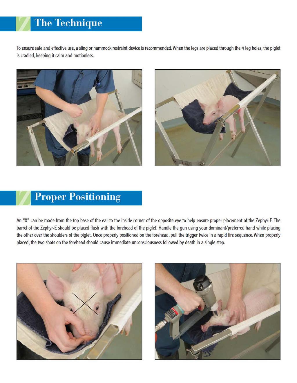
141 132
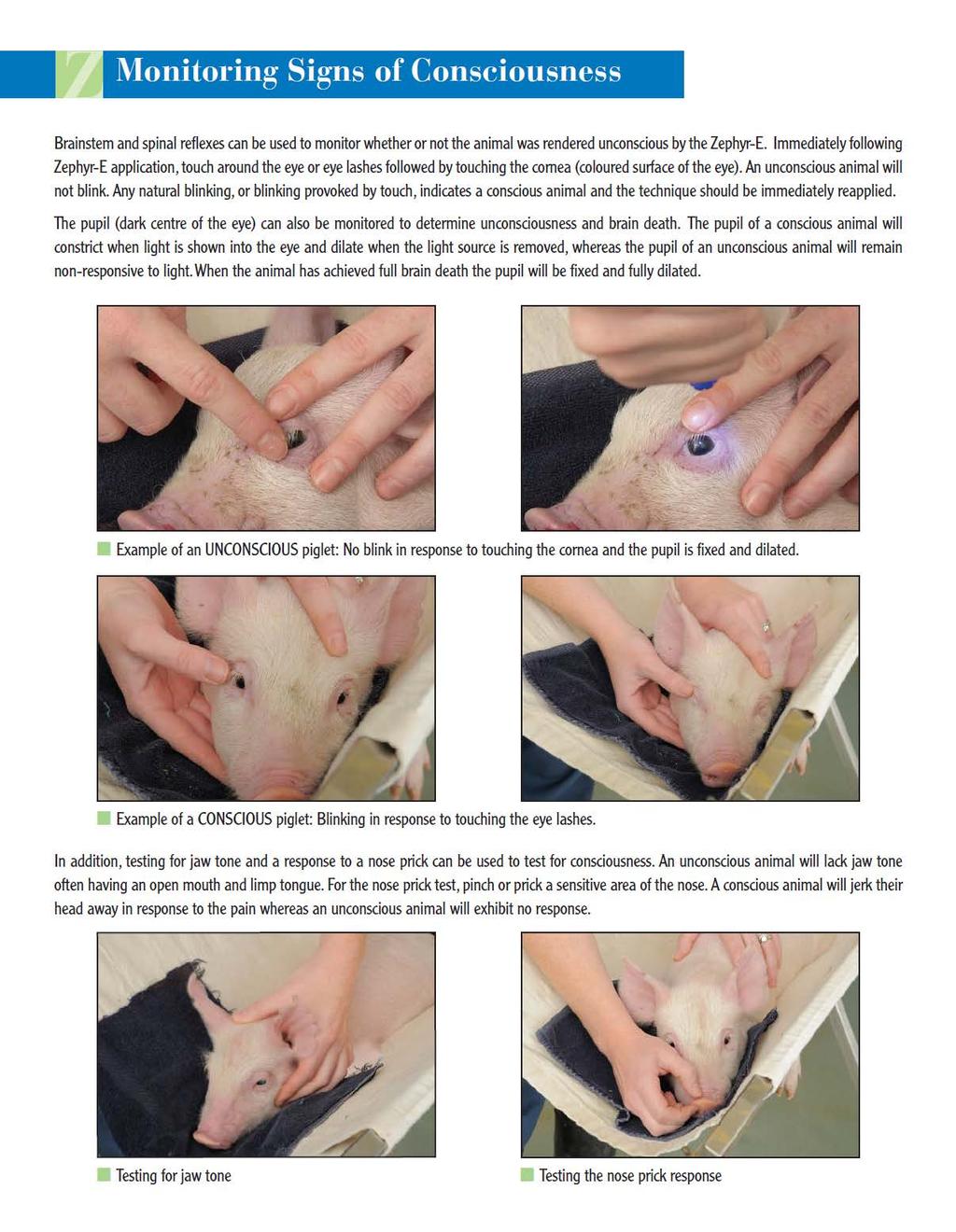
142 133
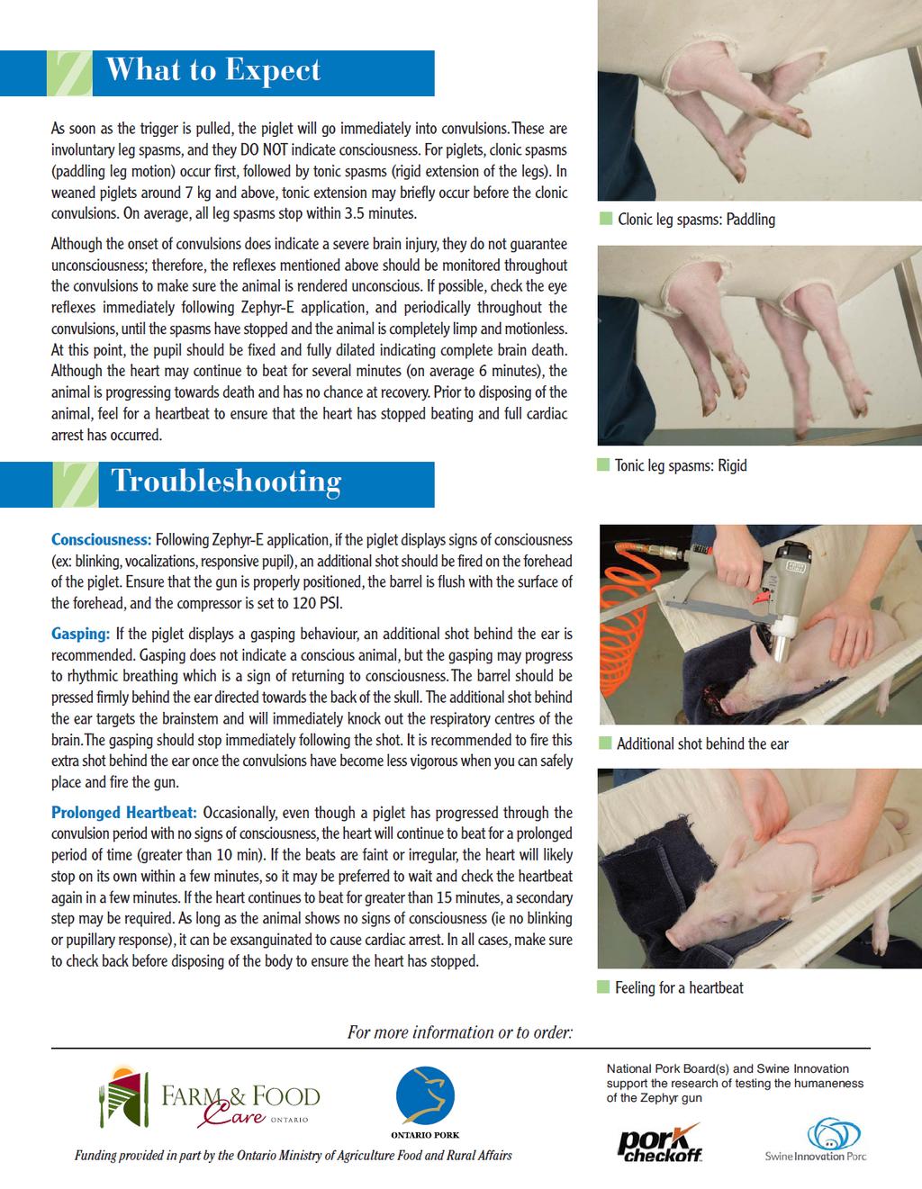
143 134
Effectiveness of a non-penetrating captive bolt for the euthanasia of piglets from birth to 9 kg
 Effectiveness of a non-penetrating captive bolt for the euthanasia of piglets from birth to 9 kg T.M. Casey-Trott 1 ; R. Brooks 2 ; P.V. Turner 1 ; S.G. Nykamp 1 ; M. Litman 1 ; S.M. Millman 2 ; T.M. Widowski
Effectiveness of non-penetrating captive bolt (Zephyr) and restraint for euthanasia of piglets from birth to 9 kg NPB #
 and restraint for euthanasia of piglets from birth to 9 kg NPB #") Title: Investigator: Institution: Effectiveness of non-penetrating captive bolt (Zephyr) and restraint for euthanasia of piglets from birth to 9 kg NPB # 09-190 Dr. Tina Widowski University of Guelph Co-investigators:
Title: Investigator: Institution: Effectiveness of non-penetrating captive bolt (Zephyr) and restraint for euthanasia of piglets from birth to 9 kg NPB # 09-190 Dr. Tina Widowski University of Guelph Co-investigators:
Pre-Slaughter Stunning: Why it is important
 A Greener World Technical Advice Fact Sheet No. 18 Pre-Slaughter Stunning: Why it is important Certified Animal Welfare Approved by A Greener World (AGW) has the most rigorous standards for farm animal
Animal Welfare at Slaughter
 Animal Welfare at Slaughter The purpose of any method of stunning is to render the animal immediately unconscious until it is dead Haluk ANIL - The Welfare of Animals (Slaughter or Killing) Regulations
Indicators for assessing unconsciousness and death during slaughter without stunning
 Indicators for assessing unconsciousness and death during slaughter without stunning Antonio Velarde Parma, January, 30th 2013 References - EFSA opinions of 2004 and 2006 on the stunning and killing of
On-Farm Euthanasia of Swine
 On-Farm Euthanasia of Swine Sherrie Niekamp National Pork Board 1 Outline What is euthanasia Implications of euthanasia Importance of timeliness Available methods Confirming insensibility and death Ongoing
Analysis of the Cash Euthanizer system in commercial production settings
 Analysis of the Cash Euthanizer system in commercial production settings J.A. Woods; J.H. Hill; K.J. Schwartz; L.J. Sadler; R.L. Parsons; T. Grandin; S.T. Millman The objective of this study was to assess
Determining Insensibility for Accurate Stunning and On-Farm Euthanasia
 Determining Insensibility for Accurate Stunning and On-Farm Euthanasia Recognition that animals can perceive pain is fundamental to animal welfare. Euthanasia implies that death occurs with minimal pain
WHAT ARE the COMPONENTS OF THE NERVOUS SYSTEM?
 The Nervous System WHAT ARE the COMPONENTS OF THE NERVOUS SYSTEM? The nervous system is made of: the brain & the spinal cord the nerves the senses There are lots of proteins and chemicals in your body
Stamping out procedures (domestic pigs)
") Stamping out procedures (domestic pigs) Sixth meeting of the Standard Group of Experts on ASF in the Baltic and Eastern Europe Region Vilnius, Lithuania 28-29 November 2016 Jorgen M. Westergaard OVERVIEW
Stamping out procedures (domestic pigs) Sixth meeting of the Standard Group of Experts on ASF in the Baltic and Eastern Europe Region Vilnius, Lithuania 28-29 November 2016 Jorgen M. Westergaard OVERVIEW
V. CENTRAL NERVOUS SYSTEM TRAUMA
 V. CENTRAL NERVOUS SYSTEM TRAUMA I. Concussion - Is a clinical syndrome of altered consiousness secondary to head injury - Brought by a change in the momentum of the head when a moving head suddenly arrested
Myers Psychology for AP*
 Myers Psychology for AP* David G. Myers PowerPoint Presentation Slides by Kent Korek Germantown High School Worth Publishers, 2010 *AP is a trademark registered and/or owned by the College Board, which
Unit 3: The Biological Bases of Behaviour
 Unit 3: The Biological Bases of Behaviour Section 1: Communication in the Nervous System Section 2: Organization in the Nervous System Section 3: Researching the Brain Section 4: The Brain Section 5: Cerebral
8.3 The Central Nervous System. SBI4U Ms. Ho-Lau
 8.3 The Central Nervous System SBI4U Ms. Ho-Lau The Central Nervous System the structural and functional centre for the entire nervous system the site of neural integration and processing The Central
Nervous System: Part IV The Central Nervous System The Brain
 Nervous System: Part IV The Central Nervous System The Brain Can you survive when part of your brain is destroyed? 2 Essential Knowledge 3.D.2 2. Cells communicate with each other through direct contact
fmri (functional MRI)
") Lesion fmri (functional MRI) Electroencephalogram (EEG) Brainstem CT (computed tomography) Scan Medulla PET (positron emission tomography) Scan Reticular Formation MRI (magnetic resonance imaging) Thalamus
Lesion fmri (functional MRI) Electroencephalogram (EEG) Brainstem CT (computed tomography) Scan Medulla PET (positron emission tomography) Scan Reticular Formation MRI (magnetic resonance imaging) Thalamus
Neurology study of the nervous system. nervous & endocrine systems work together to maintain homeostasis
 Nervous System Neurology study of the nervous system nervous & endocrine systems work together to maintain homeostasis Nervous System works very fast Uses electrical signals called nerve impulses Short-lived
W. Berry, D. Bourassa, J. Davis, J. Hess, J. Johnson, A. Morey, R. Wallace Auburn University Department of Poultry Science
 Advancements in Poultry Stunning W. Berry, D. Bourassa, J. Davis, J. Hess, J. Johnson, A. Morey, R. Wallace Auburn University Department of Poultry Science Purpose and Goals of Stunning An effective stunning
Understanding Neurological Death
 Understanding Neurological Death Special thanks to Trillium Gift Of Life Network s Donor Family Advisory Council, whose dedication and commitment never fail to profoundly move us all. Understanding Neurological
Nervous System and Brain Review. Bio 3201
 Nervous System and Brain Review Bio 3201 Dont worry about: glial cells Oligodendrocytes Satelite cells etc Nervous System - Vital to maintaining homeostasis in organisms - Comprised of : brain, spinal
Activity Overview. A Piece of Your Mind: Brain Anatomy Teacher Pages Activity 1A. The Brain: It s All In Your Mind
 A Piece of Your Mind: Brain Anatomy Teacher Pages Activity Objectives: Using diagrams of the brain, students will be able to: Observe the basic anatomy of the brain and its components Identify the parts
THE CENTRAL NERVOUS SYSTEM. The Brain & Spinal Cord
 THE CENTRAL NERVOUS SYSTEM The Brain & Spinal Cord Review: Nervous System Parallel Distributed Processing Composition of the CNS Nuclei: Clusters of neurons in the CNS ( neighborhoods ) Fiber Tracts/Pathways:
THE NERVOUS SYSTEM CONCEPT 2: THE VERTEBRATE BRAIN IS REGIONALLY SPECIALIZED
 THE NERVOUS SYSTEM CONCEPT 2: THE VERTEBRATE BRAIN IS REGIONALLY SPECIALIZED Images of the human brain in popular culture almost always focus on the cerebrum, the part of the brain whose surface lies just
Biology 3201 Unit 1: Maintaining Dynamic Equilibrium II
 Biology 3201 Unit 1: Maintaining Dynamic Equilibrium II Ch. 12 The Nervous System (Introduction and Anatomy) The Nervous System - Introduction Cells, tissues, organs and organ systems must maintain a biological
LOSS OF CONSCIOUSNESS & ASSESSMENT. Sheba Medical Center Acute Medicine Department MATTHEW WRIGHT
 LOSS OF CONSCIOUSNESS & ASSESSMENT Sheba Medical Center Acute Medicine Department MATTHEW WRIGHT OUTLINE Causes Head Injury Clinical Features Complications Rapid Assessment Glasgow Coma Scale Classification
CHAPTER 48: NERVOUS SYSTEMS
 CHAPTER 48: NERVOUS SYSTEMS Name I. AN OVERVIEW OF NERVOUS SYSTEMS A. Nervous systems perform the three overlapping functions of sensory input, integration, and motor output B. Networks of neurons with
Head Trauma Inservice (October)
") John Tramell - Head Trauma Inservice, October 2005.doc Page 1 Head Trauma Inservice (October) Head trauma is the leading cause of death in trauma patients. Having a basic understanding of the anatomy and
John Tramell - Head Trauma Inservice, October 2005.doc Page 1 Head Trauma Inservice (October) Head trauma is the leading cause of death in trauma patients. Having a basic understanding of the anatomy and
Basic Brain Information
 Basic Brain Information Brain facts Your brain weighs about 3lbs, or just under 1.5Kg It has the texture of blancmange Your brain is connected to your spinal cord by the brain stem Behind your brain stem
Nervous System - PNS and CNS. Bio 105
 Nervous System - PNS and CNS Bio 105 Outline I. Central Nervous System vs Peripheral Nervous System II. Peripheral Nervous System A. Autonomic Nervous Systems B. Somatic Nervous Systems III. Autonomic
Biology. Slide 1 of 37. End Show. Copyright Pearson Prentice Hall
 Biology 1 of 37 35-3 Divisions of the Nervous 2 of 37 The Nervous The human nervous system has two major divisions: central nervous system peripheral nervous system 3 of 37 The Central Nervous The Central
III. Studying The Brain and Other Structures
 III. Studying The Brain and Other Structures 1. Accidents (case study) In 1848, a railroad worker named Phineas Gage was involved in an accident that damaged the front part of his brain. Gage s doctor
3/15/17. Outline. Nervous System - PNS and CNS. Two Parts of the Nervous System
 Nervous System - PNS and CNS Bio 105 Outline I. Central Nervous System vs Peripheral Nervous System II. Peripheral Nervous System A. Autonomic Nervous Systems B. Somatic Nervous Systems III. Autonomic
BRAIN: CONTROL CENTER
 BRAIN: CONTROL CENTER ORCHESTRA Scientists now believe the brain functions much like an orchestra, where different instruments each play a different part. Scans show that the brain divides different aspects
The Nervous System PART B
 7 The Nervous System PART B PowerPoint Lecture Slide Presentation by Jerry L. Cook, Sam Houston University ESSENTIALS OF HUMAN ANATOMY & PHYSIOLOGY EIGHTH EDITION ELAINE N. MARIEB Central Nervous System
1. Processes nutrients and provides energy for the neuron to function; contains the cell's nucleus; also called the soma.
 1. Base of brainstem; controls heartbeat and breathing 2. tissue destruction; a brain lesion is a naturally or experimentally caused destruction of brain tissue 3. A thick band of axons that connects the
Chapter 17. Nervous System Nervous systems receive sensory input, interpret it, and send out appropriate commands. !
 Chapter 17 Sensory receptor Sensory input Integration Nervous System Motor output Brain and spinal cord Effector cells Peripheral nervous system (PNS) Central nervous system (CNS) 28.1 Nervous systems
14 - Central Nervous System. The Brain Taft College Human Physiology
 14 - Central Nervous System The Brain Taft College Human Physiology Development of the Brain The brain begins as a simple tube, a neural tube. The tube or chamber (ventricle) is filled with cerebrospinal
Traumatic brain injuries are caused by external mechanical forces such as: - Falls - Transport-related accidents - Assault
 PP2231 Brain injury Cerebrum consists of frontal, parietal, occipital and temporal lobes Diencephalon consists of thalamus, hypothalamus Cerbellum Brain stem consists of midbrain, pons, medulla Central
Basic Brain Structure
 The Human Brain Basic Brain Structure Composed of 100 billion cells Makes up 2% of bodies weight Contains 15% of bodies blood supply Uses 20% of bodies oxygen and glucose Brain Protection Surrounded by
Page 1. Neurons Transmit Signal via Action Potentials: neuron At rest, neurons maintain an electrical difference across
 Chapter 33: The Nervous System and the Senses Neurons: Specialized excitable cells that allow for communication throughout the body via electrical impulses Neuron Anatomy / Function: 1) Dendrites: Receive
Parts of the Brain. Hindbrain. Controls autonomic functions Breathing, Heartbeat, Blood pressure, Swallowing, Vomiting, etc. Upper part of hindbrain
 Parts of the Brain The human brain is made up of three main parts: 1) Hindbrain (or brainstem) Which is made up of: Myelencephalon Metencephalon 2) Midbrain Which is made up of: Mesencephalon 3) Forebrain
CNS composed of: Grey matter Unmyelinated axons Dendrites and cell bodies White matter Myelinated axon tracts
 CNS composed of: Grey matter Unmyelinated axons Dendrites and cell bodies White matter Myelinated axon tracts The Brain: A Quick Tour Frontal Lobe Control of skeletal muscles Personality Concentration
Ch 13: Central Nervous System Part 1: The Brain p 374
 Ch 13: Central Nervous System Part 1: The Brain p 374 Discuss the organization of the brain, including the major structures and how they relate to one another! Review the meninges of the spinal cord and
Organization of the nervous system. The withdrawal reflex. The central nervous system. Structure of a neuron. Overview
 Overview The nervous system- central and peripheral The brain: The source of mind and self Neurons Neuron Communication Chemical messengers Inside the brain Parts of the brain Split Brain Patients Organization
TABLE OF CONTINENTS. PSYC1002 Notes. Neuroscience.2. Cognitive Processes Learning and Motivation. 37. Perception Mental Abilities..
 TABLE OF CONTINENTS Neuroscience.2 Cognitive Processes...21 Learning and Motivation. 37 Perception.....54 Mental Abilities.. 83 Abnormal Psychology....103 1 Topic 1: Neuroscience Outline 1. Gross anatomy
Chapter 26 Head and Spine Trauma The Nervous System The nervous system controls virtually all of our body activities including reflex, voluntary and
 1 2 3 4 5 Chapter 26 Head and Spine Trauma The Nervous System The nervous system controls virtually all of our body activities including reflex, voluntary and involuntary activities Voluntary activities
Traumatic Brain Injury TBI Presented by Bill Masten
 1 2 Cerebrum two hemispheres and four lobes. Cerebellum (little brain) coordinates the back and forth ballet of motion. It judges the timing of every movement precisely. Brainstem coordinates the bodies
Functional Organization of the Central Nervous System
 Functional Organization of the Central Nervous System Hierarchical orgnization CNS consists of the brain and the spinal cord The brain analyzes and interprets the information Response messages are
The Nervous System. The right half of the brain controls the left half of the body. This means that only left handed people are in their right mind.
 The Nervous System The right half of the brain controls the left half of the body. This means that only left handed people are in their right mind. Central Nervous System Made up of brain and spinal cord
Neural Integration I: Sensory Pathways and the Somatic Nervous System
 15 Neural Integration I: Sensory Pathways and the Somatic Nervous System PowerPoint Lecture Presentations prepared by Jason LaPres Lone Star College North Harris An Introduction to Sensory Pathways and
Electrical and CO 2 Stunning, Handling, and Determining Insensibility in Pigs and Sheep, 2 nd Edition
 Electrical and CO 2 Stunning, Handling, and Determining Insensibility in Pigs and Sheep, 2 nd Edition Temple Grandin Department of Animal Sciences Colorado State University Humane Slaughter Act All animals
PARA210 SUMMARY Hyperglycaemia (DKA & HHS) Brain & Nervous System Anatomy & Physiology Degenerative Neurological Disorders
 Brain & Nervous System Anatomy & Physiology Degenerative Neurological Disorders") PARA210 SUMMARY Page Topic 01-03 Diabetes Mellitus 04-05 Hyperglycaemia (DKA & HHS) 06-13 Toxicology 14-18 12 Lead ECG 19-21 Brain & Nervous System Anatomy & Physiology 22-24 Degenerative Neurological
PARA210 SUMMARY Page Topic 01-03 Diabetes Mellitus 04-05 Hyperglycaemia (DKA & HHS) 06-13 Toxicology 14-18 12 Lead ECG 19-21 Brain & Nervous System Anatomy & Physiology 22-24 Degenerative Neurological
Biology 3201 Nervous System #2- Anatomy. Components of a Nervous System
 Biology 3201 Nervous System #2- Anatomy Components of a Nervous System In any nervous system, there are 4 main components: (1) sensors: gather information from the external environment (sense organs) (2)
On-farm poultry euthanasia technologies. Phase 1 Lab Based Evaluation
 On-farm poultry euthanasia technologies Phase 1 Lab Based Evaluation 1 Evaluation of Euthanasia Technologies Institute for Applied Poultry Technologies (IAPT) project: Applicability of Commercial Euthanasia
Principles of Anatomy and Physiology
 Principles of Anatomy and Physiology 14 th Edition CHAPTER 14 The Brain and Cranial Nerves Introduction The purpose of the chapter is to: 1. Understand how the brain is organized, protected, and supplied
Somatic Nervous Systems. III. Autonomic Nervous System. Parasympathetic Nervous System. Sympathetic Nervous Systems
 7/21/2014 Outline Nervous System - PNS and CNS I. II. Two Parts of the Nervous System Central Nervous System vs Peripheral Nervous System Peripheral Nervous System A. B. Brain and Spinal Cord III. Autonomic
Chapter 9. Nervous System
 Chapter 9 Nervous System Central Nervous System (CNS) vs. Peripheral Nervous System(PNS) CNS Brain Spinal cord PNS Peripheral nerves connecting CNS to the body Cranial nerves Spinal nerves Neurons transmit
Class X: Biology. Chapter 2: Control and coordination.
 Class X: Biology Chapter 2: Control and coordination. Chapter Notes Key learnings: 1) A system of control and coordination is essential in living organisms so that the different body parts can function
Unit Six The Nervous System
 Unit Six The Nervous System I. Introduction A. Definition a coordinating system of the body, composed of highly specialized cells that conduct nerve impulses to a center so responses can be made. The nervous
Chapter 7: The Nervous System
 Name: Block: Chapter 7: The Nervous System Lesson 1: How the Nervous System Works What is the Role of the Nervous System? Your nervous system receives information about what is happening both inside and
Homework Week 2. PreLab 2 HW #2 Synapses (Page 1 in the HW Section)
") Homework Week 2 Due in Lab PreLab 2 HW #2 Synapses (Page 1 in the HW Section) Reminders No class next Monday Quiz 1 is @ 5:30pm on Tuesday, 1/22/13 Study guide posted under Study Aids section of website
Homework Week 2 Due in Lab PreLab 2 HW #2 Synapses (Page 1 in the HW Section) Reminders No class next Monday Quiz 1 is @ 5:30pm on Tuesday, 1/22/13 Study guide posted under Study Aids section of website
The Nervous System. Functions of the Nervous System input gathering To monitor occurring inside and outside the body Changes =
 The Nervous System Functions of the Nervous System input gathering To monitor occurring inside and outside the body Changes = To process and sensory input and decide if is needed output A response to integrated
Module 5 : Anatomy The nervous system
 Module 5 : Anatomy The nervous system In this module you will learn: The main parts of the nervous system The different sections of the brain and how it functions The structure and function of the spinal
Nervous system, integration: Overview, and peripheral nervous system:
 Nervous system, integration: Overview, and peripheral nervous system: Some review & misc. parts [Fig. 28.11B, p. 573]: - white matter --> looks white due to the myelinated sheaths, which are quite fatty.
action potential afferent neuron Weblike; specifically, the weblike middle layer of the three meninges. arachnoid astrocytes autonomic nervous system
 action potential A large transient depolarization event, including polarity reversal, that is conducted along the membrane of a muscle cell or a nerve fiber. afferent neuron Nerve cell that carries impulses
Pediatric Abusive Head Trauma
 Pediatric Abusive Head Trauma Rebecca Girardet Associate Professor of Pediatrics Director, Division of Child Protection Pediatrics McGovern Medical School at The University of Texas Health Science Center
The nervous system regulates most body systems using direct connections called nerves. It enables you to sense and respond to stimuli
 The nervous system regulates most body systems using direct connections called nerves. It enables you to sense and respond to stimuli The basic function of nervous system are: Receive sensory input internal
Nervous System: An Introduction. HAP Susan Chabot Lemon Bay High School
 Nervous System: An Introduction HAP Susan Chabot Lemon Bay High School Function of the Nervous System 3 overlapping functions SENSORY INPUT - Monitor changes inside and outside of the body; these changes
The Nervous System. Divisions of the Nervous System. Branches of the Autonomic Nervous System. Central versus Peripheral
 The Nervous System Divisions of the Nervous System Central versus Peripheral Central Brain and spinal cord Peripheral Everything else Somatic versus Autonomic Somatic Nerves serving conscious sensations
TRAUMATIC BRAIN INJURY. Moderate and Severe Brain Injury
 TRAUMATIC BRAIN INJURY Moderate and Severe Brain Injury Disclosures Funded research: 1. NIH: RO1 Physiology of concussion 2016-2021, Co-PI, $2,000,000 2. American Medical Society of Sports Medicine: RCT
Anatomy & Physiology Central Nervous System Worksheet
 1. What are the two parts of the CNS? 2. What are the four functions of the CNS Anatomy & Physiology Central Nervous System Worksheet 3. What are the four functions of the meninges? (p430) 4. Starting
The Human Brain: Anatomy, Functions, and Injury
 The Human Brain: Anatomy, Functions, and Injury Main Menu Brain Anatomy Brain Functions Injury Mechanisms Brain Anatomy Menu Skull Anatomy Interior Skull Surface Blood Vessels of the Brain Arteries of
Head Injury: Classification Most Severe to Least Severe
 Head Injury: Classification Most Severe to Least Severe Douglas I. Katz, MD Professor, Dept. Neurology, Boston University School of Medicine, Boston MA Medical Director Brain Injury Program, HealthSouth
Primary Functions. Monitor changes. Integrate input. Initiate a response. External / internal. Process, interpret, make decisions, store information
 NERVOUS SYSTEM Monitor changes External / internal Integrate input Primary Functions Process, interpret, make decisions, store information Initiate a response E.g., movement, hormone release, stimulate/inhibit
THE ESSENTIAL BRAIN INJURY GUIDE
 THE ESSENTIAL BRAIN INJURY GUIDE Neuroanatomy & Neuroplasticity Section 2 Contributors Erin D. Bigler, PhD Michael R. Hoane, PhD Stephanie Kolakowsky-Hayner, PhD, CBIST, FACRM Dorothy A. Kozlowski, PhD
Cephalization. Nervous Systems Chapter 49 11/10/2013. Nervous systems consist of circuits of neurons and supporting cells
 Nervous Systems Chapter 49 Cephalization Nervous systems consist of circuits of neurons and supporting cells Nervous system organization usually correlates with lifestyle Organization of the vertebrate
CNS pathology Third year medical students. Dr Heyam Awad 2018 Lecture 5: disturbed fluid balance and increased intracranial pressure
 CNS pathology Third year medical students Dr Heyam Awad 2018 Lecture 5: disturbed fluid balance and increased intracranial pressure ILOs Understand causes and symptoms of increased intracranial pressure.
EFFECTIVE TREATMENT AND HANDLING OF POOR DOING PIGS IN THE FINISHING BARN
 EFFECTIVE TREATMENT AND HANDLING OF POOR DOING PIGS IN THE FINISHING BARN Tim Blackwell Ontario Ministry of Agriculture and Food Wellington Place, RR #1 Fergus, Ontario N1M 2W3 E-mail: tim.blackwell@omaf.gov.on.ca
Neural Basis of Motor Control
 Neural Basis of Motor Control Central Nervous System Skeletal muscles are controlled by the CNS which consists of the brain and spinal cord. Determines which muscles will contract When How fast To what
The CNS and PNS: How is our Nervous System Organized?
 Honors Biology Guided Notes Chapter 28 Nervous System Name 28.10 28.19 The CNS and PNS: How is our Nervous System Organized? ANIMAL NERVOUS SYSTEMS Define Cephalization and Centralization. What type of
 BRAIN AND ITS VITAL FUNCTIONS 1 Brain and Its Vital Functions Student s Name Institution Name Professor s Name Course Title BRAIN AND ITS VITAL FUNCTIONS 2 The brain is the integral organism and all its
The Nervous System: Sensory and Motor Tracts of the Spinal Cord
 15 The Nervous System: Sensory and Motor Tracts of the Spinal Cord PowerPoint Lecture Presentations prepared by Steven Bassett Southeast Community College Lincoln, Nebraska Introduction Millions of sensory
Chapter 3. Structure and Function of the Nervous System. Copyright (c) Allyn and Bacon 2004
 Allyn and Bacon 2004") Chapter 3 Structure and Function of the Nervous System 1 Basic Features of the Nervous System Neuraxis: An imaginary line drawn through the center of the length of the central nervous system, from the
Chapter 3 Structure and Function of the Nervous System 1 Basic Features of the Nervous System Neuraxis: An imaginary line drawn through the center of the length of the central nervous system, from the
WHITE PAPER: A GUIDE TO UNDERSTANDING SUBARACHNOID HEMORRHAGE
 WHITE PAPER: A GUIDE TO UNDERSTANDING SUBARACHNOID HEMORRHAGE Subarachnoid Hemorrhage is a serious, life-threatening type of hemorrhagic stroke caused by bleeding into the space surrounding the brain,
BRAIN DEATH. Frequently Asked Questions 04for the General Public
 BRAIN DEATH Frequently Asked Questions 04for the General Public Neurocritical Care Society BRAIN DEATH FAQ s FOR THE GENERAL PUBLIC NEUROCRITICAL CARE SOCIETY 1. Q: Why was this FAQ created? A: Several
Introduction to Emergency Medical Care 1
 Introduction to Emergency Medical Care 1 OBJECTIVES 31.1 Define key terms introduced in this chapter. Slides 13 15, 17, 19, 28 31.2 Describe the components and function of the nervous system and the anatomy
Chapter 6. Body and Behavior
 Chapter 6 Body and Behavior Section 1 The Nervous System: The Basic Structure How the nervous system works Central nervous system (CNS)- the brain and spinal cord Spinal cord- nerves that run up and down
CHAPTER 13&14: The Central Nervous System. Anatomy of the CNS
 CHAPTER 13&14: The Central Nervous System Anatomy of the CNS in human consists of brain and spinal cord as stated earlier neurons have little support from their extracellular matrix and depend on glial
Keeping your born alive alive! A focus on fostering
 Keeping your born alive alive! A focus on fostering Keelin O Driscoll, Amy Quinn, Julia Calderón Díaz and Laura Boyle Teagasc, Moorepark Introduction Birth to weaning period is most dangerous in a pig
Bellringer: The central nervous system is comprised of: What is the name of the outermost layer of the brain? a. Brain. b.
 Bellringer: The central is comprised of: a. Brain b. Spinal cord c. Sensory receptors d. Both a and b What is the name of the outermost layer of the brain? a. Pia mater b. Dura mater c. Arachnoid d. Pons
Five Levels of Organization Cell Tissue Organ Organ System Organism
 28.1 35.1 Levels Human of Body Organization Systems Five Levels of Organization Cell Tissue Organ Organ System Organism ORGANS ORGAN SYSTEM ORGANISM 28.1 35.1 Levels Human of Body Organization Systems
1. NERVOUS SYSTEM FUNCTIONS OF THE NERVOUS SYSTEM. FUNCTION The major function of the nervous system can be summarized as follows (Figure 1-1).
.") 1. NERVOUS SYSTEM FUNCTION The major function of the nervous system can be summarized as follows (Figure 1-1). Sensory input. Multiple signals from both, internal and external environment are detected
1. NERVOUS SYSTEM FUNCTION The major function of the nervous system can be summarized as follows (Figure 1-1). Sensory input. Multiple signals from both, internal and external environment are detected
15-2: Divisions of the Nervous System. 7 th Grade Life Science
 7 th Grade Life Science Purpose Students will be able to: 1. Identify the functions of the nervous system. 2. Describe the structure of a neuron and the kinds of neurons found in the body. 3. Explain how
Introduction to Emergency Medical Care 1
 Introduction to Emergency Medical Care 1 OBJECTIVES 6.1 Define key terms introduced in this chapter. Slides 11, 15, 17, 26, 27, 31, 33, 37, 40 42, 44, 45, 51, 58 6.2 Describe the basic roles and structures
meninges Outermost layer of the meninge dura mater arachnoid mater pia mater membranes located between bone and soft tissue of the nervous system
 membranes located between bone and soft tissue of the nervous system meninges Outermost layer of the meninge dura mater middle layer of the meninges, contains no blood vessels arachnoid mater Innermost
Psychology in Your Life
 Sarah Grison Todd Heatherton Michael Gazzaniga Psychology in Your Life SECOND EDITION Chapter 2 The Role of Biology in Psychology 1 2016 W. W. Norton & Company, Inc. 2.1 How Do Our Nervous Systems Affect
Unit Three. The brain includes: cerebrum, diencephalon, brain stem, & cerebellum. The brain lies within the cranial cavity of the skull.
 Human Anatomy & Physiology 11 Divisions of the Nervous System Karen W. Smith, Instructor Unit Three BRAIN & SPINAL CORD Refer to the following URLs. Be sure to study these along with your book. http://www.sirinet.net/~jgjohnso/nervous.html
Stunning, Handling, and Determining Insensibility in Pigs. Temple Grandin Department of Animal Sciences Colorado State University
 Stunning, Handling, and Determining Insensibility in Pigs Temple Grandin Department of Animal Sciences Colorado State University Trouble Shooting Handling 1. Distractions that cause balking 2. Slick floor
b. The groove between the two crests is called 2. The neural folds move toward each other & the fuse to create a
 Chapter 13: Brain and Cranial Nerves I. Development of the CNS A. The CNS begins as a flat plate called the B. The process proceeds as: 1. The lateral sides of the become elevated as waves called a. The