4/30/2009 FN PARALYSIS HEMIFACIAL SPASM
|
|
|
- Rosamund Paul
- 6 years ago
- Views:
Transcription


1 FN PARALYSIS HEMIFACIAL SPASM 1
2 Hemifacial spasm (Involuntary twitches) Etiology: Vascular loop compressing the FN at the root exit zone in the CPA = Neuro-vascular conflict Diagnosis: CT & MRI, Electrophysiology Treatment: Medical Surgical: Microvascular decompression via Retrosigmoid approach). FACIAL PALSY 2


3 7000 axons: Motor 3000 axons: Secretomotor 3
4 Three nuclei supply the FN: A- The motor nucleus: in the pons. Sup. Part: bilateral innervations from the motor cortex, inferior part: unilateral crossed cortical innervations. B- The superior salivatory nucleus: parasympathetic secretory fibers to lacrimal glands, nasal glands, submandibular, sublingual and palatal salivary glands. C- The nucleus of the tractus solitarius: receives taste sensation. It arises from the inferior border of the pons (pontomedullary sulcus) crosses the CPA to enter the IAC. Anatomical segments: 1- Intracranial segment 2- IAC segment 3- Labyrinthine segment which ends by the geniculate ganglion 4- Tympanic (horizontal) segment 5- Mastoid (vertical) segment which ends by the stylomastoid foramen. 4


5 5
 2- Nerve to stapedius muscle 3- Chorda tympani")

6 Branches: A- Intra-temporal branches: 1- Greater superficial petrosal (parasympathetic secretory) 2- Nerve to stapedius muscle 3- Chorda tympani (Taste) B- Extra-temporal branches: p 1- Post auricular nerve 2- Nerve to stylohyoid & posterior belly of digastric 3- Cranio-temporal division 4- Cranio-cervical division 6


7 7


8 8
9 1. Neuropraxia: Physiologic block, No anatomical disruption, temporary, lasts few days and full return of function is expected. The axoplasm can still be transported from and to the cell body across the blocked area and so; the nerve fiber distal to the site of injury retains normal electrical response. 2. Axonotmesis: Axon sheath is intact but the axon is divided. i.e. more severe lesion. Distal degeneration (Wallerian) occurs. Epineurium is intact. 3. Neurotmesis: Epineurium enclosing all nerve fascicles is also torn. Regeneration is the worst and traumatic neuroma may develop. Seddon Classification Sunderland Classification Pathophysiologic Process Neuropraxia I Physiologic block Axonotmesis II III IV Axons disrupted Axons and endoneurium disrupted Axons, endoneurium, & perineurium disrupted Neurotmesis V Complete transection 9


10 Wh i h l i? - What is the lesion? - Where is the lesion? - Degree of the lesion: - Causes of the lesion: 10
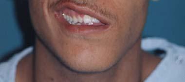
11 What is the lesion? Is the face paralyzed or not? Inspection of the face movement. Testing for facial movement and emotional movement. (Laughing; blinking, wrinkling, closing eyes, whistling, grinning and blowing of the cheeks). - Where is the lesion? Lesions can be either : 1. Supra-nuclear (UMNL), 2. Nuclear, 3. Infra-nuclear (LMNL). 11


12 Difference Supra-Nuclear UMNL Infra-Nuclear LMNL Type of paralysis Spastic Flaccid Distribution Lower face Whole face Emotional Movement Free Paralyzed Other signs Crossed hemiplegia -ve 12
13 Where is the lesion? i- Nuclear lesions are associated by crossed hemiplegia, crossed hemianesthesia, other cranial nerves palsies specially VI. Lacrimation, salivation and taste are normal. ii- LMNL : FN may be affected at different sites of its course. 1 Schirmer s tear test 1. Schirmer s tear test 2. Stapedius reflex 3. Evaluation of Taste 4. Submandibular salivary flow 13

 3.")
14 Degree of the lesion 1. Maximum nerve excitability test (Hilgers test) 2. Electroneuronograpy (ENoG) 3. Electromyography yg py( (EMG) 14
15 1- Maximum nerve excitability test (Hilger s test): The response on the involved side is compared to the normal side. The idea is that the distal segment of a completely cut nerve may continue to conduct when stimulated by a faradic current (distal to the cut) for as long as hours. The response then disappear due to Wallerian degeneration. Ideally testing is started on the third post onset day. 2- Electroneuronography (ENOG) = Evoked electromyography: Similar to maximum stimulation test in which the evoked CAP are recorded. The height of the action potential is compared with the good side and a percentage comparison is made. It is a quantitative test. Degeneration of more than 90% means recovery is poor. 15


16 3- Electromyography (EMG): The paralyzed side is explored by the EMG needle while the patient is attempting to move it. In the first 10 days, the presence of voluntary CAP indicates less than total transaction. After days, the presence of fibrillation indicates denervation. After 10 weeks have passed, the test is prognostic. These action potentials can be found a week or two before the clinical evidence of recovery. 16
17 Supra-nuclear or nuclear: Hemorrhage; vascular; encephalitis; tumors, cerebral palsy;. Infra-nuclear: 1. Congenital: poor facial muscular development 2. Inflammatory: Malignant ext. otitis; AOM, Ch.OM, cholesteatoma, Herpes zoster oticus (Ramasy Hunt syndrome), spirochetes (Lyme disease) 3. Traumatic: Fracture base, forceps delivery, 4. Toxic: Diphteria, tetanus,.. 5. Metabolic: Diabetes 6. Neoplastic: 1ry tumor: FN neuroma; 2ry tumors of middle ear and parotid and CPA. 7. Iatrogenic: mastoid, parotid, CPA surgery 8. Idiopathic: Bell s palsy A- Complications due to FN paralysis: 1- Drooling 2- Eye complication: exposure keratitis; corneal ulcer; panophthalmitis. 3- Facial contractures (fixed shortening of the muscles) 4- Psychological disturbance, depression, and suicidal tendencies. B- Complications due to abnormal regeneration 1. Facial tics and spasms: due to short-circuiting between nerve fibers 2. Abnormal mass movement of the face: Synkinesis. 3. Crocodile tears: due to misdirected salivation fibers to the lacrimal gland. 4. Frey s syndrome: facial flushing and sweating over the parotid during mastication = misdirected salivary fibers to the sweat glands. 17


18 1- Treatment of the cause 2- Treatment of the complication: eg. Gold weight for protection of the cornea; Facial plastic surgery for the contractures. 3- Treatment of the facial paralysis by facial re-animation surgery meant to: a- Restoration of anatomical integrity of the FN. Eg. FN decompression; anastomosis and grafting. May be indicated in Bell s palsy, Herpes zoster oticus, acute or chronic otitis media, trauma, and tumors b- Restoration of the functional integrity of the neuromuscular system of the face: includes: i. nerve substitution technique e.g. Facial-hypoglossal anastomosis; and ii. muscle substitution techniques e.g. Masseter or temporalis muscle transfer. 4- Facial rehabilitation: e.g. Massage, heat and facial exercises. Facial Nerve Reconstruction End-to-End Anastomosis Line of Anastomosis FN-d. FN-Pr. Tumor bed 18


19 Facial Nerve Reconstruction Sural Nerve Cable Graft FN Graft FN Tumor bed 19
20 Defined as the sudden occurrence of a flaccid facial paralysis (LMNL) in an apparently healthy individual. It is the commonest cause of intratemporal causes of facial paralysis. - Incidence: 1/5000 per year. It is the most common form of FN paralysis. It is attributed to sudden exposure to cold. - Etiology: Due to VIRAL causes (Activation of Herpes Simplex). - Pathogenesis: Segmental demyelination followed by 2ry edema and compression. - Clinical picture: Sudden onset of FN paralysis of LMN type. Pain in the ear is noted in over half of patients. Recovery starts even without treatment in most cases within few days up to 3 weeks. 20
21 - Prognosis: 71% complete recovery; 25% recovers with slight sequalae; and 4% with severe sequelae. Not a single patient who does not show some recovery. - Diagnosis: It s a diagnosis of exclusion. A- Conservative treatment is helpful in 96% of cases. 1. Zovirax (800 mg 5 x daily for ONE week) + Cortisone therapy 2. Physiotherapy treatment: massage, heat, and facial exercises. 3. Dark eye glasses and artificial tears. Protection of the eye. B- Surgical treatment: For complete paralysis with more than 90% degeneration by ENOG within 6 days: Decompression of the FN (specially the entry of the nerve into the Fallopian canal) through Middle Cranial Fossa. 21


22 5-15% of acute facial palsy Palsy appears 2-7 days 30-50% show incomplete recovery Diffuse lymphocytic infiltration..? Vesicles may also affect palate & anterior 2/3 of tongue 22
23 The FN appears to be more severely damaged in herpes zoster oticus than with idiopathic facial palsy. Only 30% of patients will recover normal function, and thus 70% experience some sequelae. Ramsay Hunt Syndrome consists of Facial paralysis; Auditory and vestibular dysfunction associated with Painful herpetic vesicles of the auricle and the EAC. 23
 + 2) Oral steroids 1m/kg/day for 2 weeks 3) Decompression if degeneration is more than 90% in 2 weeks.")
24 MRI + Gadolinium shows enhancement at labyrinthine segment 1) Acyclovir (Zovirax) should be started as early as possible (800 mg 5 times daily for a week) + 2) Oral steroids 1m/kg/day for 2 weeks 3) Decompression if degeneration is more than 90% in 2 weeks. 24
25 1. Accidental: eg. Fracture of the petrous temporal bone: 2. Iatrogenic (Unintentional surgical injury) 3. Surgical (unavoidable injury during surgery) 25


26 Accidental: eg. Fracture of the petrous temporal bone: a- 10% of cases of longitudinal fractures b- 40% of cases of transverse fractures. The site of the lesion is commonly at the geniculate ganglion. c- Extratemporal trauma is not common. Occasionally, direct trauma may damage the nerve. 26
27 Iatrogenic (Unintentional surgical injury) a- Incision of a postauricular abscess in infant. Incision must be placed high. b- Classical cortical & radical mastoidectomies c- Congenital ear surgery d- Surgery of CPA. Intraoperative Facial Nerve Monitoring (IOM) is of great help to prevent surgical trauma during difficult otological/neurotological surgery. Surgical (unavoidable injury during surgery) Impossible to preserve the FN during surgery as in some cases of malignancies, Tumors of the CPA, extensive cholesteatomas, Management: Diagnosis:. electric testing; Management: Diagnosis:. electric testing; Medical steroids; Surgical Exploration.. 27


28 28
Functional components
 Facial Nerve VII cranial nerve Emerges from Pons Two roots Functional components: 1. GSA (general somatic afferent) 2. SA (Somatic afferent) 3. GVE (general visceral efferent) 4. BE (Special visceral/branchial
ASSESSMENT AND TREATMENT OF FACIAL PALSY. Michael J. LaRouere, M.D. Michigan Ear Institute Farmington Hills, Michigan
 ASSESSMENT AND TREATMENT OF FACIAL PALSY Michael J. LaRouere, M.D. Michigan Ear Institute Farmington Hills, Michigan FACIAL PARALYSIS - ETIOLOGY Bells Palsy Ramsay Hunt Syndrome Infection (Acute/Chronic)
By : Prof Saeed Abuel Makarem & Dr.Sanaa Alshaarawi
 By : Prof Saeed Abuel Makarem & Dr.Sanaa Alshaarawi OBJECTIVES By the end of the lecture, students shouldbe able to: List the nuclei of the deep origin of the trigeminal and facial nerves in the brain
Cranial Nerve VII - Facial Nerve. The facial nerve has 3 main components with distinct functions
 Cranial Nerve VII - Facial Nerve The facial nerve has 3 main components with distinct functions Somatic motor efferent Supplies the muscles of facial expression; posterior belly of digastric muscle; stylohyoid,
CHAPTER 13. FACIAL NERVE PARALYSIS
 CHAPTER 13. FACIAL NERVE PARALYSIS Introduction Facial nerve paralysis, whilst not a disease of the ear itself, commonly arises within the ear due to its anatomical course, and often as a result of ear
Laith Sorour. Facial nerve (vii):
:") Laith Sorour Cranial nerves 7 & 8 Hello, there are edited slides please go back to them to see pictures, they are not that much important in this lecture but still, and yes slides are included :p Let s
Laith Sorour Cranial nerves 7 & 8 Hello, there are edited slides please go back to them to see pictures, they are not that much important in this lecture but still, and yes slides are included :p Let s
Cranial Nerve VII & VIII
 Cranial Nerve VII & VIII Lecture Objectives Follow up the course of facial nerve from its point of central connections, exit and down to its target areas. Follow up the central connections of the facial
14, 2007 RESIDENT PHYSICIAN:
 TITLE: Bell s Palsy SOURCE: Grand Rounds Presentation, UTMB, Dept. of Otolaryngology DATE: February 14, 2007 RESIDENT PHYSICIAN: Ki-Hong Kevin Ho, MD FACULTY PHYSICIAN: Shawn D. Newlands, MD, PhD, MBA
Facial Paralysis: Objectives: Discuss the anatomy of the facial nerve. Look at common patterns of facial nerve palsy
 Facial Paralysis: Objectives: Discuss the anatomy of the facial nerve Look at common patterns of facial nerve palsy Discuss imaging appearance of lesions that lead to facial paralysis. Lindell R. Gentry,
Parotid Gland, Temporomandibular Joint and Infratemporal Fossa
 M1 - Anatomy Parotid Gland, Temporomandibular Joint and Infratemporal Fossa Jeff Dupree Sanger 9-057 jldupree@vcu.edu Parotid gland: wraps around the mandible positioned between the mandible and the sphenoid
Facial Paralysis Facial Nerve Subcommittee of the American Academy of Otolaryngology-Head & Neck Surgery
 Facial Paralysis Facial Nerve Subcommittee of the American Academy of Otolaryngology-Head & Neck Surgery Editor: Peter S Roland MD Contributors: Peter S Roland MD, Larry Lundy MD, Jacques Herzog MD, Fred
Imaging of facial paralysis
 Imaging of facial paralysis Poster No.: C-2151 Congress: ECR 2013 Type: Educational Exhibit Authors: N. Martinez Molina, L. Aleman Romero, L. A. Sanchez Alonso, A. Puerta Sales, V. Garcia Medina; Murcia/ES
The Seventh Cranial Nerve The Facial By Prof. Dr. Muhammad Imran Qureshi
 The Seventh Cranial Nerve The Facial By Prof. Dr. Muhammad Imran Qureshi Functional Components: SVE: Fibers originate from nucleus of facial nerve, and supply facial muscles GVE: Fibers derived from superior
Dr.Ban I.S. head & neck anatomy 2 nd y. جامعة تكريت كلية طب االسنان املرحلة الثانية أ.م.د. بان امساعيل صديق 6102/6102
 جامعة تكريت كلية طب االسنان التشريح مادة املرحلة الثانية أ.م.د. بان امساعيل صديق 6102/6102 Parotid region The part of the face in front of the ear and below the zygomatic arch is the parotid region. The
Evaluation and Management of Bell's Palsy January 2002
 TITLE: Evaluation and Management of Bell s Palsy SOURCE: Grand Rounds Presentation, UTMB, Dept. of Otolaryngology DATE: January 29, 2002 RESIDENT PHYSICIAN: Russell D. Briggs, MD FACULTY PHYSICIAN: Byron
K. J. Lee: Essential Otolaryngology and Head and Neck Surgery (IIIrd Ed) Chapter 10: Facial Nerve Paralysis. Evaluation
 Chapter 10: Facial Nerve Paralysis. Evaluation") K. J. Lee: Essential Otolaryngology and Head and Neck Surgery (IIIrd Ed) Chapter 10: Facial Nerve Paralysis Evaluation Evaluation of patients with facial nerve paralysis must be a careful process including
K. J. Lee: Essential Otolaryngology and Head and Neck Surgery (IIIrd Ed) Chapter 10: Facial Nerve Paralysis Evaluation Evaluation of patients with facial nerve paralysis must be a careful process including
Dr. Sami Zaqout Faculty of Medicine IUG
 Auricle External Ear External auditory meatus The Ear Middle Ear (Tympanic Cavity) Auditory ossicles Internal Ear (Labyrinth) Bony labyrinth Membranous labyrinth External Ear Auricle External auditory
Brainstem and Cranial Nerves II. Nerves covered in other lectures. A reminder about embryology. Prof. Stuart Bunt
 Brainstem and Cranial Nerves II Prof. Stuart Bunt Nerves covered in other lectures 1 Olfactory 2 Optic 3,4,6 Extraocular eye muscles 8 Vestibulo-cochlear 5 Motor and Sensory to the face and muscles of
Facial nerve, more than any other cranial nerves, Intratemporal Facial Nerve Paralysis- A Three Year Study. Case Series
 Case Series Intratemporal Facial Nerve Paralysis- A Three Year Study Anirban Ghosh, 1 Sankar Prasad Bera, 2 Somnath Saha 3 ABSTRACT Introduction This study on intratemporal facial paralysis is an attempt
POST TRAUMATIC FACIAL PARALYSIS - A REVIEW
 POST TRAUMATIC FACIAL PARALYSIS - A REVIEW Pages with reference to book, From 105 To 107 Naresh K. Panda, Y. N. Mehra, S.B.S. Mann, Satish K. Mehta ( Deptt. of Otolaryngology, Postgraduate Institute of
For the following questions, indicate the letter that corresponds to the SINGLE MOST APPROPRIATE ANSWER
 GROSS ANATOMY EXAMINATION May 15, 2000 For the following questions, indicate the letter that corresponds to the SINGLE MOST APPROPRIATE ANSWER 1. Pain associated with an infection limited to the middle
Facial Nerve Palsy in Three HIV/AIDS Patients
 Facial Nerve Palsy in Three HIV/AIDS Patients By A.I. Shugaba, R.M. Mathew, C. B. Uzokwe, F.Shinku, Y.M. Usman B. M. Mohammed, A. M. Rabiu, I. M. Gambo and M.B.T. Umar ISSN 0970-4973 (Print) ISSN 2319-3077
Major Anatomic Components of the Orbit
 Major Anatomic Components of the Orbit 1. Osseous Framework 2. Globe 3. Optic nerve and sheath 4. Extraocular muscles Bony Orbit Seven Bones Frontal bone Zygomatic bone Maxillary bone Ethmoid bone Sphenoid
VII NERVE PALSY EVALUATION AND MANAGEMENT
 VII NERVE PALSY EVALUATION AND MANAGEMENT The eye cannot close and constantly weeps. The mouth dribbles, the speech is interfered with and mastication impaired. The delicate shades of continence are lost.
Lec [8]: Mandibular nerve:
![Lec [8]: Mandibular nerve:](/thumbs/94/121295776.jpg "Lec [8]: Mandibular nerve:") Lec [8]: Mandibular nerve: The mandibular branch from the trigeminal ganglion lies in the middle cranial fossa lateral to the cavernous sinus. With the motor root of the trigeminal nerve [motor roots lies
Lec [8]: Mandibular nerve: The mandibular branch from the trigeminal ganglion lies in the middle cranial fossa lateral to the cavernous sinus. With the motor root of the trigeminal nerve [motor roots lies
INTRODUCTION: ANATOMY UNDERLYING CLINICAL TESTS OF CRANIAL NERVES
 INTRODUCTION: ANATOMY UNDERLYING CLINICAL TESTS OF CRANIAL NERVES CRANIAL NERVE I - OLFACTORY I - OLFACTORY NERVE - SMELL TEST: SMELL ODORS (note: not ammonia; pain in nasal cavity CN5 DAMAGE: LOSS OF
CNS CRANIAL NERVES STEPS OF EXAMINATION
 CNS CRANIAL NERVES STEPS OF EXAMINATION (1) APPROACH THE PATIENT Read the instructions carefully for clues Approach the right hand side of the patient, shake hands, introduce yourself Ask permission to
PTERYGOPALATINE FOSSA
 PTERYGOPALATINE FOSSA Outline Anatomical Structure and Boundaries Foramina and Communications with other spaces and cavities Contents Pterygopalatine Ganglion Especial emphasis on certain arteries and
Via Transmastoid Approach. Mitul Chaitan Bhatt. Maharashtra University Miraj India
 ISSN: 2250-0359 Volume 6 Issue 1 2016 Evaluation Of Patients Of Traumatic Facial Palsy Treated By Facial Nerve Decompression Via Transmastoid Approach Mitul Chaitan Bhatt Maharashtra University Miraj India
STUDY OF VARIOUS CAUSES OF FACIAL PALSY AND ITS MANAGEMENT PROTOCOL
 STUDY OF VARIOUS CAUSES OF FACIAL PALSY AND ITS MANAGEMENT PROTOCOL S. Venkataramana Rao 1, V. S. Sharma 2, M. V. Subba Rao 3, B. J. Prasad 4, Kalyan 5, Pravin Tez 6, Vijay Kumar 7, G. S. Keerthi 8 1Professor,
A Study of Etiopathology and Management of Facial Palsy at a Tertiary Care Institute
 IOSR Journal of Dental and Medical Sciences (IOSR-JDMS) e-issn: 2279-0853, p-issn: 2279-0861.Volume 15, Issue 7 Ver.VII (July 2016), PP 71-75 www.iosrjournals.org A Study of Etiopathology and Management
Temporal region. temporal & infratemporal fossae. Zhou Hong Ying Dept. of Anatomy
 Temporal region temporal & infratemporal fossae Zhou Hong Ying Dept. of Anatomy Temporal region is divided by zygomatic arch into temporal & infratemporal fossae. Temporal Fossa Infratemporal fossa Temporal
MRI ANATOMY OF THE CRANIAL NERVES. Alexandra Borges Radiology Dpt. Instituto Português de Oncologia de Lisboa
 MRI ANATOMY OF THE CRANIAL NERVES Alexandra Borges Radiology Dpt. Instituto Português de Oncologia de Lisboa SENR 2014 CRANIAL NERVES Olfactory: I Optic: II Oculomotor nerves: III, IV, VI Trigeminal nerve:
Unit VIII Problem 9 Anatomy of The Ear
 Unit VIII Problem 9 Anatomy of The Ear - The ear is an organ with 2 functions: Hearing. Maintenance of equilibrium/balance. - The ear is divided into 3 parts: External ear. Middle ear (which is also known
Case Report Bilateral Facial Paralysis Caused by Bilateral Temporal Bone Fracture: A Case Report and a Literature Review
 Case Reports in Otolaryngology Volume 2015, Article ID 306950, 4 pages http://dx.doi.org/10.1155/2015/306950 Case Report Bilateral Facial Paralysis Caused by Bilateral Temporal Bone Fracture: A Case Report
The Ear The ear consists of : 1-THE EXTERNAL EAR 2-THE MIDDLE EAR, OR TYMPANIC CAVITY 3-THE INTERNAL EAR, OR LABYRINTH 1-THE EXTERNAL EAR.
 The Ear The ear consists of : 1-THE EXTERNAL EAR 2-THE MIDDLE EAR, OR TYMPANIC CAVITY 3-THE INTERNAL EAR, OR LABYRINTH 1-THE EXTERNAL EAR Made of A-AURICLE B-EXTERNAL AUDITORY MEATUS A-AURICLE It consists
Parotid Gland. Parotid Gland. Largest of 3 paired salivary glands (submandibular; sublingual) Ramus of Mandible. Medial pterygoid.
 Ramus of Mandible. Medial pterygoid.") Parotid region Parotid Gland Largest of 3 paired salivary glands (submandibular; sublingual) Ramus of Mandible Medial pterygoid Cross section of mandible Masseter D S SCM Parotid Gland Mastoid Process
Parotid region Parotid Gland Largest of 3 paired salivary glands (submandibular; sublingual) Ramus of Mandible Medial pterygoid Cross section of mandible Masseter D S SCM Parotid Gland Mastoid Process
Learning Outcomes. The Carotid 20/02/2013. Scalp, Face, Parotid. Layers of the Scalp. The Parotid Gland. The Scalp. The Carotid The Facial Artery
 Learning Outcomes The Scalp Layers of the Scalp Bleeding from the Scalp The Carotid The Facial Artery Major Muscles of the Face and Jaw(s) Muscles of Mastication Muscles of Facial Expression The Parotid
CHAPTER 11 FACIAL PARALYSIS. Shailesh Agarwal, MD and Arash Momeni, MD
 CHAPTER 11 FACIAL PARALYSIS Shailesh Agarwal, MD and Arash Momeni, MD The facial nerve innervates a total of 23 paired muscles and the orbicularis oris muscle. The majority of muscles innervated by the
12 Anatomy and Physiology of Peripheral Nerves
 12 Anatomy and Physiology of Peripheral Nerves Introduction Anatomy Classification of Peripheral Nerves Sensory Nerves Motor Nerves Pathologies of Nerves Focal Injuries Regeneration of Injured Nerves Signs
Contents. Facial nerve re- animation techniques Conclusions. Microsurgical resection Radiosurgery
 Contents Introduction Anatomy of facial nerve Imaging Intra- op landmarks and intra- op nerve monitoring Grading of facial palsy Factors associated with preservation of facial nerve Microsurgical resection
Facial Palsy Management by the Multidisciplinary Team
 Facial Palsy Management by the Multidisciplinary Team Catriona Neville and Vanessa Venables Extended Scope Practitioner Therapists in Facial Palsy Queen Victoria Hospital East Grinstead 2013 MDT members
Imaging of Hearing Loss
 Contemporary Imaging of Sensorineural Hearing Loss Imaging of Hearing Loss Discussion Outline (SNHL) Imaging Approaches Anatomic Relationships Lesions: SNHL KL Salzman, MD University of Utah School of
Cranial nerves.
 Cranial nerves eaglezhyxzy@163.com Key Points of Learning Name Components Passing through Peripheral distribution Central connection Function Cranial nerves Ⅰ olfactory Ⅱ optic Ⅲ occulomotor Ⅳ trochlear
Trigeminal Nerve (V)
") Trigeminal Nerve (V) Lecture Objectives Discuss briefly how the face is developed. Follow up the course of trigeminal nerve from its point of central connections, exit and down to its target areas. Describe
Trigeminal Nerve (V) Lecture Objectives Discuss briefly how the face is developed. Follow up the course of trigeminal nerve from its point of central connections, exit and down to its target areas. Describe
Anatomy #9. Rashed AL-Jomared. The Cranial Nerves IX. Amneh Hazaimeh & Alanood Bostanji
 Anatomy #9 The Cranial Nerves IX Rashed AL-Jomared Amneh Hazaimeh & Alanood Bostanji السالم عليكم This lecture talks about the cranial nerves IX & X:: *Glossopharyngeal nerve : The nerve gets out of the
SCHOOL OF ANATOMICAL SCIENCES Mock Run Questions. 4 May 2012
 SCHOOL OF ANATOMICAL SCIENCES Mock Run Questions 4 May 2012 1. With regard to the muscles of the neck: a. the platysma muscle is supplied by the accessory nerve. b. the stylohyoid muscle is supplied by
Skull Base Course. Dissection with fresh temporal bones and half heads
 Skull Base Course Dissection with fresh temporal bones and half heads 711 November 2016 Gruppo Otologico Via Emmanueli 42 Piacenza 29122 t +39 0523 754 362 fax +39 0523 453 708 www.gruppootologico.com
DR SHRENIK M SHAH SHREY HOSPITAL AHMEDABAD
 DR SHRENIK M SHAH SHREY HOSPITAL AHMEDABAD Surgical anatomy Physiology of healing Classification Pre-operative evaluation OVERVIEW Ultrastructure of the nerve Fragile handle with care Damaged by pressure,
Facial Paralysis. A Comprehensive Rehabilitative Approach. Mark K. Wax, MD
 Facial Paralysis A Comprehensive Rehabilitative Approach Mark K. Wax, MD Contents Introduction Acknowledgments Contributors vii viii ix 1 Facial Nerve Anatomy and Mastoid Surgery in the Management 1 of
A Clinical Analysis of Facial Nerve Paralysis due to Inflammatory Diseases of the Middle Ear and the Role of Early Decompression
 Original Article Print ISSN: 2321-6379 Online ISSN: 2321-595X DOI: 10.17354/ijss/2018/311 A Clinical Analysis of Facial Nerve Paralysis due to Inflammatory Diseases of the Middle Ear and the Role of Early
7. Anatomy and physiology of the vestibular system. Harmonic and disharmonic vestibular syndrome.
 7. Anatomy and physiology of the vestibular system. Harmonic and disharmonic vestibular syndrome. 8. Fundamental examination tools of otoneurology. 20. Ménière s syndrome and Ménière s disease. Therapeutic
Face and Scalp 解剖學科鄭授德
 Face and Scalp 解剖學科鄭授德 本教材之圖片取自於 1 Gray s Anatomy for Students, 3rd ed, 2015, by Drake, Vogl, and Mitchell 2 Clinically Oriented Anatomy, 7th ed, 2014, by Moore, Dalley, and Agur 3 Clinically Oriented
Trigeminal Nerve Anatomy. Dr. Mohamed Rahil Ali
 Trigeminal Nerve Anatomy Dr. Mohamed Rahil Ali Trigeminal nerve Largest cranial nerve Mixed nerve Small motor root and large sensory root Motor root Nucleus of motor root present in the pons and medulla
Temporal fossa Infratemporal fossa Pterygopalatine fossa Terminal branches of external carotid artery Pterygoid venous plexus
 Outline of content Temporal fossa Infratemporal fossa Pterygopalatine fossa Terminal branches of external carotid artery Pterygoid venous plexus Boundary Content Communication Mandibular division of trigeminal
Features of Facial Asymmetry Following Incomplete Recovery from Facial Paralysis
 Original Article DOI.49/ymj..5.6.94 pissn: 5-5796, eissn: 976-47 Yonsei Med J 5(6):94-948, Features of Facial Asymmetry Following Incomplete Recovery from Facial Paralysis Jin Kim, Hyung Rok Lee, Jun Hui
Ramsay Hunt syndrome a case report and review of literature
 From the SelectedWorks of Balasubramanian Thiagarajan January 1, 2013 Ramsay Hunt syndrome a case report and review of literature Balasubramanian Thiagarajan Available at: https://works.bepress.com/drtbalu/27/
1. What is the embryologic origin of the major salivary glands, and when do they develop? 2. What is the embryologic origin of minor salivary glands?
 Salivary Gland Chapters 37, 38, 39, 40 Shapiro 1. What is the embryologic origin of the major salivary glands, and when do they develop? Outpouchings of oral ectoderm in the 6th 8 th week 2. What is the
Delayed facial palsy after head injury
 Journal ofneurology, Neurosurgery, and Psychiatry, 1977, 40, 342-350 Delayed facial palsy after head injury K. PUVANENDRAN, M. VITHARANA, AND P. K. WONG From the University Department of Medicine, and
human anatomy 2016 lecture fifteen Dr meethak ali ahmed neurosurgeon
 Cranial Nerves Organization of the Cranial Nerves The cranial nerves are named as follows: I. Olfactory II. Optic III. Oculomotor IV. Trochlear V. Trigeminal VI. Abducent VII. Facial VIII. Vestibulocochlear
C h a p t e r PowerPoint Lecture Slides prepared by Jason LaPres North Harris College Houston, Texas
 C h a p t e r 15 The Nervous System: The Brain and Cranial Nerves PowerPoint Lecture Slides prepared by Jason LaPres North Harris College Houston, Texas Copyright 2009 Pearson Education, Inc., publishing
Johnson. THE TWITCHING SCAR
 THE TWITCHING SCAR By JOHN MARQUIS CONVERSE, M.D., F.A.C.S. and RICHARD J. COBURN, M.D., D.M.D. From the Institute of Reconstructive Plastic Surgery, New York University Medical Center, 550 First Avenue,
Embryology The main pattern of the nerve complex course, branching pattern and the relationship is established during the first 3 months of
 Embryology The main pattern of the nerve complex course, branching pattern and the relationship is established during the first 3 months of gestation. during this period the muscles of facial expression
Chorley and South Ribble Clinical Commissioning Group and Greater Preston Clinical Commissioning Group. Policies for the Commissioning of Healthcare
 Chorley and South Ribble Clinical Commissioning Group and Greater Preston Clinical Commissioning Group Policies for the Commissioning of Healthcare Policy for Rehabilitation after Damage to the Facial
Introduction to Head and Neck Anatomy
 Introduction to Head and Neck Anatomy Nervous Tissue Controls and integrates all body activities within limits that maintain life Three basic functions 1. sensing changes with sensory receptors 2. interpreting
Tracing the Cranial Nerves Osteologically
 CN I II III IV V 1 Supra-orbital ethmoidal nn. Ext. nasal V 2 Tracing the Cranial Nerves Osteologically Nucleus of Origin Olfactory tracts of frontal lobe of cerebrum Optic tracts from optic chiasma and
Faculty of Dental Medicine and Surgery. Sem 4 Cranial Nerves Dr. Abbas Garib Alla
 Faculty of Dental Medicine and Surgery Sem 4 Cranial Nerves Dr. Abbas Garib Alla Cranial Nerves I through XII FUNCTIPONAL CLSSIFICATION OF THE CN parasympathetic nerves 1973 PHARYNGEAL ARCHES nerves 1975
Trigeminal nerve. Slide in bold and please go back to see the pictures, if I skipped any part of record that because it wasn t clear to me
 Trigeminal nerve Slide in bold and please go back to see the pictures, if I skipped any part of record that because it wasn t clear to me Hala nsour 2/26/2018 P a g e 1 this lecture contain two topics
Michigan Ear Institute
 Michigan Ear Institute Facial Nerve Problems www.michiganear.com 15-56111-110 BOOK Facial Nerve Problems.indd 1 2/13/18 10:09 A DOCTORS Dennis I. Bojrab, MD Seilesh C. Babu, MD John J. Zappia, MD, FACS
REHABILITATION AND REANIMATION. Facial Plastic Surgery. University of Missouri Columbia, Missouri
 FACIAL NERVE PARALYSIS: REHABILITATION AND REANIMATION Matthew A. Kienstra, MD, FACS Facial Plastic Surgery Mercy Health Care Springfield, Missouri University of Missouri Columbia, Missouri Incidence and
Biology 323 Human Anatomy for Biology Majors Week 10; Lecture 1; Tuesday Dr. Stuart S. Sumida. Cranial Nerves and Soft Tissues of the Skull
 Biology 323 Human Anatomy for Biology Majors Week 10; Lecture 1; Tuesday Dr. Stuart S. Sumida Cranial Nerves and Soft Tissues of the Skull FOREBRAIN MIDBRAIN HINDBRAIN Forebrain: Cerebrum Perception,
function - sensory & postganglionic sympathetic [communication from the internal carotid plexus in the cavernous sinus] innervation of the mucosa of
![function - sensory & postganglionic sympathetic [communication from the internal carotid plexus in the cavernous sinus] innervation of the mucosa of](/thumbs/74/71276096.jpg "function - sensory & postganglionic sympathetic [communication from the internal carotid plexus in the cavernous sinus] innervation of the mucosa of") Nerves I. Cranial nerves A. Olfactory (CN I) 1. Olfactory bulb 2. Olfactory tract B. Optic n. (CNII) function - carries visual sensory information from the neural retina to the diencephalon & midbrain
Nerves I. Cranial nerves A. Olfactory (CN I) 1. Olfactory bulb 2. Olfactory tract B. Optic n. (CNII) function - carries visual sensory information from the neural retina to the diencephalon & midbrain
Bell s Palsy Stephen G. Reich, MD, FAAN
 Review Article Downloaded from https://journals.lww.com/continuum by maxwo3znzwrcfjddvmduzvysskax4mzb8eymgwvspgpjoz9l+mqfwgfuplwvy+jmyqlpqmifewtrhxj7jpeo+505hdqh14pdzv4lwky42mcrzqckilw0d1o4yvrwmuvvhuyo4rrbviuuwr5dqytbtk/icsrdbt0hfryk7+zagvaltkgnudxdohhaxffu/7kno26hifzu/+bcy16w7w1bdw==
Mohammad Hisham Al-Mohtaseb. Lina Mansour. Reyad Jabiri. 0 P a g e
 2 Mohammad Hisham Al-Mohtaseb Lina Mansour Reyad Jabiri 0 P a g e This is only correction for the last year sheet according to our record. If you already studied this sheet just read the yellow notes which
List the tumours that may arise in CPA:
 List the tumours that may arise in CPA: 1. Vestibular schwannoma: 75-90% 2. Meningioma: 5-10% 3. Epidermoid 5% 4. Cholesteatoma: 5% 5. Other schwannomas 2-5%: trigeminal is the most common (0.3% of intracranial
Post Traumatic Delayed Bilateral simultaneous symmetrical facial palsy-a rare presentation
 ISSN 2250-0359 Volume 2 Issue 1 2012 Post Traumatic Delayed Bilateral simultaneous symmetrical facial palsy-a rare presentation Prof.Dr.R.Muthukumar MS DLO DNB Post graduate Dr.S.Raghukumaran DLO Madras
Oral cavity : consist of two parts: the oral vestibule and the oral cavity proper. Oral vestibule : is slit like space between.
 Oral cavity Oral cavity : consist of two parts: the oral vestibule and the oral cavity proper Oral vestibule : is slit like space between the teeth, buccal gingiva, lips, and cheeks 1 Oral cavity Oral
The Best Candidates for Nerve-Sparing Stripping Surgery for Facial Nerve Schwannoma
 The Laryngoscope VC 2014 The American Laryngological, Rhinological and Otological Society, Inc. The Best Candidates for Nerve-Sparing Stripping Surgery for Facial Nerve Schwannoma Soon H. Park, MD; Jin
Unit VIII Problem 3 Neuroanatomy: Brain Stem, Cranial Nerves and Scalp
 Unit VIII Problem 3 Neuroanatomy: Brain Stem, Cranial Nerves and Scalp - Brain stem: It is connected to the cerebellum and cerebral hemispheres. Rostral end of brain stem: diencephalon is the area which
PERIPHERAL NERVOUS SYSTEM
 CHAPTER 13 PERIPHERAL NERVOUS SYSTEM Functional division of nervous system = afferent info to the CNS ascending spinal cord = efferent info from CNS descending spinal cord somatic skin, muscles visceral
General Sensory Pathways of the Face Area, Taste Pathways and Hearing Pathways
 General Sensory Pathways of the Face Area, Taste Pathways and Hearing Pathways Lecture Objectives Describe pathways for general sensations (pain, temperature, touch and proprioception) from the face area.
Face. Definition: The area between the two ears and from the chin to the eye brows. The muscles of the face
 Face Definition: The area between the two ears and from the chin to the eye brows. The muscles of the face The muscle of facial expression (include the muscle of the face and the scalp). All are derived
Omar Sami. Aseel Abdeen. Muhammad Al-Salem. 1 P a g e
 Omar Sami Aseel Abdeen Muhammad Al-Salem 1 P a g e Using only section 2 record, I wrote this sheet; as the video is not ready yet. Despite pointing the structures, I ve tried to include all the scientific
International Journal of Research and Review E-ISSN: ; P-ISSN:
 International Journal of Research and Review www.ijrrjournal.com E-ISSN: 2349-9788; P-ISSN: 2454-2237 Original Research Article Effectiveness of Facial Nerve Stimulation with Kabat Technique in Bell s
Brain Injuries. Presented By Dr. Said Said Elshama
 Brain Injuries Presented By Dr. Said Said Elshama Types of head injuries 1- Scalp injuries 2- Skull injuries 3- Intra Cranial injuries ( Brain ) Anatomical structure of meninges Intra- Cranial Injuries
CN I Olfactory. CN II Optic. CN III Oculomotor. Special Sensory Efferent fibers to Olfactory Bulb. Cribiform Plate of Ethmoid
 CN I Olfactory Efferent fibers to Olfactory Bulb Olfactory Tract Olfactory Bulb Cribiform Plate of Ethmoid Anosmia Loss of sense of smell Uncinate Fits olfactory hallucinations To Olfactory Epithelium
Tikrit University collage of dentistry Dr.Ban I.S. head & neck anatomy 2 nd y. Lec [5] / Temporal fossa :
![Tikrit University collage of dentistry Dr.Ban I.S. head & neck anatomy 2 nd y. Lec [5] / Temporal fossa :](/thumbs/88/115294566.jpg "Tikrit University collage of dentistry Dr.Ban I.S. head & neck anatomy 2 nd y. Lec [5] / Temporal fossa :") Lec [5] / Temporal fossa : Borders of the Temporal Fossa: Superior: Superior temporal line. Inferior: gap between zygomatic arch and infratemporal crest of sphenoid bone. Anterior: Frontal process of the
Lec [5] / Temporal fossa : Borders of the Temporal Fossa: Superior: Superior temporal line. Inferior: gap between zygomatic arch and infratemporal crest of sphenoid bone. Anterior: Frontal process of the
Brain and spinal nerve. By: shirin Kashfi
 Brain and spinal nerve By: shirin Kashfi Nervous system: central nervous system (CNS) peripheral nervous system (PNS) Brain (cranial) nerves Spinal nerves Ganglions (dorsal root ganglions, sympathetic
-Ibrahim Al-Naser. -Dr Al- Muhtaseb. 1 P a g e
 -1 -Ibrahim Al-Naser - -Dr Al- Muhtaseb 1 P a g e The Digestive System The doctor started the lecture by talking about the class rules. The GI system is an organ system, it is divided into: The Alimentary
Lab Activity 19 & 20. Cranial Nerves General Senses. Portland Community College BI 232
 Lab Activity 19 & 20 Cranial Nerves General Senses Portland Community College BI 232 Cranial Nerves Nerves that originate from the brain rather than the spinal cord Part of the peripheral nervous system
Clinical Policy Bulletin: Bell's Palsy
 Go Clinical Policy Bulletin: Bell's Palsy Number: 0745 Policy *Pleasesee amendment forpennsylvaniamedicaidattheendofthiscpb. I. Aetna considers blink reflex testing medically necessary for the diagnosis
Anatomy of the Trigeminal Nerve
 19 Anatomy of the Trigeminal Nerve.1 Introduction 0. The Central Part of the Trigeminal Nerve 1..1 Origin 1.. Trigeminal Nuclei.3 The Peripheral Part of the Trigeminal Nerve 4.3.1 Ophthalmic Nerve 4.3.
CN modalities Sensory: SSA (Vision) Mixed: GSE, proprioceptive. Mixed: GSE, proprioceptive
 Mixed: GSE, proprioceptive. Mixed: GSE, proprioceptive") CN 2 3 4 6 modalities Sensory: SSA (Vision) course Rods and cones of the retina bipolar neurons gangli on cells Optic nerve optic foramen Optic chiasm Optic tracts Sup colliculi LGN Optic radiation cortex
CN 2 3 4 6 modalities Sensory: SSA (Vision) course Rods and cones of the retina bipolar neurons gangli on cells Optic nerve optic foramen Optic chiasm Optic tracts Sup colliculi LGN Optic radiation cortex
V1-ophthalmic. V2-maxillary. V3-mandibular. motor
 4. Trigeminal Nerve I. Objectives:. Understand the types of sensory information transmitted by the trigeminal system.. Describe the major peripheral divisions of the trigeminal nerve and how they innervate
Omran Saeed. Luma Taweel. Mohammad Almohtaseb. 1 P a g e
 2 Omran Saeed Luma Taweel Mohammad Almohtaseb 1 P a g e I didn t include all the photos in this sheet in order to keep it as small as possible so if you need more clarification please refer to slides In
Cranial Nerves and Spinal Cord Flashcards
 1. Name the cranial nerves and their Roman numeral. 2. What is Cranial Nerve I called, and what does it 3. Scientists who are trying to find a way to make neurons divide to heal nerve injuries often study
Principles Arteries & Veins of the CNS LO14
 Principles Arteries & Veins of the CNS LO14 14. Identify (on cadaver specimens, models and diagrams) and name the principal arteries and veins of the CNS: Why is it important to understand blood supply
Corticosteroids: Still a Cornerstone in Treatment of Bell s Palsy - A Case Report
 I J Pre Clin Dent Res 2015;2(1):94-98 January-March All rights reserved International Journal of Preventive & Clinical Dental Research Corticosteroids: Still a Cornerstone in Treatment of Bell s Palsy